Review How RNA-Binding Proteins Interact with RNA: Molecules and Mechanisms - Yeo Lab
←
→
Page content transcription
If your browser does not render page correctly, please read the page content below
Molecular Cell

Review

How RNA-Binding Proteins Interact with RNA:
Molecules and Mechanisms
Meredith Corley,1 Margaret C. Burns,1,2 and Gene W. Yeo1,2,3 ,*
1Department    of Cellular and Molecular Medicine, University of California, San Diego, La Jolla, CA, USA
2Biomedical    Sciences Graduate Program, University of California, San Diego, La Jolla, CA, USA
3Institute for Genomic Medicine, University of California, San Diego, La Jolla, CA, USA

*Correspondence: geneyeo@ucsd.edu
https://doi.org/10.1016/j.molcel.2020.03.011

RNA-binding proteins (RBPs) comprise a large class of over 2,000 proteins that interact with transcripts in all
manner of RNA-driven processes. The structures and mechanisms that RBPs use to bind and regulate RNA
are incredibly diverse. In this review, we take a look at the components of protein-RNA interaction, from the
molecular level to multi-component interaction. We first summarize what is known about protein-RNA molec-
ular interactions based on analyses of solved structures. We additionally describe software currently avail-
able for predicting protein-RNA interaction and other resources useful for the study of RBPs. We then review
the structure and function of seventeen known RNA-binding domains and analyze the hydrogen bonds
adopted by protein-RNA structures on a domain-by-domain basis. We conclude with a summary of the
higher-level mechanisms that regulate protein-RNA interactions.

RNA-binding proteins (RBPs) potently and ubiquitously regulate          seventeen well-characterized RNA-binding domains use to
transcripts throughout their life cycle (Lorkovic, 2012). RBP inter-    achieve RNA binding. Furthermore, we present an analysis of
actions with RNA range from single-protein-RNA element inter-           the preferences in protein-RNA hydrogen bonds for eight of
action to the assembly of multiple RBPs and RNA molecules               these domain types.
such as the spliceosome. How RBPs selectively bind their tar-              RBP binding ultimately achieves a range of cellular goals (Gerst-
gets is not always understood, although there are currently             berger et al., 2014; Glisovic et al., 2008), but many mechanisms—
many techniques used to study these interactions. X-ray crystal-        and many chances for regulation—lie in between binding and bio-
lography and nuclear magnetic resonance (NMR) experiments               logical consequence. These mechanisms we categorize into
facilitate precise study of the amino acids and nucleotides that        several layers: protein-RNA assembly, combined action of the
interact in protein-RNA complexes, and numerous such data-              ribonucleoprotein (RNP), and modifications and interactions that
sets have been generated for RBP domains in complex with                regulate the previous two (Lovci et al., 2016; Lunde et al., 2007;
RNA (Berman et al., 2000). Analyses of these data have inferred         Thapar, 2015). Here we describe these high-level processes and
the number and types of intermolecular interactions and                 provide functional examples (Fiorini et al., 2015; Jackson et al.,
preferred amino acids that characterize specific protein-RNA                   " "z and Jinek, 2016), all of which were discovered by
                                                                        2010; Sled
binding (Han and Nepal, 2007; Pérez-Cano and Fernández-Re-            intense and detailed biochemical work, including by insights
cio, 2010). Furthermore, numerous studies have built on pro-            from protein-RNA structures. These summaries intersect the
tein-RNA structural data to develop increasingly accurate soft-         areas of study that enable a mechanistic understanding of RBP
ware that predicts which residues in proteins interact with             regulation and we hope serve as a useful and timely resource.
RNA. We include a description of up-to-date software and
data resources for the purpose of predicting and studying how           Protein-RNA Molecular Interaction
RBPs interact with RNA.                                                 To understand RBP regulation of RNA targets, one must under-
   RNA-binding domains in protein are the functional units              stand the biochemical underpinnings that facilitate exact and
responsible for binding RNA. Multiple such domains often occur          specific interaction with these sites. RBPs bind their RNA targets
in a single RBP and these modular arrangements can coordinate           through the molecular interactions of chemical moieties between
and enhance binding to RNA (Cléry and Allain, 2012; Lunde et al.,      protein residues and RNA nucleotides. At this resolution the
2007). Additionally, RBPs tend to be enriched in intrinsically          distinction between RNA and protein begins to blend, as the
disordered regions, which themselves act as RNA-binding do-             same intermolecular forces that shape protein and RNA tertiary
mains but limit the structural study of RBPs to ordered domains         structures also stitch the two molecules together. These interac-
rather than full-length protein (Ja€rvelin et al., 2016). Several or-   tions occur dynamically, with sometimes quite large rearrange-
dered domains have been studied for decades, although it is             ments in RNA and protein (Hainzl et al., 2005; Leulliot and Varani,
important to note that RNA-binding domains are remarkably het-          2001). In this section, we will provide a detailed description of the
erogeneous and can be difficult to classify (Gerstberger et al.,        molecular interactions that occur in protein-RNA structures and
2014). Additionally, many domains remain to be characterized,           overview trends determined by previous research. We will also
where hundreds of RBPs lack known RNA-binding domains                   catalog software that uses molecular-level interaction data to
(Castello et al., 2016). Here we overview the strategies that           predict protein-RNA binding.

                                                                                    Molecular Cell 78, April 2, 2020 ª 2020 Elsevier Inc. 9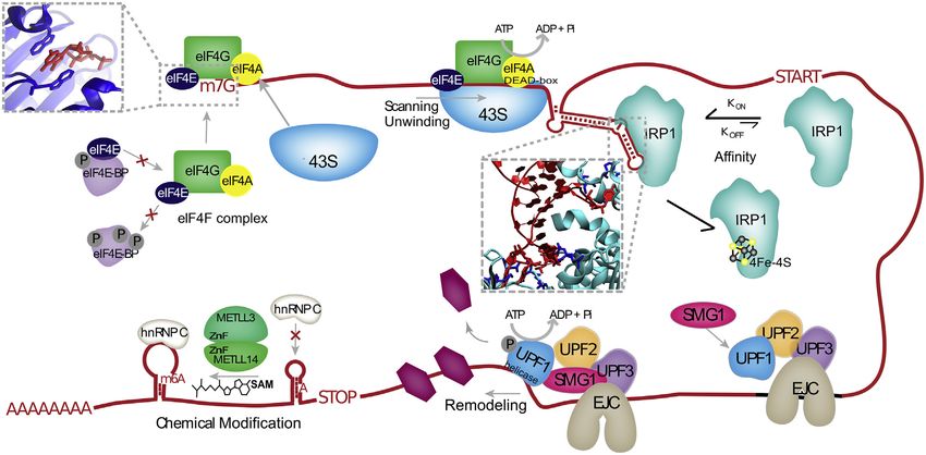
Molecular Cell
Review
Hydrogen Bonds and Van der Waals Interactions
Hydrogen bonds and Van der Waals (VdW) interactions have
been extensively analyzed in protein-RNA interactions (Gupta
and Gribskov, 2011; Han and Nepal, 2007; Hoffman et al.,
2004; Pérez-Cano and Fernández-Recio, 2010; Treger and
Westhof, 2001). Hydrogen bonds form between an electronega-
tive atom bound to a hydrogen atom, whose partial positive
charge attracts an electronegative partner. Hydrogen bonds
can be formed by both neutral and ionic groups, and can be co-
ordinated by water molecules (Figure 1B). They generally form at
distances of 2.4–3.0 A˚, contributing 0.5–4.5 kcal/mol per bond
(Auweter et al., 2006). The weakest hydrogen bonds are consid-
ered to be VdW interactions, which are weak (0.5–1 kcal/mol)
electrostatic interactions that occur above !3.0 A˚. All the studies
that analyze VdW interactions and hydrogen bonds in protein-
RNA structures identify hydrogen bonds with HBPLUS
(McDonald and Thornton, 1994) and identify VdW interactions
as the hydrogen bonds above a threshold donor-acceptor dis-
tance (Allers and Shamoo, 2001; Ellis et al., 2007; Han and Nepal,
2007; Hu et al., 2018; Jones et al., 2001; Morozova et al., 2006;
Treger and Westhof, 2001). All RNA bases, the 20 OH, and the
phosphodiester backbone can form hydrogen bonds and VdW
interactions with protein (Figures 1A–1C) (Teplova et al., 2011).
Multiple analyses of hydrogen-bond types in protein-RNA struc-
tures have found that hydrogen bonds with base, 20 OH (sugar),
and phosphate (RNA backbone) account for an average of
35.5%, 23.5%, and 41% of protein-RNA hydrogen bonds,
respectively (Figure 2A) (Gupta and Gribskov, 2011; Han and
Nepal, 2007; Hoffman et al., 2004; Treger and Westhof, 2001).
Studies of VdW percentages with base, sugar, and phosphate
are more variable (Figure 2B), perhaps reflecting inconsistent
thresholds in categorizing VdW interactions.
Proteins can interact with RNA using the main chain of any res-
idue and the side chains of most residues. Studies have consis-
tently found that the protein side chain, versus the main chain, is
employed in 71.5% of hydrogen bonds and 76% of VdW interac-
tions with RNA (Figures 2A and 2B). Polar amino acids Ser and
Asn and positively charged amino acids Lys and Arg, which
form strong ionic hydrogen bonds (salt bridges), predominate
these interactions (Gupta and Gribskov, 2011; Han and Nepal,
2007; Hoffman et al., 2004; Pérez-Cano and Fernández-Recio,
2010; Treger and Westhof, 2001). VdW interactions generally
share the same preferences for amino acids that are observed
for hydrogen bonds (Ellis et al., 2007; Han and Nepal, 2007;
Jones et al., 2001; Treger and Westhof, 2001). In the overall
set of interactions that occur at a protein-RNA interface, VdW in-
teractions are thought to predominate, although estimates of the
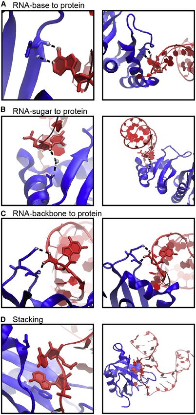
Figure 1. Examples of Protein-RNA Hydrogen Bonds and Stacking ratio of VdW-to-hydrogen-bond interactions per protein-RNA
Interactions complex vary quite a bit (Figure 2C).
The KH domain of human NOVA1 (PDB: 2ANR) (Teplova et al., 2011) and
Hydrophobic and p Interactions and Stacking
human U1A (PDB: 1AUD) (Oubridge et al., 1994) visualized with VMD (Hum-
phrey et al., 1996) in detailed (left) and zoomed-out (right) perspectives. RNA is Hydrophobic interactions occur at distances of 3.8 –5.0 A˚(Moro-
in red, and protein is in blue. zova et al., 2006; Onofrio et al., 2014) and contribute 1–2 kcal/
(A) Main-chain atoms of a Leu form hydrogen bonds with adenine. mol per interaction (Dill et al., 2008). Hydrophobic interactions
(B) Hydrogen bonds form between Gln and the 20 OH of a cytosine, bridged by
a water molecule. between RNA bases and hydrophobic side chains can be impor-
(C) Two hydrogen bonds form between the phosphate backbone atoms of tant stabilizing factors at protein-RNA interfaces by sequestering
guanine and Ser and Lys. hydrophobic residues and bases from solvent to form a ‘‘hydro-
(D) An adenine and cytosine in an unpaired loop stack between Asp and Phe.
phobic core’’ (Akopian et al., 2013; Allain et al., 1997; Yang et al.,
2002; Yu et al., 2014). For example, the SRP54 ‘‘M’’-binding
10 Molecular Cell 78, April 2, 2020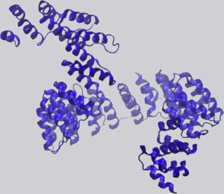
Molecular Cell

Review

A                                         B                                             C                  Figure 2. Meta-analysis of Seven Studies
                                                                                                           Analyzing Hydrogen Bonds and Van der
                                                                                                           Waals Interactions in Protein-RNA
                                                                                                           Structures
                                                                                                           (A) Reports across studies of the percent of
                                                                                                           hydrogen bonds in protein-RNA structures that
                                                                                                           occur with the RNA backbone (phosphate), sugar
                                                                                                           (20 OH), or base. The percent of hydrogen bonds
                                                                                                           that occur with the protein side chain (as opposed
                                                                                                           to the main chain). Averages are shown above each
                                                                                                           category.
                                                                                                           (B) Reports of the percent of VdW interactions in
                                                                                                           protein-RNA structures that occur with the RNA
                                                                                                           backbone (phosphate), sugar (20 OH), or base. The
percent of VdW interactions that occur with the protein side chain (as opposed to the main chain). Averages are shown above each category.
(C) Reports across studies of the average ratio of VdW interactions to hydrogen bonds per protein-RNA structure.

domain forms a methionine-rich hydrophobic surface with SRP                     available for protein-DNA interactions (Hoffman et al., 2004)
RNA (Akopian et al., 2013). Hydrophobic interactions have                       (Figure 2). The base-pairing moieties in RNA bases are also
been surveyed more sparsely in protein-RNA structures, but                      much more extensively contacted by protein than in DNA as
may account for up to 50% of the interactions at the protein-                   the Watson-Crick base face is normally base paired in DNA (Al-
RNA interface, depending on the RBP (Hu et al., 2018).                          lers and Shamoo, 2001; Luscombe et al., 2001). In this same
   p interactions can form between any nitrogenous base ring                    vein, protein-DNA interactions more frequently use the phospho-
and a p-containing amino acid, which include the aromatic res-                  diester backbone (Hoffman et al., 2004; Jones et al., 2001). DNA-
idues Trp, His, Phe, and Tyr as well as the charged residues Arg,               binding proteins tend to surround their target DNA helix, but this
Glu, and Asp (Wilson et al., 2016). These interactions are rela-                mode of binding is not always available to RBPs, which must
tively strong at !2–6 kcal/mol per interaction (Brylinski, 2018;                accommodate a diverse range of stem loops, bulges, and other
Wilson et al., 2016), and often prefer to be stacked (referred to               complex structures (Jones et al., 2001). Additionally, double-
as p stacking), occurring most frequently with an inter-atom dis-               stranded RNA (dsRNA) adopts a different helix from the standard
tance of 2.7–4.3 A˚ (Auweter et al., 2006; Brylinski, 2018; Moro-               DNA B form helix (Bercy and Bockelmann, 2015), explaining why
zova et al., 2006; Wilson et al., 2016). Analyses of p interactions             RBPs that interact with dsRNA are best suited to the RNA helix
occurring in protein-RNA crystal structures find multiple such in-              (Vukovic " et al., 2014).
teractions on average per structure (Hu et al., 2018; Wilson et al.,               Despite these overall differences, numerous proteins bind
2016). These interactions can contribute considerable stability to              both DNA and RNA (Hudson and Ortlund, 2014). A canonical
protein-RNA binding, where some p interactions are demon-                       example is the CCHH zinc-finger (ZnF) protein TFIIIA, which
strably crucial to binding function (Auweter et al., 2006; Liao                 binds both the 5S rRNA gene and 5S rRNA with at least six tan-
et al., 2018; Oubridge et al., 1994). Such is the case with the                 dem ZnF domains (Hall, 2005). In binding 5S DNA, TFIIIA ZnFs 1,
extensive stacking interactions cementing human U1A spliceo-                    3, and 5 interact with the major groove whereas ZnFs 4 and 6
somal protein with an RNA polyadenylation inhibition element,                   serve as spacers. When binding RNA, ZnF 4 and 6 interact
including two consecutive bases sandwiched between Phe                          with unpaired 5S rRNA bases and ZnF 5 binds the RNA major
and Asp residues (Figure 1D) (Oubridge et al., 1994). Stacking in-              groove, albeit by a different mode from how it contacts the
teractions also occur between bases and hydrophobic residues,                   DNA major groove. Similarly, human ADAR1 can bind both
and these may be mixed with p-p stacking interactions. For                      dsRNA and Z-form DNA, but uses separate domains for each
example, a single cytosine in a bulge in bacterial 4.5S SRP                     (Barraud and Allain, 2012). Thus, even a dual DNA- and RNA-
RNA stacks with both Phe and Leu in FtsY (Bifsha et al., 2007).                 binding protein may still use unique strategies for contacting
More exotic p-stacking configurations include bases that stack                  each molecule.
on the protein main chain between residues (Auweter et al.,                     Binding Dynamics
2006) and perpendicular ‘‘T stacks’’ between protein side chains                Protein-RNA interactions occur through dynamic rearrange-
and bases. Stacking interactions with RNA are overall quite                     ments of both molecules (Leulliot and Varani, 2001). NMR and
crucial and varied, possibly occurring at higher rates than in pro-             molecular dynamics (MD) simulations as well as crystal struc-
tein-DNA interactions (Wilson et al., 2016).                                    tures with and without ligand all shed light on the dynamic pro-
Differences from Protein-DNA Interactions                                       cess of protein-RNA interaction (Loughlin et al., 2019; Tian
Similar analyses of protein-DNA structures allow comparison                     et al., 2011; Yu et al., 2014). RNA and protein exhibit mostly local
with protein-RNA interactions (Jones et al., 2001; Luscombe                     rearrangements during binding, which often entail backbone
et al., 2001; Wilson et al., 2016). RBPs and DNA-binding proteins               shifts and bases and residues that ‘‘flip out’’ (Hainzl et al.,
show many of the same preferences for interacting residues, that                2005; Leulliot and Varani, 2001; Matthews et al., 2016; Yang
is, positively charged and polar residues (Hoffman et al., 2004;                et al., 2002). Upon binding, the site of interaction becomes rigid,
Jones et al., 2001). However, the chemical and structural                       locking the molecules together, whereas adjacent elements in
differences between DNA and RNA molecules result in observ-                     the two molecules loosen to balance the decrease in entropy.
able differences in interactions. Approximately 20% of protein                  In this way, nucleotides or residues that do not directly interact
interactions with RNA occur with the 20 OH, whereas this is not                 can still be instrumental in binding if they direct the necessary

                                                                                                                    Molecular Cell 78, April 2, 2020 11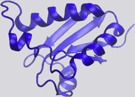
Molecular Cell

                                                                                                                    Review

compensatory changes for binding (Leulliot and Varani, 2001;            Lunde et al., 2007). Recognition of bipartite sequence motifs is
Ravindranathan et al., 2010). Unstructured loops in both protein        a common occurrence, mediated by multiple RNA-binding do-
and RNA are common sites of rearrangement during binding,               mains and their linkers and RNA structural arrangements that
such as disordered linker regions between well-ordered binding          present bipartite sequences to the RBP (Dominguez et al.,
domains in proteins. In fact, a large fraction of residues interact-    2018; Loughlin et al., 2019; Lunde et al., 2007; Tan et al., 2014;
ing with RNA tend to be in unstructured loops themselves (Barik         Walden et al., 2012). Additionally, some RNA-binding domains
et al., 2015; Cléry and Allain, 2012; Han and Nepal, 2007; Treger      are capable of mediating protein-protein interactions (PPIs),
and Westhof, 2001), and these regions adopt structure upon              including in concert with RNA binding (Cieniková et al., 2015;
binding RNA (Balcerak et al., 2019; Leulliot and Varani, 2001).         Yang et al., 2002). This multi-layered approach allows almost
Lastly, the large tertiary flexibility of RNA is a crucial functional   limitless combinations, such that there exist hundreds of
feature in protein binding (Flores and Ataide, 2018; Leulliot and       different RBPs that conduct a wide and diverse number of func-
Varani, 2001). Computational modeling of protein-RNA binding            tions. Here we describe in detail seventeen structurally charac-
found more success with simultaneous RNA folding and docking            terized domains that have been described to bind RNA in multi-
to a protein interface as opposed to RNA folding and then dock-         ple proteins (Table 2).
ing (Kappel and Das, 2019), reflecting the importance of the ter-       RNA Recognition Motif
tiary rearrangements RNA requires to bind protein.                      RRMs are the most common and well-studied RNA-binding
Protein-RNA Prediction and Resources                                    domain. A search in the Protein Data Bank (PDB) for ‘‘RRM’’
A great deal of interest lies in predicting RNA binding sites in pro-   yields over 500 structures (Table 2), and RRMs are estimated
teins. For example, the above-mentioned analyses of protein-            to occur in 1% of all human proteins (Cléry and Allain, 2012).
RNA structures indicated that protein-RNA interfaces prefer             RRMs average 90 amino acids in size and adopt a b1a1b2b3a2b4
positively charged residues, distinguishing them from protein-          topology forming two a helices against an antiparallel b sheet,
protein interfaces, which prefer polar residues (Treger and West-       which houses the conserved RNA-binding RNP1 and RNP2 mo-
hof, 2001). These sorts of metrics and the growing number of            tifs in the central b1 and b3 strands (Cléry and Allain, 2012). RRMs
solved protein-RNA structures have enabled machine-learning-            interact with 2–8 nt in single-stranded RNA (ssRNA) commonly
based attempts at predicting protein-RNA interaction. A meta-           through several sequential stacking interactions and hydrogen
analysis of these algorithms finds that the most successful fea-        bonds with RNP motifs, often with nanomolar affinities (Auweter
tures for the prediction of RNA binding sites in protein are residue    et al., 2006; Cléry et al., 2008). Each RRM has its own sequence
composition, conservation, and solvent accessibility (Zhang             preferences, often for degenerate sequences such as GU-rich
et al., 2019). Additionally, improved thermodynamic models              tracts (Cléry et al., 2008). The combination of consecutive
enable docking simulations helpful for understanding the tertiary       RRMs in an RBP dramatically increases binding affinity and
dynamics of protein-RNA binding (Huang et al., 2013; Kappel             specificity (Maris et al., 2005). For example, the dual binding of
and Das, 2019). We provide an up-to-date list of these algorithms       both RRM domains in heterogeneous nuclear RNPA1
and how to access them, along with a few key RBP database re-           (hnRNPA1) is crucial to its overall binding ability and function in
sources (Table 1). Algorithms published for the purpose of RBP          repressing splicing (Beusch et al., 2017). RRMs have also been
prediction whose source code or web server is no longer avail-          observed interacting with other protein domains, such as
able are not included. We should note that these algorithms             hnRNPC, whose single RRM domain drives multimerization
are biased by available structural data, which are dominated            with other hnRNPC molecules (Cieniková et al., 2015; Safaee
by the most abundant and readily crystallized RNA-binding do-           et al., 2012).
mains such as the RNA recognition motif (RRM). Thus, predictive         K Homology
algorithms should greatly benefit in the future from the character-     The K homology (KH) domain was first discovered in heteroge-
ization of novel RNA-binding domains and data from alternative          neous nuclear ribonucleoprotein K (hnRNPK). At 70 amino acids,
structural techniques such as cryogenic electron microscopy.            the KH domain is even smaller than the RRM domain, and typi-
                                                                        cally recognizes 4 nt in ssRNA or ssDNA (Cléry and Allain,
RNA-Binding Domains                                                     2012; Valverde et al., 2008). KH domains adopt either a type I
RBPs typically contain discrete domains for the purpose of bind-        b1a1a2b2b0 a0 topology (in eukaryotes) or the reverse type II
ing RNA. Many RNA-binding domains are quite small (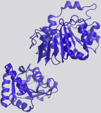
Molecular Cell

Review

Table 1. Summary of Studies and Software that Catalog or Predict RBPs and Their Targets
Name                Link                                        Reference               Description
3dRPC               http://biophy.hust.edu.cn/3dRPC.html        Huang et al., 2013      Command line software accepts PDB
                                                                                        structures of a protein and an RNA and
                                                                                        docks them.
aaRNA               https://sysimm.ifrec.osaka-u.ac.jp/aarna/   Li et al., 2014         Web server accepts PDB structure or
                                                                                        protein sequence to predict residues that
                                                                                        bind RNA. Includes graphical output of the
                                                                                        binding propensity of each residue.
Arpeggio            http://biosig.unimelb.edu.au/arpeggioweb/   Jubb et al., 2017       Web server and command line software
                                                                                        accept PDB file and chain ID and return all
                                                                                        interactions that occur with the given chain.
                                                                                        Ionic, polar, hydrogen bonds, aromatic ring
                                                                                        stacking, etc.
ATtRACT             https://attract.cnic.es/index               Giudice et al., 2016    Database of RBPs with experimental data
                                                                                        and their inferred bound sequence motif.
beRBP               http://bioinfo.vanderbilt.edu/beRBP/        Yu et al., 2019         Web server and command line software,
                    predict.html                                                        given protein and RNA sequence, predict
                                                                                        their interaction.
BioLiP              https://zhanglab.ccmb.med.umich.edu/        Yang et al., 2013       Database of PDB protein structures with
                    BioLiP/                                                             ligands (including RNA) that annotates
                                                                                        atoms at the binding interface in a given
                                                                                        structure.
catRAPID            http://s.tartaglialab.com/page/             Bellucci et al., 2011   Command line software (requires licensing)
                    catrapid_group                                                      and web server. Accept protein and RNA
                                                                                        sequences and return a heatmap of
                                                                                        interaction propensity at each residue-
                                                                                        nucleotide pair.
DR_bind1            http://drbind.limlab.ibms.sinica.edu.tw     Chen et al., 2014       Web server accepts a single protein chain in
                                                                                        PDB format and predicts which residues
                                                                                        bind RNA. Also produces a Jmol image of
                                                                                        the protein structure.
DRNApred            http://biomine.cs.vcu.edu/servers/          Yan and Kurgan, 2017    Web server accepts (up to 100) a protein
                    DRNApred/                                                           sequence(s) and assesses each residue for
                                                                                        its RNA (and DNA) interaction probability.
ENTANGLE            On request                                  Morozova et al., 2006   Software assesses hydrogen bonds and
                                                                                        Van der Waals, stacking, and hydrophobic
                                                                                        interactions between RNA and protein in the
                                                                                        given PDB structure.
HBPLUS              http://www.ebi.ac.uk/thornton-srv/          McDonald and            Command line software that lists hydrogen
                    software/HBPLUS/                            Thornton, 1994          bonds in a given PDB structure.
hybridNAP           http://biomine.cs.vcu.edu/servers/          Zhang et al., 2019      Web server accepts (up to 10) a protein
                    hybridNAP/                                                          sequence(s) and calculates each residue’s
                                                                                        interaction probability with RNA, DNA, and/
                                                                                        or protein. Also returns the feature values
                                                                                        that determine this probability.
KYG                 http://cib.cf.ocha.ac.jp/KYG/               Kim et al., 2006        Web server accepts a single-chain PDB
                                                                                        structure and predicts the RNA interface
                                                                                        propensity of each residue. Outputs graph,
                                                                                        table, and downloadable PDB file with
                                                                                        scores.
ndb                 http://ndbserver.rutgers.edu                Berman et al., 1992;    Database of solved DNA and RNA
                                                                Coimbatore Narayanan    structures.
                                                                et al., 2014
NUCPLOT             https://www.ebi.ac.uk/thornton-srv/         Luscombe et al., 1997   Command line software accepts a protein-
                    software/NUCPLOT/                                                   RNA/DNA PDB structure and returns a
                                                                                        graphic of protein interaction occurring at
                                                                                        each nucleotide.
                                                                                                              (Continued on next page)

                                                                                                Molecular Cell 78, April 2, 2020 13
Molecular Cell

                                                                                                                        Review

Table 1.     Continued
Name                     Link                                          Reference                  Description
OPRA                     https://life.bsc.es/pid/opra/default/index    Pérez-Cano and            Web server scores residues in PDB
                                                                       Fernández-Recio, 2010     structures for interaction probability with
                                                                                                  RNA. Includes Jmol output of structures
                                                                                                  with residues colored by predicted value.
PDBsum                   http://www.ebi.ac.uk/thornton-srv/            Laskowski et al., 2018     Provides an overview of a given PDB
                         databases/cgi-bin/pdbsum/GetPage.pl?                                     structure, including protein sequence,
                         pdbcode=index.html                                                       defined structural regions, sequence of
                                                                                                  bound RNA/DNA, NUCPLOT depiction of
                                                                                                  bound DNA/RNA, etc.
PLIP                     https://projects.biotec.tu-dresden.de/plip-   Salentin et al., 2015      Web server and command software accept
                         web/plip/index                                                           PDB structures of protein-ligand and list
                                                                                                  each hydrogen bond, salt bridge, p
                                                                                                  interaction, and hydrophobic interaction
                                                                                                  with ligand.
PPRInt                   https://webs.iiitd.edu.in/raghava/pprint/     Kumar et al., 2008         Web server accepts a protein sequence
                         index.html                                                               and predicts RNA-binding residues.
PredPRBA                 http://PredPRBA.denglab.org/                  Deng et al., 2019          Web server accepts a PDB file of protein-
                                                                                                  RNA structure and predicts the free energy
                                                                                                  of binding.
PRince                   http://www.facweb.iitkgp.ac.in/!rbahadur/     Barik et al., 2012         Web server accepts a PDB structure, given
                         prince/home.html                                                         a protein and RNA chain IDs, and will list
                                                                                                  atoms at the protein-RNA interface.
RAIDv2.0                 http://www.rna-society.org/raid2/             Yi et al., 2017            Database of known RNA-RNA and protein-
                         index.html                                                               RNA interactions at the full transcript/
                                                                                                  protein level (not nucleotide/residue detail).
RBP prediction           https://www.iitm.ac.in:443/bioinfo/RNA-       Nagarajan and              Online tool based on the benchmark of
selection tool           protein/                                      Gromiha, 2014              various RBP prediction software. Shows
                                                                                                  the best software (limited selection) to use
                                                                                                  for a given RBP/RNA type.
RBPDB                    http://rbpdb.ccbr.utoronto.ca                 Cook et al., 2011          Database of RBPs with available
                                                                                                  experimental data, categorized by
                                                                                                  organism or RBP domain.
RBPmap                   http://rbpmap.technion.ac.il/index.html       Paz et al., 2014           Web server and command line software
                                                                                                  search for given RBP-binding motifs in a
                                                                                                  given RNA sequence.
RCSB PDB                 https://www.rcsb.org                          Berman et al., 2000        Search parameters for RBPs with RNA:
                                                                                                  ‘‘macromolecule type: contains protein
                                                                                                  AND contains RNA.’’
RNAbindPlus              http://ailab1.ist.psu.edu/RNABindRPlus/       Terribilini et al., 2007   Web server predicts residues that bind RNA
                                                                                                  in a given protein sequence.
RNAbindRv2.0             http://ailab-projects2.ist.psu.edu/           Terribilini et al., 2007   Web server predicts residues that bind RNA
                         RNABindR/                                                                in a given protein sequence.
RPISeq                   http://pridb.gdcb.iastate.edu/RPISeq/         Muppirala et al., 2011     Web server accepts protein and RNA
                                                                                                  sequences and predicts their interaction
                                                                                                  probability.
RsiteDB                  http://bioinfo3d.cs.tau.ac.il/RsiteDB/        Shulman-Peleg              Database searches for PDB structure (if
                                                                       et al., 2008               published before 2008) and describes
                                                                                                  protein-RNA interactions: Jmol image,
                                                                                                  which base, etc.
SPOT-Seq-RNA             https://sparks-lab.org/server/SPOT-RNA/       Yang et al., 2014          Web server and command line software
                                                                                                  predict whether a given protein sequence is
                                                                                                  an RBP.
SPOT-Struct-RNA          https://sparks-lab.org/yueyang/server/        Zhao et al., 2011          Web server and command line software
                         SPOT-Struct-RNA/                                                         predict whether a given PDB structure is
                                                                                                  an RBP.
TriPepSVM                https://github.com/marsicoLab/                Bressin et al., 2019       Command line software accepts a protein
                         TriPepSVM                                                                sequence and predicts whether it is
                                                                                                  an RBP.

14     Molecular Cell 78, April 2, 2020
Table 2. Summary of described RNA binding domains and example structures.
                                                                                                           PDB
                                                                                              PDB          Structures                 Protein Families Containing   Example Structure
                                      Domain Name                PDB Search Term              Structures   with RNA     Size (a.a.)   Domain                        PDB ID Code         Example Domain Structure
                                      Cold shock domain (CSD)    ‘‘Cold shock domain’’        46           13           70            Cold shock proteins, Y-box    4A4I
                                                                                                                                      proteins
                                                                                                                                                                                                                                  Molecular Cell

                                                                                                                                                                                                                         Review

                                      Double-stranded RNA        ‘‘dsRBD’’                    59           27           65            RNases, ADARs, Dicer          3LLH
                                      Binding Domain (dsRBD)

                                      Helicase                   ‘‘dead’’ OR ‘‘deah’’         701          114          350-400       DExH/D-box, Ski2-like, RIG-   2I4I
                                                                 OR ‘‘helicase domain’’                                               I-like, NS3, UPF1-like RNA
                                                                                                                                      binding helicases

                                      Intrinsically disordered   ‘‘Intrinsically disordered   NA           NA           varies        Most RBPs                     NA                  NA
                                      region (IDR)               region’’
                                      K homology (KH)            ‘‘KH domain’’                117          20           70            hnRNPs, translation           1WH9
                                                                                                                                      regulation proteins, very
                                                                                                                                      common

                                                                                                                                                                                              (Continued on next page)

Molecular Cell 78, April 2, 2020 15
16
                                   Table 2.   Continued
                                                                                                   PDB
                                                                                      PDB          Structures                 Protein Families Containing   Example Structure
                                   Domain Name             PDB Search Term            Structures   with RNA     Size (a.a.)   Domain                        PDB ID Code         Example Domain Structure
                                   La motif (LAM)          ‘‘La motif AND NOT rrm’’   54           5            90            La proteins, La-related       1S29
                                                                                                                              proteins (LARPs)

Molecular Cell 78, April 2, 2020
                                   Piwi-Argonaute-Zwille   ‘‘PAZ domain’’             71           44           170           Argonaute proteins, Dicer     3O6E
                                   (PAZ)

                                   P-element Induced       ‘‘PIWI domain’’            74           33           290           Argonaute proteins            1X4Q
                                   Wimpy Testis (PIWI)

                                   Pentatricopeptide       ‘‘pentatricopeptide        32           9            35 * n,       RNA editing proteins          4M59
                                   repeat (PPR)            repeat’’                                             1 > n > 30

                                                                                                                                                                                      (Continued on next page)
                                                                                                                                                                                                                 Review
                                                                                                                                                                                                                          Molecular Cell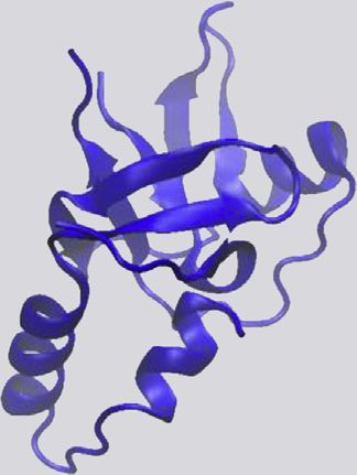
Table 2.   Continued
                                                                                                             PDB
                                                                                                PDB          Structures                 Protein Families Containing       Example Structure
                                      Domain Name                 PDB Search Term               Structures   with RNA     Size (a.a.)   Domain                            PDB ID Code         Example Domain Structure
                                      Pseudouridine synthase      ‘‘Pseudouridine synthase      119          28           66-98         RNA modifying enzymes,            1SQW
                                      and archaeosine             and archaeosine                                                       metabolic enzymes
                                                                                                                                                                                                                                        Molecular Cell

                                      transglycoslyase (PUA)      transglycoslyase’’ OR ‘‘PUA
                                                                  domain’’
                                                                                                                                                                                                                               Review

                                      Pumillo-like repeat (PUM)   ‘‘Pumilio’’ OR ‘‘PUM          60           49           334           PUF proteins                      1M8W
                                                                  domain’’ OR ‘‘Puf protein’’

                                      Ribosomal S1-like (S1)      ‘‘S1 RNA binding domain’’     25           2            70            Ribosomal proteins,               2EQS
                                                                                                                                        Translation initiation factors,
                                                                                                                                        RNase II, PNPase

                                      RNA Recognition             ‘‘RNA Recognition Motif’’     554          119          90            hnRNPs, splicing factors,         2MTG
                                      Motif (RRM)                 OR ‘‘RRM’’                                                            very common

                                                                                                                                                                                                    (Continued on next page)

Molecular Cell 78, April 2, 2020 17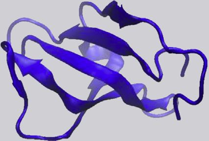
18
                                   Table 2.    Continued
                                                                                            PDB
                                                                               PDB          Structures                 Protein Families Containing   Example Structure
                                   Domain Name              PDB Search Term    Structures   with RNA     Size (a.a.)   Domain                        PDB ID Code         Example Domain Structure
                                   Sm and Like-Sm           ‘‘Sm RNA binding   31           23           80            U1 spliceosomal               2VC8
                                   (Sm / Lsm)               domain’’                                                   proteins, Hfq

Molecular Cell 78, April 2, 2020
                                   thiouridine synthases,   ‘‘THUMP domain’’   12           3            100-110       tRNA modifying enzymes        2DIR
                                   RNA methylases and
                                   pseudouridine
                                   synthases (THUMP)

                                   YT521-B homology         ‘‘YTH domain’’     28           14           100-150       YTH family m6A readers        4RCI
                                   (YTH)

                                   Zinc Finger (ZnF)        ‘‘Zinc Finger’’    2677         63           30            Transcription factors,        5ZC4
                                                                                                                       METTL enzymes, Very
                                                                                                                       common
                                                                                                                                                                                                    Review
                                                                                                                                                                                                             Molecular Cell
Molecular Cell
Review
with quaking (QUA) domains as part of the larger signal transduc- as several other RNA-modifying and metabolic enzymes
tion and activation of RNA (STAR) domain, which greatly extends (Pérez-Arellano et al., 2007). PUA domains range from 67 to 94
the binding surface and accommodates 7 or 8 nt with 0.07 mM amino acids in length, with a b1a1b2b3b4b5a2b6 architecture
affinity (Sharma and Anirudh, 2017; Teplova et al., 2013). that forms a pseudobarrel encased by two a helices. PUA do-
Zinc Finger mains have been characterized contacting dsRNA and its adja-
ZnFs describe a large family of proteins that average 30 amino cent loops or overhangs through extensive hydrogen bonds
acids in size and form a simple bba topology in which residues with all parts of the RNA. These contacts are typically formed
in the b hairpin turn and a helix are coordinated by a Zn2+ ion (Cléry by a glycine-rich loop between a1 and b2 or a2 and b6. Unlike
and Allain, 2012). Most ZnFs bind DNA, but have been additionally many other domains, PUA domains are not found as tandem re-
shown to bind RNAs, proteins, and small molecules (Lai et al., peats (Pérez-Arellano et al., 2007).
2000). ZnF subtypes that interact with RNA include CCHC (zinc THUMP
knuckle), CCCH, CCCC (RanBP2), and CCHH subtypes, where Named for thiouridine synthase, methyltransferase, and pseu-
C and H refer to the interspersed cysteine and histidine residues douridine synthase, the THUMP domain is found in numerous
that coordinate the zinc atom, respectively (Cléry and Allain, tRNA-modifying enzymes. About 100 amino acids long, THUMP
2012). These subtypes display a range of sequence and structural domains are always found in proximity to RNA-modifying do-
specificities. Zinc knuckles recognize stem-loop elements in RNA mains and often in proximity to an N-terminal ferredoxin-like
(or ssDNA) through contacts with bases in the loop and the phos- (NFLD) domain (Neumann et al., 2014). THUMP domains display
phate backbone of the stem. CCCH and CCCC subtypes tend to a a1a2b1a3b2b2 topology that forms parallel a helices flanking a b
recognize 3-nt repeats through multiple such ZnFs in one RBP sheet (Fislage et al., 2012). The first structure of a THUMP
(Font and Mackay, 2010; Hall, 2005; Lai et al., 2000). These con- domain bound by RNA, bacterial 4-thiouridine synthetase in
tacts are formed through hydrogen bonds with bases and the complex with tRNA, reveals a 3-dimensional fold that specifically
insertion of aromatic side chains that stack between bases. The recognizes the 30 -CCA tail and adjoining stem of tRNA. Several
versatile and abundant CCHH ZnFs interact with both single- hydrogen bonds and VdW contacts correctly position the tRNA
stranded and dsRNA as well as DNA (Font and Mackay, 2010; for modification by the accompanying pyrophosphate domain
Hall, 2005). Modular arrays of CCHH ZnFs have been successfully (Neumann et al., 2014).
engineered to bind desired DNA sequences. Thus, designer ZnFs YT521-B Homology
are thought to have potential for directed binding of RNA se- The YT521-B homology (YTH) domain is found in the YTH family
quences, a goal that has been achieved with the much larger Pum- of proteins that ‘‘read’’ N6-methyladenosine (m6A) marks in RNA.
ilio homology domains (Font and Mackay, 2010). The YTH domain ranges from 100 to 150 amino acids in length
Pumilio Homology Domain and forms a six-stranded b barrel surrounded by four or five a he-
The Pumilio and FBF (PUF) family of proteins occurs in most eu- lices. Three residues in the hydrophobic core of the b barrel trap
karyotes and is defined by the Pumilio homology domain (PUM- the methyl group of m6A in an ‘‘aromatic cage’’ consisting of
HD), or the PUF domain. The PUF domain is very large, consist- hydrogen bonds with the adenosine and p interactions between
ing of eight a-helical repeats of a highly conserved 36-amino tryptophan rings and the methyl group (Liao et al., 2018; Xu et al.,
acid sequence that forms a concave RNA-binding surface 2014). The YTH domain specifically binds m6A over unmodified
(Wang et al., 2018). Each repeat recognizes one unpaired adenosines. Affinity of YTHDC1 for consensus DRm6(A)CH mo-
base through hydrogen bonds and a stabilizing stacking inter- tifs was measured at 0.3 mM, whereas no binding was detected
action, where the full domain recognizes up to 8 nt in ssRNA for the unmethylated sequence (Xu et al., 2014). Similarly,
with low-nanomolar affinity (Zhao et al., 2018). Wild-type PUF YTHDF2 affinity for methylated RNA was measured as
repeats do not specifically recognize cytosine; however, pro- 2.54 mM, with 10-fold lower affinity for the unmethylated target
tein engineering has produced repeats that do (Zhao et al., (Zhu et al., 2014).
2018). These advances combined with the PUF domain’s pre- Double-Stranded RNA-Binding Domain
dictable base recognition code allow modular design of pumilio Double-stranded RNA-binding domains (dsRBDs), or motifs
proteins that recognize 8- to 10-nt sequences containing all (dsRBMs), consist of 65–70 amino acids and are the third most
RNA bases (Zhao et al., 2018). common RNA-binding domain (Masliah et al., 2013). dsRBDs
Pentatricopeptide Repeat specifically recognize and bind dsRNA and are found in proteins
Very similar to PUF repeats, eukaryotic pentatricopeptide re- with roles in viral protection, RNAi, and cellular transport (Masliah
peats (PPRs) are each !35 residues in length and form two anti- et al., 2013). dsRBDs often appear as tandem repeats or in com-
parallel a helices. 2–30 repeats form a solenoid-shaped scaffold bination with other functional RNA-binding domains, such as
that binds specific ssRNA sequences with nanomolar affinity (Ke RNA-editing or helicase domains (Cléry and Allain, 2012; Ranji
et al., 2013; Spa˚hr et al., 2018). Two residues in each repeat et al., 2011). The domain is made up of an a1b1b2b3a2 fold that
determine base-specific binding through hydrogen bonds, forms an antiparallel b sheet flanked by a helices on one face
enabling the development of designer PPRs that bind specified (Cléry and Allain, 2012; Masliah et al., 2013). dsRBDs specifically
ssRNA or ssDNA sequences (Spa˚hr et al., 2018). recognize the structure of an A-form RNA helix, spanning up to
Pseudouridine Synthase and Archaeosine 16 bp with hydrogen-bond contacts to the phosphodiester back-
Transglycosylase bone and 20 OH (Cléry and Allain, 2012; Ramos et al., 2000). In
The pseudouridine synthase and archaeosine transglycosylase some cases, dsRBDs have demonstrated base-specific contacts,
(PUA) domain is found in the aforementioned enzymes as well such as to bases in adjacent loops (Cléry and Allain, 2012; Masliah
Molecular Cell 78, April 2, 2020 19Molecular Cell
Review
A
B
Figure 3. Amino Acid and Base Preferences in Protein-RNA Hydrogen Bonds Observed in over 200 Structures, Organized by Domain
(A) The average frequency of each amino acid in forming hydrogen bonds with RNA across eight RNA-binding domain types (left). The frequency of each amino
acid (one-letter abbreviations) in forming hydrogen bonds with RNA in multiple structures, separated by domain type (right, smaller plots), is shown. CSDs use Trp
more frequently than other domains (p = 2.38 3 10-5). KH domains use Leu and Ile more frequently than other domains (p = 7.26 3 10-6, 6.16 3 10-9).
(B) The average frequency of each RNA nucleotide in forming hydrogen bonds with protein across eight RNA-binding domain types, as well as the average
frequency of each base in sequence motifs from Bind-n-Seq data (Dominguez et al., 2018) (left). The frequency of each RNA nucleotide in forming hydrogen
bonds with protein in multiple structures, separated by domain type (right, smaller plots), is shown. PUF domains contact cytosine least frequently (p = 0.001). KH
domains contact adenosine most frequently (p = 0.002).
et al., 2013). Stacking interactions are rare, potentially explaining et al., 2010). Multiple nucleotides are typically contacted simulta-
the low affinities (high-nanomolar-to-micromolar range) of neously; DEAH/DEAD-box helicases, for example, tend to
dsRBDs to RNA targets (Stefl et al., 2010; Wang et al., 2011). accommodate at least 5 single-stranded or base-paired nucleo-
Helicase tides (Jiang et al., 2011; Linder and Jankowsky, 2011; Weir et al.,
Helicase domains are found in all forms of life in helicase pro- 2010). Affinities to RNA are often in the nanomolar range,
teins, which unwind both DNA and dsRNA. Helicases comprise although they vary greatly by helicase and are modulated by
six superfamilies (SFs), of which SF1 and SF2 contain all the eu- other subdomains of the helicase. ATP binding generally pro-
karyotic RNA and DNA helicases. RNA-binding helicases include motes higher affinity to RNA by causing the helicase RNA-bind-
the Upf1-like family in SF1 and the DEAD-box, DEAH, RIG-I-like, ing regions to ‘‘clamp.’’ ATP hydrolysis subsequently promotes
Ski2-like, and NS3 families in SF2. The remaining SFs, 3–6, conformational changes that cause the helicase to translocate
contain bacterial and viral helicases that form multimeric rings 1 nt and/or unwind its substrate (Iost et al., 1999; Jankowsky,
(Jankowsky, 2011). Helicase domains are very large, containing 2011; Jiang et al., 2011; Kainov et al., 2003).
350–400 amino acids. In SF1 and SF2, the helicase domain is Cold Shock Domain
composed of two ‘‘recombinase A (recA)-like’’ subdomains, The cold shock domain (CSD) is found in a large family of pro-
each of which contains an ATP-catalytic core, a nucleic-acid- teins associated with cold adaptation found in all domains of
binding region, and subdomains that coordinate the two. Within life. CSDs are composed of !70 amino acids (more in eukary-
families of helicases these subdomains are quite conserved. otes) and five antiparallel b strands that form a common b barrel
Helicase monomers in the ring-forming SFs of helicases are simi- structure known as an oligosaccharide/oligonucleotide-binding
larly quite large and composed of multiple subdomains (Gai (OB) fold. CSDs contain the conserved RNP1 and RNP2 motifs
et al., 2004; Kainov et al., 2008). Bound RNA is surrounded by common to RRMs, which bind ssRNA and ssDNA (Amir et al.,
recA-like domains or, in the case of multimeric helicases, RNA 2018). CSDs contact 3 or 4 nt through sequential stacking inter-
is pulled through the center of the ring. Contacts with RNA are actions and hydrogen bonds with bases, achieving nanomolar
dominated by hydrogen bonds to phosphate and sugar moieties, affinities (Kljashtorny et al., 2015; Sachs et al., 2012). CSD-con-
but contacts with bases are occasionally observed (Jankowsky, taining proteins vary greatly in the types of sequences they
2011; Kainov et al., 2008; Linder and Jankowsky, 2011; Weir recognize. Bacterial CspB is reported to bind pyrimidine-rich
20 Molecular Cell 78, April 2, 2020Molecular Cell
Review
A B Figure 4. Assessment of Protein-RNA
Hydrogen Bonds in over 200 Structures,
Organized by RNA-Binding Domain
Averages for each statistic are listed above each
domain’s violin plot and medians are indicated
with black horizontal bars.
(A) The percent of protein-RNA hydrogen bonds
that are formed using protein side chains (as
opposed to the main chain). KH domains use
sidechains to hydrogen bond with RNA sign-
ficantly less than PUF domains (p = 2.25 3 10-13).
(B) The percent of protein-RNA hydrogen bonds
that are formed with RNA backbone atoms. PUF
domains hyrogen bond with the RNA backbone
the least (p = 1.60 3 10-13) and DEAD domains the
most (p < 1 3 10-307).
(C) The percent of protein-RNA hydrogen bonds
C D that are formed with RNA sugar atoms. dsRBDs
hydrogen bond most frequently with the 2’ OH
(p = 1.90 3 10-10).
(D) The percent of protein-RNA hydrogen bonds
that are formed with RNA base atoms. PUF do-
mains hydrogen bond most frequently with the
RNA base (p = 1.31 3 10-7) and least frequently
with dsRBDs and DEAD domains (p = 5.44 3 10-8,
p < 1 3 10-307).
sequences and prefer ssDNA to ssRNA by up to 10-fold (Sachs strands b4 and b5 in two Sm motifs. For example, Sm-Sm inter-
et al., 2012). Y box proteins contain the most well-studied eu- actions link the seven human Sm proteins that make up the
karyotic CSDs, showing a preference for G-rich ssRNA se- protein core of small nuclear ribonucleoproteins (snRNPs) in
quences over ssDNA (Kljashtorny et al., 2015). the spliceosome (Thore et al., 2003). The Sm multimers bind
S1 RNA with nanomolar affinity. Two Sm motifs form a 6-nt binding
The S1 RNA-binding domain was originally discovered in S1 ri- surface that binds specific bases, often uridines, through
bosomal protein, which binds both mRNA and rRNA. The !70- hydrogen bonds and stacking interactions (Schumacher et al.,
amino acid S1 domain forms a 5-stranded antiparallel b barrel 2002; Thore et al., 2003).
in the same OB-fold family as the CSD (Mihailovich et al., La Motif
2010). Despite sharing a common tertiary structure, the two do- The small !90-residue La motif (LAM) is found in eukaryotic La
mains show no sequence similarity, suggesting that their shared and La-related proteins (LARPs). The LAM consists of five a
tertiary structure was achieved through convergent evolution helices and three b strands that form a small antiparallel b
(Mihailovich et al., 2010). S1 domains are additionally found in sheet against a modified ‘‘winged-helix’’ fold (Bousquet-Anto-
several exoribonucleases and eukaryotic translation initiation nelli and Deragon, 2009). The winged-helix structure itself is
factors and in combination with other RNA-binding domains common to several other RNA- and DNA-binding proteins
such as the KH domain or CSDs (Amir et al., 2018; Chekanova (Teichmann et al., 2012). LAMs are always found adjacent to
et al., 2002; Hossain et al., 2016; Worbs et al., 2001). Despite at least one RRM, where the combination of these two do-
their abundance, very little structural information is available mains likely evolved as a unit (Bousquet-Antonelli and Dera-
for S1 domains in complex with RNA. S1 domains interact with gon, 2009). In La proteins, the dual LAM-RRM region tightly
both ssRNA and dsRNA in the context of the RNA-binding chan- binds the UUU-OH elements at the 30 ends of polymerase-
nel of exoribonucleases (Hossain et al., 2016). Similarly, S1 do- III-transcribed small RNAs. Binding occurs in a cleft between
mains of the ribosomal S1 protein likely interact with mRNA at the LAM and RRM rather than the traditional RNA-binding sur-
the entry channel of the ribosome (Loveland and Koroste- faces of either the RRM or the LAM winged-helix fold. Several
lev, 2018). uracil bases stack with highly conserved aromatic residues in
Sm the LAM, and hydrogen bonds from both the LAM and RRM
The Sm RNA-binding motif is found in Sm and like-Sm (Lsm) coordinate bases, phosphates, and the terminating 20 OH.
proteins in eukaryotes and archaea and in Hfq protein in pro- These contacts result in low-nanomolar affinities of the LAM
karyotes (Schumacher et al., 2002; Thore et al., 2003). The for 30 -terminal UUU-OH elements (Teplova et al., 2006). The
Sm motif consists of !70 residues with an a1b1b2b3b4b5 topol- other LAM-containing proteins, LARPs, bind a diverse set of
ogy that forms a curved antiparallel b sheet. Sm-containing RNAs with as-yet uncharacterized structural mechanisms
proteins readily multimerize through interactions between (Schenk et al., 2012).
Molecular Cell 78, April 2, 2020 21Molecular Cell
Review
Figure 5. Examples of Mechanisms Controlling RBP Binding, Interactions with RNA, and Their Regulation
eIF4E (dark blue) interacts with the 7-methyl-guanosine cap (m7G), in part through stacking interactions (inset) and binds to RNA as part of eIF4F, which includes
the RBPs eIF4G and eIF4A. eIF4E association with eIF4F is prevented by sequestration to hypo-phosphorylated eIF4E-BP. The 43S ribosomal subunit is recruited
to the eIF4F complex and processively scans the 50 UTR, aided by ATP-driven helicase activity of the eIF4A DEAD-box domain. The RBP IRP1 specifically binds
hairpin elements in the 50 UTR with high affinity through specific residues (inset, dark blue) that hydrogen bond with the bulge and apical loop of the RNA. RNA
binding by IRP1 is prevented by 4Fe-4S ligand binding to IRP1. UPF1 is recruited to the exon junction complex (EJC), where its helicase activity is activated by
interactions with SMG1 and UPF2. Driven by ATP, UPF1 removes both RNA structures and other bound RBPs in the 50 /30 direction. The METTL3-METTL14
complex, which contains zinc fingers (ZnF), deposits methyl groups donated by S-adenosyl methionine (SAM) on targeted adenosines (m6As). m6A modifications
reduce base pairing in RNA, such that some locations become available for hnRNPC binding.
Piwi-Argonaute-Zwille and PIWI suggesting that its activity may be modulated by the conforma-
Piwi-Argonaute-Zwille (PAZ) and PIWI RNA-binding domains tional state of the PAZ domain (Boland et al., 2011).
define the Argonaute family of proteins found in eukaryotes Intrinsically Disordered Region
(Ho¨ck and Meister, 2008). Found on opposite sides of the Argo- Intrinsically disordered regions (IDRs) are unstructured and often
naute protein, both domains facilitate binding of small interfering consist of repeats of arginine/serine (RS repeat), arginine/glycine
RNA and microRNA guides to mRNA targets (Ho¨ck and Meis- (RGG box), arginine- or lysine-rich patches (R/K basic patches),
ter, 2008). or short linear motifs of amino acids (Balcerak et al., 2019;
PAZ domains occur in Dicer proteins in addition to Argonaute Ja€rvelin et al., 2016). Despite their lack of structure, IDRs have
proteins (Ho¨ck and Meister, 2008). Crystal structures of the PAZ been found to dominate the composition in over 20% of RBPs
domain display a six-stranded b barrel topped with two a helices (Ja€rvelin et al., 2016). It is increasingly observed that IDRs can
and flanked on the opposite side by a special appendage con- be the sole RNA-binding domain in an RBP and may actually
taining a b hairpin and short a helix (Ma et al., 2004; Tian et al., drive the majority of protein-RNA interactions in the cell (Hentze
2011; Yan et al., 2003). A binding pocket formed between this et al., 2018). Like globular RNA-binding domains, IDRs are
appendage and the b barrel binds the 2-nt 30 overhang in guide conserved, often occur multiple times in one RBP, and can coor-
RNAs (gRNAs) with low-micromolar affinity (Ma et al., 2004; Tian dinate RNA binding in concert with other domains (Balcerak
et al., 2011). Binding is coordinated mostly by conserved tyro- et al., 2019; Ja €rvelin et al., 2016; Loughlin et al., 2019). IDRs
sine residues that form hydrogen bonds with the phosphate have been shown to drive higher affinity to RNA in RBPs that
backbone and sugar hydroxyls of the two terminal nucleotides contain ordered RNA-binding domains and can themselves tran-
(Ma et al., 2004; Tian et al., 2011). sition to an ordered state once bound to RNA (Balcerak et al.,
The PIWI domain tertiary structure forms an RNase H-like fold 2019; Cruz-Gallardo et al., 2019; Ja €rvelin et al., 2016; Leulliot
consisting of a five-stranded b sheet flanked by a helices on both and Varani, 2001). IDRs show little RNA sequence dependence,
faces (Boland et al., 2011). The PIWI domain has endonucleolytic however, suggesting that these regions’ high affinity for RNA is
activity in some cases, but primarily stabilizes the gRNA-mRNA predominantly driven by electrostatic attraction to the phospho-
duplex seed region through hydrogen bonds with the gRNA diester backbone (Balcerak et al., 2019; Ja €rvelin et al., 2016).
backbone of nucleotides 3–5 and the 50 overhang base (Boland Other RNA-Binding Domains
et al., 2011; Ma et al., 2005; Miyoshi et al., 2016). The PIWI The domains detailed above represent a mere fraction of the RNA-
domain also contacts the PAZ domain in certain conformations, binding domains in existence, 23% of the 2,685 RNA-bound
22 Molecular Cell 78, April 2, 2020Molecular Cell
Review
structures in the PDB, whereas many hundreds more RNA-bind- repeatedly involved in forming hydrogen bonds with RNA among
ing domains await characterization (Hentze et al., 2018). Several KH domains but no other domain (p = 7.26 3 10"6, 6.16 3 10"9).
domains, such as the Brix domain, sterile alpha motif (SAM), This agrees with previous descriptions of salient RNA contacts in
and SAF-A/B, Acinus, and PIAS (SAP) domain, are mostly known crystal structures of KH domains being formed via the main chain
as protein- or DNA-binding domains but have one or two protein of Ile residues (Cléry and Allain, 2012). Interestingly, a single-
members shown to bind RNA. For example, the SAM domain is point mutation replacing an Ile in the KH domain of FMRP is
a well-known a-helical PPI domain, but the SAM-containing pro- known to cause fragile X syndrome (De Boulle et al., 1993).
teins Smaug and its homolog VTS1p bind the pentaloop of an Preferences for RNA nucleotides in protein-RNA hydrogen
RNA hairpin element called the Smaug recognition element bonds were assessed as well. Note that even with small RNAs,
(SRE) with nanomolar affinity (Aviv et al., 2006; Ravindranathan not necessarily all of the nucleotides interact with the RBP in a
et al., 2010). Conservation of the RNA-interacting residues among hydrogen bond. Previous analyses have varied in whether they
Smaug homologs suggests RNA-binding function exists in other report RBP preferences for interaction with specific nucleotides
SAM-containing proteins (Aviv et al., 2006). Many other RNA- (Pérez-Cano and Fernández-Recio, 2010), and the types of RNA
binding domains are utterly unique, i.e., not yet found to resemble sequences that have been successfully co-crystallized with
any other domain (Gerstberger et al., 2014; Hentze et al., 2018; RBPs could be biased by technical reasons. Nevertheless, as-
Tan et al., 2013; Walden et al., 2012). The stem-loop-binding pro- sessing our data, we observe a preference for interactions with
tein domain is found only in the protein of the same name (SLBP) uracil and an under-enrichment for cytosine (Figure 3B).
that exclusively binds a conserved stem loop at the ends of his- Sequence motifs derived from RNA Bind-n-Seq experiments
tone mRNAs (Tan et al., 2013). IRP1, also known as ACO1, is an (Dominguez et al., 2018) also follow this pattern of base fre-
aconitase with a unique fold that tightly binds iron response ele- quencies (r2 = 0.89, Pearson), providing orthogonal agreement
ments in the UTRs of iron-metabolism-related transcripts (Walden for the observed base preferences. PUF domain structures
et al., 2012). Most ribosomal proteins each contain a unique RNA- exhibit the lowest percentage of hydrogen bonds with cytosine
binding domain, the S1 domain excepted (Gerstberger et al., (p = 0.001), reflecting the lack of cytosine recognition in wild-
2014). Structural data of viral RBPs reveal incredibly diverse and type PUF repeats (Zhao et al., 2018). The KH domain re-asserts
unique structures specialized for binding highly structured viral its status as an oddball, as it forms hydrogen bonds with aden-
RNA elements. Recent large-scale studies have discovered hun- osines more frequently than any other domain (p = 0.002).
dreds of novel RNA-binding regions in proteins, including in many We were also interested in statistics summarizing the fre-
well-characterized enzymatic proteins such as GAPDH, that sur- quency of hydrogen bonds with the side chain of amino acids
prisingly ‘‘moonlight’’ as RBPs (Hentze et al., 2018; Hudson and and with the base, backbone, or sugar of RNA (Figure 4). We
Ortlund, 2014). Overall, the diversity of RBPs is an astounding tes- observe pronounced domain differences with the percent usage
tament to their all-encompassing cellular roles in many domains of amino acid side chains (versus the main chain). For example,
of life. KH domains use side chains in 43.6% of hydrogen bonds with
Domain Differences in Hydrogen-Bond Formation RNA, whereas PUF domains use side chains in 90.1% of interac-
with RNA tions (p value of difference = 2.25 3 10"13) (Figure 4A). In fact,
We additionally assessed the type and number of hydrogen side-chain rather than main-chain hydrogen bonds typically
bonds that proteins form with RNA in over 200 structures from dominate protein-RNA interactions (Figure 2A), but KH domains
eight common domain types (KH, dsRBD, RRM, ZnF, PUF/ are the only domain analyzed here that violate this trend
PUM-HD, DEAD helicase, YTH, and CSD). This includes the fre- (Figure 4A). The percentages of hydrogen bonds forming with
quency of each amino acid used for protein-RNA hydrogen either the backbone, sugar, or base of RNA nucleotides were
bonds, the frequency of each RNA base that contacts are formed also calculated for each protein structure (Figures 4B–4D). PUF
with, and the moieties used to facilitate those bonds (Figures 3 domains predictably hydrogen bond with the RNA backbone
and 4). This hydrogen-bond analysis of protein-RNA structures with the lowest frequency on average (p = 1.60 3 10"13)
has been conducted many times before (Figure 2) (Allers and (Figure 4B), instead interacting with the RNA base in 65.4% of
Shamoo, 2001; Ellis et al., 2007; Han and Nepal, 2007; Hu its hydrogen bonds—the highest average of all domains (p =
et al., 2018; Jones et al., 2001; Morozova et al., 2006; Treger 1.31 3 10"7) (Figure 4D). Also, somewhat predictably, dsRBDs
and Westhof, 2001), but without assessing domains separately. and DEAD-helicase domains hydrogen bond least frequently
We immediately observe that across all domain types the posi- with the RNA base (p = 5.44 3 10"8, p < 1 3 10"307), and
tively charged amino acids Lys and Arg most frequently facilitate DEAD domains most frequently with the RNA backbone (p <
hydrogen bonds with RNA (Figure 3A), which directly agrees with 1 3 10"307). This reflects the known ability of PUF domains to
previous analyses (Pérez-Cano and Fernández-Recio, 2010). recognize sequences hyper-specifically, and of dsRBDs and
Asp, Gln, His, and Ser are also frequently used, but are more DEAD domains to generally bind RNA without sequence prefer-
dependent on domain type. Non-polar amino acids Ala, Cys, ences. dsRBDs also hydrogen bond most frequently—43.3% of
Met, and Pro are universally avoided. Trp is strongly avoided in the time—with the 20 -OH moiety (p = 1.90 3 10"10) (Figure 4C), a
hydrogen bonds with RNA by all domains except CSDs (p = testament to these domains’ specific recognition of dsRNA
2.38 3 10"5). Our analysis of amino acid frequencies considers rather than DNA (Vukovic " et al., 2014).
the main-chain atoms of each residue as belonging to that Overall, our analysis highlights how domains differ and rein-
particular amino acid. Thus, we observe that Leu and Ile, whose forces what is known about how certain domains form hydrogen
side chains are not capable of forming hydrogen bonds, are bonds with RNA in service of their specific biology. In the future,
Molecular Cell 78, April 2, 2020 23You can also read