Synaptic Vesicle Endocytosis in Different Model Systems
←
→
Page content transcription
If your browser does not render page correctly, please read the page content below
REVIEW
                                                                                                                                             published: 28 June 2018
                                                                                                                                       doi: 10.3389/fncel.2018.00171

                                            Synaptic Vesicle Endocytosis in
                                            Different Model Systems
                                            Quan Gan 1 and Shigeki Watanabe 1,2 *
                                            1
                                              Department of Cell Biology, Johns Hopkins University School of Medicine, Baltimore, MD, United States, 2 Solomon
                                            H. Snyder Department of Neuroscience, Johns Hopkins University School of Medicine, Baltimore, MD, United States

                                             Neurotransmission in complex animals depends on a choir of functionally distinct
                                             synapses releasing neurotransmitters in a highly coordinated manner. During synaptic
                                             signaling, vesicles fuse with the plasma membrane to release their contents. The rate
                                             of vesicle fusion is high and can exceed the rate at which synaptic vesicles can be
                                             re-supplied by distant sources. Thus, local compensatory endocytosis is needed to
                                             replenish the synaptic vesicle pools. Over the last four decades, various experimental
                                             methods and model systems have been used to study the cellular and molecular
                                             mechanisms underlying synaptic vesicle cycle. Clathrin-mediated endocytosis is
                                             thought to be the predominant mechanism for synaptic vesicle recycling. However,
                                             recent studies suggest significant contribution from other modes of endocytosis,
                                             including fast compensatory endocytosis, activity-dependent bulk endocytosis, ultrafast
                                             endocytosis, as well as kiss-and-run. Currently, it is not clear whether a universal
                                             model of vesicle recycling exist for all types of synapses. It is possible that each
                                             synapse type employs a particular mode of endocytosis. Alternatively, multiple modes of
                           Edited by:        endocytosis operate at the same synapse, and the synapse toggles between different
                       Dion Dickman,
     University of Southern California,
                                             modes depending on its activity level. Here we compile review and research articles
                        United States        based on well-characterized model systems: frog neuromuscular junctions, C. elegans
                       Reviewed by:          neuromuscular junctions, Drosophila neuromuscular junctions, lamprey reticulospinal
                       Ling-Gang Wu,
                                             giant axons, goldfish retinal ribbon synapses, the calyx of Held, and rodent hippocampal
    National Institute of Neurological
      Disorders and Stroke (NINDS),          synapses. We will compare these systems in terms of their known modes and kinetics
                         United States
                                             of synaptic vesicle endocytosis, as well as the underlying molecular machineries. We will
     Henrique Prado von Gersdorff,
 Oregon Health & Science University,         also provide the future development of this field.
                         United States
                    C. Andrew Frank,         Keywords: synaptic vesicle recycling, synaptic vesicle endocytosis, molecular mechanisms, kinetics of
    University of Iowa, United States        endocytosis, model systems

                  *Correspondence:
                    Shigeki Watanabe        INTRODUCTION
          shigeki.watanabe@jhmi.edu
                                            Synaptic vesicle recycling has been studied extensively for over 40 years in various model
         Received: 06 January 2018          systems. These studies led to the discovery of five distinguishable mechanisms: clathrin-mediated
           Accepted: 01 June 2018           endocytosis, fast compensatory endocytosis, activity-dependent bulk endocytosis, ultrafast
          Published: 28 June 2018
                                            endocytosis, and kiss-and-run (von Gersdorff and Matthews, 1994a; Saheki and De Camilli,
                            Citation:       2012; Rizzoli, 2014). The clathrin-mediated endocytosis model proposes that synaptic vesicles
             Gan Q and Watanabe S           fuse with and fully collapse into the plasma membrane; vesicle components then diffuse
 (2018) Synaptic Vesicle Endocytosis
         in Different Model Systems.
                                            to endocytic zones and are retrieved via clathrin scaffolds. Likewise, ultrafast endocytosis,
       Front. Cell. Neurosci. 12:171.       fast compensatory endocytosis and bulk endocytosis involve full collapse fusion of synaptic
     doi: 10.3389/fncel.2018.00171          vesicles, but the vesicle components are thought to be retrieved from the plasma membrane

Frontiers in Cellular Neuroscience | www.frontiersin.org                           1                                              June 2018 | Volume 12 | Article 171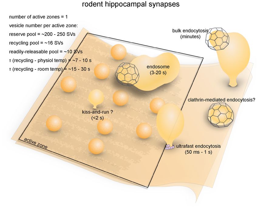
Gan and Watanabe                                                                                                    Synaptic Vesicle Endocytosis

via clathrin-independent mechanisms. Synaptic vesicles are              FROG NEUROMUSCULAR JUNCTIONS
then regenerated from the internalized membrane or synaptic
endosomes. The most notable features that distinguish these             Anatomical and Functional Overview
three pathways are the sizes of the endocytic membrane,                 The neuromuscular junctions of the sartorius or cutaneous
stimulation conditions, and kinetics. Ultrafast endocytosis             pectoris muscles of frogs (Rana pipiens) were the model systems
generates somewhat uniformly-sized vesicles (diameter of                used to study synaptic vesicle dynamics. The anatomical and
60–80 nm) within 30−1000 ms of action potentials and occurs             functional features of these neuromuscular junctions have been
with mild stimulation at physiological temperature (Watanabe            well characterized since initial work by Katz and colleagues
et al., 2013a,b). Like ultrafast endocytosis, fast compensatory         in the 1950s (Fatt and Katz, 1951, 1952; del Castillo and
endocytosis is triggered by mild stimulation but is relatively          Katz, 1954, 1956). Sartorius and cutaneous pectoris motor
slower (τ = 1–2 s; von Gersdorff and Matthews, 1994a;                   neurons are anatomically similar (Grinnell and Herrera, 1980).
Renden and von Gersdorff, 2007; Soykan et al., 2017). This              Cholinergic motor neuron axons extend elongated unmyelinated
endocytic pathway is also temperature-sensitive (Renden and             branch terminals (total length 100–300 µm; Pawson et al.,
von Gersdorff, 2007) and generates endocytic vesicles several           1998) running parallel to the muscle fiber (Birks et al.,
times larger than synaptic vesicles (Paillart et al., 2003). Both       1960). Fusion sites, or active zones (Couteaux and Pécot-
ultrafast endocytosis and fast compensatory endocytosis retrieve        Dechavassine, 1970), are organized in periodic 1.0 µm-long
all exocytosed membrane in a short period, and thus both                ‘‘stripes’’ running transverse to the terminal, with roughly
mechanisms are compensatory in nature. In contrast, activity-           0.8–1.1 µm spacing in between (Propst et al., 1986; Pawson
dependent bulk endocytosis is induced at both physiological             et al., 1998). The total number of active zones per neuromuscular
and non-physiological temperatures by high-frequency (typically         junction ranges between 50–250 (110 on average). Along the
non-physiological) stimulation or application of high potassium         central axis of each active zone, the plasma membrane curves
(Clayton and Cousin, 2009). This endocytic pathway occurs on a          out to form a 75–90 nm-wide central ‘‘ridge’’ (Figure 1;
slower time scale (8–20 s; Wu and Wu, 2007) and internalizes            Heuser et al., 1974; Stoschek et al., 2001). Flanking the ridge
large pieces of membrane of random size commensurate to                 on both sides are 30–40 vesicles in physical contact (or
the amount of fusion. Unlike the other four mechanisms,                 ‘‘docked’’) with the plasma membrane (Couteaux and Pécot-
the kiss-and-run endocytosis does not involve full collapse             Dechavassine, 1970; Heuser et al., 1979; Pawson et al., 1998).
fusion—neurotransmitter is instead thought to be released from          Other vesicles in the terminals also tend to cluster above
a transient fusion pore (He and Wu, 2007). Upon release,                the active zone (Heuser et al., 1974). Active zones are often
the pore closes, and the very same vesicle is retrieved. This           directly apposed to 50–100 nm-wide invaginations on the
mechanism has been well-established in secretory cells such as          post-junctional membrane of the muscle, known as junction
chromaffin cells (Artalejo et al., 1998; Elhamdani et al., 2006).       folds (Heuser and Reese, 1973). Acetylcholine receptors are
Several studies have demonstrated the existence of kiss-and-run         concentrated at the crests of junction folds (Matthews-Bellinger
at conventional synapses, yet its occurrence under physiological        and Salpeter, 1978). This architecture allows close alignment of
conditions remains under extensive debate (He et al., 2006; He          postsynaptic acetylcholine receptors with presynaptic fusion sites
and Wu, 2007; Zhang et al., 2009; Wu et al., 2014). Nevertheless,       and facilitates efficient activation of the receptors (York and
these five mechanisms are thought to be the core recycling              Zheng, 2017).
pathways for synaptic vesicles.                                             The presynaptic bouton of frog neuromuscular junctions
    What factors determine the mode of endocytosis at a                 contains many vesicles, which are divided into functional pools
particular synapse? At any given presynaptic terminal, one form         (for definition of pools, see Table 1). There are roughly 200,000 to
of endocytosis might predominate. Alternatively, multiple forms         500,000 synaptic vesicles at each terminal (Heuser and Reese,
of endocytosis might cooperate, depending on the availability           1973). Of these, an estimated 4400 (0.8%–2%) are docked
of vesicles at the terminals and the activity level, to meet the        (assuming 40 docked vesicles per active zone and active zones
demand for synaptic vesicle recycling. Here, we review the              per junction, Pawson et al., 1998). These vesicles likely represent
literature concerning synaptic vesicle endocytosis in a number          the vesicles in the readily-releasable pool (RRP), since a brief
of well-characterized model systems: frog neuromuscular                 high-frequency stimulation train (30 Hz for 0.5 s triggers fusion
junctions, Caenorhabditis elegans (C. elegans) neuromuscular            of roughly the same number of vesicles (about 2 vesicles
junctions, Drosophila neuromuscular junctions, lamprey                  per active zone per action potential; Rizzoli and Betz, 2004).
reticulospinal giant axons, ribbon synapses of goldfish                 In addition, about 15% (30,000–75,000) of vesicles at each
retinal bipolar neurons, the calyx of Held, and rodent                  terminal constitute the recycling pool. This pool of vesicles
hippocampal synapses. For each synapse, we will first describe          sustains synaptic transmission indefinitely under low-frequency
its anatomical and functional features, then review what is             stimulation (2–5 Hz) but is depleted at a time constant of
known about endocytosis and its molecular requirements                  4 s during 30 Hz stimulation, which overwhelms the rate
in each system. Furthermore, we will discuss whether some               of synaptic vesicle recycling at this terminal (Richards et al.,
earlier observations may be explained by ultrafast endocytosis,         2003). The rest of the vesicles likely belong to the reserve
since this pathway was only discovered in 2013. We will                 pool, which is only used during intense, prolonged stimuli.
close with perspectives on the future development of this               The reserve pool is depleted at a time constant of 40 s
field.                                                                  during 30 Hz stimulation (Richards et al., 2003). Given the

Frontiers in Cellular Neuroscience | www.frontiersin.org            2                                         June 2018 | Volume 12 | Article 171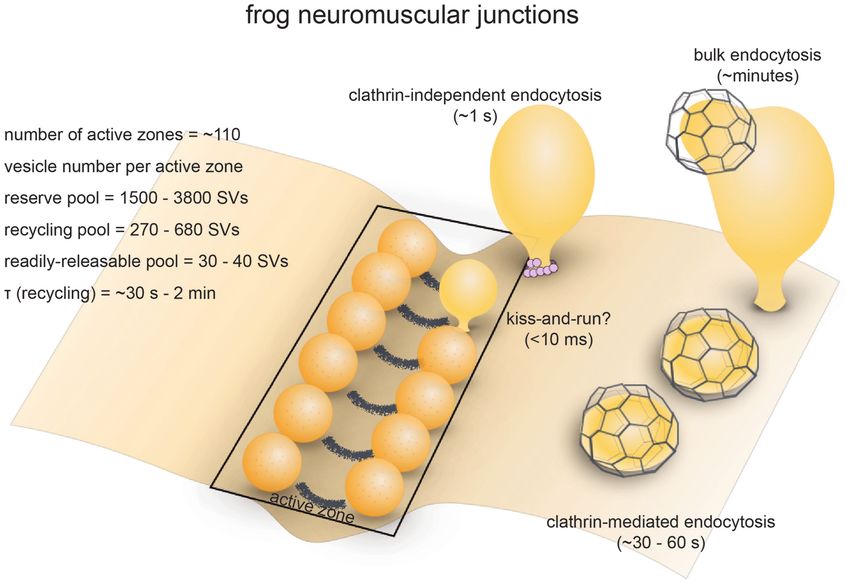
Gan and Watanabe                                                                                                                           Synaptic Vesicle Endocytosis

  FIGURE 1 | Vesicle pools and endocytic pathways at frog neuromuscular junctions. Frog neuromuscular junctions possess on average 110 active zones. At each
  active zone, 30–40 vesicles are docked on either side of a central “ridge.” These vesicles represent the readily-releasable pool (RRP). The number of recycling pool
  vesicles per active zone is 270–680, and the number of reserve pool vesicles per active zone is 1500–3800. During physiological stimulation, vesicle recycling takes
  place on a time scale of 30 s−2 min. Two endocytic pathways are found at frog neuromuscular junctions: a fast pathway that internalizes vesicles and cisternae
  within a minute, and a slow pathway that internalizes vesicles at ∼8 min during intense stimulation by high K+ . The fast pathway is mediated by clathrin-independent
  endocytosis (potentially ultrafast endocytosis or fast compensatory endocytosis) and clathrin-mediated endocytosis. The slow pathway represents
  activity-dependent bulk endocytosis. An alternative fast pathway kiss-and-run is also suggested. This pathway is predicted to occur within 10 ms. However, the
  existence of kiss-and-run at frog neuromuscular junctions is still controversial.

number of vesicles and the rate of fusion, the recycling pool                           potentials (EPPs) can be recorded (Betz and Bewick, 1993).
would be consumed completely within 50 s without endocytic                              Low-dose curare prevents muscle twitching during recordings
processes when these neurons are stimulated at 5 Hz. Thus, the                          by attenuating nicotinic acetylcholine receptor-mediated EPPs
time course of the entire vesicle recycling process cannot be                           without affecting presynaptic function (Auerbach and Betz,
longer than 50 s.                                                                       1971). Finally, the presynaptic vesicle pool can sustain several
                                                                                        minutes of high-frequency activity before it is depleted due
Strengths as a Model System                                                             to its sheer size (Ceccarelli et al., 1973). These features
Frog neuromuscular junctions were so widely used in early                               make it possible to monitor synaptic vesicle dynamics by a
studies of synaptic vesicle dynamics owing to their numerous                            combination of electrophysiology, optical imaging and electron
advantages as a model system. First, frog cutaneous pectoris                            microscopy.
or sartorius nerve-muscle preparations are easy to dissect, and
the activity at the neuromuscular junctions can be conveniently                         Endocytic Pathways
controlled and monitored in vitro (Heuser et al., 1974). Second,                        Optical imaging with lipophilic dyes and electrophysiological
active zones and docking sites are highly compartmentalized                             recordings suggest the existence of fast and slow pathways
and distinguishable from endocytic sites, and thus endocytic                            at frog neuromuscular junctions (Figure 1). Fei Mao (FM)
events can be identified easily using freeze-fracture and ultrathin-                    dyes, which fluoresce strongly when bound to membrane
section electron microscopy (Heuser and Reese, 1973; Heuser                             (Gaffield and Betz, 2007), can be taken up via activity-dependent
et al., 1974). This organization also results in a very distinct                        endocytosis (staining) and then released by subsequent stimulus-
and easily recognizable pattern upon FM dye staining (Betz                              induced exocytosis (de-staining). Since the mid-1990s, FM dyes
and Bewick, 1992, 1993). Third, innervated muscles treated with                         have been used to measure the kinetics of synaptic vesicle
curare can be impaled with a microelectrode so that endplate                            endocytosis and recycling at frog neuromuscular junctions

Frontiers in Cellular Neuroscience | www.frontiersin.org                            3                                               June 2018 | Volume 12 | Article 171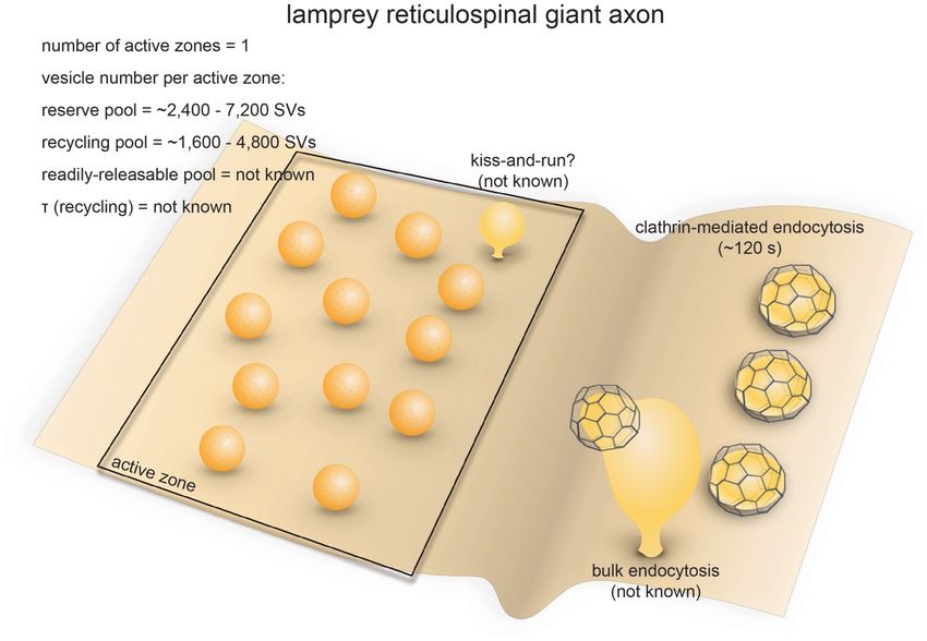
Gan and Watanabe                                                                                                                   Synaptic Vesicle Endocytosis

TABLE 1 | Definition and characteristics of major synaptic vesicle pools.

                                 Readily-releasable pool (RRP)              Recycling pool                              Reserve pool

Functional definition            Can be consumed by brief stimulation       Consumed only after the RRP has been        Released only during intense activity
                                 (
Gan and Watanabe                                                                                                     Synaptic Vesicle Endocytosis

Molecular Requirements                                                   Strengths as a Model System
Frogs were not commonly used for genetic studies until recently          Five features of C. elegans allow investigation of vesicle dynamics
(Wang et al., 2015). As a result, the roles of many of the               in molecular detail. First, the nervous system of C. elegans
well-known endocytic proteins (e.g., α-adaptin, endophilin,              is remarkably simple and its synaptic connectivity has been
Epsin etc.) have not been explored in frog neuromuscular                 determined by serial-section electron microscopy (White et al.,
junctions. What little data are available come from studies using        1986). Second, C. elegans colonies are easy to maintain, with a
chemical probes. For example, the actin polymerization blocker           rapid generation cycle of 3 days. In addition, extensive molecular
cytochalasin-D prevents the initiation of activity-dependent bulk        and genetic resources are available and widely shared in the
endocytosis (Nguyen et al., 2012). In contrast, the dynamin              community. Third, the C. elegans genome contains single copies
GTPase inhibitor Dyngo-4a does not affect the initiation of              of genes, whereas multiple copies are often found in vertebrates
bulk endocytosis, but traps endocytic intermediates on the cell          (Harris et al., 2001). This lack of genetic redundancy, combined
surface (Nguyen et al., 2012). Analogs of cyclic-GMP (cGMP)              with the surprising viability of the worm without a functional
accelerate the vesicle cycle at frog neuromuscular junctions,            nervous system (Avery and Horvitzt, 1989; Richmond et al.,
while inhibitors of guanylate cyclase have the opposite effect           2001), makes it possible to probe the molecular mechanisms
(Petrov et al., 2008). The accelerating effect of cGMP on the            underlying synaptic functions in intact adult animals. Fourth, its
vesicle cycle might be due to an enhancement of the fast                 small size and optical transparency makes C. elegans amenable
endocytic pathway, although direct evidence is lacking. A similar        to optical stimulation and high-pressure freezing experiments
cGMP-dependent regulation of the vesicle cycle mediated by               (flash-and-freeze). Finally, despite initial technical challenges,
cGKII (cyclic GMP Kinase II) has been found in cerebellar                protocols for performing whole-cell patch clamping of motor
granule cells (Collado-Alsina et al., 2014). In addition, treating       neurons and the muscles of C. elegans are now well-developed
motor neurons with methyl-β-cyclodextrin (MβCD) inhibits                 (Richmond and Jorgensen, 1999). Thus, functional studies can
the uptake of FM1–43 during and after prolonged stimulation,             be correlated with ultrastructural analysis in various genetic
suggesting that membrane cholesterol might play a role in                backgrounds.
synaptic vesicle endocytosis (Rodrigues et al., 2013). These
results, though limited, suggest that synaptic vesicle endocytosis
at frog neuromuscular junctions involve complex molecular                Endocytic Pathways
machineries and are impacted by a wide range of cellular                 Little is known about the kinetics of synaptic vesicle recycling
signaling pathways.                                                      at C. elegans neuromuscular junctions. Measuring the kinetics
                                                                         using optical methods in motor neurons has been extremely
                                                                         difficult due to autofluorescence from gut granules and refraction
CAENORHABDITIS ELEGANS                                                   of light by the thick cuticle. Based on pHluorin imaging of
NEUROMUSCULAR JUNCTIONS                                                  an odor-responsive sensory neuron, the τ for endocytosis is
                                                                         8 –18 s (Ventimiglia and Bargmann, 2017), which is similar to
Anatomical and Functional Overview                                       the time constant of endocytosis in mammalian central synapses
The soil nematode Caenorhabditis elegans (C. elegans) is                 (Granseth and Lagnado, 2008; Soykan et al., 2017). This time
an excellent model system for cell biological studies. Adult             constant is much faster than the appearance of clathrin-coated
animals are roughly 1 mm in length and are composed                      pits in frog neuromuscular junctions (Heuser and Reese, 1973;
of about 1000 somatic cells, of which exactly 302 are                    Miller and Heuser, 1984) or the kinetics of clathrin-mediated
neurons (Jorgensen and Nonet, 1995). The motor neurons                   endocytosis in mammalian cell lines (Kirchhausen et al., 2014).
of C. elegans form en passant neuromuscular junctions on                 Whether this τ is similar to the time constant in motor neurons
ventral and dorsal body wall muscles and innervate muscle                is questionable. Nonetheless, a relatively fast mechanism is
cells at multiple locations (Jorgensen and Nonet, 1995). A               expected in C. elegans neurons.
typical synapse contains a single large active zone with                     What are the fast mechanisms that drives synaptic vesicle
a proteinaceous electron-dense projection (dense projection;             endocytosis at C. elegans neuromuscular junctions? Like in other
Ackermann et al., 2015). The total number of vesicles at a               systems, the main pathway was originally thought to be clathrin-
single terminal is roughly 300, of which an average of 34                mediated endocytosis (Harris et al., 2001). Consistent with this
(11%) are docked (Hammarlund et al., 2007), and these vesicles           idea, clathrin adaptor proteins AP2 and AP180 were shown to
comprise the recycling pool (Figure 2; Watanabe et al., 2013a).          be required for synaptic vesicle recycling (Nonet et al., 1999).
Interestingly, instead of action potentials, graded potentials           In addition, stonin (UNC-41), which sorts synaptotagmin into
trigger neurotransmitter release at C. elegans neuromuscular             the AP2-depedent pathway, contributes to the regeneration of
junctions (Liu et al., 2009). During sporadic motor neuron               synaptic vesicle pools (Jorgensen et al., 1995; Mullen et al., 2012),
activity, the rate of fusion is ∼25 vesicles/s per muscle, which         suggesting that clathrin-mediated processes are likely essential.
is about 1–2 vesicles/s per active zone (Liu et al., 2009).              However, later studies using more advanced electron microscopy
During evoked activity, ∼120 vesicles per muscle (∼12 vesicles           techniques suggested that endocytic processes in C. elegans
per active zone) fuse in response to each stimulus (Figure 2;            neuromuscular junctions are clathrin-independent.
Watanabe et al., 2013a). These vesicles presumably represent                 Recent morphological studies suggest that clathrin-
the RRP.                                                                 independent ultrafast endocytosis is the primary endocytic

Frontiers in Cellular Neuroscience | www.frontiersin.org             5                                         June 2018 | Volume 12 | Article 171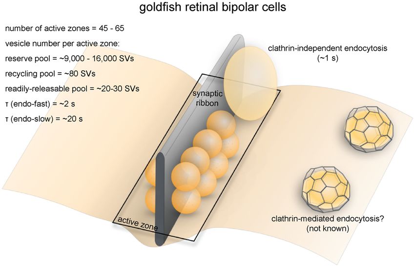
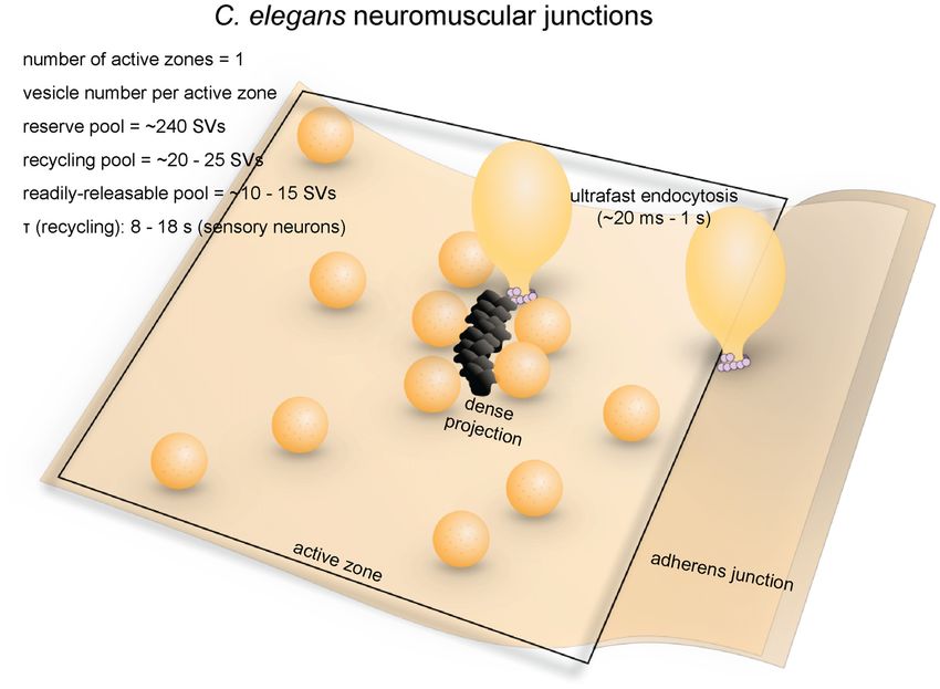
Gan and Watanabe                                                                                                                             Synaptic Vesicle Endocytosis

  FIGURE 2 | Vesicle pools and endocytic pathways at C. elegans neuromuscular junctions. C. elegans neuromuscular junctions usually possess a single active zone
  with a dense projection. An average of 35 vesicles are docked, of which 10–15 can fuse in response to a single stimulus, and therefore represents the RRP. The rest
  of the docked vesicles (15–20) represent the recycling pool. Roughly 240 vesicles are in the reserve pool. Following single stimuli, ultrafast endocytosis occurs on a
  time scale of 20 ms–1 s both at the dense projection or at adherens junctions. Bulk endocytosis might also occur at this synapse following hyperstimulation (not
  shown in figure).

pathway in C. elegans (Figure 2). Experiments combining                                  Molecular Requirements
optogenetics with high-pressure freezing (flash-and-freeze) of                           Dynamin is essential for synaptic vesicle recycling at C. elegans
whole animals showed that hyperstimulation (30 s continuous                              neuromuscular junctions. A temperature-sensitive mutant
stimulation) of cholinergic neurons resulted in accumulation                             similar to the Drosophila shibirets mutant, dyn-1(ky51), displays
of endosome-like structures at synapses ∼6 s after stimulation,                          severe locomotion defects (Clark et al., 1997). Following
presumably via activity-dependent bulk endocytosis. The                                  optogenetic hyperstimulation, the neuromuscular junction
diameters of these structures range from 50 nm to 200 nm                                 boutons of dyn-1 mutants contain large membrane involutions
(100 nm on average). No pits were trapped on the surface                                 that are continuous with the plasma membrane, suggesting that
even in synaptojanin and endophilin mutants, suggesting                                  dynamin is involved in fission of bulk endocytic intermediates
that endocytosis can complete without these clathrin-associated                          (Kittelmann et al., 2013). Occasionally, synaptic vesicle-sized
proteins (Kittelmann et al., 2013). In the same year, with the finer                     buds formed on these endocytic intermediates. However,
temporal resolution of the flash-and-freeze approach, another                            the budding of synaptic vesicles from these large vesicles
group observed formation of smaller non-coated endocytic                                 is also blocked in the mutant (Kittelmann et al., 2013).
vesicles (40 nm on average) adjacent to dense projections within                         Similarly, dyn-1(ky51) displays large vesicles linked to the
50 ms or by adherens junctions within 1 s following a single                             plasma membrane by narrow necks close to the dense
stimulus (Watanabe et al., 2013a). Because of its kinetics, this                         projection 1 s after a single stimulation pulse, indicating
endocytic pathway was named ultrafast endocytosis. Coated pits                           a defect in ultrafast endocytosis (Watanabe et al., 2013a).
and vesicles were not observed in either study, suggesting that                          These results highlight the essential role of dynamin in
clathrin itself may not be essential in synaptic vesicle recycling                       clathrin-independent endocytosis at C. elegans neuromuscular
at these terminals. It is likely that synaptic vesicle endocytosis                       junctions.
at C. elegans neuromuscular junctions is primarily mediated by                              Clathrin-associated proteins are essential in synaptic vesicle
ultrafast endocytosis.                                                                   recycling at C. elegans neuromuscular junctions. However, it

Frontiers in Cellular Neuroscience | www.frontiersin.org                             6                                                June 2018 | Volume 12 | Article 171
Gan and Watanabe                                                                                                       Synaptic Vesicle Endocytosis

is unclear whether they function on the plasma membrane                     the residual amount of clathrin might be sufficient to
or at endosomes. Complete loss of the AP2 complex subunits                  maintain synaptic vesicle recycling. Experiments using a
leads to a significant reduction in the number of synaptic                  conditional null background will be needed to resolve this
vesicles (∼70% reduction) and an accumulation of endosomes                  uncertainty.
(Gu et al., 2008; Mullen et al., 2012). Similarly, AP180 is not
essential for endocytosis, but regulates the size and protein               DROSOPHILA NEUROMUSCULAR
compositions of synaptic vesicles (Nonet et al., 1999). These
results are consistent with the notion that clathrin-mediated
                                                                            JUNCTIONS
endocytosis is not required for membrane retrieval from the cell
surface but for the resolution of endosomes. However, synaptic              Anatomical and Functional Overview
vesicle markers diffusively localize on the plasma membrane                 The neuromuscular junction of fruit flies (Drosophila) is an
in these mutants (Nonet et al., 1999; Mullen et al., 2012),                 excellent model for studying the synaptic development and
suggesting that adaptor proteins may also function at the plasma            function (Keshishian et al., 1996). Like excitatory synapses in the
membrane to sort vesicular proteins. Likewise, homologs of                  mammalian central nervous system, Drosophila neuromuscular
the clathrin-associated proteins Esp15 (EHS-1) and intersectin-1            junctions use L-glutamate as the neurotransmitter (Jan and
(ITSN-1) contribute to synaptic vesicle regeneration (Salcini               Jan, 1976). They have large presynaptic boutons (type Is
et al., 2001; Wang et al., 2008). Paradoxically, loss of EHS-1              boutons, ∼1–3 µm in diameter; type Ib boutons, ∼2–5 µm
leads to complete depletion of synaptic vesicles without apparent           in diameter (Atwood et al., 1993), with each neuromuscular
formation of endosomes, suggesting its function at the plasma               junction containing roughly ∼180 boutons (Schuster et al.,
membrane, while loss of ITSN-1 results in accumulation of large             1996), and each bouton containing 7–41 active zones (Atwood
vesicles in the terminals (Salcini et al., 2001). The amphiphysin           et al., 1993). The center of the active zone is marked
and syndapin homologs AMPH-1 and SDPN-1 act on early                        with a dense projection known as the ‘‘T-bar’’ (due to its
endosomes to regulate cargo recycling (Pant et al., 2009; Gleason           apparent shape in electron micrographs from chemically-fixed
et al., 2016). They are therefore not likely to be directly involved        specimens; Figure 3). The area around the T-bar is enriched
in synaptic vesicle endocytosis from the plasma membrane,                   in synaptic vesicles. On average, the total number of synaptic
although their exact roles are yet to be determined. Thus,                  vesicles at a single bouton is around 460 (assuming 83,000
although these proteins all interact with clathrin, their functional        vesicles/neuromuscular junction, Delgado et al., 2000) in muscles
domains do not necessarily overlap. This suggests that either               6/7 synapse and 180 boutons per neuromuscular junction in
clathrin-mediated processes occur both at the plasma membrane               muscles 6/7 (Schuster et al., 1996). Of these, roughly 0.7% are
and endosomes or these proteins have clathrin-independent                   in the RRP (Müller et al., 2012), ∼14% are in the recycling pool
functions. Further investigation is required to resolve these               (Delgado et al., 2000) and the rest constitute the reserve pool
apparent contradictions.                                                    (Delgado et al., 2000). FM dye staining/de-staining experiments
    Similarly, mutations in endophilin (unc-57) and synaptojanin            suggest that the recycling pool is located in the periphery of
(unc-26) block synaptic recycling in C. elegans (Harris et al., 2000;       the bouton while the reserve pool resides primarily in the
Schuske et al., 2003). Initial experiments using conventional               core region (Ramaswami et al., 1994; Kuromi and Kidokoro,
chemical fixation at 4◦ C showed accumulation of both coated                1998). The maximal rate of synaptic vesicle recycling at a
and non-coated pits on the membrane (Schuske et al., 2003).                 typical neuromuscular junction has been estimated to be around
However, later studies employing high-pressure freezing at                  1000/s (3–5/s per active zone; Delgado et al., 2000), implicating
room temperature reported that endosomes accumulate in the                  that the recycling pool could be completely turned over in
terminals (Kittelmann et al., 2013). This temperature effect is             roughly 10 s.
consistent with the observation in mouse hippocampal synapses
that ultrafast endocytosis fails at lower temperatures (Watanabe            Strengths as a Model System
et al., 2014). Thus, clathrin-mediated endocytosis may be able              Drosophila neuromuscular junctions are conducive to
to compensate for the loss of ultrafast endocytosis in C. elegans           optical, electrophysiological and genetic approaches. Optical
neuromuscular junctions.                                                    imaging using pHluorin and FM dyes can be performed
    While clathrin-associated proteins are essential in C. elegans          on Drosophila larva, allowing functional measurement
neuromuscular junctions, clathrin itself may not be involved                of endocytic processes. Post-synaptic responses can be
in synaptic vesicle recycling. In mutants with a temperature-               measured with relative ease by performing two-electrode
sensitive allele of clathrin heavy chain (CHC), shifting to                 voltage clamp on innervated muscles. Most of the synaptic
the non-permissive temperature removes almost all CHC                       proteins implicated in mammalian synaptic transmission
but does not significantly reduce the number of synaptic                    are well-conserved in Drosophila and are expressed at the
vesicles at steady state or the amplitude of postsynaptic                   neuromuscular junction. Methods for genetic manipulations
miniature currents (Sato et al., 2009). However, the                        are well-developed, including an extensive toolkit of GAL4/UAS
diameter of synaptic vesicles is smaller, suggesting that                   targeted expression lines. These advantages have made the
clathrin is likely needed to maintain the size, but not the                 Drosophila neuromuscular junction one of the most popular
number, of synaptic vesicles. Nevertheless, the hypomorphic                 model systems for studying molecular mechanisms of synaptic
nature of this temperature-sensitive mutant means that                      vesicle endocytosis.

Frontiers in Cellular Neuroscience | www.frontiersin.org                7                                        June 2018 | Volume 12 | Article 171
Gan and Watanabe Synaptic Vesicle Endocytosis
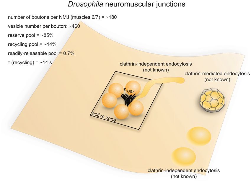
FIGURE 3 | Vesicle pools and endocytic pathways at Drosophila neuromuscular junctions. Drosophila neuromuscular junctions possess 200–300 active zones. The
center of each active zone is marked by a dense projection commonly referred to as a “T-bar”, around which synaptic vesicles tend to cluster. In each bouton, 0.7%
of the vesicles on average are readily releasable; ∼14% of the vesicles are in the recycling pool; and roughly ∼85% are in the reserve pool. Synaptic vesicle
endocytosis occurs with a time constant of 14 s as measured by pHluorin. Both clathrin-mediated and clathrin-independent endocytosis occurs at Drosophila
neuromuscular junctions. One particular form of clathrin-independent endocytosis might occur adjacent to the T-bar. The exact time courses of these endocytic
pathways have not been determined.
Endocytic Pathways was noted to be low (data not quantified; Kosaka and Ikeda,
The time constant for synaptic vesicle recycling has been 1983). Nonetheless, these results suggest the involvement of
determined by optical imaging in Drosophila neuromuscular both clathrin-dependent and clathrin-independent mechanisms
junctions. In wild-type flies, pHluorin recovery following 50 Hz in synaptic vesicle recycling at these synapses (Figure 3). Another
stimulation for 10 s has a time constant of ∼14 s (Poskanzer study in the shits mutant revealed the possible existence of
et al., 2006). Similarly, FM dye de-staining experiments suggested two separate endocytic pathways at retinula cell terminals:
a similar time constant for the synaptic vesicle cycle (Delgado a faster pathway occurring directly at the dense projection,
et al., 2000). Thus, like in C. elegans neurons, a relatively fast and a slower pathway occurring away from the active zone
mechanism is expected at Drosophila neuromuscular junctions. (Koenig and Ikeda, 1996; Kidokoro, 2006). A similar distinction
The mechanisms underlying synaptic vesicle recycling at supposedly exists at neuromuscular junction terminals, given
Drosophila neuromuscular junctions have been extensively that endocytic structures are trapped at dense projections as well
studied since the discovery of the shibirets (shits1 ) mutation as peri-active zone membrane (Kosaka and Ikeda, 1983). It is
(Poodry et al., 1973). Shibirets inhibits the function of possible that the earlier pathway at the active zone represents
dynamin in a temperature-dependent manner (Chen et al., ultrafast endocytosis or fast compensatory endocytosis, while
1991; van der Bliek and Meyerowrtz, 1991), thereby stalling the later pathways represent clathrin-mediated endocytosis and
endocytosis and allowing endocytic intermediates to be captured activity-dependent bulk endocytosis (Figure 3). However, due to
by electron microscopy (Kosaka and Ikeda, 1983). In shits the limited temporal resolution associated with chemical fixation,
flies, synaptic vesicles in the bouton are depleted under the exact time course of these pathways could not be determined.
non-permissive temperatures (30◦ C, 5−10 min), with both Nevertheless, the shits mutant shows synaptic fatigue within
coated and uncoated endocytic intermediates stalled throughout 20 ms of repetitive stimulation at non-permissive temperature
the plasma membrane, although the number of coated pits while wild type flies maintain their synaptic transmission
Frontiers in Cellular Neuroscience | www.frontiersin.org 8 June 2018 | Volume 12 | Article 171Gan and Watanabe Synaptic Vesicle Endocytosis
(Kawasaki et al., 2000). This effect is not due to the availability LRRK2, which regulates the phosphorylation cycle of endophilin
of release-ready vesicles, implying that a fast dynamin-mediated A, is also required for normal synaptic vesicle endocytosis
process is required to clear fusion sites for incoming vesicles. (Matta et al., 2012). The polyphosphoinositide phosphatase
Since no evidence has been found to support the existence of synaptojanin-1 is recruited to endocytosed vesicles at Drosophila
kiss-and-run at these synapses (Dickman et al., 2005), such a neuromuscular junctions and mediates the uncoating of
fast process probably represents ultrafast endocytosis. Future those vesicles (Verstreken et al., 2003). Interestingly, the
studies using rapid freezing techniques might help resolve this N-BAR domain protein amphiphysin and the F-BAR domain
issue. protein syndapin, both of which participate in clathrin-
mediated endocytosis (Takei et al., 1999; Qualmann and
Molecular Requirements Kelly, 2000), are not strictly required for synaptic vesicle
The genetic tractability of Drosophila makes it an ideal model endocytosis at Drosophila neuromuscular junctions (Razzaq
organism for investigating the molecular requirements of vesicle et al., 2001; Kumar et al., 2009). Thus, these membrane-
recycling. As mentioned above, dynamin is required for vesicle modifying proteins seem to play differential roles in synaptic
recycling at Drosophila neuromuscular junctions (Kosaka and vesicle recycling.
Ikeda, 1983). Clathrin and α-adaptin (a component of the
AP2 complex) are also both required for vesicle recycling LAMPREY RETICULOSPINAL SYNAPSES
(González-Gaitán and Jäckle, 1997; Heerssen et al., 2008),
although it is unclear whether they are directly needed for Anatomical and Functional Overview
endocytosis from the plasma membrane. Photoinactivation of The nervous system of lampreys, a class of jawless fish belonging
clathrin leads to a failure of synaptic vesicle recycling at these to the order Petromyzontiformes, represents the most primitive
terminals, but FM dye can be loaded into the terminals by form among vertebrates (Xu et al., 2016). The giant axons of
activity (Heerssen et al., 2008). Similarly, mutation of the reticulospinal neurons (also known as Müller cells) in adult
stoned proteins (Stoned A and Stoned B), which are hubs lampreys (usually Lampetra fluviatilis or Petromyzon marinus)
for Synaptotagmin-1 (Syt I) and AP2, leads to accumulation can reach up to 80 µm in diameter. These axons are specialized
of fully internalized large endocytic vesicles and a reduced in transmitting phasic signals by firing brief high-frequency
density of synaptic vesicles (Fergestad et al., 1999). Similarly, spike trains (usually lasting less than 2 s; Brodin et al., 1997,
point mutations in the poly-lysine motif of Syt I, which binds 1999), with maximal firing frequencies in the range of 20–30 Hz
the AP2 complex, results in an accumulation of large vesicles (average 4.4 ± 4.9 Hz; Zelenin, 2011). They form en passant
without affecting the rate of endocytosis (Poskanzer et al., 2006). chemical (glutamatergic) and electrical synapses on the dendrites
Likewise, null mutation of syt I and rapid photoinactivation of spinal neurons in the lateral column (Vesselkin et al.,
of Syt I both lead to defect in endocytosis (Poskanzer et al., 1995; Vinay et al., 1998). Most of the synapses have a single
2003), suggesting that Syt I, a calcium sensor for exocytosis, oval-shaped active zone (simple synapses) while a fraction of
may also play a role in endocytosis (Poskanzer et al., 2003). them have 2–3 active zones (complex synapses). For simple
This endocytic function of Syt I seems to depend on its ability synapses, the diameter of the active zone ranges from 0.8 µm
to bind calcium, since point mutations in the C2B, but not to 1.8 µm (1.2 µm on average; Gustafsson et al., 2002). The
C2A, domain of Syt1 result in slowed endocytic rate (Poskanzer number of synaptic vesicles per synapse ranges from 4000 to
et al., 2006). These results suggest that clathrin and associated 12,000 depending on active zone area (Figure 4; Gustafsson
proteins are needed for regeneration of vesicles from endosomes, et al., 2002). Vesicles can be divided into two separate pools:
but not for endocytosis at the plasma membrane, and that a synapsin-independent proximal pool immediately adjacent
calcium may control the rate of endocytosis at the plasma to the active zone membrane and a synapsin-dependent distal
membrane. pool residing >200 nm from the plasma membrane (Pieribone
The clathrin-associated proteins Eps15 (an EH domain et al., 1995). The synapsin-dependent pool contains ∼60%
adaptor-like protein) and intersectin-1/Dap60 (Koh et al., 2004; of all vesicles within 500 nm of the active zone and is
Majumdar et al., 2006) are also required for synaptic vesicle thought to represent the reserve pool, while the synapsin-
recycling. Mutations in Eps15 lead to defects in FM dye uptake independent pool represents vesicles in the readily-releasable
and an accumulation of membrane invaginations, suggesting and recycling pools (Pieribone et al., 1995; Vesselkin et al.,
that it functions at the plasma membrane (Majumdar et al., 1995).
2006). This phenotype is rescued by an Eps15 construct
lacking its α-adaptin-interacting domain (Koh et al., 2007), Strengths as a Model System
suggesting that Eps15 might be involved in clathrin-independent Vesicle dynamics have been studied extensively in reticulospinal
endocytosis. giant synapses in lampreys mainly for the following four
Several membrane-remodeling proteins are required for reasons (Rovainen, 1974). First, the remarkable size of lamprey
synaptic vesicle recycling at Drosophila neuromuscular junctions. reticulospinal axons permits microinjection of antibodies and
Mutations in the BAR domain protein endophilin A, which active peptides into the cytoplasm to sequester and disrupt
senses and modifies membrane curvature, lead to synaptic specific molecular targets (Gustafsson et al., 2002; Brodin
vesicle depletion and an accumulation of early endocytic and Shupliakov, 2006). This approach compensates for the
intermediates (Guichet et al., 2002; Rikhy et al., 2002). lack of genetic manipulation tools in lampreys. Second, the
Frontiers in Cellular Neuroscience | www.frontiersin.org 9 June 2018 | Volume 12 | Article 171Gan and Watanabe Synaptic Vesicle Endocytosis
FIGURE 4 | Vesicle pools and endocytic pathways at lamprey reticulospinal giant synapses. Lamprey reticulospinal giant synapses usually possess only one active
zone. The exact number of vesicles in the RRP is unknown. Vesicles at this synapse can be divided into a synapsin-independent (recycling) pool of
1600–4800 vesicles and a synapsin-dependent (reserve) pool of 2400–7200 vesicles. The exact time course of vesicle recycling has not been measured.
Clathrin-mediated endocytosis occur 40–120 s following prolonged stimulation. Activity-dependent bulk endocytosis also occur. Kiss-and-run has been suggested,
but direct morphological evidence is lacking.
active zones of reticulospinal synapses are well-separated from blocked by placing the spinal cord in a Ca2+ -free solution
each other, making it possible to study vesicle dynamics immediately after stimulation (20 Hz, 20 min) but resumes
at individual active zones (Gustafsson et al., 2002). Third, upon re-exposure to Ca2+ (Gad et al., 1998). Based on the
by microinjecting Ca2+ -sensitive dyes into the axons, Ca2+ increase in the number of coated pits over time after Ca2+
influx at individual active zones can be monitored over re-exposure, the rate of vesicle recycling through clathrin-
time (Brodin and Shupliakov, 2006). Fourth, action potentials mediated endocytosis is around 0.4 vesicles/s per synapse
in reticulospinal axons as well as postsynaptic currents in (Gad et al., 1998). Most of the coated pits are shallow at
the lateral column cells they innervate can be conveniently the 40 s time point following Ca2+ re-exposure, while they
recorded using intracellular or extracellular electrodes (Brodin are deeply invaginated by the 120 s time point. The kinetics
et al., 1994). The reticulospinal giant synapses are thus a of coated pit maturation is similar to that of the receptor-
convenient model for studying the synaptic vesicle cycle in mediated endocytosis (Kirchhausen et al., 2014), suggesting
vertebrates. that lamprey reticulospinal synapses employ slow clathrin-
mediated endocytosis as the primary route for synaptic vesicle
Endocytic Pathways endocytosis.
Synaptic vesicle recycling at lamprey reticulospinal synapses is In addition to coated pits, plasma membrane invaginations
mainly governed by clathrin-mediated endocytosis (Figure 4). and endosome-like vacuoles connected to the plasma membrane
Time constants for endocytosis at these synapses have not also emerge following prolonged stimulation. Clathrin-coated
been measured using optical imaging approaches. Instead, pits occasionally form at the tip of these deep invaginations
time-resolved electron microscopy has been employed to (Figure 4; Gad et al., 1998). These observations are consistent
monitor the endocytic events. Prolonged high-frequency with the presence of activity-dependent bulk endocytosis. Thus,
stimulation (20–30 Hz, 15–30 min) causes clathrin-coated in addition to clathrin-mediated endocytosis, synaptic vesicle
pits to appear on the plasma membrane (Kershaw and recycling at lamprey reticulospinal synapses involves activity-
Christensen, 1980). The formation of these coated pits is dependent bulk endocytosis.
Frontiers in Cellular Neuroscience | www.frontiersin.org 10 June 2018 | Volume 12 | Article 171Gan and Watanabe Synaptic Vesicle Endocytosis
In contrast, little work in lamprey has explored fast is organized in a donut-shape at lamprey reticulospinal synapses,
endocytosis. The large reserve vesicle pool per active zone, presumably surrounding the active zone (Shupliakov et al.,
combined with the phasic activity pattern, likely results in 2002). Stimulation increases F-actin signal in the adjacent
a low demand on vesicle recycling kinetics at reticulospinal endocytic zone (Shupliakov et al., 2002). Acutely disrupting actin
synapses (Brodin et al., 1999). Based on the modulation polymerization leads to an accumulation of coated pits with a
of quantal size by serotonin, a role has been proposed wide-open neck at the plasma membrane, whereas blocking actin
for kiss-and-run (Photowala et al., 2006), although direct depolymerization causes recycled vesicles to be trapped between
morphological evidence is still lacking. No ultrastructural the endocytic site and the active zone vesicle cluster (Shupliakov
evidence has been found for ultrafast endocytosis. However, et al., 2002). These results indicate that actin dynamics are
this could be due to the limited temporal resolution of involved at multiple stages of the vesicle recycling process at
existing studies or the stimulation paradigms used. Almost reticulospinal synapses.
all electron microscopy studies are performed following In addition to clathrin-mediated endocytosis, activity-
prolonged high-frequency stimulation, lasting 15–30 min, dependent bulk endocytosis operates under high frequency
whereas intrinsic activity pattern is phasic (0.3–34 Hz, lasting stimulation; this process is, in part, mediated by syndapin.
seconds; Zelenin, 2011). How endocytosis takes place under a Microinjecting an antibody against the SH3 domain of lamprey
more physiological activity pattern has not been explored in this syndapin leads to no detectable defect in vesicle recycling
system. following low-frequency stimulation (Andersson et al., 2008).
However, under intense stimulation, disrupting syndapin
Molecular Requirements function leads to a decrease in vesicle number and an
Since genetic approaches are not possible in lamprey, molecular accumulation of large VAMP-containing cisternae, some of
mechanisms are probed by microinjection of antibodies which are connected to the plasma membrane (Andersson et al.,
and small peptides against candidate proteins implicated in 2008). These results suggest that syndapin is not strictly required
clathrin-mediated endocytosis (Brodin and Shupliakov, 2006). for clathrin-mediated endocytosis, but may help regenerate
These antibodies and peptides specifically disrupt protein- synaptic vesicles from membrane intermediates during activity-
protein interactions. Following application, clathrin-mediated dependent bulk endocytosis. In addition, adding excessive
endocytosis is perturbed at various stages, indicating the amounts of synuclein, a protein implicated in Parkinson’s
step at which each protein acts. For example, disrupting the disease that regulates synaptic vesicle endocytosis at mammalian
intersectin-AP2 interaction leads to an accumulation of shallow synapses (Lautenschläger et al., 2017), depletes synaptic vesicles
coated pits, implying a block at the early stage of clathrin- and causes cisternae and clathrin-coated pits to accumulate
mediated endocytosis (Pechstein et al., 2010). Microinjecting under intense stimulation (20 Hz, 5 min; Busch et al., 2014;
Epsin1 antibodies yields a similar phenotype (Jakobsson Medeiros et al., 2017). These results indicate that synuclein
et al., 2008), suggesting that Epsin1 is also an early factor. plays a regulatory role in clathrin-mediated endocytosis and
Similarly, disrupting the CLAP domain-mediated interaction activity-dependent bulk endocytosis at lamprey reticulospinal
between amphiphysin and clathrin depletes the vesicle pool synapses.
and causes accumulation of coated pits that are abnormally-
shaped (Evergren et al., 2004), suggesting that amphiphysin
plays an essential role in proper maturation of clathrin-coated RIBBON SYNAPSES OF RETINAL BIPOLAR
pits. Furthermore, disrupting the SH3 domain on amphiphysin NEURONS
leads to a depletion of synaptic vesicles and an accumulation
of clathrin-coated pits with a narrow neck, suggesting that the Anatomical and Functional Overview
SH3 domain of amphiphysin is needed to recruit dynamin Ribbon synapses are found in vertebrate sensory systems
(Shupliakov et al., 1997). Likewise, disrupting the function of that use graded potentials instead of action potentials for
the SH3 domain of endophilin causes accumulation of clathrin- neurotransmission (Sterling and Matthews, 2005). They are
coated intermediates with a narrow neck as well as coated vesicle characterized by a striking ultrastructural feature: the synaptic
that are fully internalized (Ringstad et al., 1999; Gad et al., ribbon, which is typically a 30 nm-thick plate jutting ∼200 nm
2000). The accumulation of clathrin-coated pits likely results from each active zone into the cytoplasm (Figure 5). Prominent
from the endophilin failing to recruit dynamin, which may be examples of ribbon synapses include those of photoreceptors,
compensated by other proteins like amphiphysin (Sundborger retinal bipolar neurons, auditory or vestibular hair cells, and
et al., 2011). On the other hand, accumulation of coated vesicles electrosensory receptors (Sterling and Matthews, 2005). These
is likely due to a loss of endophilin-synaptojanin interaction, synapses are anatomically and functionally diverse, although
since synaptojanin is essential for uncoating (Gad et al., 2000). they invariably use L-glutamate as the primary neurotransmitter
Thus, proteins associated with clathrin-mediated endocytosis (Sterling and Matthews, 2005). For the sake of simplicity, here
form a complex network of interactions and cooperatively we will focus on the ribbon synapses of goldfish retinal bipolar
promote the formation and maturation of clathrin-coated cells.
vesicles. Retinal bipolar cells relay information from photoreceptors
The actin cytoskeleton also contributes to clathrin-mediated to ganglion cells and amacrine cells. Goldfish Mb1-type bipolar
synaptic vesicle recycling in lamprey. Filamentous actin (F-actin) cells have large bulbous axonal terminals (8–12 µm in diameter;
Frontiers in Cellular Neuroscience | www.frontiersin.org 11 June 2018 | Volume 12 | Article 171Gan and Watanabe Synaptic Vesicle Endocytosis FIGURE 5 | Vesicle pools and endocytic pathways at goldfish bipolar neuron terminals. The axonal terminals of goldfish bipolar neurons possess 45–65 active zones. At each active zone, there is a large electron-dense body known as the synaptic ribbon. Twenty to 30 vesicles are docked at the base of the synaptic ribbon and are readily releasable. Another 80 or so are tethered to the ribbon; these vesicles belong to the recycling pool. Nine-thousand to 16,000 vesicles per active zone are in the reserve pool. Two endocytic pathways exist at these terminals: a fast pathway (τfast ≈ 2 s) following weak stimulation and a slow pathway (τslow ≈ 20 s with delayed initiation) following repeated strong stimulation. The fast pathway is likely operated by clathrin-independent endocytosis (potentially ultrafast endocytosis or fast compensatory endocytosis), while the slow pathway could represent either clathrin-mediated endocytosis or activity-dependent bulk endocytosis. Lagnado et al., 1996; von Gersdorff and Matthews, 1999), which depletion (Lagnado et al., 1996; Rouze and Schwartz, 1998). This makes them a very popular system for studying the synaptic continuous vesicle cycle entails balanced exo- and endocytosis vesicle dynamics of ribbon synapses (Figure 5). Each terminal at a maximal rate of 900 vesicles/s (Rouze and Schwartz, contains 45–65 ribbons (von Gersdorff et al., 1996). The total 1998; or 14–20/s per active zone), equivalent to the turnover number of vesicles per terminal is estimated to range from of the entire surface area of the terminal every 2 min. ∼500,000 to 900,000 depending on the size of the terminal Another study found that with sustained depolarization using (von Gersdorff et al., 1996). Among these vesicles, 1000–1800 50 mM K+ at 2.5 mM Ca2+ , the continuous recycling rate (0.15%–0.3%) belong to the RRP while 4500 (0.5%–0.9%) can be as high as 3800/s (Lagnado et al., 1996; or 69/s belong to the recycling pool, adding up to about 5500 vesicles per active zone). In contrast, conventional synapses between that are available for depolarization-evoked fusion (Neves and amacrine cells have a maximal fusion rate of roughly 20/s per Lagnado, 1999; von Gersdorff and Matthews, 1999). This number active zone (Stevens and Tsujimoto, 1995). Thus, functional corresponds well with the ultrastructural observation that at each properties of synaptic transmission are well characterized, terminal roughly 6000 vesicles on average are tethered to ribbons, making these neurons ideal model systems for studying synaptic and of these about 1200 are docked (22 per active zone; von vesicle recycling in synapses that engage in high-rate graded Gersdorff et al., 1996). neurotransmission. Capacitance measurements and FM dye imaging indicate that vesicle fusion at ribbon synapses occurs at exceptionally high Strengths as a Model System rates (von Gersdorff et al., 1996). In response to depolarizing Axonal terminals of goldfish bipolar neurons possess unique pulses, vesicles of the RRP are depleted within 20 ms (Neves advantages for studying synaptic vesicle dynamics. Bipolar and Lagnado, 1999), giving a fusion rate of 900–1600/s per neurons can be easily extracted from dissociated retinal tissue. active zone. The recycling pool is depleted within 200 ms Due to their large size, high fusion rate and simple morphology, (von Gersdorff and Matthews, 1999), giving a fusion rate of bipolar neuron terminals are well-suited for direct measurements 410/s per active zone. Vesicles can also fuse continuously at of membrane capacitance changes (Mennerick et al., 1997). low intracellular calcium concentrations without any apparent Whole-cell patch clamp recording can be performed either on Frontiers in Cellular Neuroscience | www.frontiersin.org 12 June 2018 | Volume 12 | Article 171
Gan and Watanabe                                                                                                     Synaptic Vesicle Endocytosis

the cell body or directly on the synaptic bouton. A sinusoidal            size of a synaptic vesicle are observed at bipolar terminals
voltage can be superimposed on the holding potential, and the             either during spontaneous calcium spiking or after a brief
resulting current can be analyzed to derive the time-resolved             high-[K+ ]o pulse (Paillart et al., 2003). The shape of endocytosed
membrane capacitance of the cell (von Gersdorff and Matthews,             vesicles is similar to ultrafast endocytic intermediates (Watanabe
1999). The change in membrane capacitance following the                   et al., 2013b). However, the fast pathway at bipolar neuron
fusion of a single vesicle roughly equals to the capacitance              terminals has slower kinetics (1–2 s) than ultrafast endocytosis
of that vesicle (roughly 3 × 10⁻¹⁷ F; Matthews, 1999; von                 (50–1000 ms) at conventional synapses (Watanabe et al.,
Gersdorff and Matthews, 1999). Due to the small size of this              2013b). Moreover, disrupting F-actin, which is essential for
change, exo- and endocytosis can only be accurately assayed               ultrafast endocytosis, does not affect the fast component
by capacitance measurement at synapses where the rates of                 of endocytosis (Holt et al., 2003), suggesting that this
these processes are very high (Matthews, 1999). The advantage             fast pathway may be driven by a mechanism distinct from
of capacitance measurement over other electrophysiological                ultrafast endocytosis. Nevertheless, detailed temporal analysis
approaches and pHluorin imaging is that it can monitor                    is likely required to determine the exact nature of this fast
membrane flux independent of other steps of the vesicle cycle             pathway.
such as endosomal sorting and reacidification. In addition, the               The slow pathway is most likely to be operated by activity-
superior time resolution of capacitance measurement makes it              dependent bulk endocytosis (Figure 5), given that both appear
more suitable at determining the kinetics of exo- and endocytosis         under sustained stimulation. The presence of large ‘‘vacuoles’’
than pHluorin and FM dye imaging studies. Although goldfish               near the sites of exocytosis further supports this conclusion
is not conventionally used for genetic studies, molecular                 (Holt et al., 2003). Bulk endocytosis is also observed at
mechanisms of the synaptic vesicle cycle can still be investigated        the ribbon synapses of frog saccular hair cells (Lenzi et al.,
in goldfish bipolar neurons using purified protein domains and            2002). In addition to bulk membrane uptake, clathrin-mediated
antibodies in the internal pipette solution during patch clamp            endocytosis may also contribute to the slow pathway, as FM
experiments.                                                              dyes are taken up into small vesicles (Holt et al., 2003).
                                                                          However, clathrin-coated vesicles are rarely observed at goldfish
Endocytic Pathways                                                        bipolar neuron terminals (Paillart et al., 2003; LoGiudice and
Membrane capacitance measurements revealed two kinetically                Matthews, 2007), questioning whether those small vesicles
distinct endocytic pathways at bipolar neuron ribbon synapses             represent endocytic intermediates from the clathrin-mediated
(Figure 5; von Gersdorff and Matthews, 1994a). In response                pathway. Interestingly, at the finely-branched axon terminals
to a single 250 ms depolarizing pulse, membrane capacitance               of mouse retinal bipolar neurons, prolonged stimulation by
rapidly increases due to vesicle fusion, followed by fast recovery        high [K+ ]o at room temperature triggers clathrin-mediated
back to the original level due to compensatory endocytosis at a           endocytosis instead, with little or no sign of bulk retrieval
time constant (τ) of 2 s at room temprautre (von Gersdorff and            (LoGiudice et al., 2009). It is not clear what underlies the
Matthews, 1994a,b). Capacitance recovery following a weaker               difference between mammalian retinal bipolar neurons and those
(10 ms) stimulus has a similar rate (τ = 1.4 s; von Gersdorff             of other vertebrates.
and Matthews, 1994b). In contrast, after a train of strong
stimuli, capacitance recovery initiates 2 s later (von Gersdorff          Molecular Requirements
and Matthews, 1994b) and proceeds much more slowly (τ ≈                   Proteins implicated in clathrin-mediated endocytosis are
20 s; von Gersdorff and Matthews, 1994a,b). The slow pathway              involved in vesicle recycling at retinal bipolar neuron terminals.
might be recruited due to the fast pathway being inhibited by             Both dynamin and clathrin are found in the inner plexiform
high intracellular [Ca2+ ] (von Gersdorff and Matthews, 1994b)            layer of the goldfish retina, where the ribbon synapses of
during strong stimuli (Llobet et al., 2011). Endocytosis is also          bipolar neurons are located (Sherry and Heidelberger, 2005).
slowed by high [Ca2+ ]i at the ribbon synapses of mouse rod               Intracellular dialysis of a non-hydrolyzable GTP analog, GTP-
bipolar cells (Wan et al., 2008). A similar phenomenon is                 γ-S, inhibits both the fast and slow components of synaptic
found in conventional central synapses (Leitz and Kavalali, 2011;         vesicle endocytosis (Jockusch et al., 2005), indicating that
Armbruster et al., 2013; see later sections), but might only be           dynamin is involved in vesicle retrieval from the plasma
physiologically significant at synapses that normally experience          membrane. On the other hand, intracellular dialysis of
sustained high-frequency activity.                                        the clathrin- and AP2-interacting domains of amphiphysin
   The fast pathway of membrane retrieval at bipolar neuron               specifically affects the slow component of endocytosis following
terminals is operated by fast compensatory endocytosis                    a 100 ms depolarizing pulse (Jockusch et al., 2005) despite
(Figure 5). In fact, bipolar neuron ribbon synapses are the               the absence of clathrin-coated vesicles after prolonged
model system where this form of endocytosis was originally                stimulation in the wild-type terminals (Paillart et al., 2003).
discovered (von Gersdorff and Matthews, 1994a). FM dye                    It is possible that clathrin-mediated endocytosis only operates
imaging and interference reflection microscopy studies indicate           at low [Ca2+ ]i , while bulk endocytosis takes over at high
that synaptic vesicles fully collapse during exocytosis at retinal        [Ca2+ ]i in response to sustained activity. The roles of other
bipolar neuron terminals (Zenisek et al., 2002; Llobet et al.,            proteins involved in clathrin-mediated endocytosis, such as
2003), thereby precluding kiss-and-run as an alternative for              stonin, EPS-15 and intersectin, have not been studied in this
the fast pathway. Non-coated large vesicles several times the             system.

Frontiers in Cellular Neuroscience | www.frontiersin.org             13                                        June 2018 | Volume 12 | Article 171
You can also read