Crosstalk of Microorganisms and Immune Responses in Autoimmune Neuroinflammation: A Focus on Regulatory T Cells - Frontiers
←
→
Page content transcription
If your browser does not render page correctly, please read the page content below
REVIEW
                                                                                                                                          published: 07 October 2021
                                                                                                                                    doi: 10.3389/fimmu.2021.747143

                                           Crosstalk of Microorganisms and
                                           Immune Responses in Autoimmune
                                           Neuroinflammation: A Focus on
                                           Regulatory T Cells
                           Edited by:
                       Cinthia Farina,
                                           Christina B. Schroeter 1, Niklas Huntemann 1, Stefanie Bock 2, Christopher Nelke 1,
       San Raffaele Scientific Institute
                                           David Kremer 1, Klaus Pfeffer 3, Sven G. Meuth 1 and Tobias Ruck 1*
                        (IRCCS), Italy

                        Reviewed by:       1 Department of Neurology, Medical Faculty, Heinrich Heine University Düsseldorf, Düsseldorf, Germany, 2 Department of

                   Claudio Procaccini,     Neurology With Institute of Translational Neurology, University of Münster, Münster, Germany, 3 Institute of Medical
        Istituto per l’Endocrinologia e    Microbiology and Hospital Hygiene, Heinrich-Heine-University Düsseldorf, Düsseldorf, Germany
     l’oncologia “Gaetano Salvatore”
                           (CNR), Italy
                          Wakiro Sato,     Regulatory T cells (Tregs) are the major determinant of peripheral immune tolerance. Many
    National Center of Neurology and       Treg subsets have been described, however thymus-derived and peripherally induced
                     Psychiatry, Japan
                                           Tregs remain the most important subpopulations. In multiple sclerosis, a prototypical
               *Correspondence:
                       Tobias Ruck
                                           autoimmune disorder of the central nervous system, Treg dysfunction is a pathogenic
 tobias.ruck@med.uni-duesseldorf.de        hallmark. In contrast, induction of Treg proliferation and enhancement of their function are
                                           central immune evasion mechanisms of infectious pathogens. In accordance, Treg
                   Specialty section:
         This article was submitted to
                                           expansion is compartmentalized to tissues with high viral replication and prolonged in
                      Multiple Sclerosis   chronic infections. In friend retrovirus infection, Treg expansion is mainly based on
              and Neuroimmunology,         excessive interleukin-2 production by infected effector T cells. Moreover, pathogens
               a section of the journal
              Frontiers in Immunology      seem also to enhance Treg functions as shown in human immunodeficiency virus
           Received: 25 July 2021          infection, where Tregs express higher levels of effector molecules such as cytotoxic T-
     Accepted: 20 September 2021           lymphocyte-associated protein 4, CD39 and cAMP and show increased suppressive
       Published: 07 October 2021
                                           capacity. Thus, insights into the molecular mechanisms by which intracellular pathogens
                            Citation:
Schroeter CB, Huntemann N, Bock S,
                                           alter Treg functions might aid to find new therapeutic approaches to target central nervous
        Nelke C, Kremer D, Pfeffer K,      system autoimmunity. In this review, we summarize the current knowledge of the role of
        Meuth SG and Ruck T (2021)         pathogens for Treg function in the context of autoimmune neuroinflammation. We discuss
         Crosstalk of Microorganisms
          and Immune Responses in          the mechanistic implications for future therapies and provide an outlook for new
     Autoimmune Neuroinflammation:          research directions.
      A Focus on Regulatory T Cells.
         Front. Immunol. 12:747143.        Keywords: T cells, regulatory T cells, microorganism, pathogens, neuroinflammation, autoimmunity,
   doi: 10.3389/fimmu.2021.747143          microbiome, immunometabolomics

Frontiers in Immunology | www.frontiersin.org                                     1                                      October 2021 | Volume 12 | Article 747143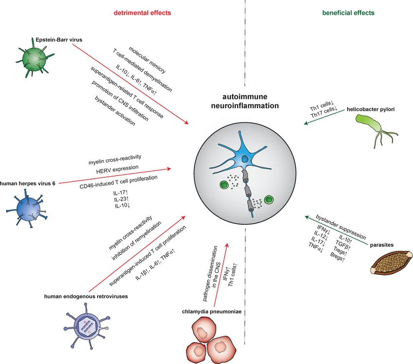
Schroeter et al.                                                                                                      Crosstalk of Microorganisms and Tregs

1 INTRODUCTION                                                             mediated diseases and in autoimmune neuroinflammation. We
                                                                           discuss established and potential therapeutic strategies in MS
In the context of infections, Tregs mediate beneficial and                  resulting from this new molecular understanding.
detrimental effects on short- and long-term disease outcomes.
Although many Treg subsets have been described, thymus-
derived (tTregs) and peripherally induced Tregs (pTregs)
remain the most important subpopulations (1, 2). Tregs are                 2 MANUSCRIPT/SUBSECTIONS
generally found to express CD4 and either or both the high-
affinity receptor for interleukin (IL)-2 CD25+ as well as the               2.1 Janus-Faced Nature: Duality of Tregs
forkhead box protein P3 (Foxp3) (3). Their expression of                   in Pathogen-Mediated Diseases
intracellular and surface markers, such as CD25,                           In the context of pathogen-mediated diseases, Tregs are Janus-
glucocorticoid-induced tumor necrosis factor receptor (GITR)               faced (Figure 1): On the one hand, they are the major
and the inhibitory cytotoxic T-lymphocyte-associated protein 4             determinant of peripheral immune tolerance not only
(CTLA-4) define their phenotype and function (4–6). tTregs                  controlling immune responsiveness to intrinsic antigens and
emerge with a CD4+CD25+Foxp3+ phenotype directly from the                  infective pathogens but also modulating immune capacity (9,
thymus. They are specific for self-antigens requiring continuous            11–13). On the other hand, Tregs dampen favorable pathogen-
antigenic stimulation for their survival and preservation of               directed adaptive immunity counter acting complete pathogen
self-tolerance, the lack of which may lead to autoimmune                   clearance and giving rise to chronic infections (14, 53, 54). Their
disorders (7–11). pTregs on the other hand adopt a regulatory              phenotype and functions are, among others, dependent on their
function upon expression of Foxp3 and are therefore likely to be           localization and the tissue type (55–62).
specific to an exogenous antigen (3, 12–16). In the context of
infection, Tregs can ameliorate excessive immune responses by              2.1.1 One Janus Face: Beneficial Treg Effects in
interaction with and suppression of immune cells. However, Treg            Infections
expansion and enhanced Treg function are central mechanisms of             Aside from their immunosuppressive capacity forestalling
pathogen-related immune evasion. Yet, the contribution of Tregs            pathology, Tregs have been shown to facilitate appropriate
to the pathophysiology of pathogen-mediated diseases as well as            e ff e c t o r m e c h a n i s m s . F u r t h e r m o r e , T r e g s c o n t r o l
the underlying molecular mechanisms remain largely elusive.                immunopathology detrimental to the host body arising from
    In the context of therapeutic interventions, it is important to        excessive effector immune responses.
consider the Janus-faced functions of Tregs in infections                       Tregs utilize diverse immunosuppressive mechanisms
potentially providing beneficial or detrimental effects                     depending on their microenvironment: Expression of CD25,
(Figure 1). Defining the mechanisms by which intracellular                  the a-chain of IL-2, leads to consumption of IL-2 inhibiting
pathogens alter Treg function might pave the way toward new                activation and proliferation of conventional T cells (Tconv) (63–
therapeutic approaches not only in the settings of infections, but         65). Interestingly, Chinen et al. showed that IL-2 expression
also in autoimmune neuroinflammation.                                       activates signal transducer and activator of transcription (STAT)
    With this review, we intend to give a detailed overview of             5 further boosting the suppressor function of Tregs (66).
molecular mechanisms underlying altered Treg function in                   Suppression of Tconv next to macrophages can also be
models of acute and chronic infections as well as in                       triggered by cAMP-mediated protein kinase A (PKA)
autoimmune neuroinflammation with a focus on multiple                       activation. Here, expression of the ectonucleotidase CD39 by
sclerosis (MS) (Table 1). We investigate the impact of                     Tregs leads to hydrolyzation of ATP followed by further
pathogens on immune cell distributions, profiles, and                       cleavage of AMP to adenosine by CD73 (67–69). Subsequently,
functionality - particularly Treg functions - in the setting of            activation of the adenosine receptor A2A causes intracellular
neuroinflammation. We discuss how the complex changes in                    accumulation of cAMP which in turn stimulates PKA and
Tregs lead to altered function and that the underlying                     associated downstream signaling (70–73). Tregs can induce the
mechanisms could contribute to better understand the                       death of natural killer cells (NK cells) and other effector cells,
pathophysiology of neuroinflammatory diseases and their                     such as B cells, dendritic cells (DCs), CD4+ and CD8+ cells, by
treatments. We further review the interplay of infection with              releasing granzyme resulting in perforin-dependent apoptosis of
pathogens and autoimmune processes (Figure 2) and, of                      target cells (74–76). B cells and DCs are regulated by Tregs via
particular interest, the clinical targets that result from these           CTLA-4 which binds CD80/CD86 and increases the expression
interactions (Table 2). In addition, we highlight the interplay            of indoleamine 2,3-dioxygenase (IDO) (77–79). Consecutively,
between commensal bacteria and the function/plasticity of Tregs.           binding of CD28 to CD80/CD86 is limited hampering the
In doing so, we particularly consider the implications for the             crosstalk between Tconv and antigen-presenting cells (APCs).
phenotype of autoimmune phenomena. We point out the need                   Also, accumulation of IDO can lead to starvation of Tconv and
for multi-omic approaches (functional analyses, transcriptomics,           cell cycle arrest, amongst others via degradation of the essential
proteomics, and metabolomics) to illuminate the complex                    amino acid tryptophan (80, 81). Besides that, T cells can suppress
changes in Tregs leading to altered function (Figure 3).                   T cell receptor (TCR)-induced Ca 2+ , NFAT and NF-kB
    Overall, our review focuses on past, present and future insights       downstream signaling (77). The co-inhibitory molecule T cell
into the role of Tregs in the pathophysiology of pathogen-                 immunoreceptor with Ig and immunoreceptor tyrosine-based

Frontiers in Immunology | www.frontiersin.org                          2                                        October 2021 | Volume 12 | Article 747143
Schroeter et al. Crosstalk of Microorganisms and Tregs
inhibitory motif domains (TIGIT) suppresses pro-inflammatory T cell proliferation and functions resulting in viral persistence.
T helper (Th) 1 and Th17 cells, but not Th2 cell responses (82, Treg expansion is mainly based on excessive IL-2 production by
83). Tregs can directly induce angiogenesis via vascular murine, FV-infected effector T cells (90, 91, 133, 134). In HIV,
endothelial growth factor or target tissue cells (58). Further human Tregs inhibit effector T cell responses thereby promoting
immunosuppressive effects of Tregs, e.g. on monocytes and viral chronicity and opportunistic infections acting as a viral
macrophages, are mediated via anti-inflammatory cytokines reservoir (Figure 1) (100, 135, 136).
such as IL-10, IL-35 or tumor growth factor b (TGFb), In human and murine mycobacterium tuberculosis (Mtb)
cytolysis or metabolic disruption (62, 77, 84). infection, expansion of Mtb-specific Tregs interferes with
During acute and chronic (retro-) viral infections, Tregs have priming and migration of T cells to the infected lung resulting
been shown to promote the in- and efflux of pro-inflammatory cells in deficient clearance of Mtb (Figure 1) (137–140). Here, Treg
into lymph nodes (85, 86). Also, they suppress the proliferation and numbers inversely correlate with local mycobacteria-specific
entry of infected cells (87, 88) and contribute to a memory immunity. Three human studies have shown an increase in T
formation via antigen persistence (89). In mice, Treg response is reg numbers in the blood and at sites of infection during active
locally defined controlling magnitude and duration of virus-specific disease (140–142). In human hepatitis B virus (HBV), the
cytotoxic T cell responses (90, 91). For example, in human and expansion of antigen-specific Tregs suggests their contribution
murine cytomegalovirus, vaccinia virus and influenza virus, CD8+ T to the liver pathology (143–145). Here, the frequency of Tregs
cell responses are controlled by Tregs by suppression of the immune correlates with viral load.
response to immunodominant epitopes (92, 93). In human In murine fungal infections such as Candida albicans, the
immunodeficiency virus (HIV) low Treg frequencies are strongly absence of Toll-like receptors (TLRs) and Tregs lead to an
associated with increased immune activation, accelerated increase in immunity to candida via secretion of anti-
atherosclerosis and other morbidities linked to inflammation (94– inflammatory cytokines and improved leukocyte recruitment to
103). A negative correlation between the relative amount of Tregs infection sites (146, 147).
and inflammation has also been observed for hepatitis C virus In parasitic infections [e.g., schistosoma in mice (148, 149),
(HCV) in humans and mice (104, 105). Here, Tregs suppress not leishmania in humans and mice (150–155), plasmodium, and
only the production of interferon gamma (IFNg) but also the helminths (156, 157)], Tregs are reported to favor parasite
expansion and activation-induced cell death of HCV-specific T expansion and persistence by limiting effector responses,
cells resulting in reduced CD4+ T cell reactivity and mitigation of especially of Th1 and Th2 cells, in an IL-10-dependent manner
T cell-mediated liver disease (105–109). and by suppression of antigen-specific T cell proliferation (36,
In bacterial infections, Tregs show a predominant regulatory 158). Nevertheless, while Treg frequency correlated with parasite
function controlling and limiting adaptive and innate immune pathogen load, it also accounted for reduced liver pathology and
responses as shown in different mouse studies (110): improved host survival rates. Also, in murine chronic infections
Immunosuppressive functions of Tregs have been already of the protozoan parasite Toxoplasma gondii, a nonresolving Th1
described for helicobacter hepaticus (111, 112), helicobacter myositis occurs where Treg ablation during chronic infection
pylori (H. pylori) (113–115), listeria monocytogenes (116), rescues macrophage homeostasis and skeletal muscle fiber
pneumocystis carinii (117, 118). regeneration (159).
In toxoplasmosis as a prototypical parasitic infection, Treg
depleted mice showed 50-60% mortality during acute 2.1.3 Treg/Th17 Ratio in Pathogen-Mediated
infection (119). Diseases
In general, the balance between Th17 and Tregs is crucial for the
2.1.2 Other Janus Face: Detrimental Treg Effects maintenance of immune homeostasis during pathogen-mediated
in Infections infections (160–162). By presentation of antigens via major
Immune responses to pathogens can be impaired by an overly histocompatibility complex II molecules on APCs and certain
strong suppressive effect of Tregs interfering with early cytokine environments, naïve Th cells are activated and polarized
protective immunity (84): Tregs can inhibit effector T cell into either peripherally-induced Tregs or Th17 cells to maintain
responses thereby promoting chronic inflammation. In turn, homeostasis. Among APCs, macrophages are known to promote
lack of complete eradication of pathogens leads to a reservoir Treg responses, while DCs mainly activate Th17 cell responses
function of human and murine Tregs acting as carriers for (163). In mice, Th17 differentiation is mainly dependent on the
respective pathogens promoting their expansion in the cytokines IL-6 and TGFb which induce the transcription factor
environment resulting in spread of infections (120–122). retinoic acid-related orphan receptor gamma t (RORgt) in a
Accordingly, pathogen clearance during the disease course STAT3-dependent manner (164, 165). In humans, Th17
correlates with a decrease in the suppressive capacity of Tregs differentiation mainly depends on IL-23 and IL-1ß (166–168).
(123). Vice versa, states of chronic inflammation are often Th17 cell differentiation is further stimulated by TGFb, TNF,
characterized in humans by resistance to immune regulation IL-6, and IL-21. Maintenance and expansion of Th17 cells is
by Tregs (124–126). regulated by IL-23 (168). Differentiation towards the Treg subset
In friend retrovirus (FV) (127–130) and herpes simplex virus is stimulated via induction of the transcription factor STAT5 by
(HSV) infection in mice (54, 131, 132), Tregs limit CD8+ effector TGFb and in the absence of IL-6 (169–172). IL-2 and IL-10 also
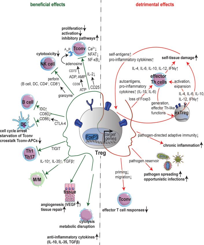
Frontiers in Immunology | www.frontiersin.org 3 October 2021 | Volume 12 | Article 747143Schroeter et al. Crosstalk of Microorganisms and Tregs FIGURE 1 | Janus-faced nature of Tregs. Schematic overview of molecular mechanisms underlying the regulation of immune cells and immune responses by regulatory T cells (Tregs). Anti-infective functions are mainly mediated by suppression of immune cells (left): Expression of CD25 leads to consumption of interleukin (IL)-2 inhibiting activation and proliferation of conventional T cells (Tconv). Suppression of Tconv can also be mediated by adenosine production via the ectoenzymes CD39 and CD73. Besides, Tregs are able to suppress T cell receptor (TCR)-induced Ca2+, NFAT and NF-kB signaling. Dendritic cells (DCs) and B cells are influenced by cytotoxic T-lymphocyte antigen 4 (CTLA-4) which binds CD80/CD86 and increases the expression of indoleamine 2,3-dioxygenase (IDO) resulting in starvation of Tconv next to cell cycle arrest and decrease in crosstalk between Tconv and antigen-presenting cells (APCs). Tregs can induce the death of effector cells (B cells, DCs, CD4+ and CD8+ cells) in a granzyme-perforin-dependent manner. The co-inhibitory molecule T cell immunoreceptor with Ig and immunoreceptor tyrosine-based inhibitory motif domains (TIGIT) suppresses T helper (Th) 1 and Th17 cell responses. Tregs can also induce angiogenesis via vascular endothelial growth factor (VEGF) or target tissue cells directly. Further immunosuppressive effects of Tregs are mediated by cytokines (IL-10, IL-35, TGFb), cytolysis or metabolic disruption. By contrast, Tregs can support inflammation (right) by a multi-layered feed-forward loop promoting the generation of ‘exTreg’ cells adapting Th-like functions, which in turn stimulate activation and expansion of autoreactive Th effector cells. Loss of immunosuppressive capacity adapting phenotype and functionality of Th cells is also reported upon loss of forkhead box protein 3 (Foxp3) in Tregs. Also, Tregs inhibit effector T cell responses thereby promoting chronic inflammation, pathogen spreading and opportunistic infections acting as pathogen reservoir. APCs, antigen-presenting cells; CTLA-4, cytotoxic T-lymphocyte antigen 4; DCs, dendritic cells; Foxp3, forkhead box protein 3; IDO, indoleamine 2,3-dioxygenase; IL, interleukin; M/M, monocytes and macrophages; TIGIT, T cell immunoreceptor with Ig and immunoreceptor tyrosine-based inhibitory motif domains; Tconv, conventional T cells; TCR, T cell receptor; TGFb, tumor growth factor b; Tregs, regulatory T cells; VEGF, vascular endothelial growth factor. play important roles in the differentiation of Tregs (173). Th17 These opposing effects are also represented on cytokine cells show pro-inflammatory effects during disease progression, level: While Th17 cells secrete mainly IL17, IL-21 and IL-22, which can result in autoimmunity. Tregs on the other hand, Tregs produce IL-10, IL-35 and TGFb (160). Via IL-17 Th17 serve as immunoregulatory cells and maintain self-tolerance. cells attract other immune cells, such as macrophages and Frontiers in Immunology | www.frontiersin.org 4 October 2021 | Volume 12 | Article 747143
Schroeter et al.                                                                                              Crosstalk of Microorganisms and Tregs

neutrophils resulting in a state of chronic inflammation. In               chronic infections (19, 25, 90). However, the molecular
contrast, Tregs regulate differentiation and activity of Th17             mechanisms by which intracellular pathogens alter Treg
and other T cells (8, 174). The Treg/Th17 ratio can contribute            functions as well as the origin of these Tregs remain
to a wide range of immune responses ranging from predominantly            incompletely understood (for detailed overview see Table 1).
regulatory to stimulatory function. Its balance is crucial for                Some pathogens directly contribute to Treg induction. In
immune homeostasis.                                                       humans, for example, hepatocytes infected with HCV or gastric
    In recently infected SARS-CoV-2 patients, the number of               epithelial cells infected with H. pylori induce Tregs via TGFb
Th17 cells was significantly increased compared to healthy                 production (Table 1) (17, 34). Upon infection with plasmodium
controls causing inflammatory responses due to the production              falciparum there is a burst of IL-2, IL-10 and TGFb associated
of pro-inflammatory cytokines (175, 176). In contrast, Treg                with Treg induction and expansion. Here, Tregs were, among
numbers were decreased and even further downregulated in                  others, induced by TLR9 signaling in mice and expressed high
the disease course (177–179). Interestingly, the Treg/Th17 cell           levels of Foxp3 suppressing inflammatory processes and
ratio and expression levels of their related cytokines were               immunity-driving mediators of effector T cells (36–39).
significantly higher in deceased patients than during                          Likewise, viral pathogens such as HSV-1, FV and Japanese
remission (175).                                                          encephalitis virus enhance Treg expansion (Table 1). In murine
    Likewise, in respiratory syncytial virus infection but also in        HSV-1, the viral binding site herpes virus entry mediator is
pulmonary infections in general, Tregs and Th17 cells have                upregulated (18). For FV infection, there are two possible
opposing features determining clinical severity (136, 177, 180,           mechanisms underlying Treg expansion: IL-2-dependent
181): Tregs promote viral clearance by recruiting CD8+ cytotoxic          stimulation versus IL-2-independent, tumor necrosis factor
T cells to the lungs and limiting inefficient or excessive T cell          (TNF) receptor 2-dependent upregulation (Table 1) (25–27).
responses (Th2, CD4+ and CD8+) (182–186). In addition, they               In both, Japanese encephalitis virus and Mtb, Treg expansion is a
control the innate immune response by neutrophils and NK cells.           result of programmed death-1 ligand 1 (PD-L1) induction
In contrast, Th17 cells hamper viral clearance by limiting CD8+           (31–33).
T cell responses and enhance Th2 immune responses resulting in                Further, pathogens can enhance Treg functions as shown in HIV
a more severe clinical picture (187, 188).                                infection. Here, upon binding of HIV glycoprotein 120 to the CD4
    In peripheral blood analysis of HBV patients, the Treg/Th17           receptor, Tregs express higher levels of effector molecules such as
ratio was decreased with reduced Treg levels and increased Th17           CTLA-4, CD39 and cAMP and show increased suppressive capacity
cell numbers (189). The latter correlated with TGFb and IL-21             next to prolonged survival rates (Table 1) (19–23). Of note,
levels. Interestingly, here, the Treg/Th17 ratio was the best             expression of Foxp3 by patients with a progressive HIV-1
marker for predicting the stage of HBV-associated liver                   infection seems to be upregulated by individual T cells due to
cirrhosis (190). In HCV patients, Th17 cells were associated              antigen stimulation. Moreover, Foxp3 expression in CD4+ T cells
with early infection and repair processes leading to liver fibrosis.       was shown to be a marker of HIV infection and potentially even a
Here, TGFb and IL-10 suppressed Th17 cells (191).                         prognostic marker of HIV progression (24). Also, in the context of
    For Mtb, the balance between Tregs and Th17 cells regulates           COVID-19 disease, an alteration in the expression of Foxp3 could
encapsulation and control of lung lesions (192). If Tregs become          be detected. More precisely, in patients with a severe disease course,
predominant over Th17, Mtb disseminates more easily and                   a downregulation of Foxp3 could be detected in T cells indicating an
recruitment of neutrophils to the infection sites gets                    impaired Foxp3-mediated feedback on T cell activation as potential
delayed (192).                                                            mechanisms underlying disease progression (198). Similarly,
    In gastrointestinal infectious diseases, Th17 are predominant         human T cell lymphotropic virus 1 associated gene products are
in the acute phase producing the cytokines IL-17A, IL-17F and             reported to inhibit Foxp3 gene expression thereby causing Treg
IL-22. In contrast, Treg/Th17 ratio increases in the chronic phase        dysfunction (Table 1) (28–30).
and infection progress because of the suppressive function of                 Interestingly, women infected with Chlamydia trachomatis
Tregs (193).                                                              displayed increased expression levels of Foxp3 in vaginal swab
                                                                          samples following the clearance of infection due to antibiotic
2.1.4 Impact of Pathogens on Tregs                                        treatment (199). In Candida albicans, prolonged Treg survival
Several pathogens impact the immune status exploiting the                 rates were achieved by TLR2 signaling and IL-10 production
regulatory T cell compartment to enhance their replication and            (Table 1) (43). Altered Foxp3 expression could also be detected
become persistent (95, 194–196). While tTregs are usually                 in the context of parasitic infections: During chronic infection
specific for self-antigens requiring continuous antigen                    with Leishmania of the Viannia subgenus, a decreased Foxp3
stimulation for their survival (8–11), pTregs are converted in            expression was detected (200). Recruitment of Tregs to infection
the periphery and therefore more likely to be influenced by and            sites of Leishmania major was improved by expression of
specific for a microorganism-derived antigen (13, 14). Induction           integrin aEb7 and CC-chemokine receptor 5 (40, 41).
of Treg proliferation and enhancement of Treg function might be           Excitingly, infection with helminth parasites mediated by
central to immune evasion mechanisms of intracellular                     parasite-secreted proteins could also induce de novo T cell
pathogens (123, 197). In accordance, Treg expansion is                    Foxp3 expression (201). This, in turn, may be a way through
compartmentalized to tissues with high viral replication (here            which parasites can evade the host immune response. Regarding
Treg frequency often correlates with viral load) and prolonged in         H. pylori infection, it is worth noting that increased expression of

Frontiers in Immunology | www.frontiersin.org                         5                                  October 2021 | Volume 12 | Article 747143
Schroeter et al. Crosstalk of Microorganisms and Tregs
TABLE 1 | Impact of pathogens on Tregs and the underlying molecular mechanisms.
Infection Pathogen (human or Molecular mechanisms Impact on Tregs Reference
murine)
Viral Hepatitis C virus (human) Production of TGFb. Treg induction. (17)
Herpes simplex virus 1 Upregulation of HEVM, a binding site for viral glycoprotein Treg expansion. (18)
(murine) HSVgD.
Human Binding of gp120 to CD4 receptor of Tregs with Prolonged survival and higher suppressive activity. (19–24)
immunodeficiency virus 1 consecutive upregulation of cAMP. Accumulation in lymph nodes and mucosal lymphoid
Upregulation of CD39/adenosine axis and functional tissue.
markers CTLA-4, TNFR, Foxp3, TGFb, IDO.
Increased expression of homing receptors CD62L and
integrin alpha 4 beta in Tregs.
Upregulation of Foxp3 expression in progressive infection.
Friend retrovirus (murine) IL-2 dependent: IL-2 production by FV-specific effector Treg expansion. (25–27)
CD4+ T helper cells. Coregulation by B cells.
IL-2 independent: Membrane bound TNFa binds to TNFR2.
TNFR2 is indirectly upregulated upon FV infection.
Human T-cell HTLV-1 associated gene products inhibit Foxp3 expression. Dysfunction of Tregs. (28–30)
lymphotropic virus 1
Japanese encephalitis Induction of PD-L1 on dendritic cells. Treg expansion. (31)
virus
Bacterial Mycobacterium Induction of PD-L1 and CISH on dendritic cells. Treg expansion. (32, 33)
tuberculosis (human and
mice)
Helicobacter pylori Production of TGFb. Treg induction. (34, 35)
Upregulation of Foxp3 expression in children.
Parasitic Plasmodium falciparum TLR9 signaling. Treg induction and expansion. Upregulation of Foxp3 (36–39)
(human and murine) Burst of IL-2, IL-10 and TGFb. Correlation between parasite expression.
expansion and Treg increase.
Leishmania major TGFb enhances expression of integrin aEb7. Recruitment and retention of Tregs to infection site. (40, 41)
Production of ligands for CCR5 by infected APCs.
Toxoplasma gondii Upregulation of GITR expression in Tregs. Increased pathogen clearance and host resistance by (42)
enhancement of cellular immune responses.
Fungal Candida albicans TLR2 signaling. Immunosuppression by increased IL-10 production (43)
and prolonged survival of Tregs.
APCs, antigen-presenting cells; CCR5, CC-chemokine receptor 5; CISH, cytokine inducible SH2-containing protein; CTLA-4, cytotoxic T-lymphocyte-associated protein 4; Foxp3,
forkhead box protein P3; FV, friend virus; GITR, glucocorticoid-induced tumor necrosis factor receptor; HTLV-1, human T-cell lymphotropic virus 1; HVEM, herpes virus entry mediator;
IDO, indoleamine 2,3-dioxygenase; IL, interleukin; PD-L1, programmed death-1 ligand 1; TGFb, tumor growth factor b; TLR, Toll-like receptor; TNFR, tumor necrosis factor receptor;
Tregs, regulatory T cell.
Foxp3 on Tregs was observed in Tregs isolated from infected turn, stimulate the activation and expansion of autoreactive Th
children, possibly contributing to an inverse association between effector cells. These effector Th cell-like functions of ‘exTreg’ cells
H. pylori infection and allergic disease through changes in Treg also directly stimulate pathogenic immune responses in local
functionality (35). tissues and promote the pathogenesis of autoimmune diseases by
participating in a feed-forward loop (Figure 1). Strikingly,
2.2 Role of Tregs in Multiple Sclerosis dysfunctional, instable or insufficient Foxp3 expression can
Immune homeostasis and self-tolerance are regulated by the also trigger autoimmunity (15, 208–212): Upon loss of Foxp3,
development, stability and function of Tregs (202). Tregs control Tregs lose their immunosuppressive capacity adapting
immune capacity thereby influencing bystander immune phenotype and functionality of Th cells (Th1, Th2, Th17), such
responses such as allergies or autoimmune diseases (11, 14). as production of IFNg and IL-17 (Figure 1) (213–215).
Tregs prevent the activation and infiltration of T cells into the In contrast, CD4+ HLA-G+ tTregs cells were shown to
central nervous system (CNS) and maintain the homeostasis of ameliorate polyclonal adaptive immune response suppressing
the immune system (203–205). By suppressing CD8+ T effector graft-versus-host disease in vivo (216). Likewise, Foxp3+ Tregs
cell responses, they limit parenchymal damage during CNS limit muscle destruction by cytotoxic T cells in dermatomyositis,
inflammation (206). polymyositis and inclusion body myositis (217).
Tregs can contribute to the pathogenesis of autoimmune Treg/Th17 imbalance is associated with autoimmune diseases
diseases by a multi-layered feed-forward loop (Figure 1) (84): such as MS, myasthenia gravis, psoriasis, inflammatory bowel
Autoantigens and pro-inflammatory cytokines (IL-1b, IL-6 etc.) diseases and rheumatoid arthritis (165, 218–223). Here, Th17
activate effector Th cells which further aggravate self-tissue cells are regarded as the main driver of autoimmune
damage by the expression of IL-4, IL-6, IL-10, IL-12 and IFNg inflammation activating other immune cells and secreting pro-
(207). Antigens and cytokines from damaged tissue promote the inflammatory cytokines (224, 225). A decrease of Tregs in
generation of ‘exTreg’ cells adapting Th-like functions which, in autoimmune and inflammatory diseases is reported to cause
Frontiers in Immunology | www.frontiersin.org 6 October 2021 | Volume 12 | Article 747143Schroeter et al. Crosstalk of Microorganisms and Tregs
disease progression (226). Therapeutic approaches targeting the preceding the diagnosis of MS was associated with alterations of
described Treg/Th17 axis are promising (227) and mainly aim at the Treg compartment in peripheral blood (255) Here, patients
neutralizing Th17-secreted cytokines, reducing Th17 cell counts, displayed lower levels of immunosuppressive CD45RA +
increasing Treg cell levels and regulating transcription factors Foxp3 low Tregs while levels of non-immunosuppressive
such as RORgt, STAT3 and Foxp3 (228–231). CD45RA- Foxp3low Tregs and Th17-like Tregs increased.
These observations suggest that progression to MS might be
2.2.1 Qualitative Treg Alterations in MS preceded by changes to the Treg compartment. A recent study
In MS pathogenesis, T cells acquire an autoreactive phenotype investigating the mechanisms driving Treg dysfunction reported
against CNS autoantigens followed by migration into the CNS an inhibitory effect of circulating exosomes from MS on Tregs
causing inflammatory lesions. Activation of T cells is induced by (256). This effect is thought to be mediated by let-7i miRNA
molecular mimicry in the periphery or by autoreactive T cells in interacting with insulin like growth factor 1 receptor and TGFb
the CNS (232). Control mechanisms that should prevent receptor 1 expressed by CD4+ T cells. Thus, miRNA profiles
autoimmunity, such as selection processes during tTreg from MS patients might directly inhibit Treg expansion (256).
development or peripheral suppression by Tregs, are often
circumvented by autoreactive T cells (233, 234). 2.3 The Interplay of Infections and
Tregs acquire a phenotype and expression profile resembling Autoimmunity - Translation in the Setting
Th1 cells, thereby contributing to disease progression (235): of Multiple Sclerosis
Lower expression levels of Foxp3, TGF, CTLA-4 and CD39 It is commonly accepted that the interaction of genetic
were accompanied by an increase in IFN secretion in relapsing- susceptibility and the exposure to certain environmental
remitting MS (RRMS) patients (236–238). Myelin-reactive T cells factors is crucial for the occurrence of autoimmunity. A major
secreted high levels of IL-17, IFNg and granulocyte-macrophage environmental factor contributing to the pathogenesis of
colony-stimulating factor compared to healthy controls (239). autoimmune diseases and, more specifically, autoimmune
Next to an upregulation of markers associated with Th1 identity, neuroinflammation is pathogen-mediated infection. One of the
Tregs expressed higher levels of the migration markers CD103 underlying mechanisms is so-called molecular mimicry. Here,
and CD49d enhancing transmigration of ‘exTregs’ into the CNS due to structural similarity of pathogen-derived peptides with
(238). High IL-17 levels have also been detected in the host molecules, autoreactive B and T cells become cross-
cerebrospinal fluid (CSF) of MS patients during relapse as well activated leading to an immune response directed against self-
as in chronic lesions (222, 240), suggesting that both, the antigens (257). Likewise, epitope spreading is involved in the
upregulation of IL-17 and down-regulation of Treg-mediated interplay of autoimmunity and infections. In this context, a new
immunity, contribute to MS pathogenesis (241, 242). infection in an ongoing autoimmune disease leads to tissue
Likewise, in experimental autoimmune encephalomyelitis damage with exposure of further self-antigens (258). APC-
(EAE), a mouse model of MS, an altered phenotype and mediated presentation of these antigens to autoreactive
impaired suppressive capacity of Tregs have been associated lymphocytes then accelerates inflammatory processes (258).
with clinical deterioration (235, 243, 244). Transfer of Th1-like Furthermore, infections can facilitate inflammatory processes
‘exTregs’ even lead to induction of EAE in naïve recipient mice. through bystander activation leading to a general immune
Interestingly, Othy et al. (245) showed that Tregs can suppress response with activation of immune cells such as NK cells or
Th17 cells by inhibition of intracellular Ca2+ signaling and their macrophages and thus release of pro-inflammatory cytokines
contact to APCs. (259). This inflammatory milieu induces an antigen-independent
Therefore, therapeutic induction of Tregs as well as activation of primed B and T lymphocytes at the inflammation
modulation of Treg/Th17-related pathways could attenuate the site and thereby enhances autoimmune damage. Lastly,
inflammatory immune response resulting in mitigation of pathogen-mediated amplification of autoimmune events
disease symptoms (246–250). Interestingly, Haas et al. showed involves bacterial or viral superantigens leading to an
that the immunosuppressive effect of Tregs after alemtuzumab extremely potent activation of polyclonal autoreactive T cells
treatment of MS patients was mainly due to an altered by binding to major histocompatibility complex II (258). These
composition and reactivity of conventional CD4+ cells after superantigens lead to a massive proliferation of T lymphocytes
immunodepletion (251). with excessive cytokine production, especially of IL-2 and IFNg,
resulting in an exacerbation of autoimmune processes (258).
2.2.2 Quantitative Treg Alterations in MS The Treg compartment is needed to control immunopathology
Treg frequency and the Treg/Th17 ratio were negatively throughout life. However, while Tregs are indispensable for
correlated with disease severity in MS patients (252, 253). In immune regulation, exuberant Treg function might prove
RRMS patients, reduced Treg numbers were observed. In EAE, detrimental for host defense. For example, in Mtb as a model
Treg plasticity was studied in detail showing an increase of for chronic bacterial infection, Tregs can delay leukocyte
‘exTreg’ counts during the preclinical phase until disease migration from lymph nodes to sites of ongoing infection (107,
maximum (212, 254). In line, remission is linked to an 260). In line, Treg ablation reduces accumulation of Mtb in lungs
increase in Treg numbers representing a recovery of Treg of infected mice (139). These observations underpin the potential
identity (254). Interestingly, clinically isolated syndrome often of Tregs to exert detrimental effects in immune-mediated
Frontiers in Immunology | www.frontiersin.org 7 October 2021 | Volume 12 | Article 747143Schroeter et al. Crosstalk of Microorganisms and Tregs
diseases. The continuum of dysfunctional Treg action is shared discussion of both modulation of peripheral and CNS-localized
between infectious conditions, characterized by exuberant immune responses. In the periphery, EBV might lead to a cross-
immunosuppression, and autoimmune conditions, characterized activation of pathogenic T cells via molecular mimicry as
by promotion of immunogenicity (84). Common to Treg described above (275). This theory is supported by the fact
dysfunction is the instability of Foxp3 (213). Foxp3 is pivotal for that EBNA-1-specific T cells react to myelin antigens more
Treg homeostasis. However, lineage tracing studies revealed that frequently than to other auto-antigens causing a release of pro-
Foxp3 is frequently lost under autoimmune conditions (213). Loss inflammatory IFNg (276). Furthermore, EBV-reactive T cells
of Foxp3 leads to generation of the so called ‘exTreg’ phenotype were isolated from the CSF of MS patients also recognizing
characterized by functions shared with effector Th cells, such as myelin basic protein (MBP) (277). In addition, a very recent
secretion of pro-inflammatory cytokines e.g. IFN-g and IL-17 (84). study found cross-reactivity between EBNA-1 and the recently
Moreover, continuous IL-2 signaling is needed to prevent loss of identified MS autoantigen called anoctamin 2 further supporting
Foxp3 (261). Intriguingly, inflammatory conditions promote loss EBV-induced molecular mimicry (278). Another hypothesis
of Foxp3 and, therefore, contribute to maintaining autoimmune proposes that EBV infection of peripheral B cells induces the
states (212). Inflamed tissue constitutes a complex micro- expression of aB-crystallin. As it is also expressed in
environment characterized by immune cell infiltration, pro- oligodendrocytes, an aB-crystallin-directed T cell response
inflammatory cytokine secretion and increased (self-)antigen might ultimately lead to demyelination (279). Furthermore,
presentation. Treg instability might therefore contribute to there is evidence for EBV infection of B cells leading to a
sustaining inflammatory conditions whereas inflammation release of predominantly pro-inflammatory cytokines such as
promotes loss of Foxp3 and generation of Tregs more closely IL-6 or TNFa and simultaneously impeding immunoregulatory
resembling effector Th cells further contributing to pro- processes by reducing IL-10 levels (280, 281). Further evidence
inflammatory stimuli in a feed-forward loop (84). Intriguingly, derives from recent data providing another pathophysiological
Tregs from patients who resolved an HCV infection reacted to a link between EBV infections and MS. Wang et al. reported an
virus-encoded peptide with substantial human homology while autoreactive CD4+ T cell clone showing cross-reactivity between
Tregs from non-infected patients did not (260). Taken together, HLA-DR-derived self-proteins, EBV antigens, as well as
the pathogenic potential and lineage instability of Tregs make autoantigens presented by HLA-DR allomorphs DR2a and
them suspects for mediating autoimmunity following DR2b (282). Thus, EBV antigens could be actively involved in
chronic infections. the activity of autoreactive CD4+ T cells. Since HLA-DR15 is one
One of the best studied pathogens involved in MS of the genetic factors most strongly associated with MS, this link
pathophysiology is Epstein-Barr virus (EBV). Based on highlights the relevance of EBV infection in the pathogenesis of
epidemiological similarities, an association of EBV infection MS (282). A theory with regard to the modulation of peripheral
and MS was suspected early on (262). Further research immune processes describes that the invasion of autoreactive T-
subsequently not only proved that virtually all MS patients and EBV-infected B cells into the CNS is forced by expression
exhibit an EBV infection (263), but also that prior infectious induction of EBV-induced G protein-coupled receptor 2 thereby
mononucleosis is associated with a 2-3 times higher risk of fostering the neuroinflammatory response (Figure 2) (275,
developing MS (264). Conversely, this risk is significantly 283–285).
reduced for individuals with a negative EBV serology (265). In the CNS, the accumulation of infected B cells within the
Particularly interesting in this context are data showing that meninges and perivascular cuffs suggests that these B cells may
initially seronegative patients experience seroconversion shortly elicit a CD8+ T cell response, leading to a multiplication of the
before the onset of MS symptoms (266). Even in pre- inflammatory response via bystander activation (272, 286).
symptomatic patients with EBV, a significant increase in anti- Expression of superantigens by EBV-infected B cells could
EBV antibodies was found over five years before disease onset further lead to an excessive T cell response (287). Finally, it is
suggesting involvement of EBV in early disease stages (267). hypothesized that EBV-induced immortalization of infected B
Interestingly, since an association between EBV serology and cells and exhaustion-induced defective elimination lead to an
early conversion of clinically isolated syndrome into clinically accumulation of EBV-infected autoreactive B cells causing a
definite MS has been demonstrated (268), EBV serology also permanent exposure to CNS antigens (Figure 2) (288–290).
appears to correlate with disease activity. Consistently, a This exposure might considerably aggravate CNS damage in
correlation between anti-EBV nuclear antigen 1 (EBNA-1) the context of autoimmune neuronal inflammation by antigen
titers, disease progression, lesion load, brain atrophy, and the expression, autoantibody production, as well as by providing
extent of demyelination in MS patients has been demonstrated survival signals to autoreactive T cells.
(269–271). Further support for an involvement of EBV in MS Besides EBV, human endogenous retroviruses (HERVs) seem
pathophysiology derives from histological studies revealing an to be significantly involved in MS pathophysiology. These
accumulation of EBV-infected B- and plasma cells in MS brain proviruses which account for circa 8% of the genome originate
meninges, in cortical as well as in white matter lesions (272–274). from exogenous infection of primate germ line cells millions
Despite this overwhelming evidence, the molecular of years ago and are today part of the human DNA (291).
mechanisms underlying the role of EBV in the While they are functionally inactive under physiological
immunopathophysiology of MS are still not properly conditions, pathological triggers such as viral infections can
understood. However, a number of hypotheses exist involving induce reactivation and thus production of viral proteins (292).
Frontiers in Immunology | www.frontiersin.org 8 October 2021 | Volume 12 | Article 747143Schroeter et al. Crosstalk of Microorganisms and Tregs FIGURE 2 | Pathogen-mediated impact on autoimmune neuroinflammation. The mechanisms by which infectious pathogens influence the processes of autoimmune neuroinflammation are diverse. Both detrimental and beneficial effects are reported. Epstein-Barr virus (EBV), for example, leads to an increase in neuronal damage via molecular mimicry, demyelination, an increase in pro- and decrease in anti-inflammatory molecules, and an augmented T cell response. Other pathways by which EBV induces amplification of the neuroinflammatory response include promotion of central nervous system (CNS) infiltration by autoreactive T and B cells next to bystander activation. Meanwhile, human herpes virus 6 (HHV-6) leads to a detrimental impact via CD8+ T cell-mediated cross-reactivity with myelin peptides and CD46-induced promotion of T cell proliferation. Furthermore, HHV-6 also triggers the expression of human endogenous retroviruses (HERVs) proteins. These in turn induce further damage via cross-reactivity with myelin antigens but also through acting as superantigens. Contributing to this is as well, HERVs trigger CD14- as well as Toll-like receptor (TLR) 4-mediated induction of proinflammatory cytokines. Interestingly, by suppression of oligodendrocyte precusor cells, HERVs also interfere with neurodegenerative processes. Finally, Chlamydia pneumoniae was shown to aggravate neuroinflammation in an animal model through pathogen dissemination into the CNS accompanied by an increase of pro-inflammatory Th1 cells. In contrast, a beneficial impact on the neuroinflammatory response was found for H. pylori and parasites. H. pylori improves the outcome in animal models of MS by reducing the proliferation of Th1 and Th17 cells. Parasites such as helminths attenuate the neuroinflammatory response by inducing bystander suppression via upregulation of regulatory B and T cells as well as anti-inflammatory cytokines. Bregs, regulatory B cells; CNS, central nervous system; HERV, human endogenous retrovirus; IFN, interferon; TGFb, tumor growth factor b; Th cell, T helper cell; TLR, Toll-like receptor; TNFa, tumor necrosis factor a; Tregs, regulatory T cells. First evidence of an involvement in MS dates back more than 30 activity and the occurrence of relapses (300). Once more, the years when retrovirus transcription was found in the possible underlying mechanisms are diverse. For example, supernatants of meningeal cell cultures of MS patients (293). reactivation of HERV-W proteins leads to an activation of Different HERVs such as HERV-H, HERV-K and HERV-W both innate and adaptive immune responses in MS (301). were subsequently associated with MS (294, 295). In addition to Thus, dysregulated expression of HERVs may contribute to an increased HERV-W expression in MS patients (296), CNS damage such as observed in a severe combined observations of higher antibody reactivity to certain HERV-W immunodeficiency mouse model (302). This dysregulated sequences in MS patients (297) and HERV-W upregulation in HERV-W activity is likely to involve binding of its envelope MS plaques correlating with disease activity support the protein (ENV) to TLR4 and its co-receptor CD14 which triggers involvement of HERVs in MS (298). There is also clinical the release of pro-inflammatory cytokines such as TNFa, IL-1b evidence of a relationship of HERVs and MS since patients or IL-6 fostering the autoimmune response (303–305). expressing high levels of HERV-W show a poorer prognosis in Furthermore, HERV-W-derived proteins such as ENV show early disease stages and increased disease progression (299). cross-reactivity with myelin antigens amplifying the Accordingly, HERV-W load also correlates with disease neuroinflammatory response (306). Aside from immune- Frontiers in Immunology | www.frontiersin.org 9 October 2021 | Volume 12 | Article 747143
Schroeter et al.                                                                                            Crosstalk of Microorganisms and Tregs

mediated mechanisms, HERV-W ENV also interferes with                      patients being seropositive for H. pylori showed reduced
remyelination via the inhibition of oligodendrocyte precursor             disability scores (325). In EAE, infection with H. pylori
cell (OPC) differentiation (307). Moreover, it also induces a pro-        resulted in reduced disease progression, milder proliferation of
inflammatory activation of myeloid cells which, in turn,                   autoreactive cells, and lower infiltration of pro-inflammatory
contributes to axonal damage and thus neurodegeneration                   effector Th1 and Th17 cells into the CNS (Figure 2) (326). A
even in long-standing MS cases (308). As there is a                       protective role of H. pylori is also assumed in other autoimmune
monoclonal antibody available that neutralizes HERV-W-                    diseases such as asthma (327) or inflammatory bowel
induced detrimental effects, endogenous retroviruses constitute           disease (328).
an attractive target for future MS therapies (Figure 2) (47, 309).            Likewise, strong evidence for a protective role in MS disease
    There is an exciting connection between EBV and HERVs.                development has been demonstrated for some parasites. First
Exposure of EBV-derived glycoprotein 350 to B cells, monocytes,           indications derive from epidemiological investigations, which
macrophages, as well as astrocytes leads to a significant                  showed an inverse relationship between parasites like Trichuris
increase in the expression of HERV-W and syncytin-1 and is                trichiura and the occurrence of MS (329). In fact, the prevalence
thus also associated with unfavorable processes (310). Similar to         of MS seemed to decrease when the contamination level
EBV, human herpes virus 6 (HHV-6) infection can trigger the               exceeded 10% (329). Of note, parasite-infected MS patients
expression of HERV-W as well as a HERV-related superantigen               showed a significantly decreased number of relapses, a minor
(Figure 2) (311, 312). HHV-6 is a neurotropic virus that is               decline in disability scores, and reduced magnetic resonance
divided into two subtypes, of which subtype A can be found in             imaging (MRI) disease activity compared to patients without
oligodendrocytes of MS white matter lesions (313). In addition to         helminthic infection (144). Parasitic infections exert an anti-
the expression of HHV-6 antigens in MS plaques (314), further             inflammatory effects both on the parasite-specific response and
evidence for an involvement of HHV-6 in MS pathogenesis                   the inflammatory response directed against other antigens in the
derives from elevated anti-HHV-6 antibodies in the CSF of MS              sense of bystander suppression (330). In mice, helminth
patients, especially in patients with an exacerbated disease              infection significantly attenuated both the incidence and
indicating HHV-6 as a trigger for disease aggravation (315–               clinical symptoms of EAE (331, 332). This amelioration was
317). Interestingly, in a non-human primate MS-like animal                accompanied by a decrease in pro-inflammatory IFNg, TNFa,
model, prior infection with HHV-6 resulted in a worse outcome             IL-17, and IL-12 with a simultaneous increase in the release of
further supporting a detrimental impact of HHV-6 on MS                    immunoregulatory IL-10 and TGFb (330–332). Also in humans,
(318). One of the HHV-6-mediated mechanisms contributing                  helminth infection was associated with induction of
to MS pathophysiology involves molecular mimicry since                    CD4+ CD25+ Foxp3 + T cells suppressing the inflammatory
cross-reactivity between HHV-6 and MBP was shown to                       response (333). Beyond Tregs, regulatory B cells secreting
induce cytotoxic T cell-mediated oligodendrocyte death (319).             IL-10 were detected in greater numbers in helminth-infected
This idea is further supported by a close sequence homology               individuals suffering from MS (334). Further, the MBP-specific
between MBP and the HHV-6-derived U24 protein (320).                      immune response was characterized by a decreased release of
Furthermore, it is suggested that HHV-6 binding to the CD46               pro- next to an enhanced release of anti-inflammatory cytokines
receptor leads to a T cell-mediated autoimmune reaction (321).            in patients with a parasitic infection (Figure 2) (333).
Also, increased IL-23 release by DCs and IL-17 production                 Interestingly, the protective effects of helminths infection were
by T cells with a concomitant decreased secretion of the                  shown to be reversed following an anthelmintic treatment
immunoregulatory IL-10 provide potential mechanisms of how                concerning both the clinical as well as radiological MS activity
also HHV-6 might exacerbate neuroinflammatory processes                    and the immunosuppressive effects in terms of the Treg activity
(Figure 2) (321).                                                         (335, 336).
    Apart from viral infections, there is also evidence for the               In summary, for many pathogens there is versatile evidence for
involvement of bacterial pathogens in MS pathophysiology. A               modulation of autoimmune processes in the context of
large meta-analysis, for instance, has shown that MS patients             neuroinflammation in MS. Nevertheless, it has not yet been
have a significantly higher incidence of Chlamydia pneumoniae              possible to conclusively define the underlying molecular mechanisms.
(C. pneumoniae) DNA and intrathecally synthesized
immunoglobulins in their CSF compared to patients with other              2.3.1 Therapeutic Targets
neurological diseases (322). In EAE, systemic infection of mice           The above findings on detrimental but also beneficial effects of
with C. pneumoniae led to dissemination of the pathogen into              pathogenic infections have led to therapeutic approaches - in some
the CNS accompanied by an aggravation of autoimmune                       cases despite continuing doubts about the mechanistic background.
neuroinflammation through reduced Th1 cell proliferation as                   In the case of EBV, attempts have been made to prevent an
well as IFNg production (Figure 2) (323). Nevertheless, the               acute EBV infection by prophylactic vaccination thereby
available data is still unclear and controversially discussed.            reducing the risk for development of MS (274). However, there
    Whereas the pathogens mentioned so far all have a negative            is currently no appropriate vaccination available. In general,
impact on the processes in MS, this is different for H. pylori. In        antibodies directed against certain EBV proteins expressed
MS cohorts for example, a reduced prevalence of the pathogen              during latency to increase anti-EBV immunity would be a
compared to controls was demonstrated (324). Even more, MS                promising strategy. Once again, however, no study results are

Frontiers in Immunology | www.frontiersin.org                        10                                October 2021 | Volume 12 | Article 747143
Schroeter et al. Crosstalk of Microorganisms and Tregs
available to date (274). In contrast, cell-based immunotherapies treatment with 2500 Trichuris suis ova every two weeks for
appear to be a more promising approach. In particular, the three months (Table 2) (49). Under treatment, there was a
application of autologous or allogenic T cells targeting EBV- reduction of new GELs by 70% compared to baseline with a
infected B cells came into focus. A first successful application of return to baseline after two months of follow-up. The reduction
this therapy was already demonstrated in a patient suffering from of lesions was also associated with increased serum levels of of
secondary progressive MS (337). Subsequently, a study on the IL-4 and IL-10 in 80% of the participants (336). The follow-up
effects of a EBV-specific autologous T cell therapy using in vitro study including 16 RRMS patients also showed a trend of
expanded T lymphocytes interfering with EBNA-1 and latent reduction of active MRI lesions compared to baseline (344).
membrane proteins 1 and 2A was initiated (Table 2) (45). Furthermore, there was an increase of Tregs observed during this
Clinical improvement occurred in 7 out of 10 included MS trial. A safety study evaluating the effect of orally administered
patients. Of note, this was only a safety trial therefore lacking a 2500 Trichuris suis eggs in 10 MS patients could not observe
placebo group. Further studies elucidating the impact of Trichuris suis ova-induced effects on disease progression
autologous or allogenic T cells attacking EBV-infected B cells (Table 2) (51, 336). In line, in this study there were no
are underway (Table 2). significant alterations detected regarding cytokine expression
HERV-targeted therapies, on the other hand, have reached a and T cell-specific transcriptions factors (336). A 9-month
more advanced stage. As already pointed out, HERV and HERV- double-blind, randomized, placebo-controlled study enrolling
related proteins such as ENV exert an unfavorable effect on 71 RRMS patients investigated the effect of transcutaneous
OPCs and myeloid cells and thus on remyelination and application of hookworm larvae on lesion burden (Table 2)
neurodegeneration (307–309). In view of the persistent lack of (50). Of note, treatment with hookworm larvae increased the
remyelinating therapies (338), it is particularly interesting that proportion of Tregs in the peripheral blood. Furthermore, the
this inhibition can be reversed by the anti-HERV-W IgG4 study showed a tendency of reduced new or enlarging lesions as
monoclonal antibody GNbAC1 (temelimab) (309). Besides well as an ameliorated MRI activity in the treatment group.
promotion of remyelination, GNbAC1 impedes the release of However, these differences were not significant (50). Given these
pro-inflammatory cytokines (339). Application of GNbAC1 led inconsistent results together with methodological limitations
to favorable effects in numerous early-phase studies (46, 340– such as small sample sizes, further studies are required to
342). Given these pre-clinical results, two phase IIa and IIb trials sufficiently address the therapeutic potential of helminth
were initiated (Table 2) (47). Therapy with 18 mg/kg resulted in infections in MS (336).
a significant reduction of the number of T1-hypointense lesions
after 48 hours. Moreover, there was a consistent trend of reduced 2.4 Microbiome - the Missing Link
brain atrophy and a magnetization transfer ratio decrease Between Biomolecular Treg Signatures
indicating a positive impact on remyelination. However, and Clinical Phenotype?
GNbAC1 failed to achieve the primary endpoint of the study, In recent years, the important role of the gut microbiome has
i.e. reduction of gadolinium-enhancing lesions (GELs), possibly been recognized in autoimmune diseases and pathogen-induced
because of underdosing. However, the beneficial MRI effects on immune responses (345, 346). The interplay of the gut
neurodegeneration raised hope and led to the initiation of a new microbiome and the immune system may explain its seemingly
phase II study (Table 2) (47). Another HERV-related approach universal impact on a great variety of diseases including
is based on the theory that antiretroviral therapies can also autoimmune diseases, cancer, vascular disease, and even
induce inhibition of HERVs in MS (343). In a baseline-versus- psychiatric disorders (347–350).
treatment phase IIb study, 20 patients with active RRMS were Importantly, a variety of factors modulate the composition of
treated with the integrase inhibitor raltegravir for 3 months the microbiome. Hence, the relationship between the host and
(Table 2) (48). However, the primary study endpoint reduction the microbiome needs to be understood as a dynamic rather than
in lesion load or development of new lesions during the static process (351). One of the most influential factors of the
treatment period compared with baseline was not met. microbiome is the diet, which under unfavorable conditions
Although to date the evidence regarding C. pneumoniae is induces dysregulation in the form of dysbiosis (351). This
quite sparse, therapy with rifampicin or azithromycin for 6 dysbiosis, in turn, contributes to an increased incidence of gut-
months were compared with placebo in newly diagnosed distal autoimmune phenomena such as autoimmune arthritis
RRMS patients with evidence of C. pneumoniae infection in (352) or type 1 diabetes (353) through alterations in Treg/Th17
the CSF (Table 2) (44). The primary endpoint, reduction of balance. In general, the dynamic balance or dysbalance of Tregs
GELs, was not reached. Only a decrease in brain atrophy was and Th17 is suggested to be a main effector mechanism by which
found under antibiotic therapy. However, given the very small the gut microbiome influences systemic immunity (354).
number of subjects, these results should be interpreted with Furthermore, antibiotic therapy has an enormous impact on
great caution. the microbiome and thus on the function of CD4+ T cells.
Recently, there are also first therapeutic approaches that For example, antibiotic therapy not only leads to an altered
exploit the protective effects of helminth infection on MS. In a colonic but also tTreg TCR repertoire (355). Likewise, antibiotic-
small phase I study, MRI activity in five treatment-naïve RRMS treated mice show a significant reduction of Tregs in the colonic
patients was compared between baseline and after probiotic lamina propria (356, 357). Similarly, also germ-free mice
Frontiers in Immunology | www.frontiersin.org 11 October 2021 | Volume 12 | Article 747143You can also read