Tackling dysfunction of mitochondrial bioenergetics in the brain by omics technologies
←
→
Page content transcription
If your browser does not render page correctly, please read the page content below
Preprints (www.preprints.org) | NOT PEER-REVIEWED | Posted: 17 June 2021
Review
Tackling dysfunction of mitochondrial bioenergetics in the
brain by omics technologies
Paola Zanfardino1, Stefano Doccini2, Filippo M. Santorelli2,* and Vittoria Petruzzella1,*
1University of Bari Aldo Moro, Department of Medical Basic Sciences, Neurosciences and Sense Organs, Bari,
70124 Italy;
2IRCCS Fondazione Stella Maris, Calambrone, 56128 Pisa, Italy;
*Correspondence: filippo3364@gmail.com and vittoria.petruzzella@uniba.it
Abstract: Oxidative phosphorylation (OxPhos) is the basic function of mitochondria although the
landscape of mitochondrial functions is continuously growing to include more aspects of cellular
homeostasis. Thanks to the application of -omics technologies to the study of the OxPhos system,
novel features emerge from the cataloging of novel proteins as mitochondrial thus adding details to
the mitochondrial proteome and defining novel metabolic cellular interrelations, especially in the
human brain. We focussed on the diversity of bioenergetics demand and different aspects of mito-
chondrial structure, functions, and dysfunction in the brain. Definition as ‘mitoexome’, ‘mitoproteome’
and ‘mitointeractome’ have entered the field of ‘mitochondrial medicine’. In this context, we reviewed
several genetic defects that hamper the last step of aerobic metabolism mostly involving the nervous
tissue as one of the most prominent energy-dependent tissues and, as consequence, as a primary
target of mitochondrial dysfunction. The dual genetic determination of the OxPhos complexes is
one of the reasons for the complexity of the genotype-phenotype correlation when facing human
diseases associated with mitochondria defects; clinically, are characterized by extremely heteroge-
neous symptoms, ranging from organ-specific to multisystemic dysfunction with different clinical
courses. Finally, we briefly discuss the future directions of the multi-omics study of human brain
disorders.
Keywords: mitochondria; mitochondrial DNA; nervous tissue; OxPhos complexes; bioenergetics;
genomics; proteomics; mitochondrial diseases
1. Introduction
The panoply of mitochondrial functions reflects on highly heterogeneous clinical
presentations when an error in a mitochondrial protein or function occurs. Mitochondria
are dynamic and mobile organelles representing a hub where exchange of information
among the nucleus and other cellular compartments takes place to modulate energy pro-
duction and metabolites provision to the cell’s specific needs and nutrient availability [1].
The basic function of mitochondria is the generation of more than 90% of cellular energy
via the oxidative phosphorylation (OxPhos) system [2] but, in addition, they play many
fold roles in the different types of cells: compartmentalize metabolites for the maintenance
of redox homeostasis; function as centers for metabolic waste management [3]; surveil
calcium homeostasis [4]; initiate caspase-dependent apoptosis and other intermediate cel-
lular stress response [5]; provide sulfur metabolism and iron-sulfur cluster biogenesis
[6,7]; house the synthesis of cardiolipin, steroids, quinone, and heme [8,9]; breakdown
fatty acids through -oxidation; and serve as a metabolic platform for the TCA and urea
cycles [10]. All these functions include homeostatic regulation of organelle morphology
and dynamics [11], quality control [12], and participation in the immune response [13,14].
Alteration of each of the above functions and activities can affect differently according to
© 2021 by the author(s). Distributed under a Creative Commons CC BY license.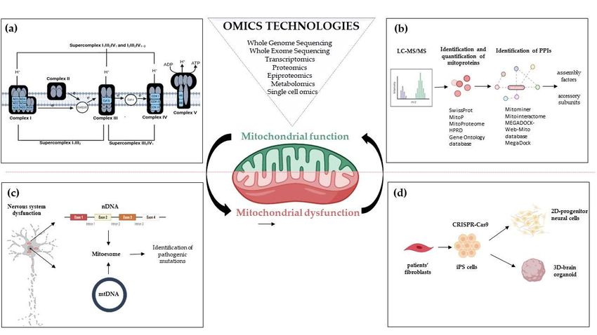
Preprints (www.preprints.org) | NOT PEER-REVIEWED | Posted: 17 June 2021
2 of 42
the specificity of the organ and cell type, but alteration of mitochondrial energy produc-
tion can impact tissues with the highest energy requirements such as the nervous either
central (CNS) and peripheral (PNS) [15,16].
The term ‘mitochondrial medicine’ categorizes the ample array of clinical presenta-
tions associated with all types of mitochondrial defects having directly or secondarily de-
fect of one or several mitochondrial functions although ‘mitochondrial diseases’ tradition-
ally indicate dysfunction of the OxPhos system [6,17]. The direct link between human dis-
ease and the genetic alteration of a mitochondrial function has taken a breakthrough with
the application of -omics technologies (i.e., genomics, transcriptomics, proteomics, metab-
olomics, and epigenomics, etc.). Rapidly, high-throughput omics techniques — that is de-
tection of massive differences in a multitude of molecular constituents in organisms sup-
ported by sophisticated bioinformatics tools — have allowed progress in cataloging the
predicted human mitochondrial proteins thus revealing new details and providing clues
to elucidating still unknown basic aspects of mitochondrial structure and function. These
novel high-throughput techniques have enhanced the final diagnosis of several mitochon-
drial disorders. This is a very relevant aspect, especially considering that mitochondrial
diseases individually are rare but are probably the most frequent genetic disorder in
adults (incidence of 1 in 5000 live births) [18]. More recently, genome editing technology
applied to neural cultures and cerebral organoids generated from patients-derived iPSCs
is revolutionizing the landscape and offering new opportunities for understanding the
pathogenetic effects of mutations in nervous tissue.
This review aims to focus on the dysfunction of OxPhos defects mostly in the nervous
system to highlighting the contributions of powerful omics technologies to mitochondrial
medicine to land from the laboratory to the clinic.
2. Mitoexome, mitochondrial proteome, and mitointeractome
Before Next-Generation Sequencing (NGS) improved our understanding of how mu-
tations cause diseases, first attempts to identify the mitochondrial proteome were based
on ‘cyberscreening’ of available genome databases. This allowed the discovery of few hu-
man mitochondrial genes presenting orthologs in lower eukaryotes. An example of the
cyberscreening strategy used Saccharomyces cerevisiae proteins as ‘probes’ to identify BCS1,
PET112, SCO1, COX15, and COX11, five yeast genes that present orthologs (respectively
BCS1L, GATB, SCO1, COX15, and COX11) in human [19]. Except for COX11, a COX-as-
sembly, all genes have been implicated in mitochondrial diseases [OMIM 603647.0001-
603647.0013; OMIM 603645.0001-603645.0002; OMIM 603644.0001-603644.0002; OMIM
603646.0001-603646.0004], see paragraphs 4.3 and 4.4. To date, whole-exome (WES) and
whole-genome (WGS) resequencing have dramatically enhanced the ability to identify the
underlying gene mutations in patients with isolated or multiple mitochondrial respiratory
chain complex defects [20,21]. The collection of mt genes and coding exons of the 1,034
nuclear genes encoding the human mitochondrial proteome is defined as “MitoExome”
[22,23]. This multigene panel is useful in performing targeted resequencing of the OxPhos
nuclear genes because it includes not only the 77 nuclear structural OxPhos subunits and
the 37 mtDNA genes [24] but also genes for mitochondrial proteins either already known
or not to be associated with a specific mitochondrial disease, including assembly factors
and electron carriers’ genes which represent a large fraction of the overall mitochondrial
genes that can cause mitochondrial dysfunction [21]. Application of MitoExome resequenc-
ing provides novel mutation candidates, enables the discovery of unusual clinical variants
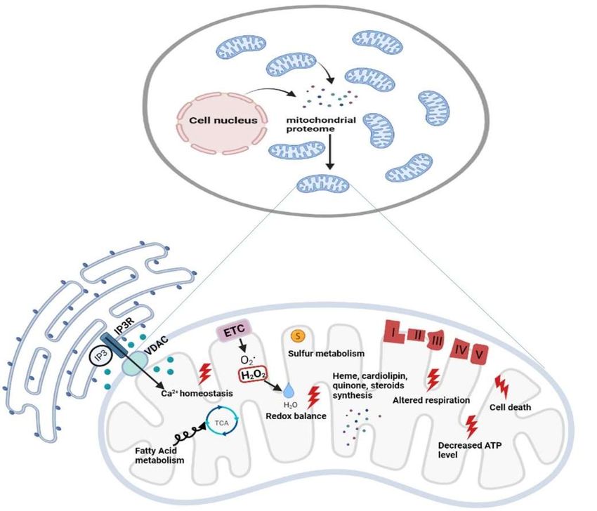
[25,26] and new clinical phenotypes [26] (Figure 1). Furthermore, the integration of Mi-
toExome sequencing with the study of mitochondrial proteome potentiates the detection
of variants causing protein destabilization and/or aberrantly low expression [27].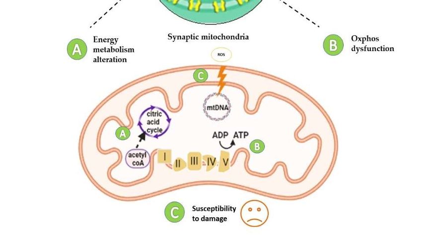
Preprints (www.preprints.org) | NOT PEER-REVIEWED | Posted: 17 June 2021
3 of 42
Figure 1. Omics strategies advance in understanding mitochondrial function and dysfunction in brain disorders related
to OxPhos gene mutations. Mitochondrial bioenergetics involves activities whose function and structure have been deeply
elucidated by omics technologies; (a) The introduction of high-resolution technologies has been resolutive to deepen the
structure of the respiratory chain complexes and supercomplexes; (b) Quantitative proteomics e.g. LC-MS/MS enable the
identification and quantification of mitoproteins and provide large amounts of data. Through Network-based approaches
analyzing protein-protein interactions, the huge amount of information allows the discovery of novel accessory subunits
and assembly factors of the five multi-subunit enzyme complexes; (c) MitoExome resequencing enhances the ability to iden-
tify gene mutations in patients and provides novel gene mutation candidates for diagnostic screening; (d) Novel multi-
omics analysis, based on single-cell omics, is applied to two-dimensional (2D) neural cultures and three-dimensional (3D)
cerebral organoids generated from patients-derived iPSCs that can be engineered by CRISPR/Cas9. Abbreviations: LC-
MS/MS: Liquid Chromatography with tandem mass spectrometry; PPIs: Protein-protein Interactions; nDNA: nuclear DNA;
mtDNA: mitochondrial DNA; iPS cells: Induced Pluripotent Stem cells.
Biochemical and ultrastructural characterizations have enhanced the heterogeneity
of mitochondria in their function, trafficking patterns, lifespan, and morphology across
cell types and different cellular compartments. Different tissues, cell types, and cellular
states have unique signatures of protein localization to mitochondria. In the proteomic
comparison of the mitochondrial proteins, almost half are found as core components in
virtually all tissues, whereas the remaining are tissue-specific [28,29]. The study of mito-
chondrial proteome starts with the isolation of mt compartment from cells and tissues and
stands behind the availability of methodologies to isolate pure mitochondria from differ-
ent sources to define exactly the function of each protein in each cell type of the human
body [30]. The performance of proteomics analysis is driven by the reduction of sample
complexity, enhancement of mass spectrometry (MS) power of resolution, and the possi-
bility to reduce the contamination of the sample with non-mitochondrial proteins owed
to chemical and physical similarities between mitochondria and other cellular compo-
nents (e.g. lysosomes). Since the initial rough estimates, it has been suggested that the
mammalian mitochondrial proteome encompassed about 1000 -1500 distinct proteins
including the 13 mtDNA-encoded proteins [24] that represents an important subset of
the ~20,000 distinct mammalian proteins [31,32] (Figure 2).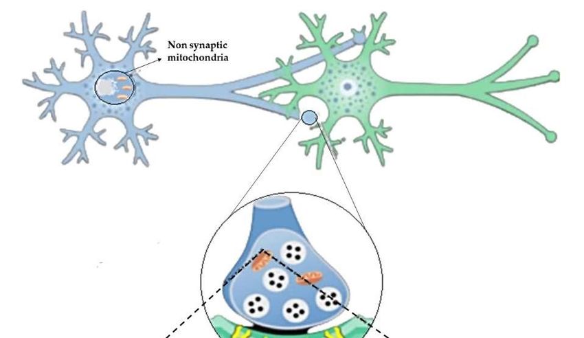
Preprints (www.preprints.org) | NOT PEER-REVIEWED | Posted: 17 June 2021

                                                                                                                                4 of 42

                                    Figure 2. Functional diversity of mitochondrial proteins and bioenergetics consequences of
                                    OxPhos system dysfunction. The mammalian mitochondrial proteome includes both mitochon-
                                    drial and nuclear DNA encoded proteins. Most of the proteins required for the various activities in
                                    which mitochondria are involved are encoded by the nuclear genome, whereas the mitochondrial
                                    energy-producing system, i.e., the OxPhos complexes, has either mitochondrial DNA (mtDNA) and
                                    nuclear DNA (nDNA) encoded components. The enlarged mitochondrion shows most of the bioen-
                                    ergetics consequences (indicated by the red bolt lightning) of genetic defects involving the OxPhos
                                    complexes. Abbreviations: IP3: Inositol Trisphosphate; IP3R: Inositol Trisphosphate Receptor;
                                    VDAC: Voltage-dependent anion channel; ETC: Electron Transport Chain; TCA: Tricarboxylic Acid
                                    Cycle; ATP: Adenosine triphosphate.

                                          Quantitative two-dimensional (2D) gels of highly purified mitochondria estimated
                                    ~1500 distinct spots [33], a number higher than the ~1000 distinct protein products en-
                                    coded by the genomes of alpha-proteobacteria, which are the closest living relatives of
                                    modern-day mitochondria [34]. Several databases have been used to integrate the experi-
                                    mental data with bioinformatic predictions based on mitochondrial localization or inter-
                                    action. For example, the MitoProteome is an object-related database developed at the
                                    UCSD Supercomputer Center, which contains information on mitochondria-localized
                                    proteins [35,36]. Each entry in the MitoProteome corresponds to a gene encoding a protein
                                    that is localized within mitochondria and its basic information, along with annotations of
                                    isoforms, splice variants, and functions of the corresponding protein. To date, the most
                                    comprehensive study elucidating the mitochondrial proteome of different mammalian tis-
                                    sues is represented by the MitoCarta inventory [29,37]. This catalog combines multiple
                                    experimental and computational approaches i.e., mass spectrometry (MS) analysis of mi-
                                    tochondria isolated from 14 mouse tissues, large-scale GFP-fusion microscopy analysis,
                                    and bioinformatics using data mining, prediction, evolutionary conservation, and a
                                    Bayesian integration of seven additional data sources. The first release was represented
                                    by MitoCarta1.0 (http://www.broadinstitute.org/pubs/MitoCarta/) which contained about
                                    1000 distinct gene loci (Pagliarini e al., 2008). Updated in 2016, MitoCarta 2.0 listed about
Preprints (www.preprints.org) | NOT PEER-REVIEWED | Posted: 17 June 2021
5 of 42
1200 genes [37]. Another dedicated database that collected, curated, and annotated infor-
mation on mitochondrial proteins is the MitoMiner database (http://mitominer.mrc-
mbu.cam.ac.uk/) [38] (version 4.0, 2018). It is based on literature and proteomics data
based on both LC-MS and 2D gel studies, antibody staining, other subcellular localization
data, and provides a collective score for each protein's probability to have the mitochon-
drial association. MitoMiner records mitochondrial proteins from 12 organisms [38]. Us-
ing the data contained within MitoMiner, the Integrated Mitochondrial Protein Index (IMPI)
was also developed (http://www.mrc-mbu.cam.ac.uk/impi). IMPI version Q2 (2018) con-
tains 1626 human genes that encode mitochondrially localized proteins, 1184 known to be
mitochondrial and 442 predicted to be mitochondrial. The large amount of information
provided by mito-databases as MitoMiner 4.0 v2018 JUN (http://mitominer.mrc-
mbu.cam.ac.uk), makes it possible to define different score systems for mitochondrial con-
fidence combining data from various mitochondrial and functional annotation databases.
These strategies allow increasing the stringency of protein accepted as inherently mito-
chondrial [39]. An exhaustive list of the major data sources loaded with the latest version
and links to the relevant resources is reported in the Data Sources section of the Mitominer
(https://mitominer.mrc-mbu.cam.ac.uk/release-4.0/dataCategories.do).
More recent advances in the experimental proteomic approaches, specifically in la-
beling and MS methods, have further expanded and defined the known mitochondrial
proteome and have simultaneously revealed the sub-mitochondrial localization of many
of them [40,41]. A novel spatial proteomics pipeline demonstrated that many proteins
cannot be classified to a single localization as they either transit between compartments
or carry out their functional role(s) in multiple locations [41]. The redundant functions, or
functions affecting multiple cellular processes, rendered difficult the study and it was es-
timated that about ~20% of mitochondrial proteins remained uncharacterized [42].
Along with technological progress that has enabled the discovery of approximately
196,111 human proteins [based on Swiss-Prot and TrEMBL entries in Universal Protein
Resource (The UniProt Consortium), as of April 15, 2021], derives the challenge of identi-
fying a large amount of potential protein-protein interactions (PPIs). An example of the
network-based approaches analyzing protein-protein interaction is represented by Mi-
toInteractome, a web-based portal containing 6,549 protein sequences extracted from
SwissProt (http://www.expasy.ch/sprot/), MitoP (http://www.mitop.de:8080/mitop2/),
MitoProteome (http://www.mitoproteome.org/), HPRD (http://www.hprd.org) and Gene
Ontology database (http://www.geneontology.org). This enables the elucidation of inte-
grative mitochondrial functions and can expedite the discovery of novel interactions
which otherwise may have been missed using traditional experimental techniques. MEG-
ADOCK [43,44], a structure-based PPI prediction method, was first developed and then
followed the MEGADOCK-Web-Mito database which is a PPI prediction data archive,
that includes prediction results for protein pairs of 654 mitochondria-related human pro-
teins [45]. All these approaches have been key in the study of PPI as a means to infer
functions for uncharacterized proteins and to enable the discovery of novel protein, e.g.
several complex I assembly factors [46,47] (Figure 1).
3. Diversity of bioenergetics demand in the brain
The brain relies on glucose metabolism for ATP generation and many other activities
and an inappropriate supply of either glucose or oxygen degrades brain function. The
high energy request of the brain is owed to neuronal signaling that accounts for ~70% of
the calculated energy expense, whereas non-signaling activities (metabolism of protein
and phospholipids, oligonucleotide turnover, axonal transport, mitochondrial proton
leak, actin cytoskeleton remodeling, etc.) expend an estimated 30%. Excitatory neurons
consume ~80–85% of calculated ATP use, whereas inhibitory neurons and glia account for
15–20% [48]. Gray matter has regionally heterogeneous metabolic rates that are consider-
ably higher than white matter [49] that have a higher fraction of non-signaling energy
demands than gray matter. In addition to ATP generation, glucose is also the precursorPreprints (www.preprints.org) | NOT PEER-REVIEWED | Posted: 17 June 2021
6 of 42
for the synthesis of many compounds within the brain, including neurotransmitters and
neuromodulators. For these reasons, mitochondria are quite heterogeneous as anatomical
localization, activity, and metabolism at regional, cellular, subcellular levels and during
differentiation, when the upregulation of mitochondrial metabolism is the basis of cell
proliferation in neuronal stem cells and progenitor cells. Although different brain regions
contain about half mitochondria to the heart, brain mitochondria are qualitatively differ-
ent to sustain the high metabolical demand that requires, for instance, tight cooperation
between neurons and astrocytes [50]. Astrocytes provide crucial metabolic and structural
support [50-52] and are key players in neurotransmission [53,54] and behavior [55,56]. The
ATP used by neurons is supplied by the OxPhos process, whereas most energy needs of
astrocytes are satisfied by glycolysis [57,58]. The mitochondrial ATP production per mol-
ecule of glucose oxidized is ~16 times more than glycolysis. The survival of neurons re-
quires OxPhos [50] and in mature neurons, the local ATP supply provided by mitochon-
dria is used to regulate axonal and dendritic development, axonal regeneration, as well as
contributing to synaptic transmission and plasticity. The different energy metabolisms of
the two cell types are closely coupled, with astrocytes releasing the glycolytic end-prod-
uct, lactate, which is used by neighboring neurons to drive OxPhos [59,60].
An example of heterogeneity of mitochondria in metabolic enzyme diversity has
been provided by a study comparing the mitochondrial proteome of the three major cer-
ebellar cell types: Granule cells (GC), the most abundant excitatory neuron; Purkinje cells
(PC), the major inhibitory neuron of the cerebellum and astrocytes. [61]. In the adult cer-
ebellum, ∼15% of the annotated mitochondrial proteome was shown to be differentially
regulated among the three cell types. Fatty acids were more efficiently metabolized by
astrocytic than neuronal mitochondria due to the enrichment of two beta-oxidation en-
zymes, i.e., short-chain-specific acyl-coenzyme A dehydrogenase and carnitine palmitoyl-
transferase 1a, a rate-limiting enzyme in long-chain fatty acids oxidation [61]. Notably, the
astrocytic mitochondrial proteome contained a substantial enrichment of peroxisomal
proteins, some of which are known to have dual-targeting (i.e. catalase) [62] or tether to
mitochondria (i.e. Eci2 and Pex11b). In the same work, the mitochondrial calcium uni-
porter (MCU) [4,61,63] and its regulators were detected mostly in GC [61]. Recent studies
suggest that the markedly different modes of ATP production in the neurons and astro-
cytes reside also in the supra-organization of the mitochondrial respiratory chain in su-
percomplexes (see 4.6 paragraph) able to regulate different rates of respiration and mito-
chondrial ROS production [64].
The brain mitochondrial proteome is not a unicum also when considering synaptic
and non-synaptic mitochondria (sMito and nsMito). Proteomic profiling of sMito vs
nsMito revealed mitochondrial complex I as an upstream regulator of degenerative pro-
cesses associated with a high range of age-related neuropathologies characterized by syn-
aptic dysfunction [65]. An accurate analysis of quantitative proteomics was performed to
differentiate sMito and nsMito in mouse brain mitochondria by stable isotope labeling with
amino acids in cell culture (SILAC). In SILAC, cells are differentially labeled by growing
them in a ‘light’ medium, containing normal amino acids, or a ‘heavy’ medium, contain-
ing a stable isotope [66]. Significant differential expression was shown for 522 proteins
involved in several pathways including the OxPhos system, mitochondrial fission/fusion,
calcium transport, and mtDNA replication and maintenance. Lower levels of Pyruvate
dehydrogenase (PDH) subunits in the synapse to other parts of the cell and reduced ex-
pression of complex I, II, and IV (expect for COX4I2) suggested decreased bioenergetic
function of sMito compared to nsMito [66]. Consistent with this finding, sMito exhibited
increased age-associated mtDNA deletions and reduced levels of TFAM and mtSod2, sug-
gesting a reduced ability of sMito to withstand ROS, thus providing insights into synaptic
mitochondrial susceptibility to damage [66] (Figure 3).Preprints (www.preprints.org) | NOT PEER-REVIEWED | Posted: 17 June 2021
7 of 42
Figure 3. Diversity of mitochondrial proteome in synaptic and non-synaptic mitochondria. Syn-
aptic mitochondria show defects in energy metabolism due to low levels of Pyruvate dehydrogen-
ase (PDH) subunits (A), reduced expression of complex I, II, and IV (B), and increased susceptibility
to damage (increased mtDNA deletions) (C), compared to non-synaptic mitochondria. Abbrevia-
tions: ADP: Adenosine diphosphate; ATP: Adenosine triphosphate.
The extreme heterogeneity of mitochondria activities and functioning has been re-
cently shown by a novel and fine imaging approach that specifically allows to label and
monitor mitochondrial translation products for microscopic fluorescent imaging. In neu-
ronal cultures, mitochondrial translation was monitored in axonal and dendritic mito-
chondria as well as in pre-and post-synaptic regions of neurites by specifically labeling
the peptides newly synthesized by mitochondrial ribosomes revealing that not all mito-
chondria translate to the same extent in different cell types [67]. Finally, the fundamental
role of mitochondria during neurogenesis has been recapitulated in the cellular and or-
ganoid model of Leigh syndrome (LS), a severe manifestation of mitochondrial disease in
children [68]. Mutations in SURF1, a complex IV assembly gene, cause neuronal impair-
ment because of defective metabolic programming of neural progenitor cells (NPCs) that
prevents the establishment of neuronal morphogenesis. Using CRISPR/Cas9 engineered
SURF1 patient-derived iPSCs, a human model of LS was developed. Single-cell RNA-se-
quencing and multi-omics analysis revealed compromised neuronal morphogenesis in
mutant 2D neural cultures and 3D brain organoids (Figure 2). The defects already
emerged at the level of NPCs, which were unable to shift toward OxPhos and retained a
proliferative glycolytic state that fails to instruct neuronal morphogenesis. Interestingly,
gene augmentation and PGC1A induction via Bezafibrate treatment inducing mitochon-
dria biogenesis supported the metabolic programming of LS NPCs, leading to restored
neuronal morphogenesis [68].Preprints (www.preprints.org) | NOT PEER-REVIEWED | Posted: 17 June 2021
8 of 42
4. Structure, assembly, and disorders of bioenergetics complexes
The development of mito-omics-based approaches has been crucial in understanding
the functional and bioenergetic consequences of mutations responsible for the onset of
primary mitochondrial diseases. The OxPhos is the process by which mitochondria pro-
duce the ATP needed by the cells. The reactions are performed by five multimeric enzyme
complexes (EC): Complex I (EC 1.6.5.3) or NADH: ubiquinone reductase, CI, 45 subunits;
Complex II (EC 1.3.5.1) or succinate dehydrogenase, CII, 4 subunits; Complex III (EC
1.10.2.2) or quinol-cytochrome c (cyt c) reductase, CIII, 10 subunits; Complex IV (EC
1.9.31) or cyt c oxidase (COX), CIV, 13 subunits; Complex V (EC 3.6.14) or FoF1-ATPase,
CV, 16 subunits; and two-electron transport carriers, namely, ubiquinone (coenzyme Q,
CoQ) and cyt c [69]. Reactions catalyzed by CI, CIII, and CIV result in the release of pro-
tons in the inner membrane space, thereby creating the proton gradient needed for ATP
synthase activity. The correct function of the OxPhos system depends on the concerted
action of several chaperones and other assembly factors that play essential roles in the
formation, regulation, and stability of the five complexes and the mobile electron carriers,
and nucleotide transporters [70]. Early-stage assembly factors, that have critical roles in
the structural assembly of individual subunits and subcomplexes, and late-stage accessory
factors, also known as the LYRM (leucine–tyrosine–arginine motif) proteins, that regulate
the incorporation and/or activation of final subunits and/or co-factors (i.e. Fe–S clusters)
provide optimal assembly of CI, CII, CIII, and CV. The human mitoproteome contains at
least 12 LYRM proteins [71].
The OxPhos system is under a dual genetic control: 13 subunits are of mtDNA origin
[24] and the remaining are encoded by the nuclear DNA (nDNA) [72]. MtDNA is a small
circular genome [24] that encodes only 13 mitochondrial proteins, 22 mt-tRNAs, and 2 mt-
rRNAs. Hence, the nuclear-encoded mitochondrial proteome requires sophisticated ma-
chinery for the transport into mitochondria [73-75]. Over the last years, a growing number
of human proteins involved in mtDNA replication, and expression have been identified
owing to the study of primary mitochondrial diseases. The coordination between the two
genomes is crucial for mtDNA integrity, copy number regulation, and mitochondrial pro-
tein synthesis because mutations in nuclear genes encoding proteins for mtDNA replica-
tion and maintenance may affect its integrity and properties [76]. Dedicated reviews on
these topics, including also the specific mechanisms regulating mtDNA replication [77],
transcription [78], and translation [79,80] are available elsewhere.
Genetically, the mitochondrial diseases associated with the OxPhos system are split
into two broad genetic categories: disorders due to mutations in the mtDNA, observing
the rules of mitochondrial genetics; disorders due to mutations in the nDNA, transmitted
as a Mendelian trait [6,81]. To date, mutations in both mitochondrial and nuclear genomes
have been reported to cause mitochondrial disease manifesting with characteristic leu-
koencephalopathy and other clinical phenotypes either multisystemic or with single tis-
sue involvement [82-84].
Since the first descriptions of mtDNA mutations [85-87], the number of mutations
has been growing more and more until it counts over 1000 heteroplasmic rearrangements
(large deletions/duplications) (http://mitobreak.portugene.com), and over 350-point mu-
tations possibly pathogenic among the 700 variants reported, which affect all mtDNA
genes (https://www.mitomap.org). A few major clinical phenotypes in adults have been
recently reviewed [88]: LHON [87,89]; Neuropathy, ataxia, retinitis pigmentosa
(NARP)/maternally inherited Leigh syndrome (MIILS) [90,91]; Maternally inherited non-
syndromic deafness, associated or not with aminoglycosides use [92]; Myoclonus, epi-
lepsy, ragged-red-fibers syndrome (MERRF) [93,94]; Mitochondrial encephalopathy, lac-
tic acidosis stroke-like syndrome (MELAS) [95,96]; Chronic progressive external ophthal-
moplegia (CPEO) spectrum [85]; Kearns–Sayre syndrome (KSS) [97,98] and Pearson’s syn-
drome [99,100]. LHON and NARP/MILS are disorders that affect single OxPhos complex,
complex I in LHON [101], and complex V in NARP/MILS [102], respectively. All these
phenotypes are maternally inherited, displaying the hallmarks of mitochondrial diseasesPreprints (www.preprints.org) | NOT PEER-REVIEWED | Posted: 17 June 2021
9 of 42
including variability of the phenotype, incomplete penetrance, and overlapping clinical
features. The exception is represented by CPEO/KSS/Pearson associated with single
mtDNA deletions, which are mostly sporadic [103,104].
Herein, we will provide some rapid information on structure, assembly, and disor-
ders related to each of the OxPhos complexes.
4.1. NADH–Ubiquinone Oxidoreductase - Complex I
NADH–Ubiquinone Oxidoreductase (Complex I, CI) couples the electron transfer of
the two electrons derived from NADH oxidation to the ubiquinone with the translocation
of four protons into the intermembrane space (IMS) [105-107]. Most of the molecular stud-
ies of mitochondrial diseases have focused on Complex I, which is the largest and most
complicated among the respiratory complexes. Of 45 subunits, seven are encoded by the
mtDNA (MT-ND1-6 and MT-ND4L), and the remaining, including the dual copy of the
acyl-carrier protein NDUFAB1 [108], are encoded by nDNA [108,109]. Structurally, CI is
an L-shaped complex that is composed of two domains: the hydrophilic head protruding
into the matrix and the hydrophobic part within the inner mitochondrial membrane
(IMM) [110]. Fourteen core subunits, conserved from bacteria to humans, perform cata-
lytic activities [108,111,112]. Seven core subunits in the hydrophilic arm contain the redox-
active centers: a non-covalently bound FMN and seven Fe–S clusters [113]. All the seven
mtDNA-encoded CI subunits are in the hydrophobic arm and form the proton channels
[109]. The remaining 30 subunits are ‘supernumerary’ but important for assembly and sta-
bility [114]. Most accessory subunits are only found in eukaryotic complex I. A notable
exception is represented by subunits NDUFS4, NDUFS6, and NDUFA12 that are already
present in complex I from -proteobacteria [115].
The complete mammalian CI structure has been elucidated [105,116] and determined
by X-ray crystallography [111,117] and cryo-EM [112,118-123]. It is organized in six inde-
pendent modules, N, Q, ND1/PP-a, ND2/PP-b, ND4/PD, and ND5/PD-b, that, assisted by spe-
cific assembly factors, are incorporated in a specific order [124]. The overall L-shaped CI
structure derives from the assembly of the N- and Q modules in the peripheral arm, and
ND1, ND2, ND4, and ND5 modules in the P part of the membrane arm forming, at the
hinge between the two arms, the channel of the CoQ binding site (Q-module) [114,113].
The N module, situated at the head of the hydrophilic part, contains the NADH-binding
site and a flavomononucleotide (FMN) cofactor which oxidizes NADH to release 2 elec-
trons [125]; the Q module for Q reduction, situated in the hydrophilic arm, contains eight
Fe–S clusters where electrons flow to reach ubiquinone [125]. The N and Q modules form
the peripheral arm containing the 7 “core” subunits (NDUFV1, NDUFV2, NDUFS1,
NDUFS2, NDUFS3, NDUFS7, and NDUFS8) whereas the 30 accessory subunits are nec-
essary to stabilize the enzyme [126]. The P-module constitutes the membrane arm and is
composed of the seven mtDNA-encoded proteins: ND1- ND4, ND4L, ND5, and ND6, in-
volved in proton translocation [127]. Specific factors assisting the preassembly of the mod-
ules and the role of protein import machinery are summarized in Table 1.
Table 1. Complex I assembly factors with interacting module/function, associated clinical phenotypes, and references. Adapted
from [70], [47] and [162].
Assembly factors CI interacting Associated clinical phenotypes References
module/function
ACAD9 ND2/PP-b module Cardiorespiratory depression, hypertrophic cardio- [128,129]
Component of MCIA com- myopathy, encephalopathy, and severe lactic aci-
plex, necessary for inser- dosis
tion of ND2
ECSIT ND2/PP-b module - [130]Preprints (www.preprints.org) | NOT PEER-REVIEWED | Posted: 17 June 2021
10 of 42
Component of MCIA com-
plex, necessary for inser-
tion of ND2
FOXRED1 ND4/PD module Leigh syndrome, congenital lactic acidosis, athetoid [125,131,132]
movements of the limbs in early
childhood, hypotonia and cerebellar atrophy, mito-
chondrial respiratory cI deficiency associated with
Leigh syndrome, encephalocardiomyopathy, or
ataxia
ATP5SL/DMAC2 ND4/PD module - [133]
TMEM70 ND4/PD module Mitochondrial complex V deficiency, nuclear type 2 [134,135]
NDUFAF1 N module, ND1 Hypertrophic cardiomyopathy, developmental de- [136,137]
Component of MCIA com- lay, lactic acidosis, hypotonia, and Wolff–Parkin-
plex, necessary for inser- son–White syndrome
tion of ND2
NDUFAF2 N module. Ataxia, lethargy, nystagmus, hypotonia, optic atro- [138]
Stabilization of pre-CI or phy, and episodic respiratory, insufficiency, ge-
830 kDa subcomplex neric encephalopathic syndromes, or Leigh syn-
drome
NDUFAF3 (C3ORF60) Q module Macrocephaly, weak cry, no eye contact, [139]
wide anterior fontanel and axial hypotonia
NDUFAF4 (C6ORF66) Q module Severe encephalopathy and antenatal [140]
cardiomyopathy
NDUFAF5 (C20ORF7) Not known Facial dysmorphism, progressive lactic acidosis [141,142]
Catalyze hydroxylation of and neurological defects, severe early-onset en-
NDUFS7 and dimethyla- cephalopathy
tion of NDUFS2 of the Q
module
NDUFAF6 Not known. Fatal infantile lactic acidosis, neonatal myopathy, [29,143]
Maintain a normal level of encephalopathy, and lactic acidosis
mt-ND1 subunit
NDUFAF7 Not known. - [144,145]
Catalyze hydroxylation of
NDUFS7 and dimethyla-
tion of NDUFS2 of the Q
module
NDUFAF8/C17ORF89 Not known. Leigh syndrome [146]
Stabilization of NDUFAF5
NUBPL Supposed to interact with Infantile onset hepatopathy, renal tubular acidosis, [147-149]
the developing developmental delay, short stature,
N module and possibly Q leukoencephalopathy, myopathy, nystagmus, and
module. ataxia
Insertion of iron-sulfur
clusters in N and Q mod-
ule subunits
TIMMDC1/C1ORF1 ND1/PP-a Infantile onset hypotonia, failure to thrive, delayed [133,150,151]
Insertion of ND1 or minimal psychomotor
development, sensorineural deafness, dysmetria,
dyskinetic movements, peripheral
neuropathy, nystagmus, and Leigh syndrome
TMEM126A ND4 module Autosomal recessive optic atrophy [152-156]
Component of MCIA com-
plex, necessary for build-
ing the intermediate ND2
module
TMEM126B ND2/PP-b module Exercise intolerance, muscle weakness, myalgia, [157-159]
Component of MCIA com- early-onset renal tubular acidosis, and hyper-
plex, necessary for build- trophic cardiomyopathy
ing the intermediate ND2
modulePreprints (www.preprints.org) | NOT PEER-REVIEWED | Posted: 17 June 2021
11 of 42
TMEM186 ND2/PP-b module- - [160]
Interact strongly with
newly synthesized ND3
DMAC1/TMEM261 ND5/PD-b - [114]
COA1/MITRAC15 ND2/PP-b module - [160]
COA7 - Autosomal recessive spinocerebellar ataxia with [161]
axonal neuropathy type 3 and mitochondrial myo-
pathy
LYRM-2 NADH-Dehydrogenase - -
module
Maturation of N-module
A wide range of pathological phenotypes of the nervous system has been found to
affect CI stability/activity both involving mitochondrial- and nuclear-encoded subunits
[6]. Many pathological variants in the seven mtDNA encoded subunits, MT-ND1-6 and
ND4L have been associated with a wide spectrum of syndromes with the age of onset oc-
curring mostly during late childhood or early adulthood [163 166]. Mutations in three MT-
ND genes are the main cause of Leber’s hereditary optic neuropathy (LHON) [OMIM 535
000], the most common mtDNA inherited disease [167]. LHON is one cause of bilateral
acute or subacute, painless loss of central vision in young men (more than 80% of LHON
patients are male, because of degeneration of retinal ganglion cell layers [168,169]. Im-
portant clues to understanding the pathogenesis of LHON, which is characterized by yet
undisclosed genetic and environmental factors affecting the incomplete penetrance, have
been obtained by analysis of mtDNA copy number and by proteomics approaches [170-
173]. Mitochondrial DNA copy number is a key factor in differentiating LHON affected
individuals from the unaffected mutation carriers [170- 173]. A mitochondrial proteomic
profile of 11778G>A mutant fibroblasts using 2-Dimensional Polyacrylamide Gel Electro-
phoresis (2-DE) and MS [174] disclosed that most of the mitochondrial proteins - including
those involved in intermediary metabolic processes, nucleoid-related proteins, chaper-
ones, cristae remodeling ones, and an antioxidant enzyme - were down-regulated, and
some OxPhos subunits were altered [174]. The major bioenergetics consequences, partic-
ularly of MT-ND4 and MT-ND1 mutations, resulted in CI-dependent reduction of ATP
synthesis and redox balance leading to increased ROS levels and decreased antioxidant
enzyme activities [175-177].
The main pathological mutations found in structural CI subunits are summarized in
Table 2.
Table 2. Complex II subunits with location, associated clinical phenotypes, and references. Adapted from [70], [47] and [162].
Subunits Location Associated clinical phenotypes References
MTND1 ND1-module Leber optic atrophy, MELAS syndrome, dystonia, spas- [178-180]
ticity, and myopathy
MTND2 ND2-module Leber optic atrophy [181]
MTND3 ND2-module Infantile encephalopathy and Leigh syndrome [182]
MTND4 ND4-module Leber optic atrophy and MELAS syndrome [183,184]
MTND4L ND2-module Leber optic atrophy [185]
MTND5 ND5-module Leber optic atrophy and MELAS syndrome [186,187]
MTND6 ND2-module Leber optic atrophy and MELAS syndrome [186,188]
NDUFV1 N-module Severe encephalopathy and neurologic abnormalities [189,190]
NDUFV2 N-module Hypertrophic cardiomyopathy, truncal hypotonia, and [191]
encephalopathy
NDUFV3 N-module Complex I deficiencyPreprints (www.preprints.org) | NOT PEER-REVIEWED | Posted: 17 June 2021
12 of 42
NDUFS1 N-module Growth retardation, axial hypotonia, hepatomegaly, dys- [190]
tonia, and persistenthyperlactatemia
NDUFS2 Q-module Neonatal lactic acidosis and hypertrophic cardiomyopa- [192]
thy
NDUFS3 Q-module Leigh syndrome, severe axial dystonia with oral and [193]
pharyngeal motor dysfunction,
dysphagia and a tetraparetic syndrome
NDUFS4 Q-module Muscular hypotonia, absence of visual and auditive at- [194]
tention, and cardiac defects
NDUFS6 Q-module Fatal infantile lactic acidosis, neonatal myopathy, en- [195,196]
cephalopathy, and lactic acidosis
NDUFS7 Q-module Leigh syndrome, feeding problems, dysarthria, and [197]
ataxia
NDUFS8 Q-module Leigh syndrome, poor feeding, and episodes of apnea [198]
and cyanosis
NDUFA11 ND2-module Fatal infantile metabolic acidosis, brain atrophy, no mo- [199]
tor development and
hypertrophic cardiomyopathy
NDUFA1 ND1-module Leigh syndrome, hypotonia, nystagmus, generalized [200]
choreoathetosis, and decreased
reflexes
NDUFA2 N-module Leigh syndrome, hypertrophic cardiomyopathy, and de- [201]
velopmental delay
NDUFA3 ND1-module -
NDUFA5 Q-module -
NDUFA6 (LYRM-6) LYR protein Auditory and optic neuropathy, mitochondrial-related [202]
infantile death, brain disorder, leukoencephalopathy
NDUFA7 N-module
NDUFA8 IMS protein Intrauterine growth retardation, respiratory insuffi- [163]
(ND1-module) ciency, lactic acidosis and
hypoglycemia
NDUFA9 Q-module Severe neonatal hypotonia, dysmorphic features, epi- [203]
lepsy, and signs of brainstem
involvement
NDUFA10 ND2-module Leigh syndrome
NDUFA11 ND2-module Encephalocardiomyopathy and fatal infantile lactic
acidemia, neuromuscular disorder
NDUFA12 N-module Respiratory and metabolic acidosis, hearing loss, apneas, [204]
and retinitis pigmentosa
NDUFA13 ND1-module Leigh syndrome, progressive loss of motor abilities, sco- [205]
liosis, and dystonia
NDUFB1 ND4-module
NDUFB2 ND5-module
NDUFB3 ND5-module Delayed development, hypotonia, poor eye contact, ab- [206]
normal eye movements, poor
feeding, encephalopathy, and hearing loss
NDUFB4 ND4-module
NDUFB5 ND4-module
NDUFB6 ND5-module
NDUFB7 ND5-module
NDUFB8 ND5-module Encephalopathy, myopathy, hypotonia, developmental [207]
delay, and lactic acidosis, mitochondrial Complex I Defi-
ciency in Individuals with Leigh-like Encephalomyopa-
thy
NDUFB9 (LYRM-3) LYR protein Leigh syndrome, respiratory failure, seizures, hypotonia, [206]
cardiac hypertrophy, failure
to thrive and severely delayed psychomotor develop-
ment
NDUFB10 IMS protein Progressive hypotonia associated with increased serum [208]
(ND4 module) lactate
NDUFB11 ND4-module Lethal complex I deficiency, X-linked microphthalmia [209-211]
with linear skin defects (MLS) syndromePreprints (www.preprints.org) | NOT PEER-REVIEWED | Posted: 17 June 2021
13 of 42
NDUFC1 ND2-module
NDUFC2 ND2-module X-linked microphthalmia with linear skin defects (MLS) [212]
syndrome, cardiomyopathy
and other congenital anomalies
NDUFS5 IMS protein
(ND2 module)
Quantitative proteomics has revealed the importance of the 30 non-catalytically ac-
tive supernumerary subunits of CI. Pathological variants causing CI deficiency have been
described in NDUFAF1 [CIA30], ACAD9, and TMEM126B that together with ECSIT,
COA1 and TMEM186, form the Mitochondrial Complex I Intermediate Assembly (MCIA)
[157] important for the biogenesis of the ND2-module. NDUFAF3 (C3ORF60) and
NDUFAF4 (C6ORF66) working together in the assembly of the Q-module, have been
found mutated in different cases of infantile mitochondrial disease [139,140,213- 216].
The gene NDUFS4 (NADH dehydrogenase [ubiquinone] iron-sulfur protein 4,
NM_002495.2), is a hotspot for pathogenic mutations. Inactivation of the NDUFS4 gene is
known to cause mostly, Leigh or Leigh-like syndrome [217-225], a rare disease with a
prevalence of roughly 1:40.000 live births [226,227]. Unfortunately, the prognosis of
NDUFS4-linked LS is poor. Loss of NDUFS4 affects complex I assembly and causes detri-
mental structural changes in assembled complex I [217,228]. Several pieces of evidence
have suggested that NDUFS4 plays a role in the late stage of complex I assembly
[218,220,229]. NDUFS4 knock out mouse models [230,231], human and murine cell lines,
and more recently induced pluripotent stem cells (iPSCs) from LS patients carrying
mtDNA mutations in the NDUFS4 [68] have been set up to explore strategies to counteract
pathophysiological consequences of complex I deficiency. LS patient-derived neural cells
have shown defective bioenergetics [232,233], decreased protein synthesis [234], impaired
mitochondrial calcium homeostasis [233,235], and abnormal corticogenesis [236]. The
presence of defective neurite outgrowth has been confirmed also in neural progenitor cells
(NPCs) carrying mutations in the NDUFS4 as well as in the SURF1 (Surfeit locus protein
1, NM_003172.2) genes, another well-known cause of LS [237-239].
Structural subunits and specific factors assisting the assembly associated with human
diseases are summarized in Tables 1 and 2.
4.2 Succinate–Ubiquinone Oxidoreductase - Complex II
Succinate dehydrogenase (SDH, complex II, CII), a ~120 kDa integral membrane com-
plex, participates in both the TCA cycle and the respiratory chain. CII transfers the elec-
trons to CoQ and does not contribute to proton pumping across the mitochondrial mem-
brane. All four subunits are encoded by the nuclear genome. The largest hydrophilic do-
main is a heterodimer composed of SDHA and SDHB that protrude toward the matrix
and contain the redox-active groups' flavin adenine dinucleotide (FAD(H2)) and three Fe–
S clusters, respectively. The smaller hydrophobic domain is composed of SDHC and
SDHD and contains two CoQ binding sites [240] providing reduction of ubiquinone to
ubiquinol, the mobile electron carrier that links to CIII. Four specific chaperones [SDH
assembly factor 1–4 (SDHAF1–4)] participate in the stabilization and incorporation of the
prosthetic groups into each of the structural subunits SDHA, SDHB, and SDHC + SDHD
[124,241]. In the late stage of assembly of CII, ACN9, similarly to LYRM-8 (also known as
SDHAF1), is important for the formation and stabilization of CII throughout the insertion
or retention of the Fe-S centers within the protein backbones and FMC1 (Formation of
mitochondrial complex V assembly factor 1) [242].
CII defects are quite rare and represent less than 10% of OxPhos deficiency cases
[243]. Different forms of encephalopathy and rare neuroendocrine tumors are the two
main pathological manifestations that can originate from mutations in CII subunits or as-
sembly factors. Mutations in SDHA, encoding the 70 kDa Flavoprotein subunit, have also
been found in rare cases of Leigh syndrome [244-249]. Ultrarare association of bi-genomicPreprints (www.preprints.org) | NOT PEER-REVIEWED | Posted: 17 June 2021
14 of 42
variants in the SDHB and mitochondrial MT-CYB genes has been described in a patient
with clinical and metabolic features of a ME-LAS-like syndrome [250].
The main pathological mutations found in CII subunits or assembly factors are sum-
marized in Tables 3.
Table 3. Complex II subunits and assembly factors with function, associated clinical phenotypes, and references. Adapted from
[264] and [70].
Subunits Function Associated clinical phenotypes References
SDHA CII subunit Leigh syndrome, neonatal dilated [244-252]
cardiomyopathy,
catecholamine-secreting extra-ad-
renal paraganglioma
SDHB CII subunit Paraganglioma, [253,254]
pheochromocytoma,
gastrointestinal stromal tumors
SDHC CII subunit Paraganglioma, gastric stromal [255,256]
sarcoma
SDHD CII subunit Paraganglioma, [256,257]
pheochromocytoma, gastric
stromal sarcoma
Assembly factors
SDHAF1/LYRM-8 Insert Fe/S clusters into Leukoencephalopathy, spastic [242]
mature SDHB quadriplegia, psychomotor re-
gression
SDHAF2 Insert FAD cofactor into Paraganglioma and [255,257-261]
apo-protein SDHA pheochromocytomas,
astrointestina cell sarcomas
SDHAF3/NDUFV1/LYRM-10 Maintain SHDB stability Familial and sporadic pheochro- [262]
mocytomas
and paraganglioma
SDHAF4 Protect the subunit from Vagal paragangliomas [263]
auto-oxidation and facili-
tates the assembly with
SDHB
4.3 Ubiquinol: cytochrome c oxidoreductase - Complex III
The ubiquinol: cytochrome c oxidoreductase (cytochrome bc1, complex III, CIII) con-
stitutes the central part of the respiratory chain. CIII accepts two electrons from reduced
CoQ (CoQH2) and donates them, one by one, to cytochrome c, using catalytic proteins:
cytochrome b (MT-CYB - human nomenclature), containing two CoQ binding sites and
two heme b groups; UQCRFS1, the Rieske Fe–S protein; and CYC1, containing heme c as
the prosthetic group. Each of the two ‘monomers’ is composed of 10 different subunits
and associate as a symmetric dimer [265]. The complex assembly starts with the synthesis,
membrane insertion, and hemylation of cytochrome b, mediated by UQCC1–3 in humans
[266-268], followed by the sequential incorporation of the remaining subunits into a di-
meric pre-CIII2 [268]. MZM1L (LYRM7), BCS1L, and tetratricopeptide repeat domain-
containing protein 19 (TTC19) are the three assembly factors, known to be involved in the
stabilization, incorporation, and metabolism of UQCRFS1 [270-277]. LYRM7 chaperone
binds the Rieske protein before its incorporation as the last step of the biogenesis of the
nascent CIII dimer (CIII2), acted by BCS1L [271,273,278].
The first mutations found in CIII were identified in MT-CYB, the only subunit en-
coded by mtDNA [279-282]. Most of these pathological variants were found in hetero-
plasmy and mainly associated with late-onset sporadic myopathy and exercise intolerance
[279-285]. Other MT-CYB mutations were associated with histiocytoid cardiomyopathy
[286], parkinsonism and MELAS overlap syndrome [281], or multisystem disorders [287-
290].Preprints (www.preprints.org) | NOT PEER-REVIEWED | Posted: 17 June 2021
15 of 42
Among the cases of CIII deficiency of nuclear origin are mutations in assembly fac-
tors [291] and the most common are nonsense and missense mutations in TTC19 [292],
LYRM7 [293], and BCS1L [294], which cause defective CIII assembly/stability and de-
creased ubiquinol:cyt c oxidoreductase activity. Interestingly, a shuttle of electrons from
NADH and/or ubiquinol to CIII, Pyocianine, has been used to efficiently recover mito-
chondrial function thus ameliorating bioenergetic efficiency in fibroblasts derived from
patients’ dysfunction due to TTC19, BCS1L, and LYRM7 [278].
The main pathological mutations found in CIII subunits or assembly factors are sum-
marized in Table 4.
Table 4. Complex III subunits and assembly factors with function, associated clinical phenotypes, and references. Adapted from
[263] and [70].
Subunits Function Associated clinical phenotypes References
UQCRC1 CIII subunit Parkinsonism with [295]
polyneuropathy
UQCRC2 CIII subunit Hypoglycemia, lactic acidosis, ke- [296]
tosis, and hyperammonemia
MT-CYB CIII subunit Leber optic atrophy, exercise in- [280,281,286,287,297,298]
tolerance, encephalomyopathy,
cardiomyopathy, and multisys-
temic disorder, histiocytosis car-
diomyopathy, parkinsonism, and
MELAS overlap syndrome
CYC1 CIII subunit Neurologic deterioration, insulin- [299]
responsive hyperglycemia, ke-
toacidosis with increased serum
lactate, liver failure, and hyper-
ammonemia
UQCRFS1 CIII subunit Cardiomyopathy and alopecia [300]
totalis
UQCRH CIII subunit - -
UQCRB CIII subunit Gastroenteritis, liver enlarge- [301]
ment, hypoglycemia, and meta-
bolic acidosis but normal
psychomotor development at age
4, hepatopathy
UQCRQ CIII subunit Severe neurologic phenotype, [302]
early-onset severe encephalopa-
thy
UQCR10 CIII subunit - -
UQCR11 CIII subunit - -
UQCRFS1 CIII subunit Decreased CIII activity, lactic aci- [300]
dosis, cardiomyopathy, and alo-
pecia totalis
Assembly factors
UQCC1 Cytochrome b assembly factor
UQCC2 Cytochrome b assembly factor 614461 Intrauterine growth retar- [267,303]
dation, neonatal lactic acidosis
and renal tubular dysfunction
UQCC3 Cytochrome b assembly factor Lactic acidosis, hypoglycemia, [268]
hypotonia, and delayed develop-
ment
VPS53 Heme lyase (Cytochrome c1) Complicated hereditary spastic [304]
paraparesis
BCS1L AAA-ATPase involved in GRACILE Syndrome, Bjornstad [26,275,294,306-308,310]
Rieske protein incorporation. Syndrome, myopathy, encephalo-
Stabilization, incorporation, pathy, proximal tubulopathy, and
and metabolism of UQCRFS1 liver failurePreprints (www.preprints.org) | NOT PEER-REVIEWED | Posted: 17 June 2021
16 of 42
MZM1L/LYRM-7 Matrix protein involved in Neurological decompensation [293,311-313]
Rieske protein incorporation. and regression, leukoencephalo-
Stabilization, incorporation, pathy and liver failure, infantile
and metabolism of UQCRFS1 CIII deficiency associated with
cavitating leukoencephalopathy
metabolic decompensation
TTC19 Rieske protein metabolism Progressive encephalopathy, [292,314- 317]
Stabilization, incorporation, ataxia, spastic paraparesis, and
and metabolism of UQCRFS1 psychiatric phenotype
4.4 Cytochrome c oxidase - Complex IV
Cytochrome c oxidase (COX, complex IV, CIV) is the terminal complex of the ETC.
The enzyme transfers electrons from cytochrome c to molecular oxygen. In humans, it is
composed of 14 subunits, with the NDUFA4, the most recently discovered subunit ini-
tially attributed to CI [318,319], found to be incorporated in the structure of monomeric
human CIV [320]. Only two, MT-CO1 and MT-CO2, are catalytical subunits. MTCO1 con-
tains three prosthetic groups: cytochrome a3 and CuB, which form the bi-nuclear center
that binds oxygen, and cytochrome a. MT-CO2 incorporates the CuA center [321]. MT-
CO3 is necessary to provide additional stability to the enzyme while it undergoes turno-
ver [322]. The remaining subunits (COX4, 5A, 5B, 6A, 6B, 6C, 7A, 7B, 7C, 8A) are supposed
to have a structural role in the stabilization of the complex. CIV is the only ETC complex
that evolved tissue, developmental and species-specific isoforms for COX subunits 4, 6A,
6B, 7A, 7B, and 8A [323,324].
CIV assembly grows with a modular process through the incorporation of modules
formed by different subunits and defined by each of the mtDNA-encoded core subunits
[124,325-326]. Any subunit of complex IV could carry mutations and rise a mitochondri-
opathy [324,327-329]. Mutations in the MT-CO1, MT-CO2, and MT-CO3 are causative of
COX deficiency and mitochondrial disease with an extreme clinical heterogeneity (Table
5).
Table 5. Complex IV subunits with associated clynical phenotypes and references. Adapted from [264] and [70].
Subunits Associated clinical phenotypes References
MTCO1 MELAS syndrome, myopathy, myoglobinu- [330-334]
ria, motor neuron disease, exercise intoler-
ance, epilepsy,
multisystem disorders, deafness, LHON, or
mitochondrial sensorineural hearing loss
Encephalomyopathy, LHON, myopathy, hy- [335-338]
MTCO2 pertrophic cardiomyopathy
MTCO3 MIDD, LHON, myopathy, Leigh disease, my- [339-344]
oglobinuria, sporadic bilateral optic neuropa-
thy,
rhabdomyolysis, encephalopathy
COX4I1 Short stature, poor weight gain, mild dys- [345,346]
morphic features, Fanconi anemia, Leigh-like
syndrome
COX4I2 Exocrine pancreatic insufficiency, dyserythro- [347]
poietic anemia, calvarial hyperostosis
COX5A Early-onset pulmonary arterial hypertension, [348]
lactic acidemia, failure to thrive
COX6A1 Charcot–Marie–Tooth disease [349]
COX6A2 Muscle weakness and hypotonia, cardiomyo- [350]
pathy
COX6B1 Severe infantile encephalomyopathy [328,329]
COX7A1 Failure to thrive, encephalopathy, hypotonia [351]
COX7B Microphthalmia with linear skin lesions [352]
COX8A Leigh-like syndrome presenting with leu- [353]
kodystrophy and severe epilepsyPreprints (www.preprints.org) | NOT PEER-REVIEWED | Posted: 17 June 2021
17 of 42
NDUFA4 Leigh syndrome [318]
Pathological variants in ‘supernumerary’ COX subunits have been reported in tissue
and development-specific isoforms [323]. Among the assembly factors, the most repre-
sentative is SURF1, the functional absence of which causes LS [237,238,261] or even Char-
cot–Marie–Tooth disease [354]. The elucidation of the pathogenetic mechanism has re-
ceived an impulse recently [68]. Mutations in COX10, which catalyzes the farnesylation of
a vinyl group of heme b, cause LS and other forms of the fatal early-onset neurological
syndrome [355-357]. Mutations in COX15, which catalyzes the subsequent step of heme
synthesis, cause variable clinical presentations [358-360]. Copper delivery to the active
sites of MT-CO1 and MT-CO2 involves factors essential for COX activity [234,361]. SCO1,
SCO2, and COA6 have been found mutated in patients showing CIV deficiency and fatal
outcomes [325,355,362-373]. Among complex IV proteins, COX6B1 assists CIV assembly,
working as a linking subunit at the dimeric interface of CIV [374].
The specific functions of the remaining proteins (all associated with human diseases,
see Table 6) are known only in part and require additional studies.
Table 6. Complex IV assembly factors with function, associated clinical phenotypes, and references. Adapted from [263]
and [70].
Assembly Factors Function Associated clinical phenotypes References
RNA stability and translation
TACO1 Translational activator of mito- Leigh syndrome [375,376]
chondria encoded MTCO1.
LRPPRC Mitochondrial mRNA stability. French Canadian type of Leigh syn- [377]
drome
FASTKD2 Involved in post-transcriptional Brain atrophy, epilepsy, delayed psycho- [378-380]
RNA maturation, ribosome bio- motor development, bilateral optic atro-
genesis and phy, spastic hemiparesis,
translation. cardiomyopathy
Heme a biosynthesis and inser-
tion
COX10 Heme a synthesis (conversion of Leigh syndrome, encephalopathy, cardi- [356,357,381,382]
heme b into heme o). omyopathy, sensorineural deafness, and
metabolic acidosis
COX15 Heme a synthesis (conversion of Leigh syndrome, encephalopathy, cardi- [356,360,383,384]
heme o into heme a). omyopathy, sensorineural deafness, and
metabolic acidosis
SURF1 Involved in the insertion or stabili- Leigh syndrome, Charcot–Marie–Tooth [237,238,261,354,385]
zation of heme a3. disease
Copper metabolism and insertion
COA5/C2ORF64 Involved in the unknown step of Fatal infantile [386]
CIV biogenesis cardioencephalomyopathy
COA6/C1ORF31?? Copper homeostasis and transport Fatal infantile cardioencephalopathy [372,373,387]
to CIV.
SCO1 Incorporation of copper atoms. (bi- Cardioencephalomyopathy, Leigh syn- [362,368,370,382,388,38
ogenesis of CuA center drome-like symptoms, spinal muscular 9]
atrophy-like presentations, Charcot–Ma-
rie–Tooth disease type 4. CIV deficiency,
neonatal hepatopathy, encephalopathy
with hepatopathy and cardiomyopathy,
pure encephalopathy, metabolic syn-
drome with exclusively fatal lactic acido-
sis
SCO2 Incorporation of copper atoms. (bi- Cardioencephalomyopathy, Leigh syn- [237,238,354,364- 368]
ogenesis of CuA center drome-like symptoms, spinal muscularYou can also read