A new Heterodontosaurus specimen elucidates the unique ventilatory macroevolution of ornithischian dinosaurs
←
→
Page content transcription
If your browser does not render page correctly, please read the page content below
RESEARCH ARTICLE

                                     A new Heterodontosaurus specimen
                                     elucidates the unique ventilatory
                                     macroevolution of ornithischian dinosaurs
                                     Viktor J Radermacher1,2*, Vincent Fernandez1,3,4, Emma R Schachner5,
                                     Richard J Butler1,6, Emese M Bordy7, Michael Naylor Hudgins8,
                                     William J de Klerk1,9, Kimberley EJ Chapelle1,10, Jonah N Choiniere1
                                     1
                                       Evolutionary Studies Institute, University of the Witwatersrand, Johannesburg,
                                     South Africa; 2Department of Earth and Environmental Sciences, University of
                                     Minnesota, Minneapolis, United States; 3European Synchrotron Radiation Facility,
                                     Grenoble, France; 4Natural History Museum, Imaging and Analysis Centre, London,
                                     United Kingdom; 5Department of Cell Biology & Anatomy, School of Medicine,
                                     Louisiana State University Health Sciences Center, New Orleans, United States;
                                     6
                                       School of Geography, Earth and Environmental Sciences, University of Birmingham,
                                     Birmingham, United Kingdom; 7Department of Geological Sciences, University of
                                     Cape Town, Cape Town, South Africa; 8Department of Biological Sciences,
                                     University of Alberta, Edmonton, Canada; 9Department of Earth Sciences, Albany
                                     Museum, Grahamstown, South Africa; 10Division of Paleontology, American Museum
                                     of Natural History, New York, United States

                                     Abstract Ornithischian dinosaurs were ecologically prominent herbivores of the Mesozoic Era
                                     that achieved a global distribution by the onset of the Cretaceous. The ornithischian body plan is
                                     aberrant relative to other ornithodiran clades, and crucial details of their early evolution remain
                                     obscure. We present a new, fully articulated skeleton of the early branching ornithischian
                                     Heterodontosaurus tucki. Phase-contrast enhanced synchrotron data of this new specimen reveal a
                                     suite of novel postcranial features unknown in any other ornithischian, with implications for the
*For correspondence:
                                     early evolution of the group. These features include a large, anteriorly projecting sternum; bizarre,
viktorsaurus91@gmail.com
                                     paddle-shaped sternal ribs; and a full gastral basket – the first recovered in Ornithischia. These
Competing interests: The             unusual anatomical traits provide key information on the evolution of the ornithischian body plan
authors declare that no              and suggest functional shifts in the ventilatory apparatus occurred close to the base of the clade.
competing interests exist.           We complement these anatomical data with a quantitative analysis of ornithischian pelvic
Funding: See page 20                 architecture, which allows us to make a specific, stepwise hypothesis for their ventilatory evolution.
Received: 22 December 2020
Accepted: 24 May 2021
Published: 06 July 2021

Reviewing editor: John A Long,
                                     Introduction
Flinders University, Australia       Ornithischia were a morphologically diverse and speciose clade of herbivorous dinosaurs that were
                                     major components of terrestrial Mesozoic ecosystems, and whose members include well-known taxa
   Copyright Radermacher et al.
                                     such as Stegosaurus, Triceratops, and Parasaurolophus. Much of the ornithischian body plan is highly
This article is distributed under
                                     derived relative to the morphology of their close dinosaurian relatives, Theropoda and Sauropodo-
the terms of the Creative
Commons Attribution License,         morpha. Although many aspects of the palaeobiology of ornithischians, such as growth strategies
which permits unrestricted use       (Redelstorff and Sander, 2009; Hübner, 2012; Horner et al., 2009), diets (Nabavizadeh, 2016;
and redistribution provided that     Ősi, 2011; Nabavizadeh, 2020), and social behaviour (Erickson et al., 2009; Meng et al., 2004),
the original author and source are   have been intensively studied, their respiratory mechanisms remain poorly understood and contro-
credited.                            versial (Norman, 2021).

Radermacher et al. eLife 2021;10:e66036. DOI: https://doi.org/10.7554/eLife.66036                                                    1 of 39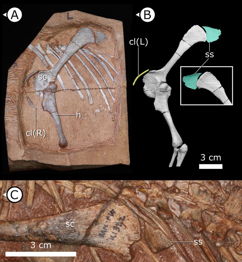
Research article                                                                                            Evolutionary Biology

                                   eLife digest The fossilised skeletons of long extinct dinosaurs are more than just stones. By
                                   comparing these remains to their living relatives such as birds and crocodiles, palaeontologists can
                                   reveal how dinosaurs grew, moved, ate and socialised. Previous research indicates that dinosaurs
                                   were likely warm-blooded and also more active than modern reptiles. This means they would have
                                   required breathing mechanisms capable of supplying enough oxygen to allow these elevated
                                   activity levels.
                                       So far, much of our insight into dinosaur breathing biology has been biased towards dinosaur
                                   species more closely related to modern birds, such as Tyrannosaurus rex, as well as the long-necked
                                   sauropods. The group of herbivorous dinosaurs known as ornithischians, which include animals with
                                   head ornamentation, spikes and heavy body armour, like that found in Triceratops and Stegosaurus,
                                   have often been overlooked. As a result, there are still significant gaps in ornithischian biology,
                                   especially in understanding how they breathed.
                                       Radermacher et al. used high-powered X-rays to study a new specimen of the most primitive
                                   ornithischian dinosaur, Heterodontosaurus tucki, and discovered that this South African dinosaur has
                                   bones researchers did not know existed in this species. These include bones that are part of the
                                   breathing system of extant reptiles and birds, including toothpick-shaped bones called gastralia,
                                   paired sternal bones and sternal ribs shaped like tennis rackets.
                                       Together, these new pieces of anatomy form a complicated chest skeleton with a large range of
                                   motion that would have allowed the body to expand during breathing cycles. But this increased
                                   motion of the chest was only possible in more primitive ornithischians. More advanced species lost
                                   much of the anatomy that made this motion possible. Radermacher et al. show that while the chest
                                   was simpler in advanced species, their pelvis was more specialised and likely played a role in
                                   breathing as it does in modern crocodiles.
                                       This new discovery could inform the work of biologists who study the respiratory diversity of both
                                   living and extinct species. Differences in breathing strategies might be one of the underlying
                                   reasons that some lineages of animals go extinct. It could explain why some species do better than
                                   others under stressful conditions, like when the climate is warmer or has less oxygen.

                                       Pulmonary ventilatory systems are highly integrated arrangements, with thoracoabdominal vol-
                                   ume change influenced by multiple anatomical regions. Unidirectional airflow is likely a synapomor-
                                   phy of diapsids (Schachner et al., 2014; Cieri et al., 2014; Schachner et al., 2013; Farmer and
                                   Sanders, 2010), but the mechanism by which air cycles through the lungs (i.e., ventilation) varies
                                   between clades. Interdependent sternocostal movement and visceral displacement drive volume
                                   changes in the compliant lungs of extant squamates (Owerkowicz et al., 1999; Brainerd et al.,
                                   2015; Cieri et al., 2018) and extant crocodilians (Claessens, 2009; Codd et al., 2019), whereas
                                   sternocostal movement and dorsoventral rocking of the sacrum ventilate the fixed, immobilized lung
                                   of birds (O’Connor, 2004). Osteological evidence of air sacs or pulmonary diverticula
                                   (O’Connor and Claessens, 2005; O’Connor, 2006; Wedel, 2006; O’Connor, 2009), and function-
                                   ally decoupled non-compliant and fixed gas-exchanging regions of the lung (Wang et al., 2018;
                                   Perry and Reuter, 1999; Schachner et al., 2009; Schachner et al., 2011; Brocklehurst et al.,
                                   2018) in the major lineages Theropoda and Sauropodomorpha, have led to the modern consensus
                                   that most dinosaurs had a ‘proto avian-like’ respiratory system. Pulmonary anatomy similar to the
                                   avian-like respiratory system is also hypothesized to have been present in pterosaurs (Butler et al.,
                                   2009; Claessens et al., 2009), leading to the hypothesis that aspects of proto-avian respiration,
                                   including air sacs, are plesiomorphic for the clade Ornithodira (Pterosauria + Dinosauromorpha)
                                   (Wedel, 2006; Brocklehurst et al., 2020).
                                       The aberrant morphology of ornithischian dinosaurs presents a fundamental challenge to this
                                   hypothesis. Ornithischians lack aspects of the abdominally mediated breathing apparatus (e.g., gas-
                                   tralia) or the sternocostal apparatus (e.g., mobile sternal ribs) that form key components of the inte-
                                   grated ventilatory systems of other diapsids. Additionally, all known ornithischians lack conspicuous
                                   evidence of postcranial skeletal pneumaticity (PSP) that is present in other ornithodiran lineages,
                                   indicating that ornithischians did not have pulmonary diverticula that invaded the skeleton

Radermacher et al. eLife 2021;10:e66036. DOI: https://doi.org/10.7554/eLife.66036                                                   2 of 39
Research article                                                                                            Evolutionary Biology

                                   (Butler et al., 2012). These observations provide at least two potential hypotheses for ornithischian
                                   breathing: (1) these taxa had avian-like air sacs that did not invade the skeleton (similar to some
                                   extant diving birds, e.g., Bucephala clangula); or, (2) they had a ventilatory strategy entirely diver-
                                   gent from other ornithodirans including living birds. Recently, the lung compliance (the elastic defor-
                                   mation capability of the lung during ventilatory cycles) of ornithischians was reconstructed as
                                   uniquely bipartite, with an inflexible, non-compliant anterior portion and a compliant posterior por-
                                   tion (Schachner et al., 2009; Schachner et al., 2011; Brocklehurst et al., 2018). This reconstruction
                                   fundamentally differs from reconstructions of other major dinosaurian lineages, which are hypothe-
                                   sized to bear a uniformly non-compliant lung as in living birds that is ventilated via expansion and
                                   contraction of anterior and posterior extrapulmonary ventilatory air sacs.
                                      Some previous studies have proposed that ornithischians evolved novel ventilation mechanisms
                                   (Norman, 2021; Brett-Surman, 1989; Carrier and Farmer, 2000), but supporting transitional mor-
                                   phological evidence and quantitative comparative analyses are lacking. For example, Brett-Surman
                                   (Brett-Surman, 1989) argued that the enlarged anterior pubic process (APP) in ornithopods was evi-
                                   dence of the presence of a muscle analogous to the hepatic piston of crocodilians (M. diaphragmati-
                                   cus), and that the elaboration of the APP in hadrosaurs was a subsequent necessity to force air
                                   through their intricate and complex narial pathways. Others have suggested instead that non-inva-
                                   sive diverticula characterized ornithischian lineages (Butler et al., 2012). However, since the more
                                   than 500 known taxa of ornithischian dinosaurs occupied similar habitats (Zanno et al., 2009;
                                   Zanno and Makovicky, 2011), size ranges (Benson et al., 2014), and postures as other dinosaur
                                   groups, the absence of PSP strongly suggests that they had a lung structure fundamentally different
                                   from other ornithodirans.
                                      Since its discovery in 1962 (Crompton and Charig, 1962), Heterodontosaurus tucki has long
                                   been recognized as a taxon crucial for resolving early ornithischian phylogenetic relationships. Most
                                   studies seeking to understand ornithischian origins have examined a single articulated skeleton,
                                   SAM-PK-K1332, which was fully prepared from its matrix nearly 50 years ago (Sereno, 1984;
                                   Sereno, 1986; Cooper, 1985; Maryanska and Osmólska, 1985; Butler et al., 2008; Boyd, 2015).
                                   Here, we present a new exquisitely preserved articulated skeleton, AM 4766 (age, stratigraphic prov-
                                   enance, and sedimentological context in Appendix 1). Cautious manual preparation and synchrotron
                                   radiation X-ray micro-computed tomography (SRmCT) using an innovative imaging protocol reveal
                                   new and unexpected elements of this taxon’s anatomy that are not preserved in any other speci-
                                   mens. Much of this new anatomical information has significant bearing on interpretations of the mac-
                                   roevolution of ornithischian respiratory biology. To further our understanding of ornithischian
                                   ventilation, we also quantitatively investigated size and shape changes in the evolution of the ornith-
                                   ischian pelvic girdle, paying special attention to the APP. Using a broad sample of ornithischian taxa,
                                   we use these data to investigate hypotheses that have implicated the APP in lung ventilation (Brett-
                                   Surman, 1989; Carrier and Farmer, 2000).

                                   Institutional abbreviations
                                   AM, Albany Museum, Makanda, Eastern Cape, South Africa; NCSM, North Carolina Museum of Nat-
                                   ural Sciences, Raleigh, North Carolina; SAM, Iziko South African Museum, Cape Town, South Africa.

                                   Results
                                   New anatomy
                                   The osteology of H. tucki has been described elsewhere (Crompton and Charig, 1962; Santa Luca
                                   et al., 1976; Sereno, 2012; Galton, 2014); we focus instead on novel anatomical features preserved
                                   in AM 4766 (Figure 1A). Visualization of these features was made possible by a high-resolution,
                                   phase-contrast enhanced SRmCT with a bespoke reconstruction algorithm developed for this particu-
                                   lar specimen (elaborated further in Appendix 1) that has recently been used elsewhere (Cau et al.,
                                   2017).

                                   Gastralia
                                   Approximately 18 gastralia are present in total and would have produced two longitudinal rows with
                                   each containing 9 gastralia. The gastralia follow the ventral abdominal midline, from the posterior

Radermacher et al. eLife 2021;10:e66036. DOI: https://doi.org/10.7554/eLife.66036                                                   3 of 39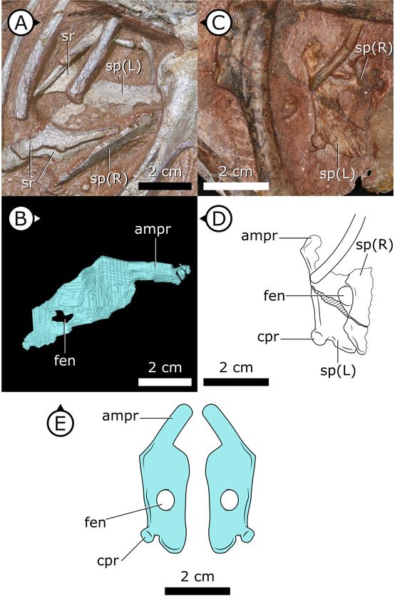
Research article                                                                                                                    Evolutionary Biology

                                   Figure 1. Overview of study specimen with emphasis on preserved gastralia. (A) Specimen AM 4766
                                   Heterodontosaurus tucki on left, with virtual anatomy reconstructed on the right. (B) Close-up of gastralia. (C)
                                   Stereopairs of anterior half of gastralial series. g: gastralia; f: femur (left); t: tibia (left); p: pubis; sr: sternal ribs.
                                   Arrows on figure labels point anteriorly.

Radermacher et al. eLife 2021;10:e66036. DOI: https://doi.org/10.7554/eLife.66036                                                                              4 of 39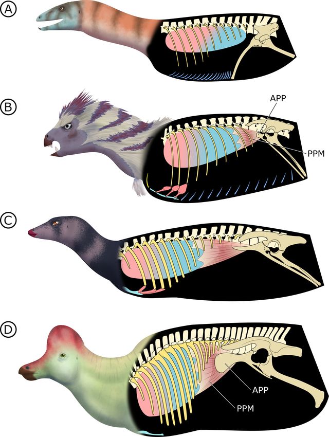
Research article                                                                                               Evolutionary Biology

                                   margin of the sternal plates to the level of the distal ends of the pubes (Figure 1B). The first two
                                   pairs of gastralia have slightly thickened medial facets that are absent from all subsequent pairs
                                   (Figure 1C), with the overall thickness of gastralia diminishing posteriorly. The gastralia are autapo-
                                   morphic among non-avian dinosaurs in lacking a lateral segment (Claessens, 2004; Fechner and
                                   Gößling, 2014; Barrett et al., 2019), which is retained in even the diminished gastral basket of early
                                   branching avialans (O’Connor et al., 2015).
                                       Fragments associated with the H. tucki specimen SAM-PK-K1332 are of comparable dimensions
                                   to the gastralia in AM 4766; however, they have been removed from context and could potentially
                                   represent displaced ossified tendons or posteriormost dorsal ribs. We tentatively identify the long,
                                   narrow bone fragments on either side of the proximal femur in the holotype specimen of Tianyulong
                                   confuciusi STMN 26-3 (Zheng et al., 2009; Appendix 1—figure 3) as gastralia based on their simi-
                                   larity with AM 4766.

                                   Sternal plates
                                   Two separate sternal plates are present, although only the left one is complete. The sternal plates
                                   are sub-rectangular, their long axes are oriented anteroposteriorly, and they are dorsoventrally thick-
                                   est on their lateral margin and progressively thin medially (Figure 2). The fenestra that perforates
                                   the centre of the sternal plate preserved in SAM-PK-K1332, identified by Sereno, 2012, is also pres-
                                   ent in AM 4766. The left sternal plate of AM 4766 bears an autapomorphic, anteromedially projec-
                                   ting, tongue-shaped process that projects abruptly from the anterolateral portion of the sternal
                                   plate (Figure 2B–E). The proximal portion of this process is partially visible in SAM-PK-K1332, but
                                   most of it is still obscured by matrix. The exact nature and function of the tongue-shaped process is
                                   currently unknown, but, when paired, they likely buttressed the region between coracoids. The pos-
                                   terolateral corner of the sternal plate of SAM-PK-K1332 has a small but distinct protuberance, identi-
                                   fied as an articulation for the sternal ribs (Sereno, 2012), herein referred to as a costal process
                                   (Figure 2C, D). While this structure appears to be missing from AM 4766, its absence cannot be con-
                                   fidently confirmed as the resolution in this region of the SRmCT data is diminished by metallic inclu-
                                   sions obscuring boundaries between bones and matrix.

                                   Sternal ribs
                                   Three pairs of sternal ribs are preserved in AM 4766, each similar in size and morphology (Figures 3
                                   and 4). The sternal ribs have a spatulate morphology, with an elongate and semi-cylindrical anterior
                                   half, and an abruptly dorsoventrally expanded posterior half that thins to a mediolaterally com-
                                   pressed, sheet-like structure, similar to the avialan Jeholornis prima (Zheng et al., 2020; Figure 4G).
                                   A thickened nub on the posterior apex of this sheet-like portion forms a monocondylar sternocostal
                                   articulation with the distal end of the corresponding dorsal rib. A similar articular relationship is pres-
                                   ent between the sternal, intermediate, and dorsal ribs of extant crocodilians (Claessens, 2009;
                                   Brocklehurst et al., 2017). Although the gross morphology of their sternal ribs differs, the sternal
                                   and dorsal ribs of pterosaurs (e.g., Rhamphorhynchus muensteri, Figure 4F) also bear monocondylar
                                   sternocostal joints that are strikingly similar to those of H. tucki (Claessens et al., 2009). The sternal
                                   ribs of AM 4766 contrast markedly with the few other ornithischian examples: for example, in The-
                                   scelosaurus neglectus (NCSM 15728) (Figure 4E) and Nanosaurus agilis (BYU 163) (identified as ‘cos-
                                   tal cartilage’ in Carpenter and Galton, 2018), the sternal ribs are comparatively shorter,
                                   subrectangular, and bearing broad butt joints rather than condylar articulations at their distal and
                                   proximal ends.

                                   Clavicles
                                   The paired clavicles (Figure 5A, B) are preserved in life position anterior to the scapulocoracoid and
                                   are proportionally long, thin, and bowed posteriorly. The proximal end of the left clavicle is margin-
                                   ally thicker than the rest of this element and gently tapers laterally. Among ornithischians, clavicles
                                   are mostly known in basal ceratopsians and neoceratopsians from the Cretaceous, for example, Psit-
                                   tacosaurus mongoliensis (Fairfield, 1924; Sereno, 1990), Psittacosaurus sibiricus (Averianov et al.,
                                   2006), Auroraceratops rugosus (Morschhauser et al., 2018a), Leptoceratops gracilis (Stern-
                                   berg, 1951), Montanoceratops cerorhynchos (Chinnery and Weishampel, 1998), and Protoceratops
                                   andrewsi (Brown and Schlaikjer, 1940) but are also present in the basal thyreophoran Scelidosaurus

Radermacher et al. eLife 2021;10:e66036. DOI: https://doi.org/10.7554/eLife.66036                                                      5 of 39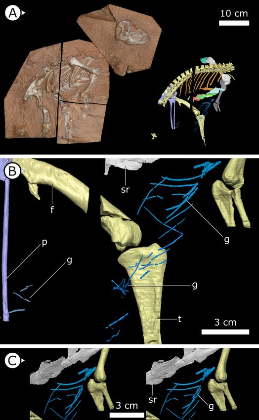
Research article                                                                                                         Evolutionary Biology

                                   Figure 2. Sternal plates of H. tucki. (A) Location of sternal plates in AM 4766, (B) segmented left sternal plate of
                                   AM 4766, and (C) sternal plates in SAM-PK-K1332. (D) Line drawing of sternal plates in SAM-PK-K1336. (E)
                                   Figure 2 continued on next page

Radermacher et al. eLife 2021;10:e66036. DOI: https://doi.org/10.7554/eLife.66036                                                                 6 of 39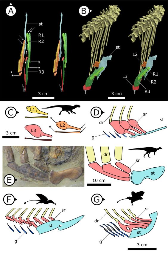
Research article                                                                                                               Evolutionary Biology

                                   Figure 2 continued
                                   Composite line drawing of H. tucki sternal plate anatomy informed by both specimens. ampr: anteromedial
                                   process; cpr: costal process; fen: fenestra; sp: sternal plate. Arrows on figure labels point anteriorly.

                                   harrisonii (Norman, 2020) as well as a new, undescribed taxon that is purported to be at the base of
                                   Ornithopoda (Spencer et al., 2020). The clavicles of H. tucki are similar to those of ceratopsians in
                                   contouring the anterior margin of the scapulocoracoid, with no apparent contact present between
                                   the clavicles along their length. The clavicles of AM 4766 differ from those of ceratopsians in overall
                                   size. AM 4766 has clavicles that are ~60% the length of the anterior margin of the body of the

                                   Figure 3. Stereopairs of segmented sternal ribs preserved in AM 4766. (A) Right lateral view and (B) left lateral
                                   view. st: sternal plate (left); L/R 1/2/3: left/right first, second, and third sternal ribs (anterior to posterior). Arrows on
                                   figure labels point anteriorly.

Radermacher et al. eLife 2021;10:e66036. DOI: https://doi.org/10.7554/eLife.66036                                                                        7 of 39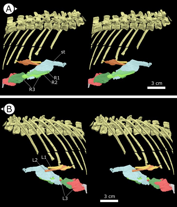
Research article                                                                                                        Evolutionary Biology

                                   Figure 4. Comparative details of sternal ribs in other ornithodirans. (A) Stereopairs of AM 4766 in (left) dorsal and
                                   (right) anteroventrolateral views. (B, C) Idealized version of sternal ribs present in AM 4766. (D, E) Photo and line
                                   drawing of sternal complex in Thescelosaurus neglectus (NCSM 15728). (F) Schematic sternal complex of
                                   Rhamphorhynchus, modified from Claessens et al., 2009. (G) Schematic sternal complex of Jeholornis, modified
                                   Figure 4 continued on next page

Radermacher et al. eLife 2021;10:e66036. DOI: https://doi.org/10.7554/eLife.66036                                                                 8 of 39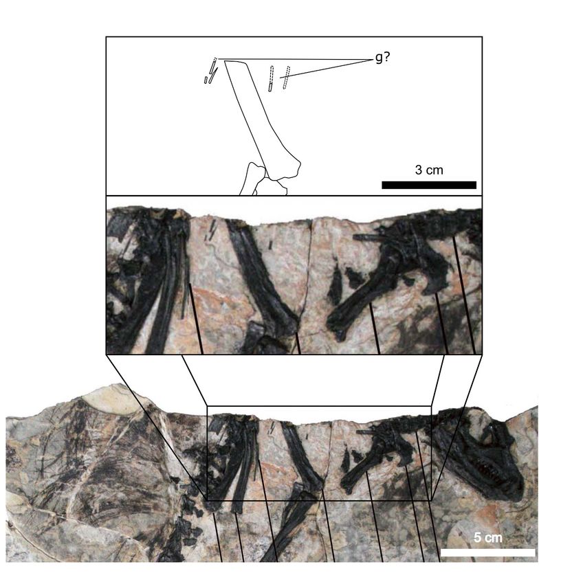
Research article Evolutionary Biology
Figure 4 continued
from Zheng et al., 2020. Arrows and asterisks point to sternal and dorsal rib articulation points, respectively. dr:
dorsal ribs; g: gastralia; L/R 1/2/3: left/right first, second, and third sternal ribs (anterior to posterior) of AM 4766;
sr: sternal ribs; st: sternal plates. Arrows on figure labels point anteriorly.
scapulocoracoid (i.e., excluding the scapular blade), where the clavicles of ceratopsians are approxi-
mately 40% of the length of the anterior margin of the scapulocoracoid.
Suprascapula
The suprascapula (Figure 5) is sub-trapezoidal in shape, being broader distally than it is proximally.
The distal margin of the suprascapula is concave and articulates with the proximal, convex margin of
the scapular blade. A suprascapula is also present in SAM-PK-K1332 and was originally described by
Santa Luca et al., 1976 as a ‘cartilaginous extension’ that capped the dorsal margin of the scapula.
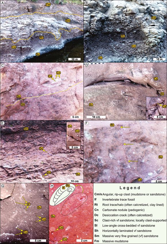
Figure 5. Accessory ossifications of pectoral girdle in H. tucki. (A, B) Clavicles and suprascapula of AM 4766 (B is
segmentation of mCT data); (C) suprascapula in SAM-PK-K1332. cl (L/R): left/right clavicle; h: humerus; sc: scapula;
ss: suprascapula. Arrows on figure labels point anteriorly.
Radermacher et al. eLife 2021;10:e66036. DOI: https://doi.org/10.7554/eLife.66036 9 of 39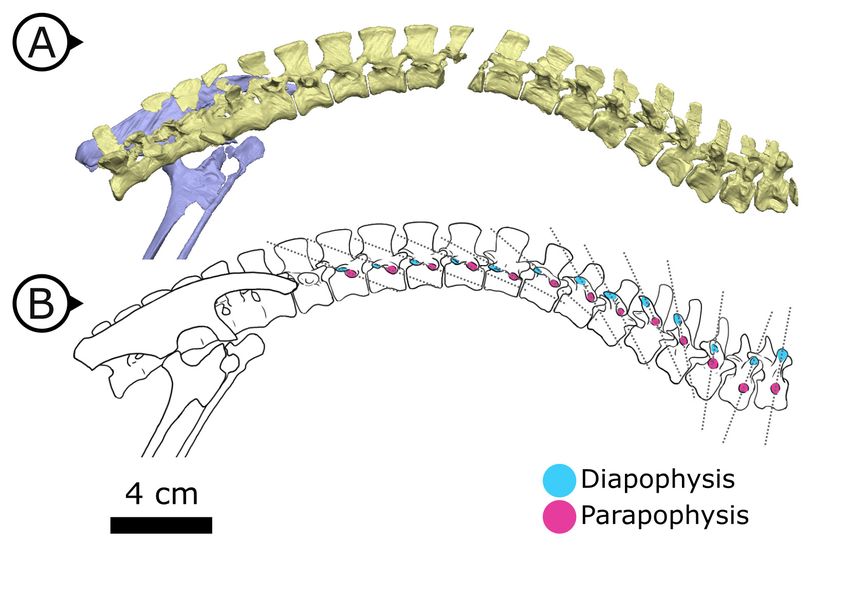
Research article                                                                                              Evolutionary Biology

                                   At present, we are unable to eliminate the possibility that this structure is indeed cartilaginous, but it
                                   is undistorted and has clearly defined margins that allow us to tentatively consider the suprascapula
                                   as an ossification (rather than a chondrification). An ossified suprascapula has only been described in
                                   one other dinosaurian taxon, the Cretaceous neornithischian Parksosaurus warreni
                                   (Sternberg, 1936).

                                   Internal thoracic ceiling and vertebral structure
                                   Synchrotron scanning of AM 4766 permitted the reconstruction of vertebral morphology previously
                                   obscured in specimen SAM-PK-K1332. Notably, the parapophyses migrate anterodorsally as the ver-
                                   tebral series progresses posteriorly (Figure 6): on the first, second, and third dorsal vertebrae, the
                                   parapophyses are located immediately ventral to the diapophyses; the fourth and fifth dorsal verte-
                                   brae mark the transition where the parapophyses migrate dorsally from the centrum and onto the
                                   neural arch; and from the sixth dorsal vertebrae and in all more posterior dorsals, the parapophyses
                                   are anterior to the diapophyses and on the same horizontal level. Our reconstruction based on
                                   SRmCT data shows that the entire internal structure of the cervical, dorsal, sacral, and proximal cau-
                                   dal vertebral column lacks pneumatic chambers or fossae, including those areas often implicated in
                                   the early evolution and development of PSP (Wedel, 2006; Butler et al., 2009; Benson et al.,
                                   2012), conclusively showing that early ornithischians lacked PSP.

                                   Quantitative analysis of ventral pelvic architecture
                                   Our measured variables show strong (log10 pubic rod length; r2 = 0.858) to very strong (log10 APP
                                   length, ischial length; r2 > 0.96) correlations with body size (represented in our analysis by log10
                                   femur length, see Materials and methods, and Appendix 1—figures 4 and 5). We therefore cor-
                                   rected for phylogenetic and allometric effects by using the residuals of phylogenetically corrected
                                   generalized least squares (pGLS) regressions of each variable against log10 femoral length. The resid-
                                   uals from our pGLS regression of each of our three variables showed poor correlation with log10
                                   femoral length. This indicates that changes in pubic and ischial dimensions are largely dissociated
                                   from the allometric effects of body size (see Appendix 1—figure 5A–F).
                                      Optimizing residuals on the phylogeny (Figure 7) shows that later branching taxa have APPs that
                                   are elongate relative to early branching taxa. APP elongation occurs at the base of Genasauria, and
                                   within this clade it is modified comparatively little over its subsequent history. There are generally
                                   declining rates of change in APP length in later-branching lineages and temporally later-appearing
                                   tips of the tree, with exactly zero known instances of reversion to the plesiomorphic relative length.
                                   Derived hadrosaurs and neoceratopsians apparently appear to have slightly shorter APPs relative to
                                   earlier-diverging taxa of their respective clades; however, it should be noted that these taxa dorso-
                                   ventrally expand the APP independently, significantly increasing the surface area for muscle
                                   attachment.
                                      Early branching ornithischians have long pubic rods, which subsequently shorten independently in
                                   ornithopods and marginocephalians well after the APP begins to elongate on the tree (i.e., after the
                                   major splits in Genasauria). Ischial length shows a more complex pattern, with most ornithischians
                                   retaining the plesiomorphic proportional length, but with stegosaurs showing large decreases in rel-
                                   ative length and ornithopods and certain ceratopsians showing modest increases.
                                      We used each set of residuals as continuous characters for an evolutionary model testing analysis
                                   using phylogenetic comparative methods (see Materials and methods). Among these, ‘Early Burst’ is
                                   strongly preferred for the evolution of APP length and performs better than other competing mod-
                                   els (Akaike Information Criterion [AICc] weight: 99.99%, likelihood ratio test p
Research article Evolutionary Biology
Discussion
Gastralia and their implications
The nearly sequentially complete gastral basket of AM 4766 is the first known in ornithischian dino-
saurs, and the tentative identification of gastralia in the holotype of the Chinese taxon T. confuciusi
suggests that gastralia may have been present in all heterodontosaurids. With gastralia being plesio-
morphically ubiquitous across a range of tetrapod clades, discovering gastralia in Heterodontosauri-
dae is not surprising as this clade is consistently recovered as the earliest branching lineage of
ornithischian dinosaurs (Butler et al., 2008; Boyd, 2015). It is more surprising, however, that these
gastralia are retained in H. tucki despite its typical ornithischian retroverted pubis. Three-dimensional
reconstruction of our SRmCT data clearly demonstrate gastralia in close association with the distal-
most point of the pubes, indicating that their complete retroversion (opisthopuby) was achieved
with the gastralia still intimately coupled. Together, these observations contest previous hypotheses
that reasoned that a divorce of the gastral basket from the pubis was a necessary prerequisite for
ornithischian pubic retroversion (Rasskin-Gutman and Buscalioni, 2001). Furthermore, the associa-
tion of the gastralia with the distal end of the pubic rod indicates that the latter structure is homolo-
gous to the pubic shaft/apron of other archosaurs (Galton, 1970, contra references therein), and
that the APP is a de novo feature.
Sternal ribs and their function
The presence of sternal ribs in H. tucki extends the occurrence of these bones from late diverging
taxa like T. neglectus and other relatively late-branching, small-bodied Late Jurassic and Cretaceous
neornithischians (Carpenter and Galton, 2018; Butler and Galton, 2008) to the basalmost mem-
bers of Ornithischia. This broader distribution strongly implies that the presence of sternal ribs may
optimize as an ornithischian plesiomorphy. However, the sternal ribs we describe in H. tucki are auta-
pomorphic in morphology, differing markedly from those of other ornithischians, and showing clear
evidence of being mobile about their dorsal rib and sternal plate joints (Figure 4A–C). The dorso-
ventrally expanded dorsal and ventral margins of these ribs were likely attachment sites for intercos-
tal musculature and in this way perhaps analogous to similar projections (sternocostapophyses) on
the sternal ribs of pterosaurs (Claessens et al., 2009), the uncinate processes of maniraptorans
(Tickle et al., 2012; Codd et al., 2008), and the remarkably similar sternal ribs of the ornithothora-
cine J. prima (Zheng et al., 2020) – all of which are adaptations hypothesized to increase lever-arm
potential and facilitate efficient deformation of the body wall to drive ventilation.
Sternum
The complex sternal plates of AM 4766 are distinct from the comparatively simple ‘hatchet-shaped’
sternal plates of iguanodontians and the ‘kidney-shaped’ (reniform) sterna of other neornithischians
but are not unique among Dinosauria. Instead, the complex sternal plates of AM 4766 bear similari-
ties with early- and late-diverging theropods such as Tawa hallae (Bradley et al., 2019) and various
avialans (Zheng et al., 2012; O’Connor et al., 2015), respectively. Features like the tongue-shaped
process of AM 4766 and the coracoid facet of T. hallae are strikingly similar in their dimensions, loca-
tion, and abrupt change in orientation relative to the posterior half of their respective sternal plates.
Further similarities include the single knob-like costal process of AM 4766 exhibiting a similar mor-
phology to the series of costal processes of T. hallae, and the analogous position of the lateral tuber-
cula of enantiornithines (Zheng et al., 2012). It is unclear whether these sternal similarities are
homologous, but they are likely functionally analogous.
Quantitative analysis of pelvic evolution
The nature of change in the relative length of the APP is conspicuous from both qualitative and
quantitative analyses of pelvic evolution. Innovation in APP length occurred early in ornithischian
evolution, before the diversification of genasaurians, and after this significant early burst (see Fig-
ure 7, Table 1), modifications of the APP were generally restricted to gross shape differences that
do not affect the relative length: derived ornithopods evolved a large, lobate APP with derived neo-
ceratopsians evolving an APP that fanned-out anteriorly. We interpret these results as rapid
Radermacher et al. eLife 2021;10:e66036. DOI: https://doi.org/10.7554/eLife.66036 11 of 39Research article Evolutionary Biology
Figure 6. Changing diapophyseal and parapophyseal relationships in AM 4766. (A) Virtual reconstruction of
cervicothoracic, thoracic, and sacral vertebrae of AM 4766; (B) Line drawing of (A) with diapophyses and
parapophyses colour-coded in cyan and magenta respectively. Dashed lines indicate shifting position of
parapophyses relative to the accompanying diapophyses. Arrows on figure labels point anteriorly.
switching of optimal phenotypes for APP size; from the plesiomorphically small condition in
H. tucki to a proportionally long APP that is then maintained across all later-branching ornithischian
lineages.
We consider this as evidence that the APP was involved in a major macroevolutionary shift in
ornithischian dinosaurs, which occurred immediately prior to their radiation in the Jurassic. More-
over, the timing of this change in the relative length of the APP cooccurs with the reduction and loss
of the gastralia and possibly the loss or reduction of sternocostal mobility. These results are consis-
tent with, but greatly expand upon, Brett-Surman’s hypothesis (Brett-Surman, 1989) that the
enlarged APP is an adaptation of ornithischians involved in driving considerable changes in abdomi-
nothoracic volume.
The pelves of published ankylosaur specimens are often obscured or incomplete and do not con-
tain sufficient measurement information to include in this analysis (Kirkland and Carpenter, 1994;
Carpenter et al., 2013; Arbour and Currie, 2013; Xu et al., 2001). Nevertheless, we observe that
despite highly derived pubic rod morphologies, including near loss in late branching taxa like Euo-
plocephalus tutus, the APP is retained as a process fused to the ventral surface of the ilium
(Carpenter et al., 2013). This strongly implies that the APP was subject to a constraint that favoured
its retention when the rest of the pubis was made redundant.
This pattern of pubic evolution is best explained by a ‘Drift’ evolutionary hypothesis and suggests
a trend away from elongate pubes. The reduction of the pubic rod is pervasive in ornithischians as it
is independently lost in derived iguanodontians, neoceratopsians, and pachycephalosaurs (as well as
ankylosaurs; Carpenter et al., 2013). This likely signifies a relaxing of a plesiomorphic constraint
between the hypaxial abdominal musculature and the pubis. This reduction of the pubic rod is
decoupled temporally and phylogenetically from the loss of gastralia and the expansion of the APP,
Radermacher et al. eLife 2021;10:e66036. DOI: https://doi.org/10.7554/eLife.66036 12 of 39Research article Evolutionary Biology
Figure 7. Phylogenetically corrected results of ornithischian pelvic element analysis. Phylogenetically corrected
generalized least squares residuals results of (A) evolution of the anterior pubic process, (B) pubic rod, and (C)
ischial length. Closed circle, Genasauria; open triangle, Ornithopoda; closed triangle, Marginocephalia.
Figure 7 continued on next page
Radermacher et al. eLife 2021;10:e66036. DOI: https://doi.org/10.7554/eLife.66036 13 of 39Research article Evolutionary Biology
Figure 7 continued
Silhouettes represent (from left to right): Heterodontosaurus, Stegosauria, Parksosauridae, Neoceratopsia, and
Hadrosauridae.
strongly suggesting that the pubic rod was rendered vestigial. It is possible that modifications like
the bowed ischium of neoceratopsians and the ‘ischial boot’ of some hadrosaurs are responses to
the ischium subsuming the myological role previously played by the pubis.
Ischial residuals are harder to interpret, and despite being best explained by a ‘Stasis’ model,
there is no statistical significance between this explanatory model or any other evolutionary models
we tested. Qualitatively, most ornithischian dinosaurs have similar-length ischia relative to their body
size. The most conspicuous departures from this are in late-branching ornithopods, where the
ischium is elongated (i.e., strong positive residuals), and in stegosaurs where the ischium is short-
ened (i.e., strong negative residuals). That the pubic rods of stegosaurs remained elongate and the
ischia short and robust almost certainly indicates that some other selective pressure such as tail-
driven defence (Mallison, 2011; Carpenter et al., 2005) was imposed on the ischium that prevented
it from supplanting the role of the pubis in anchoring abdominal musculature. Nevertheless, both
ischial modifications occur temporally and phylogenetically well after the increase in relative APP
length and the loss of gastralia.
A new model of lung ventilation in ornithischian dinosaurs
The anatomical features presented here provide consilient evidence that H. tucki preserves morphol-
ogies that reflect early steps in the evolution of a novel means of lung ventilation in ornithischian
dinosaurs. Below, we review this evidence and propose a potential model for the ornithischian venti-
lation system.
Gastralial modification
Gastralia are widespread among Palaeozoic and Mesozoic tetrapods but have never before been
unambiguously reported in Ornithischia. Among other dinosaurian lineages, theropods retain their
gastral basket until the evolution of neornithine birds, and sauropodomorphs lose their gastralia rela-
tively late in their evolutionary history at the base of Eusauropoda – potentially retaining gastralia
even during the emergence of Neosauropoda (Tschopp and Mateus, 2013).
The reduction or loss of the gastralia independently occurs in other major tetrapod lineages like
stem turtles (Lyson et al., 2013; Schoch and Sues, 2020), and eutheriodont therapsids including
mammals (Cisneros et al., 2015). Interestingly, specialized lung ventilatory mechanisms are present
in all extant clades that have lost (birds, mammals) or co-opted the gastralia (turtles). Some workers
have explicitly linked the loss of gastralia and subsequent thoracic and lumbar differentiation of ther-
ocephalian and cynodont therapsid axial skeletons to the evolution of a mammalian-style, dia-
phragm-driven ventilatory arrangement (Perry et al., 2010; Brink, 1956).
The specialized facets on the medial gastralial elements of non-avian theropods have led multiple
authors (Carrier and Farmer, 2000; Claessens, 2004; Fechner and Gößling, 2014; Codd et al.,
2008; Lambe, 1917) to hypothesize that gastralia played an important role in ventilating the lungs,
a mechanism Carrier and Farmer, 2000 termed ‘cuirassal breathing’ that was inherited from a com-
mon non-dinosaurian archosaurian ancestor. These hypotheses posit the gastralia would have
Table 1. Akaike Information Criterion weights and likelihood ratio test (p) statistics for the evolutionary models analysed here (see
Materials and methods).
Bold values indicate preferred explanatory model for each measured pelvic variable. Likelihood ratio tests are between the preferred
model and the next most preferred model. BroMo: Brownian Motion; OU: Ornstein–Uhlenbeck.
BroMo (%) OU (%) Early-burst (%) Drift (%) Stasis (%) p=
07
APP length 0.00 0.00 99.99 0.00 0.00 1.33
Pubis length 11.06 3.56 2.97 82.34 0.00 0.01
Ischium length 5.21 25.24 8.73 1.45 59.37 1.00
Radermacher et al. eLife 2021;10:e66036. DOI: https://doi.org/10.7554/eLife.66036 14 of 39Research article Evolutionary Biology
facilitated expansion and contraction of the body wall to facilitate volumetric changes in the thora-
coabdominal cavity (Carrier and Farmer, 2000; Claessens, 2004; Codd et al., 2008; Lambe, 1917).
Although in extant crocodilians the gastralia themselves only contribute a relatively small amount to
such volumetric changes in isolation (Claessens, 2009), the gastral basket is integral in bridging the
sternocostal complex and mobile pubis, serving as an attachment site for muscles fundamental to
body wall deformation and function of the ‘hepatic piston’.
The archosaurian pelvis as a respiratory locus
In archosaurian ventilation models, the involvement of the pelvis is ubiquitous, ranging from pelvic
rocking in birds (Baumel et al., 1990), to the hepatic piston in crocodilians (Farmer and Carrier,
2000), to the prepubis of pterosaurs (Claessens et al., 2009). Anterior bony projections of the pubic
region are key components of these models, including the mobile pubis of crocodilians, and the pre-
pubis and puboiliac complex in pterosaurs. Carrier and Farmer, 2000 highlighted the APP as the
integral locus for interpreting ornithischian lung ventilation, focusing their hypothesis on the major
genasaurian clades Neoceratopsia, Ornithopoda, and Stegosauria. Macaluso and Tschopp, 2018
hypothesized that pubic retroversion in dinosaurs is linked to the evolution of an innovative ventila-
tory mechanism, arguing that the plesiomorphic cuirassal breathing proposed by Carrier and
Farmer, 2000 constrains the pubis into the propubic condition, and that evolution of mesopubic
and opisthopubic conditions indicates a relaxing of those constraints as a new mechanism evolves.
The role of the APP in ventilation has been contentious, however, with other authors assigning it
a locomotory function (as the origin of the ambiens [Maidment and Barrett, 2011] or pubotibialis
[Galton, 1969] muscles). Although the locomotory and ventilatory explanations are not mutually
exclusive, the evidence gathered here makes us consider the locomotory role to be a poor explana-
tion for APP changes for the following reasons. First, our evolutionary analysis clearly shows that the
major changes in the APP residuals are phylogenetically and temporally divorced from the indepen-
dent acquisitions of quadrupedality and major postural changes (Maidment and Barrett, 2011;
Maidment and Barrett, 2012a; Maidment and Barrett, 2012b; Barrett and Maidment, 2017).
Second, although there is little available data from extant taxa, the ambiens muscle appears to have
weak negative allometry in Dromaius novaehollandiae (Lamas et al., 2014), suggesting that body
size increases in ornithischian lineages would not drive a trend of disproportional APP increase (addi-
tionally, our analysis of pubic measurements using residuals precludes this). Third, the APP is subpar-
allel to the vertebral column and medial to the ribcage, thus precluding it from being a major driver
of hindlimb extension or retraction when body wall musculature is reconstructed (principally M. obli-
quus abdominus externus; Fechner and Gößling, 2014; Fechner and Schwarz-Wings, 2013). These
lines of evidence together indicate that the factors driving the evolution of APP length and shape
are distinct from locomotory influences.
The pelvic bellows
In total, our observations here show that H. tucki has reduced gastralia, an apomorphically elaborate
sternum, well-developed and mobile sternal ribs, an incipient APP, and completely lacks PSP. We
propose a single explanatory model for these observations: that H. tucki is a transitional animal pre-
serving the early steps in the evolution of a unique ventilation mechanism in ornithischian dinosaurs.
We name this model the ‘pelvic bellows’ and elaborate on it below. This model does not require us
to make ad hoc assumptions about airflow direction (i.e., unidirectional versus tidal), but phyloge-
netic bracketing predicts intrapulmonary unidirectional airflow in Ornithischia and is fully compatible
with our model (Schachner et al., 2014; Cieri et al., 2014; Farmer and Sanders, 2010;
O’Connor and Claessens, 2005).
Stem dinosauromorphs (Figure 8A; Kammerer et al., 2020) (or stem sulcimentisaurians;
Müller and Garcia, 2020) bear the plesiomorphic archosaurian condition of a typical gastral basket
connecting to a propubic pelvis, and they lack both PSP (Butler et al., 2009) and an APP. This points
to cuirassal ventilation as the primary means of volume change. Interestingly there is evidence of a
bipartite, semi-compliant lung in Silesaurus opolensis (Schachner et al., 2011), adding potential sup-
port to a hypothesized but controversial relationship between silesaurids and Ornithischia
(Müller and Garcia, 2020; Ferigolo and Langer, 2007).
Radermacher et al. eLife 2021;10:e66036. DOI: https://doi.org/10.7554/eLife.66036 15 of 39Research article Evolutionary Biology Figure 8. Hypothesized stepwise evolution of the ornithischian pelvic bellows and accompanying skeletal modifications and myological innovations. (A) Silesaurus (outgroup), (B) Heterodontosaurus, (C) Thescelosaurus, to (D) Corythosaurus. Lung size is an approximation; red and blue portions of the lung represent hypothetical reconstructions of non-compliant and compliant lung regions, respectively. APP: anterior pubic process; PPM: puboperitoneal muscle. Not to scale. Radermacher et al. eLife 2021;10:e66036. DOI: https://doi.org/10.7554/eLife.66036 16 of 39
Research article                                                                                             Evolutionary Biology

                                       In early branching ornithischians, exemplified by H. tucki (Figure 8B), the retroverted pubes and
                                   the reduced gastralia indicate that the cuirassal breathing mechanism (Carrier and Farmer, 2000) is
                                   still present but has reduced capacity to affect changes in volume. The sternal ribs of H. tucki would
                                   have facilitated the pivoting and leveraging of the sternum and aided in its posteroventral contrac-
                                   tion, providing substantial volumetric changes. The small APP would have served as a nascent area
                                   for the origination of a muscle analogous to the dorsal component of M. diaphragmaticus in living
                                   crocodilians, which we term the ‘puboperitoneal muscle’. We hypothesize that the puboperitoneal
                                   muscle would have functioned as an accessory lung ventilator in early ornithischians, similar to the
                                   accessory ventilatory function provided by the iliocostalis musculature of some crocodilians
                                   (Codd et al., 2019). The puboperitoneal muscle would have provided an additional anteroposterior
                                   vector to the dorsoventral displacement already afforded by the cuirassal and sternocostal mecha-
                                   nisms. Upon inspiration, contraction of M. rectus abdominus would have distended the gastral bas-
                                   ket and the sternal complex posteroventrally, the puboperitoneal musculature simultaneously
                                   contracting to generate negative pressure in the posteriorly compliant half of the lung. During expi-
                                   ration, the abdomen, sternal complex, and puboperitoneal muscle relaxed and would rebound ante-
                                   rodorsally to force air out.
                                       Gastralia are seemingly lost amongst early branching ornithischians, and the cuirassal breathing
                                   mechanism is no longer present in Genasauria (Figure 8C). However, the pubic rod is still long and
                                   M. rectus abdominus is likely still present, not entirely precluding the possibility of body wall defor-
                                   mation by contractions of hypaxial musculature. The sternal ribs are greatly simplified or lost. When
                                   they are present, broad, immobile butt joints replace the condylar joints between the sternum and
                                   sternal ribs. Additionally, the well-developed processes and eminences that were features of the
                                   sternum and sternal ribs of H. tucki are lost, simplifying the sternal complex in all subsequent clades.
                                   Together, this simplification of the sternum decreases the degrees of freedom for associated skeletal
                                   components, reducing both the range of motion of the sternocostal complex (relative to the plesio-
                                   morphic condition) and its ability to contribute to changes in abdominothoracic volume. The APP is
                                   prominent and anteriorly elongated, with anteroposteriorly oriented muscle scars present on the
                                   dorsal and medial surfaces (Morschhauser et al., 2018a; Owen, 1875). In Genasauria, the puboperi-
                                   toneal muscle is now the major contributor to changes in volume, with the sternum and the abdomi-
                                   nal musculature relegated to a secondary role.
                                       Finally, in deeply nested ornithischians (Figure 8D), the gastralia remain absent, with the pubic
                                   rod shortening to a spur (derived ornithopods) or a tab (derived neoceratopsians), indicating that
                                   abdominal musculature now attaches to the ischium and mainly functions to support the viscera. The
                                   sternal plates, where present, are relatively small, show no evidence of dorsal rib interaction, and dif-
                                   fer markedly in morphology between clades, suggesting that they played no constrained role in ven-
                                   tilation. Convergently, APP area is substantially enlarged and develops clade-specific morphologies.
                                   At this stage, the puboperitoneal muscle served as the main ventilatory apparatus, sternal move-
                                   ments contribute little to no volumetric changes, and the non-puboperitoneal abdominal muscula-
                                   ture functions mainly as support for viscera.
                                       The changing vertebrocostal orientations along the axial column of H. tucki observed here (Fig-
                                   ure 6) support the bipartite and dorsally immobile lung previously reconstructed in ornithischians
                                   and silesaurids (Schachner et al., 2011; Brocklehurst et al., 2018). Considering that a dorsally
                                   immobilized, anatomically and functionally heterogeneous lung has been reconstructed for all of
                                   Ornithischia (Schachner et al., 2011; Brocklehurst et al., 2018), and that the M. diaphragmaticus of
                                   extant crocodilians is coupled with a flexible lung and shifting viscera, the proposed ventilatory
                                   mechanism for Ornithischia was likely functionally distinct from the hepatic piston model present in
                                   crocodilians, although the two may have been anatomically convergent. The crocodilian M. dia-
                                   phragmaticus originates on both the pelvis anterior to the acetabulum and the gastralia (or pubic
                                   apron, depending upon the taxon) (Gans and Clark, 1976). It then fans out anteriorly, encapsulates
                                   all of the abdominal viscera (dorsally, laterally, and ventrally), and inserts on the liver, with fibres
                                   occasionally extending to the pericardium (Gans and Clark, 1976). The proposed puboperitoneal
                                   muscle in ornithischians originating on the APP (Figure 8B–D) is reconstructed here as travelling
                                   anteriorly, and inserting on any potential number of anatomical structures, including the dorsal sur-
                                   face of the liver, pulmonary septa, posteriorly positioned air sacs (or non-invasive pulmonary divertic-
                                   ula emerging from the lung), or even the posterior aspect of the lung itself if no pulmonary
                                   diverticula existed. This putative mechanism would be distinctly different from that of extant

Radermacher et al. eLife 2021;10:e66036. DOI: https://doi.org/10.7554/eLife.66036                                                   17 of 39
Research article Evolutionary Biology
crocodilians, particularly in the larger, later-branching ornithischian taxa in that there would be no
ventral attachment due to a loss of gastralia and the shortened pubis (Figure 8D). Without bundling
the abdominal viscera into a fusiform tube, there would be no anterior-posterior translation of the
entire visceral mass within the thoracocoelomic cavity. Additionally, ventilation of the anteriorly
immobilized respiratory parenchyma by a posterior/ventral flexible region (whether air sacs, or just a
flexible sac-like expansion) would not theoretically cause the same shifts in centre of mass that the
crocodilian hepatic-piston mechanism does (see, e.g., Uriona and Farmer, 2008), and may be more
functionally analogous to the complementary integration of pelvic musculature as observed in birds
(e.g., Columba livia; Baumel et al., 1990). This hypothesis posits that only the flexible regions of the
lung linked to the pelvic bellows would be stretching with contraction of the muscle, while the ante-
rior and dorsal regions of the lung containing the respiratory parenchyma could remain fixed and
immobilized to the adjacent skeletal tissues. This type of pulmonary heterogeneity is well docu-
mented in other sauropsids outside of birds (e.g., varanids [Schachner et al., 2014], chameleons
[Klaver, 1973], snakes [Wallach, 1998]), where there is an extreme separation of the respiratory
parenchyma and more flexible sac-like structures in the posterior region of the lung, and thus sup-
ports the possibility of these characters independently evolving in this lineage if this ventilatory
mode is truly divergent from other dinosaurs.
Lung ventilation in dinosaurs is probably more complicated
Investigation into dinosaur respiration has focused on PSP, using its presence or absence as a sole
proxy for avian-like ventilation and physiology (O’Connor and Claessens, 2005; O’Connor, 2006;
Butler et al., 2012; Wedel, 2003). Recent studies showing the remarkable multiplicity of respiratory
systems employed by living reptiles (Owerkowicz et al., 1999; Cieri et al., 2018; Claessens, 2009;
Brocklehurst et al., 2017; Baumel et al., 1990; Farmer and Carrier, 2000; Lyson et al., 2014)
show that PSP is only one component of a complex suite of features that coevolve to enable lung
ventilation across a swathe of tetrapod lineages. This recent research shows that some presumed
‘bird-like’ respiratory features, such as unidirectional air flow, are actually plesiomorphies characteriz-
ing much larger groups (Schachner et al., 2014; Farmer and Sanders, 2010) and highlights the
diversity of ways in which multiple anatomical systems interlink to effectively ventilate the lungs.
New work on the pulmonary anatomy of the ostrich (Struthio camelus) has demonstrated that PSP
relationships with the respiratory system in extant birds may not be as straightforward as previously
thought (Schachner et al., 2021), and reconstructions of dinosaur lungs that directly follow a stan-
dardized avian bauplan may need to be reconsidered. Additionally, primitive features like gastralia,
simple sterna, and ‘propubic’ pelves impede attempts at completely superimposing the highly
derived physiology of birds onto comparatively less-specialized clades like non-avian theropods and
sauropods.
Inquiry into ornithischian breathing has been stunted by virtue of their phylogenetic position
between clades that have received more thorough respiratory evolution investigation
(O’Connor and Claessens, 2005; Wedel, 2006; Butler et al., 2009; Claessens et al., 2009;
Fechner and Gößling, 2014; Zheng et al., 2020; Tickle et al., 2012; Codd et al., 2008;
Wedel, 2003) paired with inferences informed by phylogenetic bracketing (Witmer, 1995). As dis-
cussed here, ornithischians are outliers among ornithodirans for many reasons – in particular, their
unique lung structure and lineage-wide lack of PSP contradict the more parsimonious inferences
made about their respiratory anatomy (i.e., that ornithischians are predicted to have conspicuous
air sacs).
Archosaurs likely demonstrate a remarkably labile respiratory evolution that has yet to be fully
appreciated, and future inquiry is at risk of overlooking a variety of ventilatory mechanisms that are
obfuscated by more parsimonious explanations. This suggests that dinosaur, and archosaur, breath-
ing should be investigated with a more nuanced view of evolution; a paradigm that is informed by
extant respiratory diversity and that is simultaneously willing to risk relaxing an insistence on phylo-
genetic bracketing in an attempt to capture increased ventilatory diversity in extinct lineages. Similar
trappings will inevitably extend beyond the topic of respiratory evolution, with the evolution and
homology of archosaurian integumentary structures being an additional area that will likely struggle
from comparable oversimplifications.
The success of ornithischians is remarkable, and the reason for the marked differences between
their body plans and those of other dinosaurs remains enigmatic. The diverging ventilatory
Radermacher et al. eLife 2021;10:e66036. DOI: https://doi.org/10.7554/eLife.66036 18 of 39Research article Evolutionary Biology
adaptations hypothesized here provide an overarching explanation for a wide range of skeletal mod-
ifications, and perhaps accompanying metabolic and physiological changes, that shaped the lineage
for 130 million years. It is likely no coincidence that the evolution of the APP and its hypothesized
role in ventilation precede dramatic and conspicuous increases in ornithischian diversity and
disparity.
Materials and methods
Statistical analysis
To quantify pelvic evolution in Ornithischia, we measured femoral length, APP (from the anterior
margin of the acetabulum), pubic rod (from in line with the anterior margin of the acetabulum to the
distalmost tip), and ischial (the contour of the posterior surface that initiates on the iliac peduncle
and terminates at the middle of the distalmost point) lengths for a phylogenetically broad sample of
ornithischian taxa (Appendix 1—table 1) through direct measurements of specimens, high-resolu-
tion photos, and published sources. We measured specimens either by hand, using digital callipers
and measuring tapes, digitally from 3D SRmCT data, or from high-resolution photos of specimens
where scale bars were available and accurate. To normalize scale, we log10-transformed all measure-
ments. We selected pubic and ischial measurements because of their hypothesized relationship with
lung ventilation (in particular, plesiomorphic models like cuirassal breathing; Carrier and Farmer,
2000). We chose femoral length as a proxy for body mass, even though it has lower correlation coef-
ficients than femoral circumference for body mass estimation (Campione and Evans, 2012;
Anderson et al., 1985). We used it here because circumference measurements were unavailable for
most of our specimens and because femoral length is frequently used in the literature and therefore
practical to collect (e.g., Christiansen and Fariña †, 2004). Although it is unlikely that this choice
greatly affects the results we present here, stegosaurs appear to have apomorphically long femora
that are likely to affect body mass corrections for these taxa specifically; we accept this localized
trade-off in error for the benefit of standardized measurements across our sampling of Ornithischia.
We analysed these data using scripts written in the R statistical software language (R Development
Core Team, 2013) and its associated packages ‘ape’ (Paradis et al., 2004), ‘ggplot2’ (Wick-
ham, 2016), ‘phytools’ (Revell, 2012), ‘strap’ (Bell and Lloyd, 2015), ‘geiger’ (Harmon et al.,
2008), and ‘nlme’ (Pinheiro et al., 2012).
To investigate evolutionary patterns in the APP, pubic rod, and ischium, we used pGLS regres-
sions of these pelvic measurements against femoral length and calculated residuals from these
regressions. This is a common means of assessing phylogenetic and size-corrected variance in mor-
phological datasets and can be used together with comparative phylogenetic methods (Rev-
ell, 2009; Hunt and Carrano, 2010). We used the residuals as continuous characters to both
qualitatively map on the ornithischian tree and to assess the fit of a variety of macroevolutionary
models implemented in the R package Geiger (Harmon et al., 2008). We used the corrected AICc
and computed likelihood ratio tests to assess whether the preferred model is significantly better
than the next-best model. ‘Source code 1’ is R code to reproduce statistical analysis; ‘Source code
2 and 3’ are phylogenetic tree files in .phy and .nex formats, respectively; and ‘Source code 4’ is
‘First Appearance Date’ and ‘Last Appearance Date’ data of taxa analysed, obtained from the Paleo-
biology Database (paleobiodb.org).
Geological context
AM 4766 was recovered from the upper Elliot Formation (uEF) in strata that correlate with the Mas-
sospondylus Assemblage Zone (Viglietti et al., 2020) and is likely Sinemurian in age (Bordy et al.,
2020). The specimen was recovered from a light red, clast-rich, very fine-grained sandstone that is
consistent with palaeo-environmental reconstructions of the uEF as a seasonally wet, fluvio-lacustrine
system (Bordy et al., 2004a). Further details of the geological context are elaborated in Appendix 1
and figured in Appendix 1—figures 1 and 2.
Digital specimen reconstruction
Volume files of AM 4766 were reconstructed using a combination of manual and semi-automated
tools (i.e., pen tool, interpolate function) in Avizo Lite version 9.0 (FEI Visualization Sciences Group,
Radermacher et al. eLife 2021;10:e66036. DOI: https://doi.org/10.7554/eLife.66036 19 of 39You can also read