Reviews and syntheses: Bacterial bioluminescence - ecology and impact in the biological carbon pump - Biogeosciences
←
→
Page content transcription
If your browser does not render page correctly, please read the page content below
Biogeosciences, 17, 3757–3778, 2020
https://doi.org/10.5194/bg-17-3757-2020
© Author(s) 2020. This work is distributed under
the Creative Commons Attribution 4.0 License.

Reviews and syntheses: Bacterial bioluminescence – ecology and
impact in the biological carbon pump
Lisa Tanet1, , Séverine Martini1, , Laurie Casalot1 , and Christian Tamburini1
1 Aix   Marseille Univ., Université de Toulon, CNRS, IRD, MIO UM 110, 13288 Marseille, France
 These   authors contributed equally to this work.

Correspondence: Christian Tamburini (christian.tamburini@mio.osupytheas.fr)

Received: 21 February 2020 – Discussion started: 19 March 2020
Revised: 5 June 2020 – Accepted: 14 June 2020 – Published: 17 July 2020

Abstract. Around 30 species of marine bacteria can emit          1   Introduction
light, a critical characteristic in the oceanic environment is
mostly deprived of sunlight. In this article, we first review
current knowledge on bioluminescent bacteria symbiosis in        Darkness constitutes the main feature of the ocean. Indeed,
light organs. Then, focusing on gut-associated bacteria, we      the dark ocean represents more than 94 % of the Earth’s hab-
highlight that recent works, based on omics methods, con-        itable volume (Haddock et al., 2017). Moreover, the surface
firm previous claims about the prominence of biolumines-         waters are also in dim light or darkness during nighttime.
cent bacterial species in fish guts. Such host–symbiont re-      Organisms living in the dark ocean biome are disconnected
lationships are relatively well-established and represent im-    from the planet’s primary source of light. They must adapt
portant knowledge in the bioluminescence field. However,         to a continuous decrease in sunlight reaching total darkness
the consequences of bioluminescent bacteria continuously         beyond a few hundred meters. Hence, it is not surprising that
released from light organs and through the digestive tracts      76 % of marine pelagic meso- and macroorganisms are bio-
to the seawater have been barely taken into account at the       luminescent from the surface to the deep sea, without vari-
ecological and biogeochemical level. For too long neglected,     ability over depth, and that bioluminescence is a major eco-
we propose considering the role of bioluminescent bacteria       logical function in interactions (Martini and Haddock, 2017).
and reconsidering the biological carbon pump, taking into        Bioluminescent species are found in most phyla from fish to
account the bioluminescence effect (“bioluminescence shunt       bacteria (Haddock et al., 2010; Widder, 2010). Amongst ma-
hypothesis”). Indeed, it has been shown that marine snow         rine light-emitting organisms, luminous bacteria are widely
and fecal pellets are often luminous due to microbial col-       distributed in oceans. Luminescent bacteria can glow contin-
onization, which makes them a visual target. These lumi-         uously under specific growth conditions (Nealson and Hast-
nous particles seem preferentially consumed by organisms of      ings, 1979), while, in contrast, eukaryotic bioluminescent or-
higher trophic levels in comparison to nonluminous ones. As      ganisms require mechanical stimulation to emit light (Had-
a consequence, the sinking rate of consumed particles could      dock et al., 2010). Most of the currently known bacterial
be either increased (due to repackaging) or reduced (due to      luminous species (about 30) are heterotrophic, copiotrophic
sloppy feeding or coprophagy/coprorhexy), which can imply        and facultatively anaerobic (Dunlap, 2014). Endowed with
a major impact on global biological carbon fluxes. Finally,      important motility and chemotactic abilities, luminous bac-
we propose a strategy, at a worldwide scale, relying on re-      teria are able to colonize a large variety of habitats (as sym-
cently developed instrumentation and methodological tools        bionts with macroorganisms, free-living in seawater or at-
to quantify the impact of bioluminescent bacteria in the bio-    tached to particles) (e.g., Dunlap and Kita-tsukamoto, 2006,
logical carbon pump.                                             and references therein). In their symbiotic forms, biolumi-
                                                                 nescent bacteria are mostly known to colonize light organs
                                                                 and guts, in which they find better growing conditions than in
                                                                 the open ocean. These symbioses lead to a continuous release

Published by Copernicus Publications on behalf of the European Geosciences Union.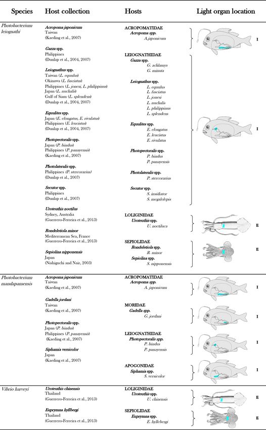
3758                                                                              L. Tanet et al.: Bacterial bioluminescence

of luminous bacteria from light organs and digestive tracts,     processes, either by aggregating or by fragmenting organic
directly to the seawater or through fecal pellets (Ramesh et     matter. We propose a synthetic representation of the biolu-
al., 1990). Bacterial bioluminescence in its free or attached    minescence shunt of the biological carbon pump and a future
forms is much less studied but is worth reconsidering, in        strategy to establish and quantify the impact of biolumines-
its prevalence as well as its ecological implications. To our    cence (Fig. 1). Figure 1 represents, throughout the text, the
knowledge, no archaea has been characterized as biolumi-         guideline of the bioluminescence shunt hypothesis of the bi-
nescent.                                                         ological carbon pump.
   The biological and physical (solubility) carbon pumps are
the main drivers of the downward transfer of carbon and play
a central role in the sequestration of carbon dioxide (Boyd
et al., 2019; Buesseler and Lampitt, 2008; Dall’Olmo et al.,     2     Symbiotic bioluminescent bacteria in light organs
2016). The biological carbon pump is defined as the pro-
cess through which photosynthetic organisms convert CO2          In Eukaryotes, light emission has two distinct origins: in-
to organic carbon, as well as the export and fate of the         trinsic or symbiotic (Haddock et al., 2010; Nealson, 1979).
organic carbon sinking from the surface layer to the dark        Intrinsic luminescence is caused by chemicals produced by
ocean and its sediments by different pathways (Siegel et al.,    the organism itself. Most bioluminescent organisms are self-
2016, and references therein). Sinking particles (bigger than    luminescent and have specialized luminous cells, i.e., pho-
0.5 mm of diameter) known as marine snow are a combi-            tocytes, grouped inside dedicated organs called photophores
nation of phytodetritus, living and dead organisms, and fe-      (Herring, 1977). Some animals, however, are capable of lu-
cal pellets (from zooplankton and fish). Marine snow, rich       minescence using symbiotic luminous bacteria housed in
in carbon and nutrients, and its surrounding solute plumes       elaborate and specialized organs.
are hot spots of microbial activity in aquatic systems (All-
dredge et al., 1990; Alldredge and Silver, 1988; DeLong          2.1    Discovery, importance, distribution and functions
et al., 1993). Marine snow is also consumed by zooplank-                of light organ symbiosis
ton, and fecal pellets are a food source through coprophagy.
When leaving the epipelagic zone and sinking to depth, or-       In the late 1880s, Raphaël Dubois was among the first to sug-
ganic particles would be utilized by microbial decomposi-        gest bacteria could be responsible for the light emitted by
tion and fish/zooplankton consumption, both considered to        some animals (Harvey, 1957). In the beginning of the twen-
be responsible for a large part of the variation in the effi-    tieth century, Balthazar Osorio (1912) provided clear and
ciency of the biological carbon pump (De La Rocha and Pas-       convincing evidence of such symbiosis, when luminescent
sow, 2007). Recently, fragmentation (potentially due to bio-     bacteria were described in high density within a dedicated
logical processes in the mesopelagic waters) has also been       fish gland, called the light organ (Balthazar Osorio was cited
shown to be the primary process controlling the sequestra-       in Hickling, 1926). Since then, luminous bacterial symbiosis
tion of sinking organic carbon, accounting for 49 ± 22 % of      has been the subject of interest among the scientific commu-
the observed flux loss (Briggs et al., 2020). Moreover, some     nity working on bioluminescence, to such an extent that, by
studies pointed out the well-adapted vision of fish or crus-     the mid-twentieth century, luminescence of many organisms
tacean to the detection of point-source bioluminescence (de      was thought to have bacterial origin. However, some of these
Busserolles and Marshall, 2017; Frank et al., 2012; Warrant      assessments have been refuted later (Herring, 1977).
and Locket, 2004). The compiled data, from all forms of ma-         Bioluminescence ability is shared by about 8 % of all
rine bacterial bioluminescence, presented and discussed in       known fish species (Paitio et al., 2016). Amongst luminous
this review bring out the uninvestigated pathway of the bio-     fishes, bacterial luminescence is the rule for almost half of
luminescence contribution into the biological carbon pump,       them (48 %) (Davis et al., 2016). To date, symbiotic bac-
through the visual attraction of consumers for luminous par-     teria are recognized as responsible for the luminescence of
ticles.                                                          some fishes and squids (Davis et al., 2016; Haygood, 1993;
   In this review, we will summarize the current knowledge       Lindgren et al., 2012). Although forms of symbiotic lumines-
on bioluminescent bacteria based on former and recent lit-       cence have been suggested for some shark species or pyro-
erature. First, we describe symbiotic bioluminescent bacte-      somes (tunicates) (Dunlap and Urbanczyk, 2013; Leisman et
ria in light organs of fish or squid, its importance, and con-   al., 1980), no evidence of luminous bacteria has been found
trols. Then, we present enteric-association occurrences. One     so far (Claes and Mallefet, 2009; Renwart et al., 2014; Wid-
of the consequences of these symbioses, in both light organs     der, 2002) and a recent study has definitely rejected a bac-
and guts, is a massive quantity of bioluminescent bacteria       terial origin in the velvet belly lanternshark (Duchatelet et
dispersed daily in the ocean. Based on this statement, we        al., 2019). Concerning luminous squids, intrinsic biolumi-
claim and demonstrate that bioluminescent bacteria have an       nescence is more common, and symbiotic light organs are
ecological and a biogeochemical importance in the biolog-        known in only two families (Sepiolidae and Loliginidae)
ical carbon pump. They catalyze and amplify the involved         (Lindgren et al., 2012; Nishiguchi et al., 2004).

Biogeosciences, 17, 3757–3778, 2020                                                 https://doi.org/10.5194/bg-17-3757-2020
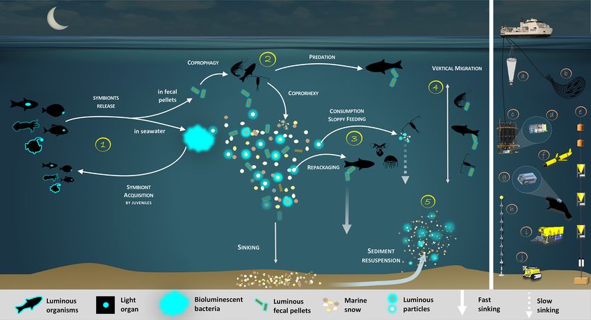
L. Tanet et al.: Bacterial bioluminescence                                                                                                3759

Figure 1. Bioluminescence shunt in the biological carbon pump in the ocean. Luminous bacteria in light organ symbioses are successively
acquired by host (squid, fish) from the seawater while they are juveniles, then regularly released into the ocean. Depending on the light
organ position, luminous bacteria are released from their guts into fecal pellets or directly into the seawater (step 1). Motile luminous
bacteria colonize organic matter sinking along the water column. Bioluminescent bacteria inseminating fecal pellets and particles influence
zooplankton consumption rates. Such visual markers increase detection (“bait hypothesis”), attraction and finally predation by upper trophic
levels (step 2). In the mesopelagic, zooplankton and their predators feed on sinking luminous particles and fecal pellets, which form either
aggregates (repackaging) of faster sinking rates or fragment organic matter (due to sloppy feeding) with slower sinking rates (step 3). Filter
feeders also aggregate sinking organic matter without particular visual detection and selection of luminous matter. Diel (and seasonal) vertical
migrators feeding on luminous food metabolize and release glowing fecal pellets from the surface to the mesopelagic zone (step 4). This
implies bioluminescent bacteria dispersion at large spatial scales, for zooplankton or even some fish actively swimming long distances.
Luminous bacteria attached to particles sink down to the seafloor, and sediment can be resuspended by oceanographic physical conditions
(step 5) and consumed by epi-benthic organisms. Instruments are (a) plankton net, (b) fish net, (c) Niskin water sampler, (d) bathyphotometer,
(e) sediment traps, (f) autonomous underwater vehicles, (g) photomultiplier module, (h) astrophysics optical modules ANTARES and (i–
j) remotely operated vehicles.

   Symbiotic luminescence seems more common in benthic                    ette and therefore avoiding dusk-active piscivorous preda-
or coastal environments for fish and squid as well (Hay-                  tors (Claes et al., 2010; Johnsen et al., 2004; Warner et
good, 1993; Lindgren et al., 2012; Paitio et al., 2016).                  al., 1979). Amongst bacterial light symbioses, counterillu-
Shallow-water fishes with luminous bacterial symbionts in-                mination has been demonstrated for the bobtail squid Eu-
clude flashlight fishes (Anomalopidae), ponyfishes (Leiog-                prymna scolopes (Jones and Nishiguchi, 2004) and some
nathidae) and pinecone fishes (Monocentridae) (Davis et al.,              leiognathids fish (McFall-Ngai and Morin, 1991) and hy-
2016; Morin, 1983). For deep-sea fishes, anglerfishes (Cera-              pothesized for other bioluminescent fishes (Dunlap et al.,
tiodei) and cods (Moridae) are among the common examples                  2009; McAllister, 1967). Less common but more striking,
of luminous-bacteria hosts.                                               some organisms found in the families Monocentridae and
   Bacterial and intrinsic light organs are predominantly in-             Anomalopidae and numerous deep-sea anglerfishes belong-
ternal, ventrally located (Paitio et al., 2016). Many lumi-               ing to the suborder Ceratoidei exhibit externally located light
nous organisms with ventral light organs likely use the                   organs colonized by bacteria (Haygood, 1993). The external
emitted light to conceal themselves by counterillumina-                   light organs of flashlight fish have been demonstrated to be
tion. This defensive strategy allows luminous species to                  used to illuminate the nearby environment and detect prey
match with the intensity, spectrum and angular distribu-                  (Hellinger et al., 2017), or schooling behavior (Gruber et al.,
tion of the downwelling light, thus obliterating their silhou-            2019), while the lure of female anglerfish is generally be-

https://doi.org/10.5194/bg-17-3757-2020                                                             Biogeosciences, 17, 3757–3778, 2020
3760 L. Tanet et al.: Bacterial bioluminescence
lieved to be used for mate-finding purposes and prey attrac- light was proved by microscopic observation and that genes
tion (Herring, 2007). from luminous bacteria were amplified (Haygood and Dis-
tel, 1993), bacterial cultivation has not yet been successful.
2.2 Symbiont selection and colonization of the light Thanks to the emergence of genome sequencing, the com-
organ plete genome of these symbionts has been reported in the
last years. Analyses revealed a genome reduction in size by
Like most symbiotic bacterial associations with animals, lu- about 50 % and 80 % for anglerfish and flashlight fish sym-
minous bacteria are acquired from the surrounding environ- bionts, respectively, compared to facultative luminous sym-
ment by individuals, independently of their ancestry (i.e., bionts or free-living relatives (Hendry et al., 2014, 2016,
horizontally transmitted) (Baker et al., 2019; Haygood, 1993; 2018). Genome reduction is a common trait shared by bacte-
McFall-Ngai, 2014). One of the best-documented symbioses ria involved in obligatory symbiosis (Moran et al., 2009) and
is the association of Aliivibrio fischeri with the bobtail squid explains the inability of these symbionts to grow in labora-
Euprymna scolopes (Nyholm and McFall-Ngai, 2004; Ruby, tory cultures. Flashlight fish and anglerfish symbionts appear
1996). Through the easy independent cultivation of both part- to be obligately dependent on their hosts for growth, as some
ners in the laboratory, this symbiosis has become a perfect metabolic capacities (e.g., genes necessary for amino acid
model for studying the process of bacterial colonization into synthesis) are absent in the genome.
the light organ and understanding bacteria–animal interac-
tions, broadly speaking (Mandel and Dunn, 2016; McFall- 2.3 Light organs are under well-established controls
Ngai, 2014).
Knowledge of the mechanisms involved in the selec- Although light organs can differ in form, size or location
tion and the establishment of bacterial symbionts in the according to the host (see Table 1), some structural and
squid–Vibrio symbiosis have considerably improved over functional features are common for all of them. Luminous
the last few decades. Harvest of the luminous symbionts bacteria are densely packed within tubules which connect
from the bacterioplankton is driven by microbial recognition to the exterior of the light organ (Haygood, 1993; Neal-
and molecular dialog (Kremer et al., 2013; Nyholm et al., son, 1979). The host provides nutrients and oxygen to the
2000; Nyholm and McFall-Ngai, 2004; Pankey et al., 2017; tubules through a highly vascularized system (Tebo et al.,
Schwartzman and Ruby, 2016; Visick and Ruby, 2006). 1979). Bioluminescent bacteria emit light continuously in
Moreover, bacterial colonization of host tissues induces the the light organ, as they do in laboratory cultures (Nealson
morphogenesis process of the light organ and appears to sig- and Hastings, 1979). However, the light intensity varies over
nal its further development and maturation (McFall-Ngai and time. As for self-luminescent fish, bacterial light organs have
Ruby, 1991; Montgomery and McFall-Ngai, 1998). The lu- evolved with a multitude of adaptations of tissue, to serve as
minescence feature is essential for a correct morphogenesis reflectors, diffusers, screens and light-conducting channels
process of the light organ and symbiont persistence inside (Haygood, 1993; Munk et al., 1998). Such anatomical fea-
(McFall-Ngai et al., 2012; Visick et al., 2000). tures assist in directing and enhancing light output (Sparks
While the bobtail squid model provides a window to un- et al., 2005). In addition, the host can control the light dif-
derstand the establishment of such symbioses, this system fusion through different mechanisms, which may be external
cannot be systematically transferred to other bacterial lumi- lids, chromatophores, organ rotation, filters, occlusion with
nous symbioses. Although less well-known, the other associ- a shutter or muscle contraction (Hansen and Herring, 1977;
ations are no less important and many questions remain un- Herring, 1977; Johnson and Rosenblatt, 1988). As an exam-
solved since they might be harder to study. ple, for counterillumination, controlling the intensity of light
To date, 11 bacterial species are known to be involved in output gives the host a better camouflage, adapting its silhou-
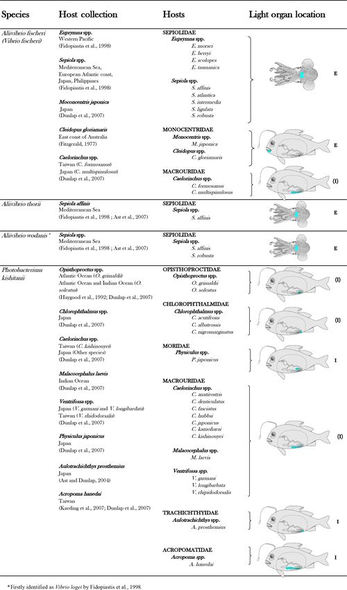
light organ symbioses (Table 1). In a light organ, the bacte- ette to environmental changes in light (Jones and Nishiguchi,
rial population is most of the time monospecific (Dunlap and 2004; McFall-Ngai and Morin, 1991). For intraspecies com-
Urbanczyk, 2013; Ruby, 1996). munication, it permits the production of sudden flashes or a
Considering that fish and squid housing luminous bacteria specific signal/rhythm of light (e.g., schooling behavior, Gru-
are never found without symbionts in nature, the symbiosis ber et al., 2019).
appears obligatory for hosts (Haygood, 1993). In contrast, In squid–Vibrio symbiosis, bacterial luminescence genes
most symbiotic bacteria are viable outside the light organ, are regulated with a quorum-sensing system, a cell-density-
and thus are considered to be facultatively symbiotic. These dependent process. When the cell density reaches a cer-
facultative symbiotic bacteria are readily culturable under tain level, autoinducers responsible for triggering the syn-
laboratory conditions, outside the host light organ. Excep- thesis of the genes involved in light emission are accumu-
tions have been highlighted for the luminous symbionts of lated in sufficient amounts, and light is emitted (Nealson et
two groups of fish, the flashlight fish and the deep-sea angler- al., 1970; Verma and Miyashiro, 2013). Interestingly, A. fis-
fish (Dunlap and Kita-tsukamoto, 2006; Haygood and Distel, cheri produces a higher level of luminescence within the light
1993). Indeed, despite the fact that the bacterial origin of the organ than in laboratory cultures, despite a similarly high
Biogeosciences, 17, 3757–3778, 2020 https://doi.org/10.5194/bg-17-3757-2020L. Tanet et al.: Bacterial bioluminescence 3761 Table 1. List of luminous bacterial species found in light organ symbiosis in fishes and squids. The diagrammatic fish, from Nealson and Hastings (1979), was used to indicate, in blue, the approximate locations of the light organ of the different families of symbiotically luminous fishes. E indicates an external expulsion of the bioluminescent bacteria, directly into the seawater. I indicates an internal expulsion of the bioluminescent bacteria, in the digestive tract. (E) or (I ) indicate a putative localization of the expulsion. https://doi.org/10.5194/bg-17-3757-2020 Biogeosciences, 17, 3757–3778, 2020
3762                                  L. Tanet et al.: Bacterial bioluminescence

Table 1. Continued.

Biogeosciences, 17, 3757–3778, 2020    https://doi.org/10.5194/bg-17-3757-2020
L. Tanet et al.: Bacterial bioluminescence                                                                                  3763

Table 1. Continued.

cell density (Boettcher and Ruby, 1990). Hence, Verma and          related with the diel pattern of the host behavior. Indeed, the
Miyashiro (2013) suggested that the light organ environment        bobtail squid expels 95 % of the luminous symbionts in the
offers specific conditions such as the levels of oxygen, iron or   surrounding environment at dawn, the beginning of its inac-
phosphate, to enhance bacterial light emission. Here again,        tive phase. The remaining 5 % of A. fischeri grow through the
while the control mechanisms of the squid–Vibrio symbio-           day and the highest concentration is reached at the end of af-
sis are well-understood, those of the other symbioses remain       ternoon, at the nocturnal active phase of the squid (Nyholm
enigmatic and there are indications that they may vary. For        and McFall-Ngai, 2004; Ruby, 1996). Currently, with the ex-
example, the absence of the quorum-sensing-gene detection          ception of the squid–Vibrio symbiosis, accurate data on the
in anglerfish and flashlight fish symbionts suggests a con-        symbiont release are still largely unknown. Indeed, the fre-
stitutive light emission by the bacteria (Hendry et al., 2016,     quency of release may vary and occur more than once a day
2018).                                                             as has been shown for some flashlight and pinecone fishes
   For all symbioses, luminous symbionts, within the light         (Haygood et al., 1984).
organ, reach a very high density which reduces the oxygen             Regular expulsion of symbionts maintains favorable con-
availability, essential for the light reaction. Such oxygen lim-   ditions in the light organ for the bacterial population, but it
itation leads to a decrease in the specific luminescence activ-    also seeds the environment with luminous symbionts for col-
ity (Boettcher et al., 1996). The bacterial population inside      onization of the next host generation. The consequence is
the light organ is regulated by the host, by coupling the re-      a release of a huge quantity of bioluminescent bacteria in
striction of the growth rate and the expulsion of symbionts.       the seawater, inducing a major contribution to the ocean mi-
Growth repression is thought to reduce the energetic cost of       crobiome. To make it more concrete and provide an order
the symbiosis to the host (Haygood et al., 1984; Ruby and          of magnitude, two examples are proposed. Using laboratory
Asato, 1993; Tebo et al., 1979). Additionally, since luminous      experiments on different fishes (Monocentridae, Anomalop-
bacteria are densely packed inside tubules communicating           idae), Haygood et al. (1984) estimated a release of between
with the exterior of the light organ (Haygood, 1993), the cell     107 and 109 bioluminescent bacterial cells per day and per
number of symbionts is regulated by the regular expulsion          individual. Another study on the Hawaiian bobtail squid (E.
of most of the bacterial population, followed by a period of       scolopes) has estimated that the squid expels about 5 × 10⁸
regrowth of the remaining symbionts. Concerning the well-          bioluminescent bacterial cells per day and per individual
known squid–Vibrio symbiosis, its daily release is highly cor-     (Lee and Ruby, 1994). These discharges lead to a regular

https://doi.org/10.5194/bg-17-3757-2020                                                   Biogeosciences, 17, 3757–3778, 2020
3764                                                                                  L. Tanet et al.: Bacterial bioluminescence

luminous-bacteria enrichment of the areas inhabited by these            Seasonal variations have been observed in both luminous
organisms.                                                           bacterial density (Liston, 1957; Ramesh and Venugopalan,
   Depending on the anatomical location of the light organ           1988) and predominant species (Bazhenov et al., 2019). Such
(see Table 1), luminous symbionts are released through pores         variability is not surprising since it is inferred from the struc-
or ducts into the surrounding seawater or into the digestive         ture and composition of the gut microbiota of fish, which are
tract (Haygood, 1993; Nealson and Hastings, 1979). An en-            influenced by a series of factors, including (i) host factors
teric lifestyle has indeed been suggested for the luminous           (e. g genetics, gender, weight, age, immunity, trophic level);
bacteria (Ruby and Morin, 1979; Nealson, 1979).                      (ii) environmental factors such as water, diet and surrounding
                                                                     environment; (iii) microbial factors (e.g., adhesion capacity,
                                                                     enzymes and metabolic capacity); (iv) and individual vari-
3   Enteric associations in marine-fish guts                         ations and day-to-day fluctuations (Nayak, 2010; Sullam et
                                                                     al., 2012; Wang et al., 2018). Interestingly, a high propor-
The gastrointestinal (GI) tract of an animal is a very com-
                                                                     tion of luminescent bacteria (>70 %) has been found in the
plex and dynamic microbial ecosystem (Nayak, 2010). Cur-
                                                                     gut of an Atlantic halibut recently fed, while an individual
rent knowledge and concepts of GI microbiota derive from
                                                                     male in spawning condition, which had not eaten recently,
studies on humans or other terrestrial mammals. In contrast,
                                                                     had a flora dominated by non-luminescent microorganisms
GI ecosystems of marine inhabitants have yet received lit-
                                                                     (Verner-Jeffreys et al., 2003). This result underlines the link
tle attention, and studies focused on farmed fish or commer-
                                                                     between food ingestion and abundance of luminous bacte-
cially important species of fish. Whether aerobes or anaer-
                                                                     ria and suggests that they do not persist within the halibut
obes are the main group in the microbiota in fish intestines
                                                                     gut once the feces are eliminated. This also suggests that lu-
is still discussed (Romero et al., 2014). For marine fish, the
                                                                     minous bacteria are then released with the feces in the wa-
dominant members seem to be facultative anaerobes (Wang
                                                                     ter column. Makemson and Hermosa (1999) have reported
et al., 2018). Considering that most of the bioluminescent
                                                                     a slightly higher proportion of culturable luminous bacteria
bacteria are facultative anaerobes (Ramesh et al., 1990; Re-
                                                                     in herbivorous fish compared to carnivorous fish. They also
ichelt and Baumann, 1973), it is not surprising to find them
                                                                     emphasized the higher incidence of luminescent bacteria in
in gut niches.
                                                                     pelagic than in reef-associated fish, and filter-feeder-fish guts
   Although luminescence of dead fish was a well-known
                                                                     contain more luminous bacteria compared to other feeding
phenomenon, one of the first mentions of the presence of
                                                                     types (e.g., predator). For bigger fishes, a potential introduc-
luminescent bacteria in fish slime and intestinal contents
                                                                     tion source of luminous bacteria into the gut could be the
is only from the beginning of the 1930s (Stewart, 1932).
                                                                     ingestion of smaller prey bearing a bacterial light organ. For
Since then, the high occurrence of luminous bacteria in
                                                                     all organisms, enteric luminous bacteria may be transferred
fish intestines has been reported in many studies (Baguet
                                                                     to the gut bacterial community of their predators.
and Marechal, 1976; Barak and Ulitzur, 1980; Liston, 1957;
                                                                        It should be emphasized that investigations on microbial
Makemson and Hermosa, 1999; O’Brien and Sizemore,
                                                                     communities of fish have long been limited by the use of
1979; Ramesh and Venugopalan, 1988; Reichelt and Bau-
                                                                     culture-dependent methods (Austin, 2006; Romero et al.,
mann, 1973; Ruby and Morin, 1979). Most hosts with an in-
                                                                     2014). The fish-gut microbiota has been reported to be par-
ternal light organ release luminous bacteria into the digestive
                                                                     ticularly of low cultivability, with less than 0.1 % of the to-
tract via ducts (Haygood, 1993; Nealson and Hastings, 1979)
                                                                     tal microbial community cultivable (Zhou et al., 2014), al-
and thus may largely contribute to their abundance in lumi-
                                                                     though the level of cultivability may be taxon dependent
nous fish intestines. However, many fishes without a light or-
                                                                     (Ward et al., 2009). Today, advanced molecular techniques
gan also harbor luminescent bacteria in their gut (Makemson
                                                                     offer a wide variety of culture-independent methods, such as
and Hermosa, 1999), which clearly demonstrates the exis-
                                                                     next-generation sequencing (NGS), for analyzing fish micro-
tence of other sources of enteric luminous bacteria. Through
                                                                     biota (Tarnecki et al., 2017).
the gut-content analysis of 109 fish species from the Gulf of
                                                                        Several studies using gene sequencing based on 16S rRNA
Oman, Makemson and Hermosa (1999) showed that the rel-
                                                                     to characterize the gut microbiome of fish have reported the
ative proportion of the occurring culturable luminous bacte-
                                                                     genus Photobacterium as the most abundant in the guts of
ria was strongly variable. While some fish guts harbor more
                                                                     salmon and trout (Bagi et al., 2018; Givens et al., 2015; Michl
than 80 % luminous bacteria, some others have between 20 %
                                                                     et al., 2019; Riiser et al., 2018), shark (Michl et al., 2019),
and 50 %, and a minority have none detected, with a substan-
                                                                     and Atlantic cod (Bagi et al., 2018; Givens et al., 2015; Michl
tial intra- and interspecies fish variability. Like other authors,
                                                                     et al., 2019; Riiser et al., 2018). Other studies reported the
Makemson and Hermosa (1999) highlighted V. harveyi and P.
                                                                     presence of Photobacterium spp. in the gut of hydrothermal
phosphoreum as the dominant luminous species found in fish
                                                                     shrimp (Durand et al., 2009), in some adult anglerfish (Freed
guts (O’Brien and Sizemore, 1979; Reichelt and Baumann,
                                                                     et al. 2019) and, seasonally variable, in the gut of Norway
1973; Ramesh and Venugopalan, 1988).
                                                                     lobster (Meziti et al., 2010). However, because not all Pho-
                                                                     tobacterium spp. have luminescence ability, it is important

Biogeosciences, 17, 3757–3778, 2020                                                     https://doi.org/10.5194/bg-17-3757-2020
L. Tanet et al.: Bacterial bioluminescence 3765
to be able to resolve dominant operational taxonomic unit scolopes was 24 to 30 times higher, in both water column
(OTU) at the species level, which, most of the time, is not and sediments, in areas inhabited by the squids than in simi-
possible with a 16S rRNA barcoding sequencing approach. lar locations where squids were not observed.
The emergence of multi-gene approaches offers more de- Bioluminescent bacteria also seem to be the cause of the
tailed insights into the taxonomic diversity of these commu- spectacular and still largely unexplained events, so-called
nities (i.e., species level). Thus, using metagenomic shotgun milky seas (Lapota et al., 1988; Nealson and Hastings, 2006).
sequencing, two independent and recent works on wild At- Milky seas are characterized by an unusual brightness on the
lantic cods also concluded Photobacterium spp. domination ocean surface and extend over such a large area that the light
and have been able to go deeper into the taxonomic identifi- emitted is detectable from space (Miller et al., 2005). The
cation. Le Doujet et al. (2019) demonstrated that the Photo- light emission pattern of milky seas is continuous and homo-
bacterium genus represents 78 % of all present genera and geneous, which is consistent with light emission from bacte-
identified the P. phosphoreum clade as the most abundant ria and easily distinguished from blooms of dinoflagellates.
Photobacterium lineage. According to Riiser et al. (2019),
the luminous species P. kishitanii constitutes over 26 % of 4.2 Bioluminescent bacteria attached to particles
the Vibrionales community, which is the dominant clade,
and the authors underlined the presence of the functional lux Outside of spatially restricted niches, such as light organ or
genes, the light-emission-involved genes. Therefore, recent gut environments, the role of the dispersed luminous cells
metagenomic studies seem to confirm the trend of a high oc- in the marine environment was a matter of debate, and it
currence of luminous bacteria in fish intestines. was thus mentioned that non-symbiotic bacteria may have
no ecological significance (Hastings and Greenberg, 1999;
Nealson and Hastings, 1979). However, Herren et al. (2004)
4 Luminous bacteria and the biological carbon pump suggested that luminous bacteria are more often attached to
particles than free-living, which was confirmed by Al Ali et
As previously discussed, light organs and guts act as a source al. (2010). Many bacteria, including bioluminescent bacte-
for luminous-bacteria persistence in the oceans. Therefore, ria (Ruby and Asato, 1993; Zhang et al., 2016), can develop
luminous bacteria are widespread in the ocean. They can be swimming behavior to colonize the sinking organic material,
found as free-living forms or attached to particles (Nealson therefore reaching a cell density 100 to 10 000 times higher
and Hastings, 1979; Ramesh and Mohanraju, 2019; Ruby et than in the water column (up to 108 to 109 cells mL−1 ) (e.g.,
al., 1980). Ploug and Grossart, 2000).
Bacteria that glow on particles can attract macroorgan-
4.1 Bioluminescent bacteria in the water column isms. After being ingested, they will find a more favorable
environment to live and grow in their gut (Andrews et al.,
Qualitative and quantitative studies showed that the lumi- 1984; Ruby and Morin, 1979). Actually, this is the preferred
nous bacteria are dynamic over time and space. Seasonal current hypothesis that supports a positive selection related
variations have been identified, in both abundance and pre- to the dispersion and propagation of the bacteria. Indeed, lu-
dominant species (O’Brien and Sizemore, 1979; Ruby and minous bacteria growing on particulate matter could produce
Nealson, 1978; Yetinson and Shilo, 1979). A wide variability enough light to be visible by other organisms. For bacterial
has been observed in species repartition over depth and be- species with light production under cell-density control (i.e.,
tween geographic areas (DeLuca, 2006; Gentile et al., 2009; under quorum-sensing regulation), the high cell concentra-
Nealson and Hastings, 1979; Ramaiah and Chandramohan, tion reached on particles can allow the sufficient accumula-
1992; Ruby et al., 1980). Horizontal, vertical and seasonal tion of the autoinducers, and thus the emission of light for
variations were presumed to reflect physiological preferences attracting predators. For species for which light production
most of the time, and particularly temperature or salinity sen- is not subject to cell-density control (i.e., not under quorum-
sitivity (Orndorff and Colwell, 1980; Ramesh et al., 1990; sensing regulation) (Tanet et al., 2019), to be able to pro-
Ruby and Nealson, 1978; Shilo and Yetinson, 1979; Yetinson duce light at a very low cell concentration could give them
and Shilo, 1979). Some works mentioned that symbiotic an advantage. Continuously glowing bioluminescent emis-
niches, such as light organs and enteric tracts, may serve sions are thought to attract predators (Nealson and Hastings,
to inoculate the planktonic population (Nealson et al., 1984; 1979). In the water column, the glowing bacteria aggregated
Nealson and Hastings, 1979; Ramesh et al., 1990; Ruby et on particles would lead to the detection, attraction, ingestion
al., 1980). To our knowledge, very few studies focused in- and decomposition of particles by larger organisms. Graz-
tensively on the contribution of species-specific symbiotic ers would consume luminous matter at a higher rate than in-
associations on the occurrence and distribution of luminous visible particles. Being consumed and ending up in the gut,
bacteria in the surrounding water. Amongst these rare stud- bacteria would benefit from a more suitable environment re-
ies, Lee and Ruby (1994) reported that the abundance of A. garding the growth conditions and the nutrient accessibility.
fischeri, the luminous symbiont of the Hawaiian squid E. In the open ocean, and particularly in deep regions, where
https://doi.org/10.5194/bg-17-3757-2020 Biogeosciences, 17, 3757–3778, 20203766 L. Tanet et al.: Bacterial bioluminescence
sparse nutrient supply prevails, nutrient-rich gut niches of the 4.3 Bioluminescent bacteria in the sediments
surrounding animals could appear as an oasis of life for bac-
teria. This dispersion hypothesis has also been strongly con- Information relative to luminous bacteria in sediment is also
solidated by field data where bacterial bioluminescence was limited. It is known that bioluminescent bacteria can be iso-
observed in freshly egested fecal pellets and in materials col- lated from sediment samples (Ramesh et al., 1990), but rare
lected from sediment traps (Andrews et al., 1984), as well as data exist about their distribution or abundance. In some sed-
by laboratory experiments where glowing zooplankton were iment samples, occurrence of luminous bacteria among total
preferentially ingested by fishes (Zarubin et al., 2012). heterotrophic bacteria could reach up to 70 %, with seasonal
The copiotrophic trait of luminous bacteria is another point variations (Ramesh et al., 1989), although less pronounced
supporting their particle-attached lifestyle. Bacterial popula- than in the water column (O’Brien and Sizemore, 1979). The
tion colonizing nutrient-rich environments (e.g., floating car- main sources of luminous bacteria in sediments are likely the
cass, marine snow, fecal pellets or the gut tract of a marine glowing sinking marine snow and benthic or demersal hosts,
eukaryote) are defined as copiotrophs, by opposition to the harboring symbiotic light organs with regular discharges.
oligotrophs which are members of free-living microbial pop- More recently, sediment resuspension events (Durrieu de
ulations (Lauro et al., 2009). All luminous Vibrionaceae, ex- Madron et al., 2017) were correlated with newly formed
cept reduced genome symbionts, possess two chromosomes deep-water events and deep-sea bioluminescent events
in their genome (Boyd et al., 2015; Zhang et al., 2016), with a recorded in the NW Mediterranean Sea (Martini et al., 2014;
high copy number of rRNA operons. Such genomic features, Tamburini et al., 2013a). Since the presence of active lu-
a large genome size and multiple rRNA operons, are consid- minous bacteria has been demonstrated on the site (Martini
ered to be an adaptation for a copiotrophic lifestyle (Klap- et al., 2016), it has been hypothesized that resuspended lu-
penbach et al., 2000; Lauro et al., 2009). Copiotrophs are minescent bacteria present in sediment can be part of these
thought to have strong adaptability skills, permitting them to luminescence events (Durrieu de Madron et al., 2017). Ad-
survive long enough between two nutrient-rich environments ditionally, dense water formation, conveying particulate or-
(Yooseph et al., 2010). ganic matter, could further increase luminous-bacteria pro-
Fish guts could also act as an enrichment vessel for the liferation and activity (Tamburini et al., 2013a).
growth of luminous bacteria, and thus enhance their prop-
agation (Nealson and Hastings, 1979; Ramesh and Venu-
gopalan, 1988). When expelled with feces, enteric luminous 4.4 How do bioluminescent bacteria impact the
bacteria can be easily isolated from the fresh fecal material. biological carbon pump?
This fecal luminescence increased in intensity over a mat-
ter of hours, proving that luminous bacteria survive the di- Based on the ecological versatility of the bacterial biolumi-
gestive process and can proliferate on such organic material nescence reviewed above, we propose reconsidering the clas-
(Ruby and Morin, 1979). Hence, fish feces appear to be an sical view of the fate of organic matter in the oceans. Fig-
important source of viable luminous bacteria in the marine ure 1 represents the guideline of the bioluminescence shunt
environment and could affect both the distribution and the hypothesis of the biological carbon pump.
species composition of luminous populations. The lumines- Bioluminescent bacterial emissions are continuous over
cence of fecal particles has been reported numerous times time and such a characteristic is thought to attract preda-
and is always associated with luminous bacteria, due to the tors. Indeed, the light from bioluminescence contrasts well
observation of continuous light emission or direct isolation against the dim or dark background of the ocean depths. In
(Andrews et al., 1984; Ramesh et al., 1990; Raymond and the bathypelagic zone (1000–4000 m), where no daylight re-
DeVries, 1976; Ruby and Morin, 1979; Zarubin et al., 2012). mains, bioluminescent emissions are considered the major
In comparison with free-living luminous bacteria, few visual stimulus (Warrant and Locket, 2004; Widder, 2002).
studies have focused on bioluminescence of marine snow and For these reasons, symbiotic associations in light organs have
fecal pellets. Yet, observations on materials collected from been selected as an advantage for hosts (fish or squid). Lu-
sediment traps revealed light emission in 70 % of all sam- minous bacterial symbionts are successively acquired by ju-
ples, with two distinct patterns of light kinetics, probably due veniles and released into the seawater to control population
to the presence of different luminescent organisms (Andrews concentration (Fig. 1, step 1). As indicated previously, the
et al., 1984). Surface-sample (above 60 m depth) analyses re- release of bioluminescent bacteria from light organs and fe-
ported that more than 90 % of the luminous-aggregate sam- cal pellets could represent a huge quantity of bioluminescent
ples exhibited bacterial luminescence (Orzech and Nealson, bacteria in the water column. On dead organisms, luminous
1984). Another study (between 2 and 17 m depth) also re- bacteria present in the gut of the host could initiate rapid
ported a large part of luminous marine snow, but more likely propagation and decomposition of the host body and result
due to dinoflagellates (Herren et al., 2004). in the formation of luminous debris in the marine environ-
ment. Based on the increase in light emission observed on
dead marine animals, Wada et al. (1995) argue that, upon the
Biogeosciences, 17, 3757–3778, 2020 https://doi.org/10.5194/bg-17-3757-2020L. Tanet et al.: Bacterial bioluminescence 3767 death of the host, enteric luminous bacteria may have an im- egested pellets of higher density are fast-sinking particles. portant saprophytic lifestyle. Filter-feeder plankton, without visual detection and food se- Recent studies underlined that fish vision is very-well- lection by light, will also passively contribute to such aggre- adapted to the detection and location of point-source biolu- gation or fragmentation of particles. For these organisms, bi- minescence (de Busserolles and Marshall, 2017; Mark et al., oluminescence can even have a negative effect since they can 2018; Musilova et al., 2019; Paitio et al., 2016; Warrant and be identified by the luminous material filtered. Additionally, Locket, 2004). Although less intensively documented than the consumption of organic material colonized by biolumi- fishes, the crustacean (copepods, amphipods, isopods, etc.) nescent bacteria increases their dispersal rate provided by mi- visual system is also reported to have a sensitivity shift to grating zooplankton and even more so by actively swimming bluer wavelengths, which aids their bioluminescence detec- fish, following the conveyor-belt hypothesis (Grossart et al., tion (Cohen and Forward, 2002; Frank et al., 2012; Marshall 2010) (Fig. 1, step 4). After being ingested, bacteria (includ- et al., 1999; Nishida et al., 2002). In the laboratory, Land et ing luminous ones), attached to the particles consumed by al. (1995) demonstrated that amphipods were attracted to a zooplankton and fish, stay in their digestive tract. At night, blue-light-emitting diode. Unfortunately, and despite these these organisms migrate in the upper part of the water col- statements, rare studies have investigated the effect of bi- umn and release feces in niches and at depth that, eventually, oluminescence on the ingestion rates of predators (Fig. 1, would not have been otherwise colonized by luminous bacte- step 2). To our knowledge, the only one known is from ria. This dispersion, due to the expelling of luminous feces, is Zarubin et al. (2012), who demonstrated that zooplankton is several orders of magnitude greater than that of waterborne attracted to luminous particles and feeds on the luminous- free bacteria. Zooming on the carbon fluxes at the level of bacteria-rich organic matter. Because of the ingestion of the a gravitational sinking particle (Fig. 2), the bioluminescence luminous bacteria, the zooplankton itself starts to glow. Then, shunt hypothesis implies that the bacterial glow of this parti- Zarubin et al. (2012) experimentally measured the 8-times- cle increases the distance of visual detection. Such a distance higher ingestion rate of glowing zooplankton by fishes, com- can be up to several tens of meters according to Warrant and pared to non-luminous zooplankton. Locket (2004) and probably depends on the bioluminescent Glowing bacteria have been observed attached to particles bacterial concentration and the visual perception of the or- of organic matter, marine snow and fecal pellets (Fig. 1, from ganisms. symbionts in guts in step 1 and through predation in step Sediment resuspension is another process implying the 2) sinking into the deep ocean. Thus, while sinking into the consumption of luminous bacteria by higher trophic levels deep, these glowing bacteria living on organic carbon par- (Fig. 1, step 5). This potentially re-inseminates bacteria into ticles (marine snow, fecal pellets, etc.) would lead to the the bioluminescence loop through the consumption by epi- detection, attraction, ingestion and decomposition of parti- benthic organisms. cles by larger organisms. Consumers would ingest luminous Considering this bioluminescence shunt hypothesis, all the matter at a higher rate than invisible particles and conse- processes described above show that bioluminescence affects quently will increase luminous-microorganism dispersion by the biological gravitational carbon pump (Boyd et al., 2019), the egestion of fecal pellets. Bioluminescent sinking mate- either by increasing the carbon sequestration into the deep rial should accelerate the consumption of organic matter by ocean or by slowing down the sinking rate of particles and attracting grazing organisms. Interestingly, bacteria associ- consequently increasing their degradation and the remineral- ated with animal guts are thought to be particularly adapted ization rate. Bioluminescence and especially luminous bac- to high hydrostatic pressure (Deming et al., 1981; Ohwada teria may therefore influence the export and sequestration of et al., 1980; ZoBell and Morita, 1957). Indeed, certain bio- biogenic carbon in the deep oceans (either positively or nega- luminescent bacteria resist high hydrostatic pressure (Brown tively). A better quantification of these processes and impacts et al., 1942), and some of them have a higher growth rate in the biological carbon pump is a requirement in future stud- and emit more light than at atmospheric pressure (Martini ies. et al., 2013). Such piezotolerance, or piezophile lifestyle, is undoubtedly an advantage for luminous bacteria attached to particles that are exposed to pressure variations during the 5 Past and future instrumentation for bioluminescence sinking-particle fluxes (Tamburini et al., 2013b). The addi- assays tion of these bioluminescent tags on particles has two indirect impacts (Fig. 1, steps 2 and 3). First, due to aggregate frag- 5.1 Previous sampling methods to describe diversity mentation by sloppy feeding and coprorhexy, fast-sinking and abundance of luminous bacteria particles are transformed into slow-sinking or suspended par- ticles. Fragmentation has been shown to be the primary pro- In the existing literature, to estimate the diversity and the dis- cess controlling the sequestration of sinking organic carbon tribution of bioluminescent bacteria, studies were based on (Briggs et al., 2020). The second possibility is that organic a restricted number of sampling methods and instruments. matter ingestion leads to aggregation by repackaging, and the These methods focused either on environmental samplings https://doi.org/10.5194/bg-17-3757-2020 Biogeosciences, 17, 3757–3778, 2020
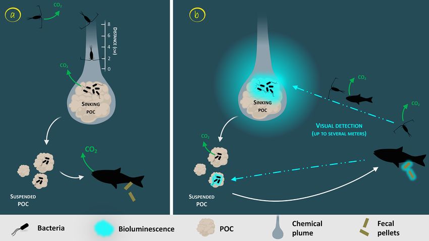
3768                                                                                        L. Tanet et al.: Bacterial bioluminescence

Figure 2. Zoom on the carbon fluxes at the level of a gravitational sinking particle (inspired by Azam and Long, 2001). The sinking POC is
moving downward followed by the chemical plume (Kiørboe, 2011). The plain white arrows represent the carbon flow. Panel (a) represents
the classical view of a non-bioluminescent particle. The length of the plume is identified by the scale on the side (Kiørboe and Jackson,
2001). Panel (b) represents the case of a glowing particle in the bioluminescence shunt hypothesis. Bioluminescent bacteria are represented
aggregated onto the particle. Their light emission is shown as a bluish cloud around it. Blue dotted arrows represent the visual detection and
the movement toward the particle of the consumer organisms. Increasing the visual detection allows a better detection by upper trophic levels,
potentially leading to the fragmentation of sinking POC into suspended POC due to sloppy feeding. The consumption of the bioluminescent
POC by fish can lead to the emission of bioluminescent fecal pellets (repackaging), which can also be produced with non-bioluminescent
POC if the fish gut is already charged with bioluminescent bacteria.

where bacteria are present or on organisms with associated               way to catch deep-sea animals is the deployment of trawls
bacteria.                                                                and more generally nets (Fig. 1a–b). They are well-adapted
   First, vertical samplings in the water column were per-               to sample squid (Zamborsky and Nishiguchi, 2011) or fish,
formed using sterile-bag samplers (Ruby et al., 1980), or later          like the anglerfish (Freed et al., 2019). One particularity of
using Niskin bottles (mounted on rosette profilers, Fig. 1c)             these methods is that the sampling covers a large section of
(Al Ali et al., 2010; Gentile et al., 2009; Kita-Tsukamoto et            the water column and combines everything into one catch
al., 2006; Martini et al., 2016; Yetinson and Shilo, 1979).              with a limited precision about depth layers. SCUBA div-
This approach is commonly set up in oceanography but re-                 ing is another method to gently select these large animals
lies on relatively small volumes of water (up to 20 L). Fur-             (Zamborsky and Nishiguchi, 2011). It has also been used to
thermore, it does not fully capture the heterogeneity of the             catch fecal pellets and sinking particles (Orzech and Nealson,
ecosystem since it provides one discreet sample over re-                 1984). Obviously, SCUBA diving has a strong depth limita-
stricted time and space. Other instruments dedicated to the              tion (generally above 50 m depth). It can be more efficient at
acquisition of sediment sampling are the multiple-core sam-              night for some migrating species and has a restricted sam-
plers, deployed onto the seafloor (Kita-Tsukamoto et al.,                pling size of organisms and number of samples carried back
2006). For particulate organic carbon and fecal pellets, in or-          to the ship.
der to describe the diversity of associated luminous bacteria,              Once environmental samples or material from an organ-
sediment traps (Fig. 1, item e) have been occasionally de-               ism’s light organ have been acquired, the objective is either
ployed from the surface down to the deep ocean (Andrews                  to describe the taxonomy and diversity of luminous bacteria
et al., 1984). Using these, fresh luminous material has been             or to quantify them. To do so, earlier studies have filtered
collected between 30 and 1900 m depth.                                   seawater samples through a polycarbonate filter with a pore
   To study the presence of luminous symbionts in guts and               size of 0.2 µm to retain bacteria. The filter is then placed with
light organs, larger organisms are caught. The most common               the bacterial side up on growth medium in Petri dishes (Kita-

Biogeosciences, 17, 3757–3778, 2020                                                           https://doi.org/10.5194/bg-17-3757-2020
L. Tanet et al.: Bacterial bioluminescence                                                                                  3769

Tsukamoto et al., 2006; Ruby et al., 1980). For symbiotic          (including observatories), oceanographic vessels, remotely
bacteria, light organs or guts are aseptically dissected shortly   operated and autonomous underwater vehicles (AUVs), and
after death, and the content is homogenized before culture         gliders (Fig. 1f, i) have considerably increased our knowl-
or microscopic observations (Dunlap, 1984). After hours of         edge of marine ecosystems and their spatial variability. For
incubation, the total colony-forming units is observed; the        temporal scales, in the last decades, the multiplication of
luminous colonies can, then, be enumerated and selected for        long-term observatories such as Ocean Network Canada
taxonomic investigation.                                           (ONC), the Ocean Observatories Initiative (OOI), the sta-
    Further investigations of symbiotic associations, in rela-     tion ALOHA, the European Multidisciplinary Seafloor and
tion to the surrounding environment, would require a reli-         water column Observatory (EMSO ERIC), or the interna-
able taxonomy of luminous bacteria and robust knowledge            tional Biogeochemical Argo program has increased global
on species-specific symbiotic associations. As an example,         ocean observations at long timescales (more than 10 years)
Photobacterium phosphoreum was thought to be the specific          and high sampling frequency. To quantitatively record biolu-
symbiont of light organs of numerous deep-sea fish (Hendrie        minescence emissions, some instruments are commercially
et al., 1970; Ruby et al., 1980; Ruby and Morin, 1978), be-        available, or have been adapted from existing sensors. Bathy-
fore a phylogenetic analysis showed distinct evolutionary lin-     photometers (Fig. 1d), a system pumping water into a closed
eages in the P. phosphoreum clade according to the colonized       chamber and measuring the emission of light by a photo-
habitat. This resolution revealed that all the P. phosphoreum      multiplier, are the most commonly used (Herren et al., 2005)
symbionts isolated from light organs should actually be iden-      and have already been implemented on AUVs (Berge et al.,
tified as P. kishitanii (Ast and Dunlap, 2005).                    2012; Messié et al., 2019; Moline et al., 2009) and other ver-
                                                                   tical profilers (Cronin et al., 2016). Other approaches have
5.2     Future strategy to quantify the role of                    been developed unexpectedly from astrophysics telescopes
        bioluminescence in the biological carbon cycle             (Fig. 1, item h) using photomultipliers with a very high sen-
                                                                   sitivity to photons embedded into optical modules. These in-
Since these first investigations on luminous bacteria in sym-      struments have been proven to be efficient to detect biolumi-
bioses or in the environment, there has been a huge im-            nescence in deep-sea environments and over long-time sur-
provement in technology and molecular-biology techniques.          veys (Aguzzi et al., 2017; Martini et al., 2014; Tamburini et
To better evaluate the role of bioluminescence and luminous        al., 2013a). Another example of quantitative records of pho-
bacteria in the biological carbon pump, further studies have       ton counts is the equipment of bio-samplers, such as elephant
to follow an efficient strategy. Such a strategy will focus on     seals, with a small, autonomous tag recording environmental
quantifying this functional trait and how it impacts the trans-    light and bioluminescence (Fig. 1g). These tags have been
fer of organic carbon between trophic levels, as well as its       shown to be a great improvement in highlighting ecologi-
sequestration into the deep ocean. This approach can be di-        cal functions such as predator–prey relationships and could
vided into several key points: (1) the assessment of the global    inform on the role of bioluminescent prey for seals (Goulet
importance of bioluminescence in the oceans, (2) the pur-          et al., 2020; Vacquié-Garcia et al., 2012). The technologi-
suit of investigations about the quantification and diversity of   cal development of high-sensitivity cameras has opened an-
luminous bacteria and their variability between ecosystems         other path for bioluminescence exploration. Low-light cam-
(free-living in the water column, on sinking particles and fe-     eras have been used to record in situ light patterns (Maxmen,
cal pellets, or in sediments), (3) the quantification of lumi-     2018; Phillips et al., 2016) and implemented on remotely op-
nous bacterial release into the surrounding environment and        erated vehicles for direct in situ observations of sinking par-
the potential impact of diel vertical migration of zooplank-       ticles, or marine luminescent creatures (Fig. 1i–j).
ton and fish, and (4) the quantification of the transfer rate         Theoretically, both bacterial light, glowing continuously,
of bacteria attached to glowing particles to zooplankton and       and eukaryotic light, emitted as flashes, could be detected.
the quantification of the effects on organic matter decompo-       All of these instruments, with the capability to record sur-
sition, sinking rate and fluxes, in comparison to non-glowing      rounding or mechanically stimulated light, have been exten-
particles. In this review, future perspectives to allow major      sively developed or adapted within the last 10 years. Their
advances on these specific key points are proposed based on        future implementation on multiple observatories and vehicles
recently developed technologies.                                   will definitely increase our knowledge on the global impor-
                                                                   tance of bioluminescence in the oceans. Long-time surveys
5.2.1    Assessment of the global importance of                    could elucidate observed extreme events, such as the bac-
         bioluminescence in the oceans                             terial abundance in water-mass movements and sediment re-
                                                                   suspension (Durrieu de Madron et al., 2017) or the frequency
In order to establish the global importance of light emitted by    of milky seas (Lapota et al., 1988; Miller et al., 2005) due to
organisms, which include glowing bacteria, quantitative sur-       luminous bacteria. Over space, profilers will provide infor-
veys are needed at large spatial scales including geograph-        mation about the role of bioluminescence in diel vertical mi-
ical variability and depth. Current existing fixed platforms       grations of zooplankton and fish. However, the future chal-

https://doi.org/10.5194/bg-17-3757-2020                                                   Biogeosciences, 17, 3757–3778, 2020
You can also read