Rampant tooth loss across 200 million years of frog evolution - eLife
←
→
Page content transcription
If your browser does not render page correctly, please read the page content below
RESEARCH ARTICLE

                                    Rampant tooth loss across 200 million
                                    years of frog evolution
                                    Daniel J Paluh1,2*, Karina Riddell1, Catherine M Early1,3, Maggie M Hantak1,
                                    Gregory FM Jongsma1, Rachel M Keeffe1,2, Fernanda Magalhães Silva1,4,
                                    Stuart V Nielsen1, Marı́a Camila Vallejo-Pareja1,2, Edward L Stanley1,
                                    David C Blackburn1
                                    1
                                     Department of Natural History, Florida Museum of Natural History, University of
                                    Florida, Gainesville, United States; 2Department of Biology, University of Florida,
                                    Gainesville, United States; 3Biology Department, Science Museum of Minnesota,
                                    Saint Paul, United States; 4Programa de Pós Graduação em Zoologia, Universidade
                                    Federal do Pará, Museu Paraense Emilio Goeldi, Belém, Brazil

                                    Abstract Teeth are present in most clades of vertebrates but have been lost completely several
                                    times in actinopterygian fishes and amniotes. Using phenotypic data collected from over 500
                                    genera via micro-computed tomography, we provide the first rigorous assessment of the
                                    evolutionary history of dentition across all major lineages of amphibians. We demonstrate that
                                    dentition is invariably present in caecilians and salamanders, but teeth have been lost completely
                                    more than 20 times in frogs, a much higher occurrence of edentulism than in any other vertebrate
                                    group. The repeated loss of teeth in anurans is associated with a specialized diet of small
                                    invertebrate prey as well as shortening of the lower jaw, but it is not correlated with a reduction in
                                    body size. Frogs provide an unparalleled opportunity for investigating the molecular and
                                    developmental mechanisms of convergent tooth loss on a large phylogenetic scale.

*For correspondence:
                                    Introduction
dpaluh@ufl.edu                      The evolution of teeth is considered a key innovation that promoted the radiation of jawed verte-
                                    brates, facilitating the transition from a passive to active predatory lifestyle (Gans and Northcutt,
Competing interests: The
                                    1983). Teeth are complex mineralized tissues that originated in stem gnathostomes more than 400
authors declare that no
                                    million years ago (Rücklin et al., 2012) and have been broadly maintained across living chon-
competing interests exist.
                                    drichthyans, actinopterygians, and sarcopterygians due to the critical role these structures play in
Funding: See page 15                the acquisition and processing of food. The shape, size, location, and number of teeth differ widely
Received: 26 January 2021           across vertebrates, especially in response to broad variation in food type. Although dentition is gen-
Accepted: 12 May 2021               erally conserved across vertebrates, teeth have been lost completely several times, resulting in
Published: 01 June 2021             toothlessness or edentulism, including in three extant clades of mammals (baleen whales, anteaters,
                                    and pangolins), turtles, and birds (Davit-Béal et al., 2009). Teeth are likely lost following the evolu-
Reviewing editor: Patricia J
Wittkopp, University of
                                    tion of a secondary feeding tool that improves the efficiency of food intake (e.g., beak, baleen, spe-
Michigan, United States             cialized tongue), leading to relaxed functional constraints on dentition (Davit-Béal et al., 2009). In
                                    contrast to other tetrapods, the evolution and diversity of teeth in amphibians has been poorly stud-
   Copyright Paluh et al. This
                                    ied, despite long recognition that frogs—one of the most diverse vertebrate orders with more than
article is distributed under the
                                    7000 species—possess variation in the presence or absence of teeth.
terms of the Creative Commons
Attribution License, which             All living salamanders and caecilians are assumed to have teeth on the upper jaw, lower jaw, and
permits unrestricted use and        palate (Duellman and Trueb, 1986), but nearly all frogs lack dentition on the lower jaw and variably
redistribution provided that the    possess teeth on the upper jaw and palate. When present, amphibian teeth are typically pedicellate
original author and source are      (each tooth consists of a crown and pedicel separated by a noncalcified dividing zone), bicuspid,
credited.                           homodont, and continuously replaced (Davit-Béal et al., 2007). Recent work suggests that dentition

Paluh et al. eLife 2021;10:e66926. DOI: https://doi.org/10.7554/eLife.66926                                                           1 of 21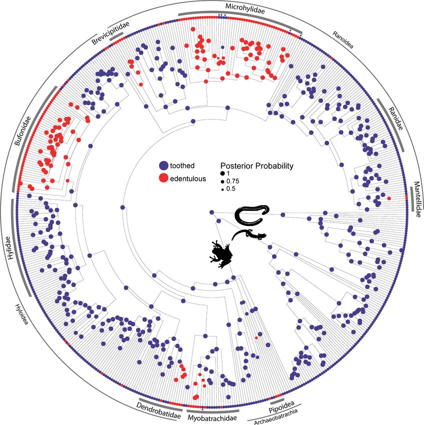
Research article                                                                                            Evolutionary Biology

                                    on the lower jaw was lost in the ancestor of frogs more than 200 million years ago and was subse-
                                    quently regained in a single species (Gastrotheca guentheri; Boulenger, 1882) during the Miocene
                                    (Wiens, 2011). The presence or absence of dentition has also been considered an important taxo-
                                    nomic character in frogs; for example, a subclass was once proposed that included all toothless spe-
                                    cies (Bufoniformia; Cope, 1867). Our understanding of the anuran tree of life has fundamentally
                                    changed with the development of molecular phylogenetics (Duellman and Trueb, 1986;
                                    Feng et al., 2017; Hime et al., 2021), but there has been no attempt to estimate the frequency of
                                    tooth loss across frog diversity or evaluate the factors that may be correlated with edentulism. Most
                                    frogs are generalist, gape-limited predators that capture prey using tongue propulsion (Regal and
                                    Gans, 1976), reducing the importance of teeth in prey capture. Tooth loss is hypothesized to occur
                                    in frogs that specialize on eating small prey (microphagy), such as ants and termites (Das and Coe,
                                    1994; Parmelee, 1999; Narváez and Ron, 2013). This may lead to relaxed functional constraints on
                                    energetically expensive teeth. Microphagous frogs are known to have shortened jaws and altered
                                    feeding cycles (Emerson, 1985), modified tongues (Trueb and Gans, 1983), and some have the abil-
                                    ity to sequester dietary alkaloids from their prey, rendering them toxic (Caldwell, 1996;
                                    Vences et al., 1998). Alternately, teeth may be reduced or lost as a byproduct of miniaturization or
                                    truncated development (paedomorphosis; Davies, 1989; Hanken and Wake, 1993; Smirnov and
                                    Vasil’eva, 1995) because the initiation of odontogenesis occurs ontogenetically late in frogs (during
                                    or after metamorphosis) compared to other vertebrates.
                                        Using the most recent species-rich phylogeny of extant amphibian species (Jetz and Pyron,
                                    2018) and our extensive taxonomic sampling via high-resolution X-ray micro-computed
                                    tomography (microCT) of over 500 of the 562 currently recognized amphibian genera (Amphibia-
                                    Web, 2021), we (1) evaluated the phylogenetic distribution of teeth and reconstructed the evolu-
                                    tionary history of dentition across all major lineages of amphibians and (2) tested whether dietary
                                    specialization, relative jaw length, and body size are correlated with the loss of teeth in frogs. Our
                                    results demonstrate that the presence and location of teeth are highly conserved in salamanders and
                                    caecilians, but labile in frogs. We found that teeth have been repeatedly lost in frogs and at a much
                                    higher frequency than in any other vertebrate group. The evolution of edentulism in anurans is corre-
                                    lated with a microphagous diet and shortening of the lower jaw but not with a reduction in body
                                    size over evolutionary time. Six reversals, from edentulous to toothed jaws, were inferred in frogs.

                                    Results
                                    Distribution of teeth in amphibians
                                    We recorded the presence or absence of teeth on each dentigerous bone of the lower jaw, upper
                                    jaw, and palate for 524 amphibian species (Figure 1; Dataset S1). Taxa were coded as ‘toothed’ if
                                    teeth were observed on any cranial element and ‘edentulous’ if teeth were entirely absent. Our sur-
                                    vey of amphibian dentition across the majority of extant genera confirmed that all salamanders and
                                    caecilians retain teeth, while 134 of the 429 frog species examined are entirely edentulous (Dataset
                                    S1). All anuran species lack dentary teeth with the exception of G. guentheri. The evolution of maxil-
                                    lary and premaxillary teeth of the upper jaw is synchronized in all frog species (Figure 1A and B),
                                    being present in 292 taxa and coordinately absent in 136 species. The vomerine teeth on the palate
                                    are the most variable in frogs, being present in 202 species and absent in 226. Many anurans have
                                    maxillary and premaxillary teeth in the absence of vomerine teeth (92 species), but only two species
                                    examined have vomerine teeth while lacking upper jaw teeth (Rhombophryne testudo, Uperodon
                                    systoma). No other anuran cranial elements possess teeth.
                                       All 65 salamander species examined have teeth on the lower jaw and palate, but three species
                                    lack upper jaw teeth on the maxilla and premaxilla (the sirenids Siren intermedia and Pseudobran-
                                    chus striatus and the salamandrid Salamandrina terdigitata). Thorius pennatulus (Plethodonidae) and
                                    two proteids (Necturus lewisi and Proteus anguinus) lack maxillary teeth but retain premaxillary
                                    teeth. All salamanders have vomerine teeth on the palate (including the paravomerine tooth patches
                                    that underlie the parasphenoid in plethodontids; Figure 1C; Lawson et al., 1971). Palatal teeth
                                    were additionally observed on the palatopterygoid (N. lewisi and P. anguinus) and palatine (S. inter-
                                    media and P. striatus). The lower jaw teeth are present on the dentary in all species, except S.

Paluh et al. eLife 2021;10:e66926. DOI: https://doi.org/10.7554/eLife.66926                                                         2 of 21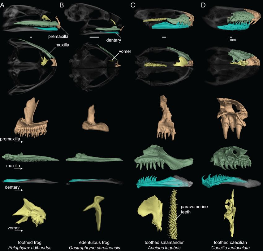
Research article                                                                                                         Evolutionary Biology

Figure 1. Dental diversity of amphibians. (A) Toothed frog, Pelophylax ridibundus (CAS:Herp:217695), (B) edentulous frog, Gastrophryne carolinensis
(UF:Herp:110645), (C) toothed salamander, Aneides lugubris (MVZ:Herp:249828), (D) toothed caecilian, Caecilia tentaculata (KU:Kuh:175441). Skulls in
lateral and ventral views: dentigerous cranial elements are colored and the remainder of the skull is semi-transparent. Isolated premaxilla (orange),
maxilla (green), dentary (blue), and vomer (yellow) in lingual views. Teeth are present on all colored elements except the dentary in P. ridibundus and
those of G. carolinensis. Scale bars = 1 mm.

                                     intermedia and P. striatus, which have mandibular teeth on the splenial. Necturus lewisi and P. angui-
                                     nus are the only two species that have lower jaw teeth on both the dentary and splenial.
                                        All 30 caecilian species examined possess marginal teeth on the lower and upper jaw and palatal
                                     teeth on multiple elements (Figure 1D). The individual elements of the lower jaw in caecilians fuse
                                     to form the pseudodentary, and this composite element varies in having either one or two rows of

Paluh et al. eLife 2021;10:e66926. DOI: https://doi.org/10.7554/eLife.66926                                                                       3 of 21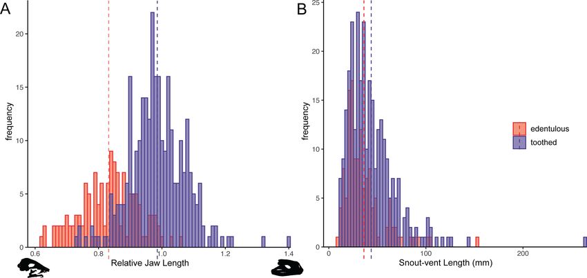
Research article                                                                                             Evolutionary Biology

                                    teeth. Upper jaw teeth are present on the nasopremaxilla (fused nasal and premaxilla) and maxillo-
                                    palatine (fused maxilla and palatine; outer row). Palatal teeth are always present on the vomer and
                                    maxillopalatine (inner row) and occur on the ectopterygoid in one species (Geotrypetes seraphini).

                                    Repeated tooth loss in frogs
                                    Teeth are absent in 134 anuran genera belonging to 19 families. We used reversible-jump Markov
                                    chain Monte Carlo (MCMC) in RevBayes (Höhna et al., 2016) to conduct ancestral state reconstruc-
                                    tions and sample all five Markov models of phenotypic character evolution in proportion to their
                                    posterior probability for toothed and edentulous states in 524 amphibian species. The maximum a
                                    posteriori model of dentition evolution was the one-rate model with a posterior probability of 0.91.
                                    The model-averaged maximum a posteriori ancestral state of Lissamphibia and Anura is toothed
                                    with a posterior probability of 0.99. Teeth have been completely lost at least 22 times in frogs (Fig-
                                    ure 2), and six reversals from edentulous to toothed upper jaws were inferred. Edentulism has
                                    evolved three times in Archaeobatrachia (in Pipoidea, Feng et al., 2017) and 19 times in Neobatra-
                                    chia (10 times in Hyloidea, six times in Ranoidea, twice in Myobatrachidae, and once in Nasikabatra-
                                    chidae). One reversal was estimated in Myobatrachidae (in Uperoleia mahonyi; Clulow et al., 2016)
                                    and five reversals were inferred in Microhylidae (in Dyscophus, Uperodon, Anodonthyla, Cophyla,
                                    and Rhombophryne + Plethodontohyla).
                                        We compared six discrete character evolution models using fitMk in phytools (Revell, 2012) for
                                    three dental states (fully toothed [on premaxilla, maxilla, vomer]; toothed upper jaw [premaxilla,
                                    maxilla] with vomerine tooth loss; edentulous) in 425 anuran species to test if vomerine tooth loss
                                    precedes complete edentulism in frogs. The all-rates-different model was the best fit (all-rates-differ-
                                    ent Akaike information criterion (AIC) weight (AICw) = 0.91, equal-rates AICw = 0.00, single-rate
                                    ordered AICw = 0.00, symmetric ordered AICw = 0.01, unsymmetric ordered AICw = 0.02, symmet-
                                    ric unordered AICw = 0.06), indicating that vomerine tooth loss does not always precede complete
                                    edentulism (Figure 3). Stochastic character mapping suggests that the number of transitions to
                                    edentulism is similar between toothed upper jaws with toothless vomers (13.8) and fully toothed
                                    states (10.3). Toothed frogs have lost vomerine teeth an estimated 59 times (Figure 3). One gain of
                                    vomerine teeth subsequent to a loss was inferred in Phlyctimantis leonardi.

                                    Relationships among tooth loss, diet, and body size
                                    We compiled published diet records for 268 frog lineages and classified 69 taxa from 20 families as
                                    microphagous (defined here as >50% of diet by number or volume consisting of ants, termites, or
                                    mites) and 199 taxa from 47 families as generalist feeders (Figure 4; Dataset S2). Of the 69 micro-
                                    phagy specialists, 54 are edentulous and 15 are toothed. Of the 199 generalists, 25 are edentulous
                                    and 174 are toothed. A BayesTrait discrete analysis indicated correlated evolution between edentu-
                                    lism and microphagy: the dependent model of trait evolution is strongly supported over the inde-
                                    pendent model (Bayes factor = 54.32; a Bayes factor >2 implies the evolution of two traits is linked).
                                    Similar results were found using a 155-taxon dataset excluding genus-level diet data (Bayes fac-
                                    tor = 26.28) and a 134-taxon dataset excluding genus-level diet data and species-level diet data
                                    based on a small sample size (less than five individuals; Bayes factor = 16.0). Of the 22 independent
                                    losses of teeth across frogs, at least 16 of these lineages contain microphagous species (Figure 4).
                                    The majority of the 25 taxa classified as both edentulous and generalist feeders are a subset of the
                                    bufonids (N = 14) and microhylids (N = 5) sampled, but also includes the fully aquatic pipids Pipa
                                    and Hymenochirus, two brevicipitids (Probreviceps and Callulina), and the Darwin’s frog,
                                    Rhinoderma.
                                        Head and body measurements were recorded for 423 anuran species. The relative jaw length in
                                    frogs ranges from 62% of head length in Synapturanus mirandaribeiroi, an edentulous microhylid, to
                                    140% of head length in Lepidobatrachus asper, a toothed ceratophryid. A phylogenetic logistic
                                    regression showed a significant relationship between edentulism and shortened jaws (alpha = 0.0011,
                                    standard error = 0.8920, p
Research article Evolutionary Biology Figure 2. Phylogeny of 524 amphibians depicting the evolution of dentition. Node point color corresponds to Bayesian model-averaged ancestral states of dentition: blue = toothed; red = edentulous. The size of each node point represents the posterior probability of the most probable ancestral state. Tip point colors correspond to dentition states for all species. Asterisks indicate inferred reversals. For species tip labels display Figure 2—figure supplement 2. Corresponding data are provided in Dataset S1. The online version of this article includes the following figure supplement(s) for figure 2: Figure supplement 1. Comparison of true teeth, odontoids, and dental anatomy of the microhylid taxa with inferred evolutionary reversals. Figure supplement 2. Phylogeny of 524 amphibians depicting the evolution of dentition with species tip labels. Figure supplement 3. Evolution of dentary teeth. Figure supplement 4. Evolution of premaxillary teeth. Figure 2 continued on next page Paluh et al. eLife 2021;10:e66926. DOI: https://doi.org/10.7554/eLife.66926 5 of 21
Research article                                                                                                      Evolutionary Biology

Figure 2 continued
Figure supplement 5. Evolution of maxillary teeth.
Figure supplement 6. Evolution of vomerine teeth.

                                    taxa have posteriorly shifted jaws (lower jaws that are longer than their heads; Figure 5A). The
                                    snout–vent length (SVL) of specimens measured ranges from 7.8 mm in the edentulous Paedophryne
                                    amauensis, the smallest known frog, to 263.9 mm in the toothed Conraua goliath, the largest known
                                    extant species. A phylogenetic logistic regression indicated that there is no relationship between
                                    edentulism and body size (alpha = 0.0016, standard error = 0.1162, p=0.09; Figure 5B). Edentulous
                                    species have an average SVL of 36.2 mm (range 7.8–152.4 mm) and toothed species have an aver-
                                    age SVL of 43.7 (range 11.2–263.9 mm). However, a relationship was found between edentulism and
                                    body size when excluding bufonids (alpha = 0.0020, standard error = 0.1539, p=0.015), a clade rep-
                                    resenting a single loss of dentition but that varies widely in size (11.7–152.4 mm SVL in our dataset).

                                    Discussion
                                    Evolution of edentulism in jawed vertebrates
                                    With at least 22 independent origins of edentulism, frogs have completely lost teeth more times
                                    than any other vertebrate clade. Based on our review of the literature, only eight other extant verte-
                                    brate lineages are entirely edentulous. There are no described edentulous chondrichthyan species
                                    (but see Mulas et al., 2020 for the first described aberrant case in a catshark). To our knowledge,
                                    teeth have been entirely lost only three times in living actinopterygian fishes in the (1) Gonorynchi-
                                    formes excluding Gonorynchidae (Kohno et al., 1996; Britz and Moritz, 2007), (2) Gyrinocheilidae

Figure 3. Estimated number of evolutionary transitions among three dental states (fully toothed, toothed upper jaw with vomerine tooth loss,
edentulous) inferred from stochastic character mapping using 1000 replicates. P = premaxilla, M = maxilla, V = vomer. Width of arrows corresponds to
estimated number of changes.
The online version of this article includes the following figure supplement(s) for figure 3:
Figure supplement 1. Discrete character evolution model comparisons for three dental states (fully toothed, toothed upper jaw with vomerine tooth
loss, edentulous) in 425 species of frogs.
Figure supplement 2. Ancestral reconstruction of three dental character states (fully toothed, toothed upper jaw with vomerine tooth loss, edentulous)
using stochastic character mapping in 425 species of frogs.

Paluh et al. eLife 2021;10:e66926. DOI: https://doi.org/10.7554/eLife.66926                                                                    6 of 21
Research article Evolutionary Biology Figure 4. Phylogeny of 268 frog species with a stochastic character map of dentition states and distribution of generalist and microphagous diet states (tip point colors) illustrating the correlated evolution of edentulism and microphagy. Diversity of edentulous frog skulls: 1. Rhinophrynus dorsalis (CAS: Herp:71766), 2. Hymenochirus boettgeri (CAS:Herp:253587), 3. Notaden bennetti (CAS:Herp:78115), 4. Myobatrachus gouldii (MCZ:Herp:A-139543), 5. Dendrobates tinctorius (YPM:VZ:HERA 016010), 6. Ameerega trivittata (UF:Herp:107200), 7. Brachycephalus ephippium (UF:Herp:72725), 8. Rhinoderma darwinii (UF:Herp:62022), 9. Telmatobius carrillae (UF:Herp:39717), 10. Melanophryniscus stelzneri (UF:Herp:63183), 11. Phrynoidis asper (USNM: Amphibians and Reptiles:586870), 12. Physalaemus nattereri (MCZ:Herp:A30113), 13. Engystomops pustulosus (CAS:SUA:21892), 14. Allophryne ruthveni (KU:Kuh:166716), 15. Nasikabatrachus sahadryensis (CES:F:203), 16. Hemisus guineensis (CAS:Herp:258533), 17. Breviceps gibbosus (AMNH: Herpetology:3053), 18. Arthroleptis schubotzi (CAS:Herp:201762), 19. Phrynomantis annectens (UF:Herp:187273), 20. Glyphoglossus molossus (CAS: Herp:243121), 21. Cacosternum namaquense (CAS:Herp:156975), 22. Mantella baroni (CAS:Herp:250387). Scale bars = 1 mm. For species tip labels display Figure 4—figure supplement 1. Corresponding data are provided in Dataset S2. The online version of this article includes the following figure supplement(s) for figure 4: Figure supplement 1. Phylogeny of 268 frog species with a stochastic character map of dentition and the distribution of diet states with species tip labels. Paluh et al. eLife 2021;10:e66926. DOI: https://doi.org/10.7554/eLife.66926 7 of 21
Research article                                                                                                       Evolutionary Biology

Figure 5. Histograms of relative jaw length (mandible length divided by skull length; A) and body size (snout–vent length [SVL]; B) in 423 frog species
plotted by dentition states (blue = toothed; red = edentulous). A phylogenetic correlation was identified between tooth loss and shortened lower jaws.
There is no association between edentulism and body size. Left skull silhouette is Hemisus guineensis (CAS:Herp:258533) and right skull is
Lepidobatrachus asper (UF:Herp:12347). Corresponding trait data are provided in Dataset S3.

                                     (Conway, 2011), and (3) Syngnathidae (seahorses and pipefish; Lin et al., 2016). Other fish lineages,
                                     such as the cyprinids, have toothless oral jaws but retain true pharyngeal teeth (Aigler et al., 2014).
                                     Five extant amniote clades are edentulous, including three lineages of mammals (baleen whales,
                                     pangolins, anteaters), all living birds, and all living turtles (Davit-Beal et al., 2009). There are several
                                     mammal clades that have lost enamel but retain reduced teeth (armadillos, sloths, aardvarks, pygmy,
                                     and dwarf sperm whales; Meredith et al., 2009). Molecular evidence suggests a single loss of teeth
                                     in the common ancestor of extant birds (Meredith et al., 2014), but complete edentulism also
                                     evolved independently in at least two extinct lineages of Mesozoic birds (Confuciusornis and Gobip-
                                     teryx; Yang and Sander, 2018; Brocklehurst and Field, 2021). Teeth were completely lost in at
                                     least two lineages of non-avian dinosaurs (ornithomimosaurs and caenagnathoids; Wang et al.,
                                     2017; Hendrickx et al., 2019) and in some pterosaurs, such as members of Azhdarchidae
                                     (Yang and Sander, 2018). All living crocodilians retain teeth, but at least two fossil suchian archo-
                                     saurs were edentulous (Shuvosaurus and Effigia; Nesbitt and Norell, 2006). There are no known
                                     edentulous squamate species, although African egg-eating snakes in the genus Dasypeltis may have
                                     a dental polymorphism, as they typically have small, short teeth but some individuals are reported to
                                     be edentulous (Visser, 1981). Emydocephalus, a genus of sea snakes that specializes on eating fish
                                     eggs, has maxillary fangs and pterygoid teeth but has lost teeth on the palatine, premaxilla, and
                                     lower jaw (Voris, 1966). Lastly, at least one extinct rhynchocephalian has been suggested to be
                                     edentulous (Sapheosaurus, Rauhut et al., 2012).
                                         The loss of teeth may be associated with the evolution of a secondary feeding apparatus (Davit-
                                     Béal et al., 2007; Wang et al., 2017), such as the keratinized beak in birds and turtles, baleen in
                                     mysticete whales, and specialized tongues in pangolins and anteaters. Nearly all frogs have a spe-
                                     cialized tongue that is used in feeding (Regal and Gans, 1976), and this adaptation might have facil-
                                     itated the repeated loss of teeth across anurans. Surprisingly, three anuran genera are both
                                     tongueless and edentulous (Hymenochirus, Pseudhymenochirus, and Pipa in Pipidae), but these spe-
                                     cies are highly aquatic and have a derived mechanism of catching prey under water through suction
                                     feeding (Dean, 2003; Cundall et al., 2017). The edentulous syngnathids (seahorses and relatives)

Paluh et al. eLife 2021;10:e66926. DOI: https://doi.org/10.7554/eLife.66926                                                                     8 of 21
Research article Evolutionary Biology
and actinopterygians with toothless oral jaws also catch prey through suction feeding (Roos et al.,
2009; Mihalitsis and Bellwood, 2019). There appears to be no size-related constraints promoting
complete tooth loss across all vertebrates. Edentulous species span the entire spectrum of verte-
brate body sizes: the smallest known vertebrate species (the microhylid frog P. amauensis,
Rittmeyer et al., 2012) and the largest (the blue whale, Balaenoptera musculus) are both edentu-
lous. The second-smallest known vertebrate, the cyprinid fish in the genus Paedocypris, retains true
pharyngeal teeth (Kottelat et al., 2006). Several edentulous vertebrate clades are thought to have
paedomorphic skulls, including toothless frogs (Smirnov and Vasil’eva, 1995), birds (Bhullar et al.,
2012), and baleen whales (Fordyce and Barnes, 1994), suggesting that truncated ontogenetic tra-
jectories may constrain the formation of teeth, but this hypothesis requires further investigation.
Tooth formation occurs ontogenetically late in frogs, during or after metamorphosis, in contrast
to during early larval or embryonic development in other vertebrates (Davit-Béal et al., 2007,
Lainoff et al., 2015). This delayed shift in odontogenesis may be linked to the evolutionary lability
of teeth in anurans. There may also be a relationship between the loss of teeth and delayed ossifica-
tion of dentigerous elements. For example, the dentary bone ossifies relatively late in frogs, and
nearly always lacks teeth, compared to being one of the first cranial elements to ossify in salaman-
ders and caecilians (Harrington et al., 2013), and these amphibians always retain mandibular denti-
tion. Truncated development is hypothesized to be associated with the repeated loss of other
mineralized structures in frogs, such as the stapes of the middle ear (Pereyra et al., 2016;
Womack et al., 2018; Womack et al., 2019). The anuran mouth undergoes dramatic restructuring
during metamorphosis while transitioning from an herbivorous tadpole with a keratinized beak and
short, cartilaginous lower jaw to a carnivorous frog with an elongated, bony lower jaw. This rapid
morphological transformation requires further study in edentulous and toothed species. Several
anuran lineages have evolved direct development (undergoing the larval stage within the egg;
Gomez-Mestre et al., 2012), and this life history transition may provide an opportunity to repattern
the jaw and alter dental development, such as the timing of tooth germ initiation. Many of the eden-
tulous frogs identified in this study are biphasic (possessing a free-swimming tadpole stage), but
edentulism is also present in some direct-developing lineages (e.g., Arthroleptis, Brachycephalus,
Brevicipitidae, Myobatrachus, asterophryine microhylids) and viviparous species (Nimbaphrynoides,
Nectophrynoides), suggesting that life history mode does not constrain patterns of tooth loss.
Amphibian dentition and tooth loss in frogs
Dentition is highly conserved in salamanders and caecilians with no identified cases of edentulism.
Teeth are present on the jaws and palate of all caecilians (Wake and Wurst, 1979), and this is also
the typical dental condition in salamanders (Gregory et al., 2016). The aquatic sirenid salamanders
(Siren, Pseudobranchus) lack maxillary and premaxillary teeth (Clemen and Greven, 1988), while the
miniaturized species of Thorius sampled here (T. pennatulus) lacks maxillary teeth but retains teeth
on the premaxilla, palate, and lower jaw. At least one species of Thorius possesses a novel dental
polymorphism in which males lack maxillary teeth but females maintain several teeth on the maxilla
(Hanken et al., 1999). To our knowledge, this is the only known case of a sexually dimorphic
presence or absence dental polymorphism in an amphibian. Larval salamanders and caecilians were
excluded in our dentition survey but differ in patterns of dentition from adults (Wake, 1976;
Clemen and Greven, 2018), such as the transient presence of teeth on the splenials, palatines, and
pterygoids that are lost during development (Schoch et al., 2019). Maxillary and premaxillary teeth
are synchronized in all anuran taxa that we sampled, but two species in the genus Telmatobius have
maxillary teeth in the absence of premaxillary teeth (Barrionuevo, 2017). Members of this genus can
be toothed or edentulous, and two species are reported to have intraspecific variation in the pres-
ence or absence of dentition (Barrionuevo, 2017). The loss of vomerine teeth is not a prerequisite
for complete tooth loss (Figure 3), and the presence or absence of teeth on the vomer is not coordi-
nated with dentition on the maxilla and premaxilla in frogs. These results suggest that dentition on
the upper jaws and palate are independent modules, and the higher lability of vomerine teeth
requires further study. Previous work has suggested that the size and number of vomerine teeth may
be correlated with diet and body size (Hedges, 1989; Estrada and Hedges, 1996). Teeth are
entirely absent in 134 anuran genera distributed across 19 families, and our ancestral state recon-
struction suggests that teeth have been lost more than 20 times during the evolution of frogs.
Paluh et al. eLife 2021;10:e66926. DOI: https://doi.org/10.7554/eLife.66926 9 of 21Research article Evolutionary Biology
We identified a phylogenetic correlation between the evolution of edentulism and a micropha-
gous diet, and these two traits co-occur in more than 50 genera belonging to 14 families (Dataset
S2; Figure 4). The majority of these species specialize on eating ants and termites, despite that
these insects have many defense behaviors (biting, stinging, chemical weapons) and low nutritional
value compared to other invertebrates (Redford and Dorea, 1984; McNab, 1984). Edentulous,
microphagous frogs inhabit biomes ranging from tropical forests (e.g., Dendrobates, Mantella, Car-
dioglossa) to arid deserts (e.g., Breviceps, Notaden) and are found on all continents, excluding Ant-
arctica. Frogs, ants, and termites evolved at roughly the same time—with important diversification
events occurring in all three groups during the Cretaceous and Cretaceous–Paleogene boundary
(Moreau and Bell, 2013; Bourguignon et al., 2015; Feng et al., 2017)—suggesting the repeated
evolution of complete edentulism in frogs may be linked to the spatiotemporal diversification of ants
and termites. Teeth have been repeatedly reduced in other tetrapods that specialize on eating ants
and termites, including multiple lineages of mammals (echidnas, numbats, aardvarks, aardwolves,
anteaters, armadillos, pangolins; Reiss, 2001) and squamates (scolecophidian blind snakes, Aprasia
worm lizards; Daza and Bauer, 2015).
The complete loss of teeth in frogs is associated with the shortening of the lower jaw (Figure 4),
a skeletal trait that is known to occur in species that eat smaller prey (Emerson, 1985; Vidal-
Garcı́a and Scott Keogh, 2017; Paluh et al., 2020). The shortening of the mandible reduces maxi-
mum gape and alters jaw biomechanics to improve the efficiency of catching many small prey items.
Frogs with a jaw length equal to or longer than the skull have an asymmetrical feeding cycle where
the time spent catching prey is short but the time spent bringing prey into the mouth is long
(Gans and Gorniak, 1982); shortened jaws result in a faster, symmetric feeding cycle where equal
amounts of time are spent catching and bringing prey into the mouth (Emerson, 1985). At least four
lineages of edentulous anurans that specialize on ants and termites have additionally evolved muscu-
lar hydrostatic tongues that can be aimed in all three dimensions and with great precision without
moving the head to improve the efficiency of small prey capture (Rhinophrynus, Trueb and Gans,
1983; Hemisus, Nishikawa et al., 1999; microhylids and brevicipitids, Meyers et al., 2004).
Recently, these same species have additionally been shown to have relatively small eyes among
frogs (Thomas et al., 2020), suggesting reduced visual sensitivity and supporting the hypothesis
that microphagous frogs may possess improved abilities to process olfactory and tactical cues in
order to detect and localize prey (Deban et al., 2001). Therefore, the evolution of microphagy may
have influenced a suite of traits in the head of frogs, including skull shape, jaw position, dentition,
tongue morphology, and the functionality of sensory organs.
The majority of the 134 edentulous frogs in our dataset are restricted to the families Bufonidae
and Microhylidae. All 48 genera of bufonids examined—the only anuran clade widely recognized as
being edentulous (Davit-Béal et al., 2007)—and 48 of 54 of microhylid genera examined lack teeth.
All remaining families have less than 10 edentulous genera. The Bufonidae and Microhylidae are two
of the most diverse frog families, comprising 637 and 697 species, respectively (18% of all frogs;
AmphibiaWeb, 2021). The evolution of edentulism in frogs may exert an influence on diversification
rates, but we refrain from testing this hypothesis using trait-dependent diversification models due to
our sparse, genus-level taxonomic sampling (429 tips representing 7299 lineages). The results of our
ancestral state reconstruction analyses indicate that teeth were independently lost in the most recent
common ancestors of both bufonids and microhylids. Once lost, teeth have not been regained in the
Bufonidae but may have re-evolved several times in microhylids. Although both clades have many
taxa that specialize on small prey, there are bufonids and microhylids with expanded, generalized
diets, and a few species that will even consume vertebrate prey (e.g., Rhinella marina, Asterophrys
turpicola). The variation in diet within bufonids and microhylids corresponds with variation in the rel-
ative length of the lower jaw (Dataset S3) and overall skull morphology (Paluh et al., 2020).
The inferred reversals in Microhylidae occur in Dyscophus, Uperodon, and four cophyline genera
(Anodonthyla, Cophyla, Plethodontohyla, Rhombophryne). Recent work has also suggested that
additional microhylid genera contain toothed and edentulous species (Mini, Scherz et al., 2019;
Glyphoglossus, Gorin et al., 2021). If teeth were entirely lost in the common ancestor of microhylids,
the repeated re-evolution of true teeth (with enamel, dentin, and pulp cavity) in this clade is unlikely
and requires histological investigation. We hypothesize the tooth-like structures in some of these
taxa, such as Rhombophryne and Uperodon (Figure 2—figure supplement 1), may be small odon-
toids (bony serrations that lack the tissue composition of true teeth; Fabrezi and Emerson, 2003),
Paluh et al. eLife 2021;10:e66926. DOI: https://doi.org/10.7554/eLife.66926 10 of 21Research article Evolutionary Biology
similar to what has been described in the bufonid Incilius alvarius (Mendelson and Pramuk, 1998),
some Brachycephalus (Ribeiro et al., 2017), and some New Guinea asterophrynine microhylids
(Zweifel, 1971). Alternatively, the most recent common ancestor of the Microhylidae possessed
teeth and several lineages then subsequently lost dentition independently, with no reversals. The
dental anatomy of Dyscophus has been examined (LaDouceur et al., 2020), and this genus does
possess true teeth. The morphology of Dyscophus, Anodonthyla, Cophyla, and Plethodontohyla indi-
cates that these taxa have true dentition based on the presence of replacement teeth and/or float-
ing teeth that are undergoing resorption (Figure 2—figure supplement 1). The phylogenetic
relationships and divergence time estimates among microhylid taxa remain controversial
(Peloso et al., 2016; Feng et al., 2017; Streicher et al., 2020), which further impedes the interpre-
tation of dental evolution in this group.
Of the ten anuran genera known to possess variation in the presence or absence of teeth, diet
data are only available for Physalaemus and Engystomops. In both genera, edentulous species have
specialized microphagous diets in comparison to toothed congeners that consume a broader array
of invertebrates (Narváez and Ron, 2013). Dietary alkaloid sequestration has evolved as a predator
defense mechanism in at least five clades of frogs that specialize on eating ants and mites, and teeth
have been lost in several of these lineages (Dendrobatinae and Ameerega [Dendrobatidae,
Saporito et al., 2004]; Pseudophryne [Myobatrachidae, Smith et al., 2002]; Mantella [Mantellidae,
Daly et al., 1997]; Melanophryniscus [Bufonidae, Hantak et al., 2013]). Teeth are retained in Phyllo-
bates, which is sister to all other edentulous genera of the Dendrobatinae, and in the Cuban Eleu-
therodactylus group known to sequester alkaloids (Rodrı́guez et al., 2011). There are other
microphagous frogs that retain teeth, such as the ant specialist Sphaenorhynchus (Parmelee, 1999).
Further work is needed to investigate the number, size, and histological anatomy of teeth across
toothed frogs that vary in diet. It remains unknown whether any anurans have lost enamel but retain
teeth, which has occurred several times in mammals (Meredith et al., 2009).
No relationship was identified between complete edentulism and body size in the 423 frog spe-
cies sampled. The smallest known species of frog, P. amauensis, lacks teeth, but some miniaturized
anurans are toothed. We examined 25 taxa with an SVL of 15 mm or less: 13 were toothed and 12
were edentulous. Several of the smallest edentulous species in our dataset are microhylids, bufonids,
and dendrobatids, and these clades have widespread tooth loss across a range of body sizes. We
identified only one case of edentulism in Brachycephaloidea, in the genus Brachycephalus, despite
this neotropical radiation of over 1000 species containing many miniaturized lineages (e.g., smallest
members of Pristimantis, Eleutherodactylus, Noblella). Within the genus Arthroleptis, several minia-
ture species lack teeth (Laurent, 1954; Blackburn, 2008), suggesting that, in some cases, a reduc-
tion in body size and tooth loss may be linked. There are several large or gigantic species within the
Bufonidae (Womack and Bell, 2020), but all true toads lack teeth regardless of size. When excluding
bufonids, reduction in body size was phylogenetically correlated with edentulism, indicating that fur-
ther work is needed to investigate the interplay between edentulism, body size evolution, and pae-
domorphism across anurans.
Tooth loss in fossil amphibians
To our knowledge, no stem tetrapods have been described as edentulous (Ruta et al., 2003;
Anderson et al., 2008; Matsumoto and Evans, 2017). Albanerpetontids, an extinct lineage of lis-
samphibians, had teeth on the premaxilla, maxilla, and dentary (Daza et al., 2020), and there is no
evidence of edentulism in fossil salamanders or caecilians. Of the stem salientians with cranial mate-
rial, teeth are present on the upper jaw in Prosalirus (Shubin and Jenkins, 1995), Vieraella
(Baez and Basso, 1996), and Liaobatrachus (Gao and Wang, 2001). No teeth are visible in Triado-
batrachus (Ascarrunz et al., 2016), the oldest known stem frog, but the maxilla and premaxilla are
poorly preserved in this impression fossil. The dentary of Triadobatrachus lacks teeth, and the
absence of dentition on the lower jaw is considered a synapomorphy of Salientia (Milner, 1988).
Several extinct crown-group anuran genera are edentulous, and the majority of these are hypoth-
esized to be members of the Pipidae (Báez and Harrison, 2005), including Saltenia from the late
Cretaceous of Argentina (Báez, 1981), Vulcanobatrachus from the late Cretaceous of South Africa
(Trueb et al., 2005), Shelania from the Paleocene-Eocene of Argentina (Baez and Trueb, 1997,
Báez and Pugener, 1998), and Singidella from the Eocene of Tanzania (Báez and Harrison, 2005).
Based on the phylogenetic analyses of Báez and Harrison, 2005 and Trueb et al., 2005, extinct
Paluh et al. eLife 2021;10:e66926. DOI: https://doi.org/10.7554/eLife.66926 11 of 21Research article Evolutionary Biology
edentulous pipids likely represent two or more instances of tooth loss: at least once in the South
American fossils (Saltenia, Shelania; sister to African Xenopus; clade Xenopodinomorpha) and once
in the African fossils (Vulcanobatrachus, Singidella; sister to South American Pipa and African Hyme-
nochirus and Pseudhymenochirus; clade Pipinomorpha). However, the clades Xenopodinomorpha
and Pipinomorpha are not supported by molecular phylogenetic analyses of living pipids
(Irisarri et al., 2011; Feng et al., 2017), and therefore the placement of these fossils requires re-
evaluation. Tooth loss likely occurred in pipids at earliest during the mid-Cretaceous because all
described pipimorph frogs from the early Cretaceous are toothed (Báez et al., 2021): Thoraciliacus
(Trueb, 1999), Cordicephalus (Trueb and Báez, 2006), Neusibatrachus, and Gracilibatrachus
(Báez, 2013). Our ancestral state reconstructions suggest that living pipid frogs have lost teeth twice
independently in the ancestor of Hymenochirus + Pseudhymenochirus and in Pipa (Figure 2). All
species of Xenopus and three of seven species of Pipa have teeth (P. carvalhoi, P. aspera, P. arrabali;
Trueb and Cannatella, 1986). Teeth may have been lost several times in Pipa, but the relationships
among species are poorly understood.
The sister clade to Pipidae is Rhinophrynidae (collectively forming the Pipoidea, Feng et al.,
2017), which contains a single living species, the edentulous Rhinophrynus dorsalis. The fossil record
of Rhinophrynidae is sparse (Blackburn et al., 2019) but includes the toothed Rhadinosteus from
the late Jurassic of Utah (Henrici, 1998) and the edentulous Chelomophrynus from the Eocene of
Wyoming (Henrici, 1991). The presence of teeth in Rhadinosteus supports the hypothesis that teeth
were lost independently in pipids and rhinophrynids (Figure 2) and indicates that edentulism may
have evolved later in rhinophrynids.
Two edentulous, non-pipoid fossil frogs have been described from the Mesozoic of the Northern
Hemisphere: Theatonius from the late Cretaceous of Wyoming (Fox, 1976) and Tyrrellbatrachus
from the late Cretaceous of Alberta (Gardner, 2015). These two crown-group anurans have an
uncertain placement in the frog tree of life but are likely distantly related to one another. Gard-
ner, 2015 hypothesized that these two taxa likely represent two independent cases of tooth loss
and possibly the oldest record of edentulism in nonpipid frogs. The majority of living, edentulous
frogs are neobatrachians (Figure 2; in Hyloidea, Ranoidea, Myobatrachidae, and Nasikabatrachidae),
but few fossil neobatrachians have been described as edentulous (Gardner, 2015).
Molecular and developmental mechanisms of tooth loss
Recent work has documented that several lineages of edentulous vertebrates have various states of
molecular tooth decay in the genes that are critical for the formation of dentin and enamel with
frameshift mutations and stop codons that result in nonfunctionalization (mammals: Meredith et al.,
2009; turtles: Meredith et al., 2013; birds: Meredith et al., 2014; syngnathids: Lin et al., 2016).
The frameshift mutation rate of these loci can be used to estimate the timing of tooth loss in the fos-
sil record (Meredith et al., 2009; Meredith et al., 2014), and the ratio of synonymous and nonsy-
nonymous substitutions can be calculated to measure selection pressure on enamel matrix proteins
(Alazem and Abramyan, 2019). Whether edentulous frogs possess similar rates of molecular tooth
decay in these loci, as demonstrated in amniotes, has yet to be tested. Recently, Lu et al., 2021
failed to identify any dentin or enamel genes in the genomes of two bufonids (R. marina and B. gar-
garizans) but Shaheen et al., 2021 demonstrated pseudogenization of the major tooth enamel gene
amelogenin in R. marina, B. gargarizans, and B. bufo. Based on selection intensity estimates and sub-
stitution rates, this gene likely became inactivated in the Bufonidae 40–60 million years ago
(Shaheen et al., 2021). We hypothesize that these tooth-specific genes have degenerated repeat-
edly across edentulous anurans by novel inactivating mutations, and the frameshift mutation rate will
indicate that teeth were lost at several different geologic times during the evolution of frogs. Anuran
enamel matrix proteins may be operating under relaxed selection, compared to purifying selection
in most mammals and reptiles (Alazem and Abramyan, 2019), due to the evolution of projectile
tongue feeding, enabling the evolutionary lability of frog teeth.
The developmental genetics of tooth formation in amphibians is almost entirely unexplored,
especially when compared to our understanding of chondrichthyan, teleost, and amniote odonto-
genesis (Fraser et al., 2004; Tucker and Sharpe, 2004; Thiery et al., 2017). It is unknown if the
genes critical for tooth formation in fishes and amniotes are also expressed during morphogenesis
of teeth in amphibians, if all frog species retain a suppressed ancestral developmental pathway of
tooth development on the lower jaw, or if the odontogenetic pathway has been disrupted via one or
Paluh et al. eLife 2021;10:e66926. DOI: https://doi.org/10.7554/eLife.66926 12 of 21Research article Evolutionary Biology
many mechanisms on the jaws of edentulous anurans. The loss of teeth on the lower jaw of frogs
could be due to the loss of a single major signal that can orchestrate odontogenesis, comparable to
the sole loss of odontogenic Bmp4 expression in living birds (Chen et al., 2000) or termination of
Msx2 expression in living turtles (Tokita et al., 2013), which arrests tooth formation early in develop-
ment. If true, potential rudimentary structures, such as tooth buds or the early thickening of the
odontogenic band, might be seen before the abortion of tooth development in the lower jaw of anu-
rans. Investigation of the developmental genetics of tooth formation in the upper and lower jaws of
frogs will fill a large gap in our understanding of vertebrate evolution and development and may elu-
cidate the mechanisms of repeated tooth loss and putative cases of the re-evolution of lost teeth in
one of the most diverse vertebrate orders.
Materials and methods
Species sampling and scanning
We collected data from high-resolution microCT scans of 523 amphibian species, representing 420
frog genera (of 460 total; AmphibiaWeb, 2021), 65 salamander genera (of 68 total), and 30 caecilian
genera (of 34 total). One recently described frog species was not microCT scanned but included in
the dataset because it is the only member of its genus with teeth (U. mahonyi; Clulow et al., 2016).
All genera are represented by one species except for nine anuran genera (Arthroleptis, Cacoster-
num, Engystomops, Gastrotheca, Physalaemus, Pipa, Telmatobius, Uperodon, Uperoleia) with two
sampled lineages that represent known dental variation within these genera (Dataset S1). All scans
were run using a 240 kv X-ray tube containing a diamond-tungsten target, with the voltage, current,
and detector capture time adjusted for each scan to maximize absorption range for each specimen.
Raw X-ray data were processed using GE’s proprietary datos|x software version 2.3 to produce a
series of tomogram images and volumes, with final voxel resolutions ranging from 1 to 147 mm. The
resulting microCT volume files were imported into VG StudioMax version 3.2.4 (Volume Graphics,
Heidelberg, Germany), the skull and skeleton were isolated using VG StudioMax’s suite of segmen-
tation tools, and then exported as high-fidelity mesh files. We deposited image stacks (TIFF) and 3D
mesh files of the skull and skeleton for each specimen in MorphoSource (see Dataset S1 for DOIs).
Survey of amphibian dentition variation and ancestral state
reconstructions
We recorded the presence or absence of teeth on each dentigerous bone of the lower jaw, upper
jaw, and palate for 524 amphibian species (Figure 1; Dataset S1). Teeth were identified by a combi-
nation of the following characteristics: conical shape, presence of distinct pedicel and crown, pres-
ence of replacement teeth, and/or presence of floating teeth that are undergoing resorption
(Figure 1, Figure 2—figure supplement 1). We conducted ancestral state reconstructions of denti-
tion (two states: toothed, edentulous) in extant amphibians using the data collected from 524 spe-
cies representing 515 genera and all 77 families using the phylogeny of Jetz and Pyron, 2018.
Bayesian ancestral state reconstructions were calculated using reversible-jump MCMC in RevBayes
(Höhna et al., 2016) to sample all five Markov models of phenotypic character evolution (one-rate,
two-rate, zero-to-one irreversible, one-to-zero irreversible, no change) in proportion to their poste-
rior probability. We accounted for model uncertainty by making model-averaged ancestral state esti-
mates (Freyman and Höhna, 2018; Freund et al., 2018). The models were assigned an equal prior
probability using a uniform set-partitioning prior, and the root state frequencies were estimated
using a flat Dirichlet prior. The rates of gain and loss of dentition were drawn from an exponential
distribution with a mean of 10 expected character state transitions over the tree. The MCMC was
run for 22,000 iterations, the first 2000 iterations were discarded as burn-in, and samples were
logged every 10 iterations. Convergence of the MCMC was confirmed using Tracer version 1.6 to
ensure that analyses had reached stationarity. We conducted additional Bayesian ancestral state
reconstructions using RevBayes to model the evolutionary history of dentition presence or absence
on individual dentigerous elements (Figure 2—figure supplements 3–6).
To test if vomerine tooth loss always precedes complete edentulism in frogs, we compared dis-
crete character evolution models using fitMk in phytools for three dental states (fully toothed,
toothed upper jaw with vomerine tooth loss, edentulous) in 425 species of frogs. The only two
Paluh et al. eLife 2021;10:e66926. DOI: https://doi.org/10.7554/eLife.66926 13 of 21Research article Evolutionary Biology
species that putatively possess vomerine teeth while lacking upper jaw teeth (R. testudo and U. sys-
toma) were excluded from this analysis. Six models were compared (equal-rates, single-rate ordered,
symmetric ordered, unsymmetric ordered, symmetric unordered, all-rates-different) using the AIC
and AICw (Figure 3—figure supplement 1). The ordered models enforced vomerine tooth loss as
an intermediate state between fully toothed and edentulous states. To estimate the posterior proba-
bilities for all nodes and the number of transitions between these three character states, we used
stochastic character mapping (MCMC sampling of character histories from their posterior probability
distribution; Huelsenbeck et al., 2003) in phytools (make.simmap function) using the best fit model
of character evolution (all-rates-different), a flat root prior, and 1000 replicates (Figure 3—figure
supplement 2).
Testing relationships among edentulism, diet, and body size
We compiled dietary data for all sampled anuran species from the literature (see Dataset S2 for
references). Species were classified as microphagous specialists if the majority (>50%) of their diet
by number or volume consists of ants, termites, or mites. Species were classified as generalists if the
majority of their diet by number or volume consists of other invertebrate groups or vertebrates. For
species with no published diet records, we searched for any existing diet records at the genus level
because dentition state (toothed/edentulous) is generally consistent within a genus. Only 10 of 461
anuran genera are known to contain both edentulous and toothed species (Arthroleptis, Cacoster-
num, Engystomops, Glyphoglossus, Mini, Physalaemus, Pipa, Telmatobius, Uperodon, Uperoleia).
Due to the disparity in existing ecological data available across all anurans, the dietary records
ranged from singular reports (one prey item in one individual) to detailed studies investigating the
stomach contents of hundreds of individuals.
We measured SVL (tip of the snout to the rear of the ischium), skull length (occiput to tip of the
snout), and mandible length (posterior to anterior tip of the lower jaw) for all sampled specimens
using the linear measurement tools in VG StudioMax and MeshLab (Cignoni et al., 2008). We calcu-
lated relative jaw length (mandible length divided by skull length) for each specimen: a jaw length
value greater than 1 indicates a posteriorly shifted jaw joint (lower jaw is longer than the head) and a
value less than 1 indicates an anteriorly shifted jaw joint (lower jaw is shorter than the head).
We used phylogenetic comparative methods to test for evolutionary correlations among denti-
tion, diet, and body size in frogs. We compiled diet records for 268 taxa, representing 259 genera
and 52 anuran families: 155 species in the dentition dataset had published diet records and the
remaining 113 lineages are represented by genus-level diet data. We excluded the remaining 161
anuran species in the dentition dataset (55 edentulous, 106 toothed) from the diet analyses due to
the lack of known diet records at the species or genus level. Because dentition (toothed/edentulous)
and diet (generalist/microphagous) were treated as binary traits, we tested for a phylogenetic corre-
lation using discrete independent and discrete dependent models with rjMCMC sampling in Bayes-
Traits version 3.0.2 (Pagel and Meade, 2006). The stepping stone sampler for marginal likelihood
reconstructions was used with 100 stones and 1000 iterations. The branch lengths were scaled to
have a mean of 0.1 using ScaleTrees. Bayes factors (log BF = 2[log marginal likelihood complex
model – log marginal likelihood simple model]) were used to compare the fit of the independent ver-
sus dependent models. Models were run using the complete 268 taxon dataset, a reduced 158
taxon dataset excluding genus-level diet data, and a reduced 134 taxon dataset excluding genus-
level diet data and species-level diet data based on a small sample size (less than five individuals).
Several previous studies have demonstrated a correlation between skull shape and diet in frogs:
species that specialize on small prey have anteriorly shifted, relatively short jaws while generalist
feeders that are capable of eating large prey have a posteriorly shifted jaw joint (Emerson, 1985;
Vidal-Garcı́a and Scott Keogh, 2017; Paluh et al., 2020). Because diet data are lacking for many
anuran genera, we additionally tested for a phylogenetic correlation between dentition and the rela-
tive length of the jaw as a morphological proxy for diet. Lastly, because teeth may be lost as a
byproduct of miniaturization (Hanken and Wake, 1993; Smirnov and Vasil’eva, 1995), we tested
for a phylogenetic correlation between dentition state and body size (SVL). Phylogenetic logistic
regression models were calculated in the phylolm R package (Ho and Ané, 2014) using dentition
and measurement data for 423 anuran species. Dentition (toothed/edentulous) was treated as the
binary response variable and the log transformed size metrics (relative jaw length, SVL) as continuous
predictor variables. We used the ‘logistic_MPLE’ method, which maximizes the penalized likelihood
Paluh et al. eLife 2021;10:e66926. DOI: https://doi.org/10.7554/eLife.66926 14 of 21Research article Evolutionary Biology
of the logistic regression, with a btol of 10, a log.alpha.bound of 10, and 1000 bootstrap replicates.
The body size analysis was run using the complete 423 taxon dataset and a reduced 377 taxon data-
set excluding bufonid toads.
Acknowledgements
We thank all of the institutions, curators, and collection managers that loaned us specimens for this
study. We thank Marta Vidal-Garcia for providing access to the Spicospina flammocaerulea scan. We
thank Kevin Conway for providing insight on the distribution of edentulism in actinopterygian fishes
and David Wake for insights into the paravomerine teeth of salamanders. We thank the research
group of DCB at the Florida Museum of Natural History and two anonymous reviewers for helpful
comments that improved an earlier version of this manuscript.
Additional information
Funding
Funder Grant reference number Author
National Science Foundation DGE-1315138 Daniel J Paluh
National Science Foundation DGE-1842473 Daniel J Paluh
National Science Foundation DBI- 1701714 Edward L Stanley
David C Blackburn
The funders had no role in study design, data collection and interpretation, or the
decision to submit the work for publication.
Author contributions
Daniel J Paluh, Conceptualization, Data curation, Formal analysis, Funding acquisition, Investigation,
Visualization, Methodology, Writing - original draft, Writing - review and editing; Karina Riddell,
Catherine M Early, Maggie M Hantak, Gregory FM Jongsma, Rachel M Keeffe, Fernanda Magalhães
Silva, Stuart V Nielsen, Marı́a Camila Vallejo-Pareja, Data curation, Investigation, Writing - review
and editing; Edward L Stanley, Resources, Data curation, Funding acquisition, Investigation, Writing
- review and editing; David C Blackburn, Conceptualization, Resources, Data curation, Funding
acquisition, Investigation, Writing - review and editing
Author ORCIDs
Daniel J Paluh https://orcid.org/0000-0003-3506-2669
Maggie M Hantak https://orcid.org/0000-0001-9469-4741
Fernanda Magalhães Silva https://orcid.org/0000-0001-7741-7232
Marı́a Camila Vallejo-Pareja https://orcid.org/0000-0002-0987-3364
Edward L Stanley http://orcid.org/0000-0001-5257-037X
David C Blackburn https://orcid.org/0000-0002-1810-9886
Decision letter and Author response
Decision letter https://doi.org/10.7554/eLife.66926.sa1
Author response https://doi.org/10.7554/eLife.66926.sa2
Additional files
Supplementary files
. Transparent reporting form
Paluh et al. eLife 2021;10:e66926. DOI: https://doi.org/10.7554/eLife.66926 15 of 21You can also read