The flagellar motor of Vibrio alginolyticus undergoes major structural remodeling during rotational switching - eLife
←
→
Page content transcription
If your browser does not render page correctly, please read the page content below
RESEARCH ARTICLE

                                    The flagellar motor of Vibrio alginolyticus
                                    undergoes major structural remodeling
                                    during rotational switching
                                    Brittany L Carroll1,2†, Tatsuro Nishikino3†, Wangbiao Guo1,2, Shiwei Zhu1,2‡,
                                    Seiji Kojima3, Michio Homma3*, Jun Liu1,2*
                                    1
                                     Department of Microbial Pathogenesis, Yale School of Medicine, New Haven,
                                    United States; 2Microbial Sciences Institute, Yale University, West Haven, United
                                    States; 3Division of Biological Science, Graduate School of Science, Nagoya
                                    University, Furo-cho, Chikusa-ku, Nagoya, Japan

                                    Abstract The bacterial flagellar motor switches rotational direction between counterclockwise
                                    (CCW) and clockwise (CW) to direct the migration of the cell. The cytoplasmic ring (C-ring) of the
                                    motor, which is composed of FliG, FliM, and FliN, is known for controlling the rotational sense of
                                    the flagellum. However, the mechanism underlying rotational switching remains elusive. Here, we
                                    deployed cryo-electron tomography to visualize the C-ring in two rotational biased mutants in
                                    Vibrio alginolyticus. We determined the C-ring molecular architectures, providing novel insights
                                    into the mechanism of rotational switching. We report that the C-ring maintained 34-fold symmetry
*For correspondence:                in both rotational senses, and the protein composition remained constant. The two structures show
g44416a@cc.nagoya-u.ac.jp (MH);     FliG conformational changes elicit a large conformational rearrangement of the rotor complex that
jliu@yale.edu (JL)                  coincides with rotational switching of the flagellum. FliM and FliN form a stable spiral-shaped base
†
 These authors contributed          of the C-ring, likely stabilizing the C-ring during the conformational remodeling.
equally to this work

Present address: ‡Howard
Hughes Medical Institute, Yale
School of Medicine, New Haven,
United States
                                    Introduction
                                    Many bacteria navigate complex environments by controlling the flagellar rotational switch between
Competing interests: The
                                    counterclockwise (CCW) and clockwise (CW). Escherichia coli and Salmonella enterica (henceforth,
authors declare that no
                                    Salmonella) use a ‘run-and-tumble’ approach for controlling movement, by which flagella rotating in
competing interests exist.
                                    a CCW sense drive the cell body forward, and when the rotation sense switches to CW the bacte-
Funding: See page 14                rium tumbles through the medium to change direction (Berg, 2003; Chevance and Hughes, 2008;
Received: 26 July 2020              Terashima et al., 2008). Vibrio alginolyticus has a unique three-step swimming pattern with forward,
Accepted: 04 September 2020         reverse, and flick motions; where CCW rotation propels the cell body forward, CW rotation drives
Published: 07 September 2020        the bacterium in reverse, and a flicking motion occurs upon CW-CCW rotation to change swimming
                                    direction, analogous to the tumble (Xie et al., 2011).
Reviewing editor: Edward H
Egelman, University of Virginia,
                                        The motor is the most intricate part of the flagellum, not only responsible for flagellar assembly
United States                       and rotation but also essential for rotational switching. Spanning from the cytosol through the outer
                                    membrane, the motor consists of a series of rings, with the L-ring at the outer membrane, the P-ring
   Copyright Carroll et al. This
                                    located within the periplasmic space, the MS-ring embedded in the cytoplasmic membrane, and the
article is distributed under the
                                    C-ring inside the cytoplasm (Homma et al., 1987; Francis et al., 1992; Ueno et al., 1992;
terms of the Creative Commons
Attribution License, which          Francis et al., 1994). The stator subunits, embedded in the cytoplasmic membrane, generate torque
permits unrestricted use and        via ion flow to rotate the C-ring (Blair, 2003; Sato and Homma, 2000a). Different from E. coli and
redistribution provided that the    Salmonella, V. alginolyticus possesses several Vibrio-specific features: H-, T-, and Orings. The O-ring
original author and source are      is located on the outside of the outer membrane (Zhu et al., 2017), the H-ring facilitates the outer
credited.                           membrane penetration of the flagellum (Terashima et al., 2010; Zhu et al., 2018), and the T-ring

Carroll et al. eLife 2020;9:e61446. DOI: https://doi.org/10.7554/eLife.61446                                                        1 of 19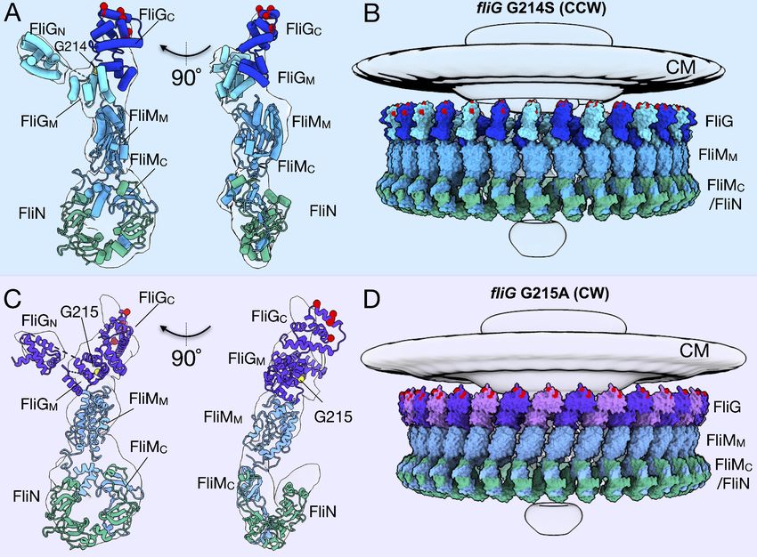
Research article                                                                                Microbiology and Infectious Disease

                                    contacts the H-ring and stators, presumably acting as a scaffold to hold the stators
                                    (Terashima et al., 2006; Zhu et al., 2019).
                                        Flagellar rotation is powered by an electrochemical gradient across the cell membrane,driving ion
                                    flow through the stator complex (Berg, 2003; Terashima et al., 2008; Kojima and Blair, 2004;
                                    Li et al., 2011). In Salmonella and E. coli, H+ ions are conducted through the stator units. In Vibrio
                                    species, Na+ ions are conducted through the stator units. The prevailing idea is that MotA and MotB
                                    (in the H+ motor) or PomA and PomB (in the Na+ motor) form a membrane-bound stator subunit
                                    (Sato and Homma, 2000a; Kojima and Blair, 2004; Braun et al., 2004; Sato and Homma, 2000b).
                                    Recent cryo-EM studies have shown that MotA and MotB assemble in 5:2 stoichiometry
                                    (Santiveri et al., 2020; Deme et al., 2020). The A and B subunits have four and one transmembrane
                                    helices, respectively, and two helices from the A subunit and one from the B subunit form an ion
                                    channel that contains an essential ion-binding aspartyl residue (Hosking et al., 2006). Inactive stator
                                    complexes diffuse through the cytoplasmic membrane and interact with the rotor to generate a
                                    series of conformational changes within PomA and PomB that open the ion channel and facilitate
                                    binding to the peptidoglycan layer (Hosking et al., 2006; Mino et al., 2019; Zhu et al., 2014;
                                    Fukuoka et al., 2009; Sudo et al., 2009; Kojima et al., 2018). However, exactly how the stator
                                    assembles around the motor and interacts with the C-ring is still not well understood.
                                        The C-ring is essential for flagellar rotation and rotational switching. The C-ring structure is con-
                                    served among diverse species, with repeating subunits consisting of FliG, FliM, and FliN, with FliN
                                    sometimes being supplemented or replaced by FliY (Kojima and Blair, 2004; Zhao et al., 1995).
                                    FliG, located closest to the cell membrane (henceforth referred to upper portion) of the C-ring, inter-
                                    acts with the MS-ring, the lower C-ring, and the stator via three domains (Lee et al., 2010). The
                                    interaction of the N-terminal domain of FliG (FliGN) with FliF tethers the C-ring to the MS-ring and is
                                    necessary both for assembly and rotation of the flagellum (Ogawa et al., 2015; Lynch et al., 2017;
                                    Xue et al., 2018). The middle domain of FliG (FliGM) interacts with FliM, holding the upper, mem-
                                    brane-proximal portion of the C-ring in contact with its lower, membrane-distal portion
                                    (Brown et al., 2002; Brown et al., 2007; Minamino et al., 2011). Lastly, the C-terminal domain of
                                    FliG (FliGC) interacts with a cytoplasmic loop of PomA (or MotA in proton-driven motors) via interac-
                                    tions of oppositely charged residues, connecting the stator and rotor (Lloyd and Blair, 1997;
                                    Yakushi et al., 2006; Takekawa et al., 2014). FliM also has three domains (Park et al., 2006). The
                                    N-terminal domain of FliM (FliMN) binds to the chemotaxis signaling protein phosphoryl CheY
                                    (CheY-P) to trigger switching from CCW to CW rotation (Paul et al., 2011; Vartanian et al., 2012).
                                    The FliMM serves as a connection between the base of the C-ring and FliG (Brown et al., 2002;
                                    Minamino et al., 2011). FliMC forms a heterodimer with FliN when fused via a flexible linker
                                    (Notti et al., 2015; Dos Santos et al., 2018). The third protein, FliN, a small single-domain protein,
                                    dimerizes with FliM or itself to form the base of the C-ring (Brown et al., 2005). Proposed models
                                    for FliG, FliM, and FliN assembly include 1:1:4 (Sarkar et al., 2010a; Sarkar et al., 2010b) or a 1:1:3
                                    (McDowell et al., 2016) stoichiometry. Two FliN homodimers form a ring at the base of each C-ring
                                    subunit in the first model (Sarkar et al., 2010a), while a FliM:FliN heterodimer and a FliN homo-
                                    dimer create a spiral base of the C-ring in the second model (McDowell et al., 2016).
                                        It has been proposed that FliG undergoes a dramatic conformational change from an open,
                                    extended form during CCW rotation to a closed, compact form during CW rotation (Lee et al.,
                                    2010). Two a-helices play an important role in determining the FliG conformation: helixMN connects
                                    FliGN and FliGM, and helixMC connects FliGM and FliGC. It was suggested that these helices are rigid
                                    and extended in the open CCW conformation, while they become disordered to switch into the
                                    compact CW conformation. Each domain contains armadillo (ARM) repeat motifs; ARMN interacts
                                    with the adjacent FliG monomer, and ARMM and ARMC interact either inter- or intramolecularly,
                                    depending upon the CCW or CW rotational sense, respectively (Lee et al., 2010; Brown et al.,
                                    2002; Minamino et al., 2011). The domain-swapping mechanism was proposed to coordinate with
                                    the conformational change in helixMC (Lee et al., 2010). Another biochemical study suggested that
                                    the domain swapping occurs during C-ring assembly, with FliG in solution existing as a monomer,
                                    and an equilibrium favoring FliG oligomers in the C-ring (Baker et al., 2016).
                                        The FliG conformational change occurs about a hinge region first predicted by in silico modeling
                                    and characterized via mutational analysis (Van Way et al., 2004). Two recently characterized fliG var-
                                    iants in V. alginolyticus (G214S and G215A), previously characterized in E. coli (G194S and G195A
                                    [Van Way et al., 2004]), appear to hinder the conformational change sterically (Nishikino et al.,

Carroll et al. eLife 2020;9:e61446. DOI: https://doi.org/10.7554/eLife.61446                                                           2 of 19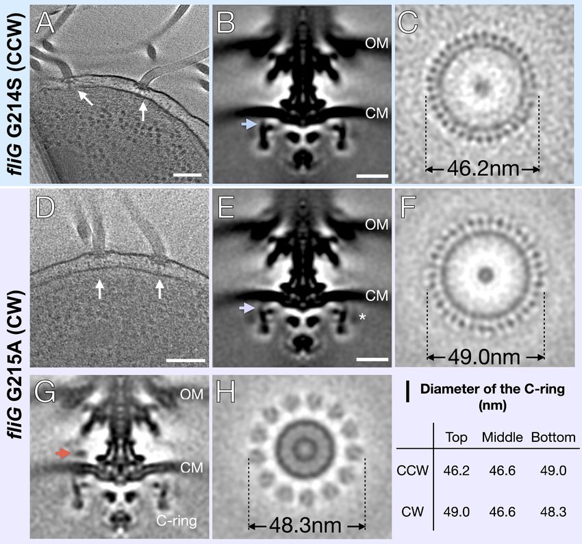
Research article                                                                                Microbiology and Infectious Disease

                                    2018; Nishikino et al., 2016), resulting in motors that rotate primarily in a single direction. The wild-
                                    type flagella rotate in a CW:CCW ratio of 1:3, while the fliG-G214S mutant produces a CCW-biased
                                    phenotype (CW:CCW 1:9), and fliG-G215A mutant produces a CW-locked phenotype
                                    (Nishikino et al., 2016). These adjacent residue substitutions create opposite motility phenotypes
                                    and are located in the Gly-Gly flexible linker, a hinge region whose conformation is believed to be
                                    dependent upon helixMC (Nishikino et al., 2016).
                                       To characterize C-ring dynamics during switching, we used cryo-electron tomography (cryo-ET) to
                                    determine in situ motor structures of the V. alginolyticus CCW-biased mutant G214S and the CW-
                                    locked mutant G215A. We found that a large conformational change of the C-ring occurs between
                                    the CCW- and CW-motors. Docking the previously solved homologous structures into our cryo-ET
                                    maps, we built two models of the C-ring in CCW and CW rotation.

                                    Results
                                    Visualization of intact flagellar motors in CCW and CW rotation states
                                    To address the mechanism of rotational switching, we utilized cryo-ET to visualize the C-rings of two
                                    FliG mutants (G214S and G215A) of V. alginolyticus. These mutants were previously shown to have
                                    CCW-biased and CW-locked flagellar motors, respectively (Nishikino et al., 2018). Each V. alginoly-
                                    ticus cell usually possesses a single, polar, sheathed flagellum, which limits the amount of informa-
                                    tion per cell for in situ motor structure determination. We therefore introduced the fliG-G214S and
                                    fliG-G215A mutations (Table 1) into the flhG KK148 strain, which produces multiple flagella at the
                                    cell pole (Kusumoto et al., 2006). Tomograms were collected using a 300kV Titan Krios electron
                                    microscope (Thermo Fisher) with Volta phase plate, a GIF energy filter and a post-GIF K2 detector
                                    electron detector (Gatan). Multiple flagella can be readily seen in a typical cryo-ET reconstruction
                                    (Figure 1A,D). In total, 2221 CCW-biased motors from G214S cells and 1618 CW-locked motors
                                    from G215A cells were used to determine in situ flagellar motor structures (Figure 1B,E and
                                    Table 1). Due to increased contrast from the Volta phase plate and focused classification, we
                                    observed 34-fold symmetry of the C-ring in both variants (Figure 1C,F and Figure 1—figure supple-
                                    ment 1). To begin to address potential differences in the switch complex, we measured the diame-
                                    ter of the C-ring in Fiji ImageJ (Schindelin et al., 2012). To describe the observed changes, we will
                                    refer to the top of the C-ring as the outermost region facing the cytoplasmic membrane. Starting
                                    from the top portion in the fliG G214S (CCW) the diameter is 46.2 nm, the middle 46.6 nm, and the
                                    bottom 49 nm (Figure 1C,I). In fliG G215A (CW) motor the diameters are 49.0 nm, 46.6 nm, and
                                    48.3 nm, respectively (Figure 1F,I). Thus, the CW-motor is about 20.8 Å wider than the CCW-motor
                                    at the top of the C-ring, even though the number of C-ring subunits remains unchanged (Figure 1).
                                    To understand the interaction between the C-ring and the stator, we also analyzed the densities on
                                    the top of the C-ring. We were able to resolve the stator ring in the CW-locked motor but not in the
                                    CCW-biased motor (Figure 1G,H). Interestingly, the diameter of the stator ring is 48.3 nm, falling in
                                    between the CCW-biased motor and CW-locked motor diameters (Figure 1H).
                                        To increase the resolution of the C-ring, we refined the C-ring structures with 34-fold symmetry
                                    applied to approximately 18 Å for the CCW and 19 Å for CW rotations (Figure 2A,D). The increased
                                    resolution allows us to reveal two unique conformations of the C-ring (Figure 2). We observed a sig-
                                    nificant lateral shift upon switching from CCW to CW, while the overall diameter of the C-ring varies
                                    slightly between the two mutants. These data demonstrate that the C-ring undergoes a large lateral
                                    conformational change during flagellar switching that leads to a difference in FliG presentation evi-
                                    dent from the diameter change at the top of the C-ring. The protein composition of the C-ring likely
                                    remains the same as both CCW- and CW-motors have 34-fold symmetry, and the middle and bot-
                                    tom regions of the C-ring are similar in diameter.

                                    Table 1. Cryo-ET data used in this study.
                                    Strain genotype                                    Tomograms                                      Motors
                                    fliG-G214S                                         259                                            2221
                                    fliG-G215A                                         220                                            1618

Carroll et al. eLife 2020;9:e61446. DOI: https://doi.org/10.7554/eLife.61446                                                           3 of 19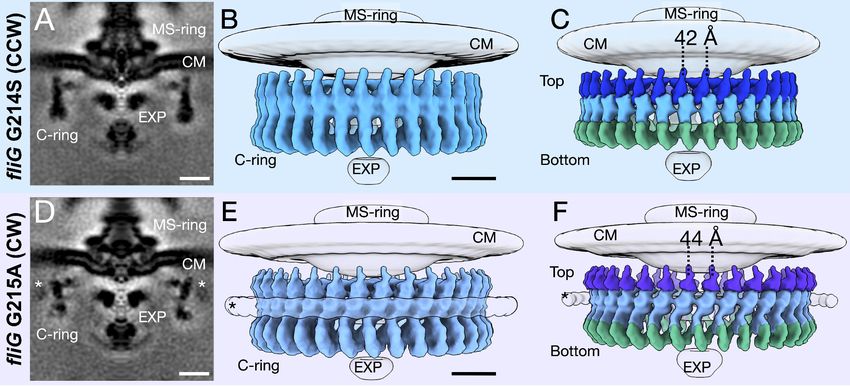
Research article                                                                                           Microbiology and Infectious Disease

Figure 1. In situ structures of V. alginolyticus flagellum motor in CCW and CW rotation. (A) A representative tomogram slice showing two motors in V.
alginolyticus fliG G214S mutant (white arrow) Scale bar 100 nm. (B) A medial cross-section of the in situ flagellar motor structure. Scale bar 25 nm. (C) A
perpendicular cross-section of the motor showing the top portion of the C-ring (blue arrow in B). (D) A representative tomogram slice of the CW-motor
in V. alginolyticus fliG G215A CW-locked variant. (white arrow) Scale bar 100 nm. (E) A medial cross-section of the in situ flagellar motor structure. Scale
bar 25 nm. (F) A perpendicular cross-section of the motor showing the top portion of the C-ring (purple arrow in E). (G) A medial cross-section of the
flagellum motor with density for the stator (orange arrow). (H) A perpendicular cross-section of the motor showing the stator ring (orange arrow in G). (I)
Diameters of the C-ring measured at the top, middle, and bottom from perpendicular cross-sections for both the CCW- and CW-motors.
Abbreviations: outer membrane (OM), cytoplasmic membrane (CM).
The online version of this article includes the following figure supplement(s) for figure 1:
Figure supplement 1. Classification of the CCW and CW flagellar motor structures reveals 34-fold symmetry of the C-ring.

Carroll et al. eLife 2020;9:e61446. DOI: https://doi.org/10.7554/eLife.61446                                                                        4 of 19
Research article                                                                                        Microbiology and Infectious Disease

Figure 2. Focused refinement of the C-ring reveals differences in the CCW- and CW-motors. (A) A medial cross-section of the focused refinement of
the C-ring in V. alginolyticus fliG G214S CCW-biased variant. Scale bar 25 nm. (B) A side view of the CCW C-ring. Scale bar 10 nm. (C) A side view of
the segmented map shows the top (blue), middle (light blue), and bottom (green) regions. (D) A medial cross-section of the C-ring in fliG G215A CW-
locked variant. Scale bar 25 nm. (E) A side view of the focused refined CW-locked C-ring. Scale bar 10 nm. (F) A side view of the focused refined C-ring
further segmented into the top (purple), middle (gray blue) and bottom (green) regions of the C-ring. The asterisk highlights the additional density
(white) observed in only the CW-motor that we speculate to be CheY-P. Abbreviations: cytoplasmic membrane (CM), export apparatus and ATPase
(EXP).

                                     Molecular architectures of the CCW and CW C-rings
                                     To understand the molecular detail, we built pseudo-atomic models of the C-ring by docking avail-
                                     able crystal structures from the C-ring proteins into the in situ maps derived from sub-tomogram
                                     averaging. First, we used homologous structures deposited in the PDB and SCWRL4 (Krivov et al.,
                                     2009) to map the V. alginolyticus amino acid sequence onto the open (PDB-3HJL [Lee et al., 2010])
                                     and closed (PDB-4FHR [Vartanian et al., 2012]) conformations of the FliG structure. Specifically, we
                                     used the full-length extended FliG with crystal packing from PDB-3HJL to build the CCW model.
                                     The monomer generated from crystal packing fits into the cryo-ET map surprisingly well. The com-
                                     pact FliG from the PDB-4FHR model was used for the CW model, with the FliGN domain from PDB-
                                     3HJL, as there are no other structures of FliGN. Second, we used I-TASSER (Roy et al., 2010;
                                     Yang et al., 2015) to generate the structures of FliM and FliN. Third, the models were placed into
                                     the cryo-ET map using UCSF ChimeraX (Goddard et al., 2018). The known protein-protein interfa-
                                     ces were preserved during docking and while the unknown protein interface between the FliMN het-
                                     erodimer and FliN homodimer was refined with the Rosetta protein-protein docking function
                                     (Lyskov and Gray, 2008) before being placed into the cryo-ET map. Phenix (Afonine et al., 2013)
                                     was used to optimize the placement and to assess the overall fit using rigid body refinement.
                                        We built the C-ring subunit with FliG at the top, FliM in the middle, and three FliN molecules at
                                     the base (see Figure 2C,F for clarification), supporting the 1:1:3 (FliG, FliM, and FliN) model in lieu
                                     of the 1:1:4 model, as there is no additional density for a fourth FliN molecule. The 1:1:3 model sug-
                                     gests a FliM-FliN heterodimer interacts with the FliN homodimer characterized by mass spectros-
                                     copy, as had been modeled into a previously solved cryo-EM map (McDowell et al., 2016;
                                     Thomas et al., 2006). To understand the differences in the CCW and CW structures, we first built
                                     the CCW model into four adjacent subunit patch. From this patch model single subunit was real
                                     space rigid body refined with the CCW subunit having CCmask of 0.69, and CCbox of 0.83 and the
                                     CW subunit had a CCmask of 0.66 and CCbox of 0.82 (Figure 3A,C). We also generated maps from

Carroll et al. eLife 2020;9:e61446. DOI: https://doi.org/10.7554/eLife.61446                                                                     5 of 19
Research article                                                                                          Microbiology and Infectious Disease

                                      the models using ChimeraX molmap at 18 Å for the CCW model and 19 Å for the CW model. The in
                                      silico generated maps exhibit features similar to the experimental map (Figure 3—figure supple-
                                      ment 1) and (Figure 3—figure supplement 2).
                                          We expanded our subunit model to the whole C-ring by applying 34-fold symmetry (Figure 3B,
                                      D). Importantly, the expanded model fits into the experimental map without clashes, and preserves
                                      the architecture of the base observed in the maps. In silico CCW and CW maps from the entire

Figure 3. Molecular architectures of the C-ring in CCW and CW rotation. (A) Two views varying by 90o of a single C-ring subunit model fitted in the in
situ map of fliG G214S (white). (B) A side view of the CCW-motor with the model shown as surface expanded for symmetry. The density map for the
MS-ring, CM, and EXP are shown in gray for reference. FliG is alternating as dark blue, and aqua, FliM is colored light blue, and FliN is shown in green.
The charged residues of FliG that interact with the stator are red spheres, and glycine 214 is shown as a yellow sphere. (C) Two views varying by 90o of
a single C-ring subunit model in cartoon fitted into the cryo-ET density of fliG G215A (white). (D) A side view of the CW model rendered as surfaces
expanded for symmetry. FliG is alternating as purple, and mauve, FliM is gray blue, and FliN is green. The charged residues of FliG that interact with
the stator are red spheres, and glycine 215 is shown as a yellow sphere. The alternating coloring of FliG reflects the different crystal structures used to
build the model. To build the CCW model, we used the extended FliG, PDB-3HJL, with crystal packing. To build the CW model, we used the compact
FliG, in PDB-4FHR. These models support the domain swapping hypothesis. Abbreviations: cytoplasmic membrane (CM), export apparatus and ATPase
(EXP).
The online version of this article includes the following figure supplement(s) for figure 3:
Figure supplement 1. In silico maps reflect similar features as experimental data.
Figure supplement 2. The important regions biochemically and structurally of the C-ring in the CCW and CW models.
Figure supplement 3. The FliG conformational change results in a tilt about FliM to change the presentation of FliGC to the stator.

Carroll et al. eLife 2020;9:e61446. DOI: https://doi.org/10.7554/eLife.61446                                                                        6 of 19
Research article Microbiology and Infectious Disease
C-ring model were also generated using ChimeraX molmap, showing that the two models yield dis-
tinct maps similar to their respective rotational sense (Figure 3—figure supplement 1).
Our pseudo-atomic models can be further substantiated from the biochemical data. The ARMM
(Figure 3—figure supplement 2, orange [Park et al., 2006]) and ARMC (Figure 3—figure supple-
ment 2, green) motif orientations have been preserved from the crystal structures. The EHPQR motif
(Glu144, His145, Pro146, Gln147, and Arg179, Figure 3—figure supplement 2, lime spheres) of
FliG is located in ARMM and oriented to interact with FliMM (Figure 3—figure supplement 2, green
sticks). Residues at the FliM-FliM interface had previously been identified in Salmonella and E. coli
correspond to Asn56, His63, Asp184, Pro185, and Met187 in V. alginolyticus and they could interact
with the neighboring FliM (Figure 3—figure supplement 2, magenta) (Park et al., 2006;
Sakai et al., 2019). The N-terminus of FliMM is oriented facing outward toward the additional den-
sity of the CW-locked mutant in a position in which it can bind to CheY-P (Figure 3—figure supple-
ment 2, asterisk and D) (Lee et al., 2001a). It is important to note that these residues were used to
identify unique location in our model, but due to limited resolution their precise location is not
known. Our models of the C-ring are thus consistent with both the previous biochemical data and
our cryo-ET maps.
FliM/FliN interactions hold the C-ring subunits together
The tilting observed between the CCW- and CW-motors is centered on FliM, which serves as a struc-
tural protein that holds the C-ring subunits together (Figure 3). However, without a full-length crys-
tal structure, the relative orientations of FliMN, FliMM, and FliMC are unknown. The previous crystal
structures and solution NMR studies showed FliMM interacting with FliG (Vartanian et al., 2012;
Dyer et al., 2009; Lam et al., 2013). FliMC shows sequence homology to FliN, and structural homol-
ogy to the FliN homodimer, as shown from the structure of a crystallized FliMC-FliN fusion protein
(Notti et al., 2015; McDowell et al., 2016). To place FliM into our map, two I-TASSER models,
FliMM and FliMC, and place the domains separately into our cryo-ET map, and then connected the
linker regions (Figure 3). We lacked sufficient information to confidently place FliMN, and we there-
fore left it out of our model. The position of FliMN, which binds CheY-P, is flexible (discussed below)
(Lee et al., 2001b). Our model shows that FliMM forms the middle portion of the C-ring and allows
for the tilting of the C-ring, as FliMC tethers FliM to FliN via the heterodimer. We observed the same
continuous helical density at the base of the C-ring as shown in Salmonella (Thomas et al., 2006)
and proposed to be formed via alternating FliMC-FliN heterodimers and FliN homodimers
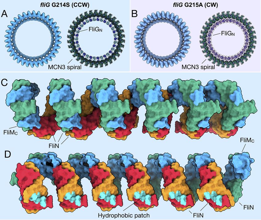
(McDowell et al., 2016) connecting adjacent subunits (Figure 4A,B). This region, known as a spiral
(McDowell et al., 2016), is uniform within both the CCW and CW cryo-ET maps, and remains largely
unaltered upon rotational switching (Figure 4C). We will refer to this as the FliMCFliN3 (MCN3) spiral
from here on. The expansion of the C-ring pseudo-atomic model preserves the continuous uniform
shape (Figure 4A,B). By building the patch model initially we were able to preserve the similarity of
the MCN3 spiral by removing potential bias from the segmentation of a monomer. The hydrophobic
patch residues, originally identified in Salmonella (McMurry et al., 2006), that interact with FliH are
facing toward the ATPase such that the interaction could occur (Figure 4D). It is important to note
that there may be slight movements of the MCN3 proteins during switching, which we would need
higher resolution to observe. Taken together, these data suggest that perhaps the MCN3 spiral is
important for keeping the C-ring subunits connected during the conformational remodeling and
rotation.
Presentation of FliGC to the stator in the CCW- and CW-motors
To characterize the observed conformational change, we created a superposition of the CCW and
CW C-ring subunits in the CCW- and CW-motors. Using UCSF Chimera (Pettersen et al., 2004) an
axis was drawn through the center of the model, FliGMC shown in red, FliGMC and FliMM in green,
and FliMM alone in orange. This revealed a 23 Å shift of FliG with a 37o tilt about FliM (Figure 3—
figure supplement 3). FliG and FliM tilt 20o outward upon switching from CCW to CW rotation (Fig-
ure 3—figure supplement 3). Furthermore, comparison of just the axes shows that the movement
characterizing the large conformational change can be attributed to FliMM (Figure 3—figure supple-
ment 3 insets). This suggests that, upon switching, FliG is presumably presented to the stator units
very differently as a result of the rearrangement of FliMM.
Carroll et al. eLife 2020;9:e61446. DOI: https://doi.org/10.7554/eLife.61446 7 of 19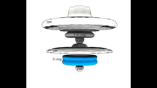
Research article                                                                                        Microbiology and Infectious Disease

Figure 4. FliMC and FliN form a continuous spiral structure at the base of the C-ring. (A) A bottom view of the fliG G214S (CCW) motor map alone
(light blue), and the model (cartoon) expanded for symmetry in the in situ map (gray transparent). (B) A bottom view of the fliG G215A (CW) motor map
alone (gray blue), and the model (cartoon) expanded for symmetry in the in situ map (gray transparent). (C) A view from the outside periphery, and (D) a
view from the center or inside looking out, are surface representations of six subunits that form a portion of MCN3 spiral from the CCW model, with
one FliMC (light blue), and three FliN molecules (green, red, and orange). The hydrophobic patch of FliN (McMurry et al., 2006), L68, A93, V111, V113,
Y118, that had been shown to interact of the FliH (cyan) points toward the center of the C-ring.

                                     Extra density around the CW C-ring
                                     We observed an extra ring around the CW-locked C-ring (Figure 2). We speculate that it is formed
                                     by CheY-P, as there are several pieces of evidences in line with this model. First, the density is above
                                     the noise level in the CW-motors but not in the CCW-motors, which biochemically makes sense as
                                     CheY-P binds to CW rotating motors (Paul et al., 2011; Vartanian et al., 2012; Dyer et al., 2009;
                                     Lee et al., 2001b). The cells should have endogenous CheY, as there has been no alteration to the
                                     CheY gene. Second, density similar in location and shape has been shown two recent studies to be
                                     CheY-P. Chang et al. have used GFP-tagged CheY-P to show that, in Borrelia burgdorferi, CheY-P
                                     occupies the same position (Chang et al., 2020). Rossmann et al. also identified similar density for

Carroll et al. eLife 2020;9:e61446. DOI: https://doi.org/10.7554/eLife.61446                                                                     8 of 19
Research article Microbiology and Infectious Disease
the CheY homolog, CleD in Caulobacter crescentus (Rossmann et al., 2020). Third, when we place
the crystal structure of CheY into the model, there is about 60 Å gap between the C-ring density
and this additional density (Figure 3—figure supplement 2). This distance may be bridged by the
42 residues of FliMN to connect CheY-P to FliMM.
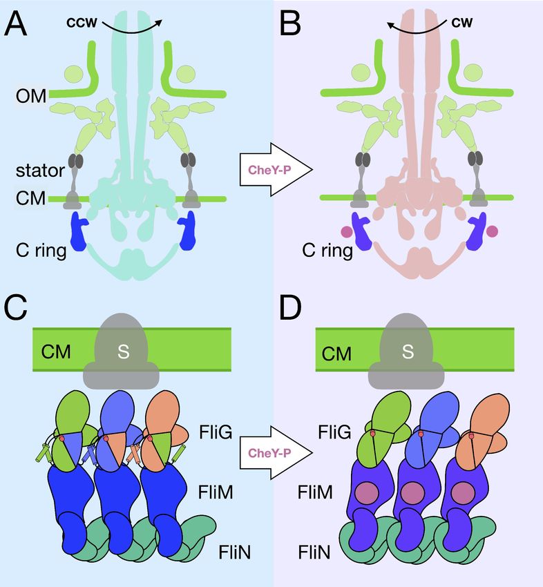
Discussion
The flagellar motor structures we determined provide direct evidence that the C-ring possesses two
distinct conformations in CCW and CW rotation. The dynamics of the C-ring appear to be confined
to the upper two-thirds of the structure, with the spiral base that connects the C-ring subunits
remaining relatively static. We observed a 37o lateral and 20o medial tilting of the C-ring (Figure 3—
figure supplement 3), which almost certainly alters its interactions with the stator. In our pseudo-
atomic model, the charged residues (Lys284, Arg301, Asp308, and Asp309 in FliGC) that are known
to interact with the stator are located at the top of the C-ring. FliGC moves about 40 Å laterally
(Video 1). This large remodeling of the C-ring presumably enables distinct interactions between the
stator and the C-ring (Video 2). FliGCN contains the ARMC motif that interacts with FliGM and FliMM,
and FliGCC contains the charged residues that interact with the stator. A flexible linker attaching the
two domains would allow for a range of movement in FliGCC relative to FliGCN and FliGM, thus sug-
gesting that the presentation of the charged residues varies greatly depending upon the conforma-
tion of FliG.
Notably, our cryo-ET model shows 34-fold symmetry for both the CCW and CW rotating C-rings,
with only a modest diameter change in FliG. It has previously been hypothesized, based on experi-
ments utilizing fluorescently tagged FliM and FliN, that the FliG, FliM, and FliN composition of the
C-ring changes during rotational switching (Delalez et al., 2014; Delalez et al., 2010; Hosu and
Berg, 2018; Lele et al., 2012). A recent single-particle cryo-EM study suggested that inter-subunit
spacing and interactions of the C-ring account for motor switching in Salmonella (Sakai et al., 2019).
That study reported a 9 Å decrease in C-ring
diameter upon switching to CW. We report iden-
tical diameters at the middle of the C-ring in
both rotational directions. However, we
observed ~21 Å increase in the diameter
and ~40 Å lateral change at FliGC of the CW-
motor. In a parallel study, Chang et al. used a B.
burgdorferi with a CheX deletion or CheY3 dele-
tion to investigate the switching mechanism of
spirochetes (Chang et al., 2020). Similar confor-
mational changes of the C-ring were observed in
both species; however, the stator complexes
were resolved in B. burgdorferi, as they appear Video 1. Focused refinement of the C-ring reveals
conformational change during rotational switching. The
to be less dynamic than those in V. alginolyticus.
video starts with a fliG G214S motor, a mutation leads
Visualization of the stator complex provided
to a strong preference for CCW rotation, without
direct evidence that the conformational change symmetry applied. The C-ring is highlighted in blue.
of the C-ring alters the rotor-stator interaction. The video then zooms in to a focused refined C-ring,
The model of stator-rotor interactions controlling with much higher resolution, and turns in the CCW
the rotational direction of the motor is further (looking down from the flagellum) direction. The
bolstered by the recent high-resolution cryo-EM pseudo-atomic model appears as cartoon in the map,
models of MotAB (Santiveri et al., 2020; as the map fades the model turns into surface
Deme et al., 2020). Santiveri et al., 2020 fur- rendering. Transparent density, that is likely CheY-P,
binds and triggers the conformational change of rotor
ther postulated that MotA rotates CCW in Cam-
into the CW architecture. The video toggles between
pylobacter jejunie by showing that the
CCW and CW pseudo-atomic models in surface
conformations of wildtype MotAB and MotAB rendering to visualize the global changes of the C-ring
trapped in the protonated state are nearly iden- architecture. It ends with the model spinning in the CW
tical, thus ruling out a dramatic conformational direction. The color scheme is the same as the paper.
change in the stator unit. Therefore, we con- The video was rendered using ChimeraX.
clude that the change in rotational direction https://elifesciences.org/articles/61446#video1
Carroll et al. eLife 2020;9:e61446. DOI: https://doi.org/10.7554/eLife.61446 9 of 19Research article Microbiology and Infectious Disease
Table 2. Strains and plasmids.
Strains or plasmids Genotype or description Reference or source
V. alginolyticus
VIO5 Wild-type strain of a polar flagellum Okunishi et al., 1996
(Rif+ Pof+ Laf-)
KK148 VIO5 flhG (Multi-Pof+) Kusumoto et al., 2008
-
NMB328 KK148 DfliG (Pof ) This study
E. coli
DH5a Host for cloning experiments Grant et al., 1990
S17-1 recA hsdR thi pro ara RP-4 2-tc::Mu-Km::Tn7 (Tpr Smr) Simon et al., 1983
b3914 b2163 gyrA462 zei-298::Tn10 Le Roux et al., 2007
(Kmr Emr Tcr)
Plasmids
pMMB206 Cmr, PtacPlacUV5 Morales et al., 1991
pNT1 fliG in pMMB206 Takekawa et al., 2014
pSW7848 Suicide plasmid for allele exchange Val et al., 2012
pHIDA3 SacI fragment of fliF-fliG (55–175 internal deletion of FliG) in pSW7848 Mino et al., 2019
occurs from a large movement of the charged residues of FliGC relative to the stator.
Our in situ structures support that FliG, FliM, and FliN interact in a 1:1:3 ratio, as suggested for
the non-flagellar homolog Spa33 in Shigella (McDowell et al., 2016). This stoichiometry favors the
model in which FliM holds the individual C-ring subunits together by interacting with FliGM and
forming a heterodimer with FliN. The FliM-FliN spiral connects adjacent C-ring subunits, allowing for
dramatic movement without dissociation of the subunits. The FliGM-FliMM interface, perhaps
dynamic in its own right, has been suggested to be involved in flagellar switching (Sakai et al.,
2019; Dyer et al., 2009; Pandini et al., 2016). It was shown, using NMR, that the CheY-P binding
to FliM displaces FliGC, and that the dramatic rearrangement of FliGMC is possible because of the
flexibility of FliG (Dyer et al., 2009). In particular, the GGPG loop in FliMM is suggested to be critical
for rotation of FliGM relative to FliMM (Pandini et al., 2016). Most recently, point mutations target-
ing FliMM were shown to restore CCW rotation in a CW-biased mutant (Sakai et al., 2019). These
results, combined with our findings, lead us to suggest that the conformational change in FliG results
in a rotation about FliMM that leads to the tilting of the C-ring subunit to alter the presentation of
the charged residues of FliGC to the stator.
Given that the putative CheY-P-ring is likely associated with the CW C-ring (Figure 1), we pro-
pose a CCW-to-CW switching model (Figure 5, Videos 1 and 3). CheY-P binding to the FliM results
in a conformational change in FliG and alters the interactions between the C-ring and stator. FliMC
and FliN create a spiral base that holds the C-ring together during these dynamic rearrangements.
FliMC and FliN may also be involved in relaying the conformational change to adjacent C-ring subu-
nits. By placing the FliG crystal structures of the open and closed variations within our cryo-ET
image, we provide additional evidence to support the proposed model in which the ARMM and
ARMC domains toggle between inter- and intramolecular interactions (Lee et al., 2010). NMR data
suggest that FliGC is the domain that moves and that FliGM remains in stable contact with FliM
(Dyer et al., 2009). These changes in the C-ring structure produce the two directions of flagellar
rotation. Understanding the interactions of the C-ring with the stator and the MS-ring is essential to
elucidate the mechanism of rotational switching and transmission of the rotation to the flagellar fila-
ment. We could not resolve the detailed interactions between the cytoplasmic portion of the stator
and the C-ring in both rotational directions, and we could not confirm directly that the orientation of
FliGCC alters the presentation of its charged residues to the charged residues of PomA. Restricting
the movement of FliGCC via truncations of the flexible linker may address the importance of FliGCC
mobility. With more data and further classification and refinement of the CCW-biased motor, we
expect to explain the FliGCC and stator interactions in great detail. With the rapid development of
Carroll et al. eLife 2020;9:e61446. DOI: https://doi.org/10.7554/eLife.61446 10 of 19Research article Microbiology and Infectious Disease
cryo-ET, it is increasingly possible to reveal motor
structure in situ at higher resolution, which will
further our understanding of the flagellar assem-
bly and function.
In summary, we have used cryo-ET to visualize
a major conformational change of the C-ring dur-
ing rotational switching due to a single point
mutant in FliG. Molecular modeling attributes the
tilt component of the conformational change to
FliM. Gyration of FliG about FliM presents the
Video 2. C-ring rearrangement leads to altered FliGC charged residues of FliG to the stator in a manner
presentation to the stator. The video starts with a side that controls the rotational sense. These move-
view of the CCW pseudo-atomic model in surface ments within the C-ring subunits may be relayed
rendering, and the view rotates so that we are looking throughout the switch complex by interactions
down on the C-ring from the flagellum. We can see the
between FliM and FliN within the spiral.
charged residues of FliGCC that interact with the stator.
The video toggles between CCW and CW models, and
asterisks appear to mark the charged residues of every
other FliG. This highlights the large change,~40 Å, in Materials and methods
FliG upon switching. A morphing of four adjacent
subunits shows the dramatic movement of FliGC Bacterial strains, plasmids, and
relative to the stator from the top view, and side view, growth condition
the CCW model is shown as transparent. The video Bacterial strains used in this study are listed in
ends with the CW pseudo-atomic model. The video Table 2. To introduce the fliG deletion, NMB328
was rendered using ChimeraX.
was constructed from KK148 using pHIDA3 by
https://elifesciences.org/articles/61446#video2
allelic exchange as previous reported
(Kusumoto et al., 2006; Le Roux et al., 2007).
V. alginolyticus strains were cultured at 30˚C on
VC medium (0.5% [wt/vol] polypeptone, 0.5%
[wt/vol] yeast extract, 3% [wt/vol] NaCl, 0.4% [wt/
vol] K2HPO4, 0.2% [wt/vol] glucose) or VPG medium (1%[wt/vol] polypeptone, 3% [wt/vol] NaCl,
0.4% [wt/vol] K2HPO4, 0.5% [wt/vol] glycerol). If needed, chloramphenicol and Isopropyl b-D-1-thio-
galactopyranoside (IPTG) were added at final concentrations of 2.5 mg/ml and 1 mM, respectively. E.
coli was cultured at 37˚C in LB medium (1% [wt/vol] Bacto tryptone, 0.5% [wt/vol] yeast extract, 0.5%
[wt/vol] NaCl). When culturing E. coli b3914 strain, 2,6-diaminopimelic acid was added to the LB
medium to a final concentration of 300 mM. If needed, chloramphenicol was added at final concen-
trations of 25 mg/ml.
Mutagenesis
To introduce mutations (G214S or G215A) in the fliG gene on plasmid pNT1 site-directed mutagene-
sis was performed using the QuikChange method, as described by the manufacturer (Stratagene).
All constructs were confirmed by DNA sequencing. Transformation of V. alginolyticus with plasmid
pNT1 was performed by conjugational transfer from E. coli S17-1, as described previously
(Okunishi et al., 1996).
Sample preparation
The methods of sample preparation, data collection, data processing, and sub-tomogram analysis
were followed as described previously (Zhu et al., 2018). V. alginolyticus cells were cultured over-
night at 30˚C on VC medium, diluted 100 with fresh VPG medium, and cultured at 30˚C. After 4 or
5 hr, cells were collected and washed twice and finally diluted with TMN500 medium (50 mM Tris-
HCl at pH 7.5, 5 mM glucose, 5 mM MgCl, and 500 mM NaCl). Colloidal gold solution (10 nm diam-
eter) was added to the diluted Vibrio sp. samples to yield a 10 dilution and then deposited on a
freshly glow-discharged, holey carbon grid for 1 min. The grid was blotted with filter paper and rap-
idly plunge-frozen in liquid ethane.
Carroll et al. eLife 2020;9:e61446. DOI: https://doi.org/10.7554/eLife.61446 11 of 19Research article Microbiology and Infectious Disease Figure 5. A model for the rotational switching. (A) A cartoon of the intact CCW-motor with the C-ring highlighted in blue, and the other conserved regions that have been resolved in teal, the Vibrio-specific proteins in light green, PomAB is colored gray as it has yet to be resolved in detail in Vibrio. (B) A cartoon of the intact motor that is rotating CW, C-ring is colored purple, the other conserved structures in coral, PomAB in gray, and CheY-P in pink. (C) A cartoon representation of the C-ring rearrangement upon CheY-P binding and rotational switching to the CW sense. In CCW rotation, the intermolecular interactions of FliG ARMM and ARMC are possible because helixNM and helixMC are ordered. The Gly-Gly flexible region is depicted by red circles. The stator (gray) is shown to interact with FliGC. Upon CheY-P binding, the C-ring undergoes a conformational change that produces CW Figure 5 continued on next page Carroll et al. eLife 2020;9:e61446. DOI: https://doi.org/10.7554/eLife.61446 12 of 19
Research article                                                                                           Microbiology and Infectious Disease

Figure 5 continued
rotation. During this transition, the center of mass tilts laterally and slightly outward. In CW rotation there are intramolecular interactions of FliG ARMM
and ARMC. CheY-P (dark purple) is shown bound to FliM, and the stator (gray) interacts with FliGC. We hypothesize that the conformational change in
FliG is initiated in vivo by CheY-P binding, and this switches the rotational direction of the C-ring by changing how FliGC interacts with the stator. FliG
undergoes a conformational change and FliM tilts about the base of the spiral. The spiral ensures that the C-ring subunits are connected while FliG and
FliM undergoes large conformational changes.

                                      Data collection and processing
                                      The frozen-hydrated specimens of NMB328 was transferred to a Titan Krios electron microscope
                                      (Thermo Fisher Scientific). The microscope is equipped with a 300-kV field emission gun (FEG), a GIF
                                      energy filter, and a post-GIF K2 Summit direct electron detector (Gatan). The images were collected
                                      at a defocus near to 0 mm using a Volta phase plate and the energy filter with a 20 eV slit. The data
                                      were acquired automatically with SerialEM software (Mastronarde, 2005). For better data collec-
                                      tion, the phase shift is normally distributed in the range of 0.33p to 0.67p. A total dose of 50 e-/Å
                                      (Chevance and Hughes, 2008) was distributed among 35 tilt images covering angles from 51˚ to
                                      +51˚ at tilt steps of 3˚. For every single tilt series collection, the dose-fractionated mode was used to
                                      generate 8 to 10 frames per projection image. Collected dose-fractionated data were first subjected
                                      to the motion correction program to generate drift-corrected stack files (Li et al., 2013). The stack
                                      files were aligned using gold fiducial markers and volumes reconstructed using IMOD and Tomo3d,
                                      respectively (Kremer et al., 1996; Agulleiro and Fernandez, 2015). In total, 259 tomograms of
                                      CCW-state motor (G214S mutation) and 220 tomograms of CW-state motor (G215A mutation) were
                                      generated.

                                      Sub-tomogram analysis with I3 package
                                      Bacterial flagellar motors were detected manually, using the I3 program (Winkler, 2007;
                                      Winkler et al., 2009). We selected two points on each motor, one point at the C-ring region and
                                      another near the flagellar hook. The orientation
                                      and geographic coordinates of selected particles
                                      were estimated. In total, 1618 and 2221 sub-
                                      tomograms of V. alginolyticus motors from CW-
                                      motor and CCW-motor, respectively, were used
                                      for sub-tomogram analysis. The I3 tomographic
                                      package was used on the basis of the ‘alignment
                                      by classification’ method with missing wedge
                                      compensation for generating the averaged struc-
                                      ture of the motor, as described previously
                                      (Zhu et al., 2017). To resolve the symmetry of
                                      the C-ring, we used focused refinement to the
                                      C-ring density. We then used multivariate statis- Video 3. A model for the rotational switching. The
                                      tical analysis for 3D classification, which allowed psudeo-atomic CCW model in surface rendering side
                                                                                          view is rotated such we are looking up the motor
                                      us to determine the 34-fold symmetry of the
                                                                                          toward the flagellum. The MCN3 spiral is in view, and
                                      C-ring. It’s important to point out that we did
                                                                                          the hydrophobic patch of FliN that interact with FliH is
                                      not resolve the 34-fold symmetry by imposing highlighted. The view then rotates back through the
                                      different symmetries. Furthermore, the C-ring side view to top view, where the charged residues of
                                      structures in the two states share the same sym- FliGC are highlighted. The view returns to side view
                                      metry, however they have different conforma- and zooms into a patch of four adjacent subunits. The
                                      tions. We did similar analysis on the stator CCW subunits morph into the CW architecture,
                                                                                          highlight the large lateral change about FliM, and the
                                      region to resolve 13 stator units in CW-motors.
                                                                                          domain swapping occurs as the FliG ARMMC
                                      However, we were unable to resolve the stator interactions change from intramolecular to
                                      units in CCW-motors. The reason behind the dif- intermolecular. The MCN3 spiral exhibits little change.
                                      ference is currently unknown.                       The video ends with the CW psudeo-atomic model.
                                                                                                  The video was rendered using ChimeraX.
                                                                                                  https://elifesciences.org/articles/61446#video3

Carroll et al. eLife 2020;9:e61446. DOI: https://doi.org/10.7554/eLife.61446                                                                        13 of 19
Research article Microbiology and Infectious Disease
Model generation
The V. alginolyticus C-ring proteins were generated using I-TASSER version 5.1 (Yang et al., 2015).
The proteins were trimmed to the homologous structures deposited in the PDB to avoid large
clashes. Using I-TASSER we have generated two models of Vibrio FliM, FliM full length, and FliMC
with the linker region before. The full-length FliM generated by I-TASSER had the correct topology
for FliMM, but the orientation FliMC relative to FliMM was incorrect. Furthermore, the folding of
FliMC is similar to the previously solved crystal structure, but different enough that the FliM-FliN het-
erodimer was unable to be modeled. To circumvent this problem, we ran I-TASSER with just the last
223 residues of FliM, including the FliMC and the flexible region immediately before. Due to the
unknown relative location of the flexible regions it was necessary to trim. FliG, FliM, and FliN were
hand guided by the literature into segmented patches of the CCW-biased and CW-locked cryo-ET
maps corresponding to four subunits, and fit using the ChimeraX (Goddard et al., 2018) fit to map
function. Mainly, four PDB models were used the full-length FliG (3HJL [Lee et al., 2010]), FliG-FliM
(4FHR [Vartanian et al., 2012]), FliM-FliN fusion (4YXB [Notti et al., 2015]), and the FliN-dimer
(1YAB [Brown et al., 2005]).
Model refinement
The patch models were refined using PHENIX-1.17.1 Real Space Refinement (Afonine et al., 2013)
to move the protein domains relative to one another while preserving the known architecture of the
C-ring subunits. The unknown protein-protein interfaces were refined in Rosetta_2019.35 using the
protein-protein docking scripts (Lyskov and Gray, 2008). The optimized single subunit model was
then rigid body refined into a single subunit segmentation of the corresponding C-ring tomography
map using PHENIX Real Space Refinement.
Acknowledgements
We thank Michael Manson for critical reading and suggestions. We thank A Abe in our laboratory
for technical support in this research. We thank Shenping Wu for her support on Krios. This work
was supported in part by JSPS KAKENHI Grant Numbers JP16H04774 and JP18K19293 (to SK),
JP18K06155 (to TK), and Program for leading Graduate Schools of Japan, Science for the Promotion
of Science (JP17J11237 to TN). TN was supported in part by the Integrative Graduate Education
and Research program of Nagoya University. BLC, SZ and JL were supported by grants GM107629
and R01AI087946 from National Institutes of Health. Molecular graphics and analyses were per-
formed with UCSF ChimeraX, developed by the Resource for Biocomputing, Visualization, and Infor-
matics at the University of California, San Francisco, with support from National Institutes of Health
R01-GM129325 and the Office of Cyber Infrastructure and Computational Biology, National Institute
of Allergy and Infectious Diseases.
Additional information
Funding
Funder Grant reference number Author
Japan Society for the Promo- JP16H04774 Seiji Kojima
tion of Science
Japan Society of Ultrasonics in JP18K19293 Seiji Kojima
Medicine
National Institute of Allergy AI087946 Jun Liu
and Infectious Diseases
National Institute of General GM107629 Brittany L Carroll
Medical Sciences Shiwei Zhu
Jun Liu
Japan Society for the Promo- JP18K06155 Tatsuro Nishikino
tion of Science
National Institutes of Health R01AI087946 Seiji Kojima
Carroll et al. eLife 2020;9:e61446. DOI: https://doi.org/10.7554/eLife.61446 14 of 19Research article Microbiology and Infectious Disease
Japan Society for the Promo- JP17J11237 Brittany L Carroll
tion of Science Shiwei Zhu
Jun Liu
Japan Society for the Promo- JP17J11237 Tatsuro Nishikino
tion of Science
The funders had no role in study design, data collection and interpretation, or the
decision to submit the work for publication.
Author contributions
Brittany L Carroll, Formal analysis, Investigation, Visualization, Methodology, Writing - original draft;
Tatsuro Nishikino, Conceptualization, Data curation, Formal analysis, Investigation, Writing - original
draft, Writing - review and editing; Wangbiao Guo, Formal analysis; Shiwei Zhu, Data curation, For-
mal analysis; Seiji Kojima, Conceptualization, Supervision, Funding acquisition, Investigation, Writing
- review and editing; Michio Homma, Conceptualization, Formal analysis, Supervision, Writing - origi-
nal draft, Writing - review and editing; Jun Liu, Conceptualization, Supervision, Funding acquisition,
Validation, Investigation, Writing - original draft, Writing - review and editing
Author ORCIDs
Seiji Kojima http://orcid.org/0000-0002-5582-8935
Michio Homma https://orcid.org/0000-0002-5371-001X
Jun Liu https://orcid.org/0000-0003-3108-6735
Decision letter and Author response
Decision letter https://doi.org/10.7554/eLife.61446.sa1
Author response https://doi.org/10.7554/eLife.61446.sa2
Additional files
Supplementary files
. Transparent reporting form
Data availability
The resulting structures have been deposited in EMDB under accession codes EMD-21819 and
EMD-21837.
The following datasets were generated:
Database and
Author(s) Year Dataset title Dataset URL Identifier
Carroll BL 2020 Structures from: The flagellar motor https://www.ebi.ac.uk/ Electron Microscopy
of Vibrio alginolyticus undergoes pdbe/entry/emdb/EMD- Data Bank, 21819
major structural remodeling during 21819
rotational switching
Carroll BL 2020 Structures from: The flagellar motor https://www.ebi.ac.uk/ Electron Microscopy
of Vibrio alginolyticus undergoes pdbe/entry/emdb/EMD- Data Bank, 21837
major structural remodeling during 21837
rotational switching
References
Afonine PV, Grosse-Kunstleve RW, Adams PD, Urzhumtsev A. 2013. Bulk-solvent and overall scaling revisited:
faster calculations, improved results. Acta Crystallographica Section D Biological Crystallography 69:625–634.
DOI: https://doi.org/10.1107/S0907444913000462, PMID: 23519671
Agulleiro JI, Fernandez JJ. 2015. Tomo3D 2.0–exploitation of advanced vector extensions (AVX) for 3D
reconstruction. Journal of Structural Biology 189:147–152. DOI: https://doi.org/10.1016/j.jsb.2014.11.009,
PMID: 25528570
Baker MA, Hynson RM, Ganuelas LA, Mohammadi NS, Liew CW, Rey AA, Duff AP, Whitten AE, Jeffries CM,
Delalez NJ, Morimoto YV, Stock D, Armitage JP, Turberfield AJ, Namba K, Berry RM, Lee LK. 2016. Domain-
Carroll et al. eLife 2020;9:e61446. DOI: https://doi.org/10.7554/eLife.61446 15 of 19Research article Microbiology and Infectious Disease
swap polymerization drives the self-assembly of the bacterial flagellar motor. Nature Structural & Molecular
Biology 23:197–203. DOI: https://doi.org/10.1038/nsmb.3172, PMID: 26854663
Berg HC. 2003. The rotary motor of bacterial flagella. Annual Review of Biochemistry 72:19–54. DOI: https://doi.
org/10.1146/annurev.biochem.72.121801.161737, PMID: 12500982
Blair DF. 2003. Flagellar movement driven by proton translocation. FEBS Letters 545:86–95. DOI: https://doi.
org/10.1016/S0014-5793(03)00397-1, PMID: 12788496
Braun TF, Al-Mawsawi LQ, Kojima S, Blair DF. 2004. Arrangement of core membrane segments in the MotA/
MotB proton-channel complex of Escherichia coli. Biochemistry 43:35–45. DOI: https://doi.org/10.1021/
bi035406d, PMID: 14705929
Brown PN, Hill CP, Blair DF. 2002. Crystal structure of the middle and C-terminal domains of the flagellar rotor
protein FliG. The EMBO Journal 21:3225–3234. DOI: https://doi.org/10.1093/emboj/cdf332, PMID: 12093724
Brown PN, Mathews MA, Joss LA, Hill CP, Blair DF. 2005. Crystal structure of the flagellar rotor protein FliN
from Thermotoga maritima. Journal of Bacteriology 187:2890–2902. DOI: https://doi.org/10.1128/JB.187.8.
2890-2902.2005, PMID: 15805535
Brown PN, Terrazas M, Paul K, Blair DF. 2007. Mutational analysis of the flagellar protein FliG: sites of interaction
with FliM and implications for organization of the switch complex. Journal of Bacteriology 189:305–312.
DOI: https://doi.org/10.1128/JB.01281-06, PMID: 17085573
Chang Y, Zhang K, Carroll BL, Zhao X, Charon NW, Norris SJ, Motaleb MA, Li C, Liu J. 2020. Molecular
mechanism for rotational switching of the bacterial flagellar motor. Nature Structural & Molecular Biology 72:
71–84.
Chevance FF, Hughes KT. 2008. Coordinating assembly of a bacterial macromolecular machine. Nature Reviews
Microbiology 6:455–465. DOI: https://doi.org/10.1038/nrmicro1887, PMID: 18483484
Delalez NJ, Wadhams GH, Rosser G, Xue Q, Brown MT, Dobbie IM, Berry RM, Leake MC, Armitage JP. 2010.
Signal-dependent turnover of the bacterial flagellar switch protein FliM. PNAS 107:11347–11351. DOI: https://
doi.org/10.1073/pnas.1000284107, PMID: 20498085
Delalez NJ, Berry RM, Armitage JP. 2014. Stoichiometry and turnover of the bacterial flagellar switch protein
FliN. mBio 5:e01216. DOI: https://doi.org/10.1128/mBio.01216-14, PMID: 24987089
Deme JC, Johnson S, Vickery O, Muellbauer A, Monkhouse H, Griffiths T, James RH, Berks BC, Coulton JW,
Stansfeld PJ, Lea SM. 2020. Structures of the Stator complex that drives rotation of the bacterial flagellum.
Nature Microbiology. In press. DOI: https://doi.org/10.1038/s41564-020-0788-8, PMID: 32929189
Dos Santos RN, Khan S, Morcos F. 2018. Characterization of C-ring component assembly in flagellar motors from
amino acid coevolution. Royal Society Open Science 5:171854. DOI: https://doi.org/10.1098/rsos.171854,
PMID: 29892378
Dyer CM, Vartanian AS, Zhou H, Dahlquist FW. 2009. A molecular mechanism of bacterial flagellar motor
switching. Journal of Molecular Biology 388:71–84. DOI: https://doi.org/10.1016/j.jmb.2009.02.004, PMID: 1
9358329
Francis NR, Irikura VM, Yamaguchi S, DeRosier DJ, Macnab RM. 1992. Localization of the Salmonella
typhimurium flagellar switch protein FliG to the cytoplasmic M-ring face of the basal body. PNAS 89:6304–
6308. DOI: https://doi.org/10.1073/pnas.89.14.6304, PMID: 1631122
Francis NR, Sosinsky GE, Thomas D, DeRosier DJ. 1994. Isolation, characterization and structure of bacterial
flagellar motors containing the switch complex. Journal of Molecular Biology 235:1261–1270. DOI: https://doi.
org/10.1006/jmbi.1994.1079, PMID: 8308888
Fukuoka H, Wada T, Kojima S, Ishijima A, Homma M. 2009. Sodium-dependent dynamic assembly of membrane
complexes in sodium-driven flagellar motors. Molecular Microbiology 71:825–835. DOI: https://doi.org/10.
1111/j.1365-2958.2008.06569.x, PMID: 19183284
Goddard TD, Huang CC, Meng EC, Pettersen EF, Couch GS, Morris JH, Ferrin TE. 2018. UCSF ChimeraX:
meeting modern challenges in visualization and analysis. Protein Science 27:14–25. DOI: https://doi.org/10.
1002/pro.3235, PMID: 28710774
Grant SG, Jessee J, Bloom FR, Hanahan D. 1990. Differential plasmid rescue from transgenic mouse DNAs into
Escherichia coli methylation-restriction mutants. PNAS 87:4645–4649. DOI: https://doi.org/10.1073/pnas.87.12.
4645, PMID: 2162051
Homma M, Ohnishi K, Iino T, Macnab RM. 1987. Identification of flagellar hook and basal body gene products
(FlaFV, FlaFVI, FlaFVII and FlaFVIII) in Salmonella typhimurium. Journal of Bacteriology 169:3617–3624.
DOI: https://doi.org/10.1128/JB.169.8.3617-3624.1987, PMID: 3301807
Hosking ER, Vogt C, Bakker EP, Manson MD. 2006. The Escherichia coli MotAB proton channel unplugged.
Journal of Molecular Biology 364:921–937. DOI: https://doi.org/10.1016/j.jmb.2006.09.035, PMID: 17052729
Hosu BG, Berg HC. 2018. CW and CCW conformations of the E. coli Flagellar Motor C-Ring Evaluated by
Fluorescence Anisotropy. Biophysical Journal 114:641–649. DOI: https://doi.org/10.1016/j.bpj.2017.12.001,
PMID: 29414710
Kojima S, Takao M, Almira G, Kawahara I, Sakuma M, Homma M, Kojima C, Imada K. 2018. The Helix
rearrangement in the periplasmic domain of the flagellar Stator B subunit activates peptidoglycan binding and
ion influx. Structure 26:590–598. DOI: https://doi.org/10.1016/j.str.2018.02.016, PMID: 29576320
Kojima S, Blair DF. 2004. The bacterial flagellar motor: structure and function of a complex molecular machine.
International Review of Cytology 233:93–134. DOI: https://doi.org/10.1016/S0074-7696(04)33003-2,
PMID: 15037363
Kremer JR, Mastronarde DN, McIntosh JR. 1996. Computer visualization of three-dimensional image data using
IMOD. Journal of Structural Biology 116:71–76. DOI: https://doi.org/10.1006/jsbi.1996.0013, PMID: 8742726
Carroll et al. eLife 2020;9:e61446. DOI: https://doi.org/10.7554/eLife.61446 16 of 19You can also read