Sleep, Cognition and Cortisol in Addison's Disease: A Mechanistic Relationship - Frontiers
←
→
Page content transcription
If your browser does not render page correctly, please read the page content below
REVIEW
                                                                                                                                         published: 27 August 2021
                                                                                                                                   doi: 10.3389/fendo.2021.694046

                                           Sleep, Cognition and Cortisol in
                                           Addison’s Disease: A Mechanistic
                                           Relationship
                                           Michelle Henry 1*, Kevin Garth Flusk Thomas 2 and Ian Louis Ross 3
                                           1 Centre for Higher Education Development, University of Cape Town, Cape Town, South Africa, 2 ACSENT Laboratory,

                                           Department of Psychology, University of Cape Town, Cape Town, South Africa, 3 Division of Endocrinology, Department of
                                           Medicine, University of Cape Town, Cape Town, South Africa

                                           Sleep is a critical biological process, essential for cognitive well-being. Neuroscientific
                                           literature suggests there are mechanistic relations between sleep disruption and memory
                                           deficits, and that varying concentrations of cortisol may play an important role in mediating
                                           those relations. Patients with Addison’s disease (AD) experience consistent and
                                           predictable periods of sub- and supra-physiological cortisol concentrations due to
                                           lifelong glucocorticoid replacement therapy, and they frequently report disrupted sleep
                                           and impaired memory. These disruptions and impairments may be related to the failure of
                                           replacement regimens to restore a normal circadian rhythm of cortisol secretion. Available
                           Edited by:
                       Alberto Falorni,    data provides support for existing theoretical frameworks which postulate that in AD and
            University of Perugia, Italy   other neuroendocrine, neurological, or psychiatric disorders, disrupted sleep is an
                         Reviewed by:      important biological mechanism that underlies, at least partially, the memory
                      Andrew Coogan,
         Maynooth University, Ireland
                                           impairments that patients frequently report experiencing. Given the literature linking
               Christopher S. Colwell,     sleep disruption and cognitive impairment in AD, future initiatives should aim to improve
 University of California, Los Angeles,
                                           patients’ cognitive performance (and, indeed, their overall quality of life) by prioritizing and
                          United States
                                           optimizing sleep. This review summarizes the literature on sleep and cognition in AD, and
                  *Correspondence:
                       Michelle Henry      the role that cortisol concentrations play in the relationship between the two.
                   m.henry@uct.ac.za
                                           Keywords: Addison’s disease, cortisol, sleep, cognition, circadian rhythm

                   Specialty section:
         This article was submitted to
          Translational Endocrinology,
                                           INTRODUCTION
               a section of the journal
            Frontiers in Endocrinology
                                           Sleep is a critical biological process, an inevitable and essential aspect of normal human physiology.
           Received: 13 April 2021         Nonetheless, questions about the functions of sleep (e.g., whether it is necessary for more than
         Accepted: 02 August 2021          simple physical and mental restoration) remained unanswered until relatively recently. Available
         Published: 27 August 2021
                                           neuroscience literature provides some of the sought-after answers, suggesting that healthy,
                             Citation:     uninterrupted sleep is vital to ensuring that, for instance, consolidation of memory traces
               Henry M, Thomas KGF         acquired during waking hours occurs smoothly and efficiently. Of particular interest here is that
 and Ross IL (2021) Sleep, Cognition
                                           cortisol appears to play a particularly important role in mediating the sleep-memory relationship,
   and Cortisol in Addison’s Disease:
          A Mechanistic Relationship.
                                           especially because of its function in maintaining the integrity of sleep architecture (1, 2).
        Front. Endocrinol. 12:694046.         Patients with Addison’s disease (AD) require lifelong glucocorticoid replacement therapy.
    doi: 10.3389/fendo.2021.694046         However, replacement medication does not restore the natural circadian rhythm of cortisol and,

Frontiers in Endocrinology | www.frontiersin.org                                 1                                       August 2021 | Volume 12 | Article 694046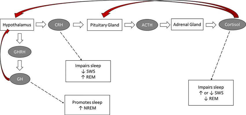
Henry et al.                                                                                                               Sleep and Cognition in AD

despite adherence, patients experience sub/supra physiological               rhythmicity and the sleep-wake cycle (9, 14–16). Circadian
cortisol concentrations, particularly during the night. Patients             rhythms are generated by the suprachiasmatic nucleus (SCN)
with AD report and experience both poor-quality sleep and                    hypothalamus, by light, and by ultradian rhythms (17, 18). The
cognitive difficulties (3–8). One possible (but as yet unexplored)            internal master clock in the SCN ensures we anticipate and
explanation for the sleep disruptions and memory deficits                     prepare for changes in our environment and act appropriately
experienced by these patients is that the periods of sub- and-               (19, 20). These oscillators are synchronized with each other by
supra-physiological cortisol concentrations they experience may              the SCN’s master clock (21). Regarding the circadian clock, its
have a specific negative impact on processes of sleep-dependent               timing mechanism is located in the SCN and incorporates three
memory consolidation.                                                        different components: (a) input pathways that transmit light and
   This review aims to summarize and integrate the literature on             other environmental signals to the clock, (b) an endogenous
the relationships between cortisol concentrations, sleep                     pacemaker that generates 24-hour rhythms, and (c) output
disruption, and cognitive functioning in patients with AD. This              pathways that project to other brain regions and peripheral
review is needed because despite expanding scientific evidence                organs (9). The circadian clock is responsible for daily
suggesting that sleep is a critical biological process and its               variations in body temperature, melatonin, and cortisol
functional value extends well beyond simple physical and                     secretion, and will align those rhythms with those of sleep and
mental restoration, very few published studies have investigated             other physiological processes (22). Circadian oscillators are also
whether disrupted sleep is a possible mechanism that underlies               located in numerous peripheral tissues, including the liver, lungs,
the memory deficits experienced by patients with AD.                          heart, and adrenal glands. These oscillators are synchronized
                                                                             with each other by the master clock (21). The hypothalamic-
                                                                             pituitary-adrenal (HPA) axis plays an important role in the
                                                                             homeostatic processes of the body, the co-ordination of the
A BRIEF OVERVIEW OF HUMAN SLEEP                                              organism’s ability to adequately cope with environmental
Human sleep is a natural state of reduced responsiveness                     stressors and sleep regulation. This physiological system
accompanied by a partial loss of consciousness. Sleep is regulated           regulates the secretion of various hormones; of particular
by three different processes: the homeostatic process, which                 concern to this review is the release of cortisol resulting from
determines its need, the circadian process that influences its timing,        HPA-axis activity.
and the ultradian process that determines its organization (9, 10).              In healthy people, cortisol has a robust diurnal secretory
    Sleep is cyclical in nature, alternating between 4 and 6 repeated        pattern. The highest concentrations occur in the early hours of
cycles of rapid eye movement (REM) and non-rapid eye                         the morning, with a peak just after waking. Concentrations then
movement (NREM) sleep, with each cycle lasting approximately                 decrease slowly throughout the day, with troughs in the mid-
90–120 120 minutes (11–13). Human sleep patterns have some                   afternoon and at midnight; the daily nadir typically happens
predictable characteristics. Sleep onset is characterized by                 several hours after initiation of nocturnal sleep. Concentrations
rhythmic alpha waves, occurring particularly in the occipital                then begin to rise from 02h00 to 03h00 and continue to rise until
regions as discerned by electroencephalogram (EEG). Sleep then               awakening. The nocturnal rise in cortisol is thought be in
follows with NREM (Stages 1-4 (N1-N4) before the first episode of             response to the greater energy demands of the brain as the
REM. The first sleep cycle usually begins with Stage 1 (N1), which            night ends (23–26). The daily rhythm of corticotropin-releasing
lasts for 1-7 minutes after sleep onset. Stage 2 (N2), which lasts for       hormone (CRH) and adrenocorticotropic hormone (ACTH)
10-25 minutes, is signaled by K-complexes. As N2 progresses,                 occur in close parallel with the daily rhythm of cortisol, all
high-voltage slow-waves appear, signaling the start of SWS.                  highest in the morning and reaching a nadir during around
Within SWS, Stage 3 (N3) lasts only a few minutes, whereas                   midnight (12, 25, 27). In contrast, melatonin is secreted from
Stage 4 (N4) lasts for 20-40 minutes. The body may re-enter                  the pineal gland and synchronized to light from retinal input and
lighter stages of sleep (N1-N3) for approximately 5 minutes,                 growth hormone (GH) concentrations and are highest during
before the first REM episode is initiated. This episode is short-             early sleep, suggesting a reciprocal relationship between the
lived, lasting only 1-5 minutes. Thereafter, NREM and REM                    HPA and hypothalamo-pituitary-somatotrophic (HPS)
continue to alternate in a cyclic manner throughout the night,               systems (21).
with REM cycles becoming longer and slow-wave sleep (SWS)
shorter as the night progresses. Brief waking episodes occur in the
later night, usually near the transitions into REM sleep (13).
                                                                             THE HPA AXIS AND SLEEP
                                                                             The HPA axis plays an important role in maintaining alertness
CIRCADIAN RHYTHMICITY – CONTROL                                              and modulating sleep [see Figure 1; (24, 28, 29)]. In fact, there is
OF HORMONE RELEASE                                                           a bidirectional relationship between sleep architecture and HPA-
                                                                             axis activity. For example, cortisol exerts specific effects on sleep,
The release of nearly all hormones follows daily oscillations,               whereas changes to sleep affect the release of this hormone (25,
which result from an interaction between 24-hour circadian                   26, 30).

Frontiers in Endocrinology | www.frontiersin.org                         2                                  August 2021 | Volume 12 | Article 694046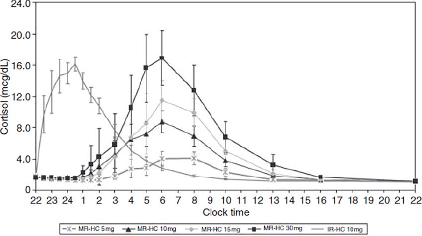
Henry et al. Sleep and Cognition in AD
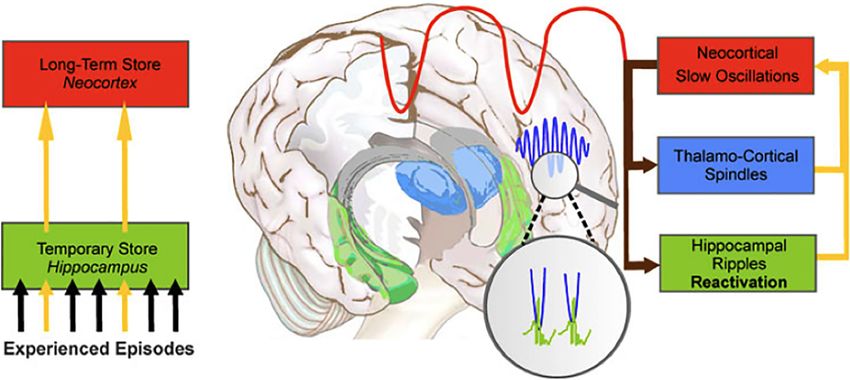
FIGURE 1 | The hormonal control of sleep. Red arrows: negative feedback on the indicated brain structure.
Endogenous HPA Hormones and pituitary-somatotrophic (HPS) and HPA systems. Confirming
Their Effect on Sleep the sleep-promoting role of GHRH and the sleep-disrupting
The circadian rhythmicity of specific hormones (e.g., cortisol, effect of CRH, in older adults the typical age-related reduction in
ACTH, CRH, GH releasing hormone, melatonin) plays an GH levels is accompanied by reduced SWS, whereas in both
essential role in sleep timing and offset and in the distribution older adults and in depressed younger adults increased CRH
of sleep stages across the night (28, 31–33). Inhibitory HPA-axis levels contribute to the typically-observed sleep disruptions
actions, particularly during SWS, are responsible for attenuated (27, 42).
cortisol activity during the first half the night. The quiescent Finally, ACTH and melatonin also play a role in sleep
period of HPA-axis activity starts prior to sleep, and continues regulation. ACTH is the prime stimulus for cortisol release
into the first half the night, when SWS occurs at a maximum. during sleep, and primarily affects sleep through its impact on
Cortisol concentrations decrease rapidly in the first 20 minutes cortisol secretion (29, 35). The secretion of melatonin, which has
after SWS onset, and there is a consistent inverse temporal a sleep-promoting effect, is dependent on the light-dark cycle and
relationship between low cortisol concentrations and high SWS is maximal during sleep periods (43). In fact, melatonin can
(15, 24, 34–37). The optimal cortisol levels during early sleep induce sleep even when there is an insufficient homeostatic drive
augments SWS via feedback inhibition of CRH (28, 33). In the to sleep. Hence, melatonin administration has been used to treat
second half of the night, when REM sleep predominates, insomnia and circadian rhythm disorders as it can preclude the
inhibitory mechanisms are attenuated and HPA secretory drive for wakefulness and produce shifts in the circadian clock so
activity slowly increases (15, 38). Cortisol, CRH, and ACTH that sleep occurs at a desired time (44).
secretion and SNS activity increase during the latter part of the
night. During the last sleep cycle, increases in cortisol are paired Effects of Sleep on HPA Hormones
with increases in REM (39). In summary, while the deepening of Sleep appears to have a direct impact on cortisol secretion.
sleep during SWS is associated with decreasing cortisol Specifically, sleep onset is associated with inhibitory effects on
concentrations and decreased sympathetic tone, high cortisol secretion. These effects persist for 1–2 hours after sleep
autonomic and high cortisol activity occur during REM cycles onset (34, 45). In contrast, awakenings and the end of sleep are
(25, 33). accompanied by cortisol increases (29, 46). Nocturnal
The reciprocal relationship between growth-hormone awakenings are associated with releases of cortisol and
releasing hormone (GHRH) and CRH also plays an important subsequent inhibition of cortisol secretion (34, 40, 46).
role in regulating sleep. GHRH inhibits HPA-axis activity during Nocturnal awakenings and final morning awakening elicit a
early sleep, stimulating NREM and promoting sleep, whereas rapid increase in both ACTH and cortisol. Unlike nocturnal
CRH inhibits SWS, enhances REM and vigilance, and disrupts awakenings, this cortisol awakening response (CAR), includes a
sleep (27, 40, 41). This pattern points to a reciprocal interaction 50–60% increase in cortisol secretion, lasting an hour, with a
between sleep architecture and hormones of the hypothalamic- peak at about 30 minutes after awakening (47–50). Some
Frontiers in Endocrinology | www.frontiersin.org 3 August 2021 | Volume 12 | Article 694046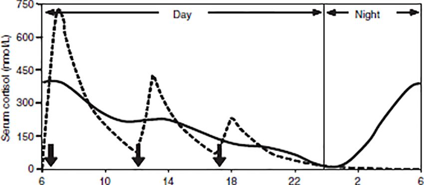
Henry et al.                                                                                                                       Sleep and Cognition in AD

research suggests that the release of ACTH and cortisol during             consolidation to take place. This is why theories regarding the
late sleep is precipitated by the physiological expectation that           function of sleep have gradually come to accept that a central
sleep will end at a certain time, and/or by the anticipation of the        aspect of this stage of consciousness is to strengthen memories
stress of waking (51–53).                                                  encoded during waking and to subsequently transfer their traces
                                                                           into long-term storage (1, 74). Sleep-dependent memory
Effects of Sleep Disruption on                                             consolidation (see Figure 2) appears to involve (a) repeated
Circadian Rhythms                                                          reactivation of information encoded during waking, and (b)
Acute shifts in the sleep-wake cycle (such as daytime sleeping or          transformation of newly acquired unstable memories into
napping or the consequences of jetlag and shift-work), reduced             stable representations that become integrated into existing
sleep quality, and sleep deprivation all lead to HPA-axis                  knowledge networks, thus forming long-term memories. In
activation and hence can alter the normal circadian pattern of             other words, during sleep the organism experiences “off-line”
cortisol secretion (34, 54–59).                                            periods (i.e., periods that do not feature the kinds of interference
    Regarding poor sleep quality, its experience (and, in fact, even       experienced during waking) during which newly encoded
its mere perception) is associated with increases in basal cortisol        memories are transferred from temporary to long-term stores
levels. Such increases stimulate arousal and suppress sleepiness,          (75, 76). The details of how these steps are accomplished, and
thus increasing sleep disturbances [which, in empirical studies,           which neural regions and neurobiological processes support
are characterised by increased wake time and reduced REM                   them, remains somewhat controversial, however [for reviews,
sleep; (16, 24, 60, 61)].                                                  see (1, 2, 77–79)].
    Regarding sleep deprivation, several studies report that                  Evidence for sleep-dependent memory consolidation is
elevated cortisol concentrations are present during both the               provided by numerous studies indicating that sleep enhances
sleep deprivation period and the subsequent day and evening                retention of information learned during waking hours, and that a
(33, 59, 62–66). Some researchers explain this physiological               sleep-filled delay enhances performance on a variety of
pattern by speculating that the initial sleep deprivation period           declarative and non-declarative memory tasks (2, 80–89). In
activates the HPA axis as part of the stress response and may also         contrast, when sleep is disrupted memory performance is poorer
reflect a decrease in the negative feedback regulation of the HPA           than when individuals are allowed to sleep uninterrupted
axis. Thereafter, prolonged wakefulness increases sleep pressure           (90–92).
(the increased need to sleep after periods of wakefulness), leads
to fatigue and sleepiness, and causes a blunting of HPA-axis
activity (34).                                                             CORTISOL: A FUNCTIONAL
    However, sleep disruptions do more than just impact the
                                                                           ROLE IN MEMORY
circadian rhythm of HPA-axis hormones. Disrupted sleep has
detrimental effects on health, quality of life, mood and cognition,        Adequate concentrations of cortisol are essential for optimal
which is not surprising given the central role of sleep in                 cognitive functioning (93–97). The hippocampus plays a vital
physiological restorative processes, emotion regulation and                role in memory consolidation and in new learning, encoding, and
memory consolidation (1, 10, 24, 67).                                      retrieval of declarative memories (97–105), while the prefrontal
                                                                           cortex (PFC) is similarly important for integrating sensory
                                                                           information, evaluating the significance of environmental
                                                                           stimuli, and processing previously encoded materials (106–110).
SLEEP AND MEMORY                                                           Because both these structures contain particularly high
                                                                           concentrations of glucocorticoid receptors (111), any alterations
The ability to effectively remember relies upon three broad
                                                                           in cortisol secretion have marked effects on their functioning. A
cognitive processes: encoding (the transformation of new
                                                                           substantial body of data indicates that elevated cortisol
information into a form that can be stored in memory),
                                                                           concentrations negatively impact performance on hippocampal-
consolidation (the stabilization of new memories in the brain),
                                                                           dependent memory tasks [e.g., word list and paragraph recall
and retrieval (68, 69). One of the most important ways in which
                                                                           tasks; (94, 112–120)] and on PFC-dependent working memory
sleep affects cognition is by helping to consolidate memories (70–
                                                                           and executive functioning tasks [e.g., tests assessing set-shifting,
72). The process of memory consolidation involves strengthening
                                                                           attention, abstract thinking, cognitive flexibility, mental rotation;
of memory traces, which represent information of our
                                                                           (120–129)].
experiences, and the parallel integration of these experiences
                                                                               Studies have consistently demonstrated that chronically
with previously acquired knowledge (69, 73).
                                                                           elevated glucocorticoids impair hippocampal-dependent
    Whereas encoding of environmental events (i.e., acquisition
                                                                           memory (130–134)1. The negative impact of elevated cortisol
of information) and retrieval of those memories (i.e.,
                                                                           on verbal declarative memory performance has been
reconstruction of previously acquired information) takes place
during waking hours, the process of memory consolidation is                1
                                                                             On the other hand, acutely elevated GCs can either enhance (114, 134–138) or
incompatible with waking consciousness (1). Hence, when the                impair memory depending on several factors, including but not limited to, the
organism effectively loses consciousness for several hours during          time of day of cognitive testing, the stage at which a stressor is applied (i.e., at
sleep, physiological conditions are optimal for memory                     encoding, consolidation or retrieval), and the dose of GC administered (139, 140).

Frontiers in Endocrinology | www.frontiersin.org                       4                                         August 2021 | Volume 12 | Article 694046
Henry et al. Sleep and Cognition in AD
A B
FIGURE 2 | The hippocampal-to-neocortical dialogue. (A) During NREM sleep, memories temporarily stored in the hippocampus are transferred to the long-term
store in the neocortex. (B) The dialogue involves the interaction between the slow oscillations, sleep spindles and hippocampal ripples to create spindle-ripple events
(magnified circle). From Born et al. (75). System consolidation of memory during sleep. Psychological Research, 76, 192-203.
demonstrated (a) following increases in endogenous levels of the (146) found that a 4-day period of oral administration of cortisol
hormone (through laboratory-based stress induction procedures; to young adults impaired their performance on a paragraph
127, 139, 141–143), and (b) in studies that featured exogenously recall task, but did not significantly affect their recall of
administered corticosteroids (128, 144–148). For example, previously presented geometric line drawings or their
Kirschbaum et al. (94) found that stress-induced cortisol performance on a spatial location task.
increases, and (separately) administration of 10 mg Regarding the PFC’s involvement in memory processing, this
hydrocortisone orally, were associated with poorer recall of brain structure plays an important role in the encoding and
verbal material. Similarly, De Quervain and colleagues (149, retrieval of declarative memories (167). Specifically, after
150) found that oral administration of 25mg of cortisone retrieval, the PFC determines whether an event occurred in a
acetate significantly impaired both free and cued recall of particular setting (168, 169), allowing accurate memories to be
verbal material, while leaving recognition memory (which is reconstructed. The PFC is also involved in working memory
not dependent on hippocampal substrates) unaffected, and that (WM). Specifically, it allows humans to (a) keep a mental
the same dose of cortisone acetate impaired cued recall of a series “sketch” of information and protect this information from
of word pairs. In that study, stress-level doses of cortisone acetate internal and external distractions, (b) inhibit inappropriate
reduced cerebral blood flow to the medial temporal lobe (MTL), responses and behaviour, and (c) regulate attention. As such,
a memory network that broadly includes the hippocampus. the PFC allows for cognitive flexibility and goal-directed
Several studies have also documented impaired performance behaviour (121, 122).
on spatial memory and navigation tasks in the presence of Chronically elevated cortisol concentrations lead to dendritic
elevated cortisol levels in humans (94, 127, 146, 151–162). atrophy in the PFC (170), and stengthens the noradrenalin
However, investigations of the impact of cortisol on spatial system, which reduces neuronal firing within the structure
memory are more abundant in the animal literature. (122, 171). Stress-induced cortisol increases also increase
Furthermore, studies investigating spatial memory and cortisol dopaminergic activity and glutamate levels in the PFC (129,
in humans have produced more variable results, compared to the 172). Glutamate receptor-mediated synaptic transmission in the
robust literature on impaired verbal memory in the presence of PFC is particularly important for WM (173, 174). While acute
elevated cortisol levels. elevations in glutamate have a positive effect on WM (129),
Although hippocampal-dependent forms of memory are excessive elevations cause impairment. Each of these hormones
impaired by increased cortisol concentrations, non- (noradrenalin, dopamine and glutamate) has an inverted-U
hippocampal forms of memory (e.g., procedural memory), influence on WM, with either too little or much impairing
appear unaffected (e.g., 117, 163, 164). For example, PFC functioning (122).
Kirschbaum and colleagues (94) found that oral administration
of 10mg cortisol to healthy subjects impaired performance on a Glucocorticoid Receptors and Memory
declarative (a word list) but not a procedural (a word priming Glucocorticoids affect the human brain by their interaction with
test) memory task. Furthermore, increased cortisol levels impair two intracellular receptors (134). Glucocorticoids that enter the
verbal declarative memory whereas non-verbal memory appears brain change gene expression by binding to type 1
unaffected (165, 166). For example, Newcomer and colleagues mineralocorticoid receptors (MRs) and type 2 glucocorticoid
Frontiers in Endocrinology | www.frontiersin.org 5 August 2021 | Volume 12 | Article 694046Henry et al. Sleep and Cognition in AD
receptors (GRs). These two receptors bind cortisol with different such mechanism may be through sleep, given that a
affinities (135). MRs have a high affinity for cortisol and become bidirectional relationship exists between circadian rhythmicity
heavily occupied at low cortisol concentrations (including the and the sleep-wake cycle, and because successful memory
evening nadir of the cortisol circadian profile, when 90% of MRs, consolidation of information learned during the day is known
but only 10% of GRs, are occupied). In contrast, GRs have a to rely on sleep (1, 90, 203).
lower affinity for cortisol and only become heavily occupied
when cortisol levels reach a peak [e.g., after a stressor or after the
post-awakening cortisol surge; (97, 134, 146, 175)].
MRs are found predominantly in the hippocampus, whereas
ADDISON’S DISEASE
GRs are distributed throughout the brain. Both play important The diagnosis of AD is based on the measured presence of low
roles in cognitive function, however (176, 177). MRs are located plasma cortisol, low aldosterone levels, high renin levels, and
in brain regions involved in behavioral reactivity to new events, elevated ACTH [loss of endogenous ACTH drive; (204)]. Patients
which enables the encoding of new information and subsequent with AD need to be on glucocorticoid (GC) replacement therapy
retrieval, whereas GRs are located in brain regions involved in for life, which is essential for survival (205). Cortisol is usually
the consolidation and storage of information learned (94, 132, replaced with oral hydrocortisone, prednisone, or cortisone
178, 179). Hence, the activation of both receptors is a necessary acetate (a ll of which a ctivate predominantly GRs,
for optimal memory functioning. For instance, de Kloet et al. predominantly), plus a mineralocorticoid (fludrocortisone) for
(135) showed that when cortisol levels were mildly elevated (and sodium and potassium regulation (206, 207). Given the
therefore all MRs, but only some GRs, were activated), long- bidirectional relationship between cortisol and the sleep-wake
term-potentiation (LTP; the reinforcement of synaptic cycle, the dosage, timing, and type of medication regimens used
connections necessary for information storage) was enhanced. by patients may impact their general well-being and sleep
However, at higher cortisol levels (when GRs were over-activated patterns (28) due to the influence of GCs on circadian
and MR occupation was low), LTP was impaired. MRs play a rhythmicity. Typically, GCs are replaced in 2-3 daily doses (see
particularly important role in hippocampal-dependent memory, Figure 3), with the total daily dose ranging from 15-30mg. The
executive function, and attention (126, 180–184). In highest dose (one-half to two-thirds) is taken in the morning, a
confirmation of the latter, Schultebraucks and colleagues (185) reduced dose is taken in the afternoon, and (if required) a third
found that, during high MR occupation, verbal memory was dose in the late afternoon/evening [typically around 5pm; (208–
significantly better and there were trends towards better 210)]. Such a dosing schedule of GC replacement is meant to
executive functioning. imitate the normal diurnal cortisol rhythm, to reflect the peak
cortisol rise in the morning, and to avoid over-replacement in the
Variations in Cortisol Concentrations and nadir of cortisol secretion during the night (208, 211). However,
Their Effects on Cognition despite efforts to find the best replacement regimen in terms of
Most studies have focused on the deleterious effects of elevated dosage and timing (211–213), none mimic the physiological
cortisol levels on cognitive functioning. The negative effects of circadian rhythm; there are still supra-physiological peaks
supra-physiological cortisol levels on brain structure and during the day and lower-than-expected concentrations during
cognitive functioning is well known and evident in both the early hours of the morning (214–219). This over- and under-
healthy individuals and in patients known to experience replacement results from the biochemical properties of
chronically elevated cortisol levels [e.g., Cushing’s syndrome, replacement medications. Oral hydrocortisone (HC) is absorbed
depression, Alzheimer’s disease; (132, 175, 186–191)]. rapidly, reaching maximum concentrations an hour after intake
Elevated cortisol concentrations impair cognitive function (220). However, HC replacement produces extremely variable
due to their effect on specific neurobiological systems. peak concentrations within a supra-physiological range, followed
Specifically, the relationship between glucocorticoids and by rapid declines toHenry et al. Sleep and Cognition in AD
FIGURE 3 | Simulated cortisol profile for a patient [broken line] following thrice-daily hydrocortisone administration [10mg at 06:00, 5mg at 12:00 and 2.5mg at
18:00, shown as solid arrows]. The normal circadian rhythm of cortisol [solid line]. From Mah et al. (211). Weight‐related dosing, timing and monitoring
hydrocortisone replacement therapy in patients with adrenal insufficiency. Clinical Endocrinology, 61(3), 367-375.
morning dose of hydrocortisone (224). Overall, conventional GCs even more impractical and, moreover, may cause more daytime
do not restore the normal circadian rhythm of hormone release fatigue as well as supra-physiological peaks. MR-HC offers a
(215, 225). Instead, patients are over-replaced immediately more practical and sustainable approach to normalizing cortisol
following therapeutic administration, and then under-replaced circadian rhythms due to its immediate and extended hormone
within a few hours of that administration (211, 226), which may release characteristics. MR-HC has been shown to mimic natural
have important implications for sleep regulation. physiological cortisol circadian rhythm (230, 231). Johannsson
and colleagues (231) demonstrated that taking a once-off
New Advancements in Treatment morning dose of either 5 or 20mg MR-HC led to a closer
As standard replacement therapy does not mimic the natural mimicking of physiological cortisol circadian rhythms, except
circadian rhythm, newer treatments aim to imitate physiological for the early-morning cortisol peak. However, if MR-HC is taken
cortisol rhythms. These new treatments attempt to improve late at night (thus allowing for a delayed and sustained release), it
biochemical control of the release of cortisol and to reduce the can mimic the rise in cortisol that typically occurs during the
long-term adverse effects typically associated with standard early hours of the morning. For example, Debono et al. (230)
replacement regimens (220). Continuous subcutaneous HC showed that taking 15-20mg of MR-HC at 23h00 and 10mg at
infusion (CSHI) and modified release HC (MR-HC) tablets are 07h00 reproduced the normal physiological cortisol circadian
two promising new treatments. rhythm in healthy controls (see Figure 4). In that study,
Infusion of HC in patients with AD has been shown to mimic participants’ cortisol concentrations peaked, on average, at
normal circadian rhythmicity and improve quality of life (QoL; 08h32 and decreased throughout the day, reaching a nadir, on
226, 227). A crossover randomized clinical trial (N = 33 patients average, at 00h18. Dual-release hydrocortisone (DR-HC;
with AD who received CSHI or thrice-daily conventional therapy Plenadren) has both an immediate-release coating and
for a 3-month period), found that a 10mg/m2 daily dose of CSHI extended-release core. This form of replacement therapy better
normalized cortisol and ACTH levels in the morning, and mimics the normal cortisol profile (218) and improves patients’
patients 24-hour cortisol curves approached normal circadian quality of life (232–234). However, despite normalizing cortisol
variation compared with conventional oral replacement. The 24- patterns, DR-HC has shown to have little effect on cognitive
hour area under the curve (AUC) did not differ between infusion functioning or sleep (235). Although Krekeler and colleagues
and conventional oral therapy, but daytime AUC (8am- (235) showed that patients with adrenal insufficiency treated
midnight) was higher for oral replacement therapy, and night- with DR-HC tended to show better executive functioning
time AUC (midnight-8am) was higher for CSHI. Infusion compared to patients on conventional HC, other cognitive
improved vitality and physical functioning (228) but did not domains appear unaffected, and they found no between-group
improve sleep (except that sleep length increased, as measured by differences in terms of sleep.
the Pittsburgh Sleep Quality Index (PSQI) and actigraphy).
Another randomized double-blind placebo-controlled clinical Sleep Disruptions in AD
trial (N = 10 patients with AD) assessed whether CSHI Numerous studies suggest that, in patients with AD, clinically
improved QoL and fatigue, compared to standard GC therapy relevant fatigue persists despite replacement therapy. For instance,
(229). CSHI did not improve health status in AD patients who Løvâs, Logeŧ, and Husebye (236) found that patients with AD self-
had mild deficits in well-being at baseline. Overall, it appears that reported reduced general health perception and vitality despite
CSHI benefits some, but not all, patients in that it restores the receiving replacement therapy with cortisone acetate and
usual circadian cortisol rhythmicity and improves QoL (226). fludrocortisone. Similarly, van der Valk and colleagues (237)
A significant disadvantage of subcutaneous infusions is their found that 48% of their patient sample (N = 328) self-reported
impracticality. An alternative is for patients using HC to wake up abnormal fatigue; 61% reported severe fatigue. Researchers
at 3am and take a dose of medication. This alternative is perhaps postulate that reports of increased daytime fatigue may be due
Frontiers in Endocrinology | www.frontiersin.org 7 August 2021 | Volume 12 | Article 694046Henry et al. Sleep and Cognition in AD
spent in REM sleep. Similarly, Garcia-Borreguero et al. (238)
reported that patients with AD who were deprived of
glucocorticoid medication for 1.5 hours prior to bedtime (and
who therefore had undetectably low levels of cortisol at bedtime)
showed increased wake after sleep onset (WASO) and REM
latency and decreased amount of time spent in REM sleep,
compared to patients who took their medication just before
bedtime. These results suggest that the initiation and
maintenance of REM sleep is facilitated by cortisol. In contrast,
high cortisol concentrations appear to reduce the amount of time
spent in SWS (27, 35, 240), and, consistent with this, relatively
lower cortisol concentrations in healthy controls (administration
of metyrapone) and patients with AD (replacement medication
was withheld) are significantly associated with increased delta
FIGURE 4 | Concentration-time profiles for modified-release hydrocortisone sleep (239).
(MR-HC) 5mg, 10mg, 15mg and 30mg compared with immediate-release
Gillin et al. (239) reported that when medication was
hydrocortisone (IRHC). Graph showing delayed and sustained release
characteristic of MR-HC (to convert values from mcg/dl to nmol/l x 27.59). administered by conventional replacement, patients with AD
From Debono et al. (230). Modified-release hydrocortisone to provide had similar sleep to controls, except that patients took
circadian cortisol profiles. The Journal of Clinical Endocrinology & Metabolism, significantly longer to fall asleep and spent significantly more
94(5), 1548-1554. time in delta sleep (i.e., SWS). More recently, Henry and
colleagues (4) found that when medication was administered
to reduced quality of sleep in patients with AD and that relatively by conventional replacement, patients with AD (compared to
increased doses of HC may contribute to these sleep disruptions healthy matched controls) spent significantly less time in SWS
(3, 236, 238). and that there was a trend towards patients experiencing
Løvâs, Husebye, Holsten, and Bjorvatn (3) found that 34% of significantly shorter REM latency and more time in Stage 2 sleep.
the 60 patients with AD in their sample self-reported weekly Overall, few studies have objectively assessed sleep in patients
sleep disturbances (difficulties falling asleep [13%], repeated with AD, despite that fact that an abundance of scientific
awakenings [14%], and early morning awakenings [20%]). evidence suggests that these patients experience disruptions to
Similarly, Henry et al. (7) found that 60 patients with AD self- the cortisol circadian rhythm and, consequently, are at risk for
reported poorer sleep quality and efficiency, longer sleep latency, experiencing negative effects on sleep architecture. For instance,
shorter sleep duration, more sleep disturbances, and greater patients with psychiatric conditions that exhibit elevated cortisol
daytime dysfunction compared to healthy controls. In terms of concentrations (e.g., depression and post-traumatic stress
objectively measured sleep quality, Henry et al. (5) found, using disorder) experience less time in SWS, shortened REM latency,
actigraphy, that patients with AD experienced interrupted sleep increased REM sleep and density, and sleep discontinuity (238,
characterized by worse sleep efficiency and a greater amount of 241–244). Similarly, in patients with Cushing’s disease (who
time spent awake compared to healthy controls, who achieved a produce excessive amounts of cortisol), findings show that SWS
fuller night of uninterrupted sleep. These data present a pattern is decreased, REM latency shortened, REM density is elevated,
showing that patients with AD report frequent sleep and aberrances in sleep continuity occur (244, 245). One study
disturbances, including difficulty falling asleep, nighttime found that elevated cortisol concentrations in 11 patients with
awakenings, and a lower sleep quality. In all of the Cushing’s disease were associated with lower REM activity, and
abovementioned studies, although patients were on more awakenings during sleep (245). Similar patterns of
replacement therapy, their cortisol secretion differed from decreased REM latency and/or increased time spent in REM
healthy individuals. Because of cortisol’s key role in regulating sleep have been found in patients with AD who took
our circadian rhythms and ensuring the transitions between hydrocortisone before bedtime and in healthy controls with
sleep stages (33), it is unsurprising patients on replacement artificially increased cortisol concentrations (41, 238). There
therapy still experience poor-quality sleep. are comparatively few data on sub-physiological cortisol levels
In terms of objectively measured sleep quality via and sleep, and similarly, few studies on sleep of patients with AD
polysomnography, limited information exists on the impact of when replacement medication is administered as part of routine
low cortisol concentrations on sleep architecture. Similarly, few replacement. Therefore, effects on sleep quality and architecture
studies report on sleep in patients with AD when replacement of the illness itself, and of replacement therapy, remains largely
medication is administered by conventional replacement. In unexplored in patients with AD.
terms of low cortisol concentrations, Gillin et al. (239) Because of the central role the HPA axis plays in sleep
reported that patients with AD whose replacement medication regulation (28, 35), either low or high night-time cortisol,
was withheld for longer than 24 hours (and who therefore had alongside high night-time ACTH and CRH, may lead to sleep
undetectably low concentrations of cortisol at bedtime), showed disturbances in patients with AD (228). However, the
increased time spent in SWS and correspondingly reduced time implications of exposure to altered circadian cortisol patterns
Frontiers in Endocrinology | www.frontiersin.org 8 August 2021 | Volume 12 | Article 694046Henry et al. Sleep and Cognition in AD
and consequent sleep disruptions have not been adequately that cognitive deficits in patients with AD are primarily in the
addressed in the available literature. For instance, disrupted domain of declarative memory (both verbal and visual memory),
sleep may impede sleep-dependent memory consolidation. but also extend to executive functioning (including attention and
Because changes in sleep and memory are associated with the processing speed).
use of corticosteroids, further research is necessary to help Impaired declarative memory performance likely emerges
understand the impact that replacement medication used by from two main sources. First, indirectly, disrupted cortisol
patients with AD has on the processes that influence sleep- secretion patterns impact circadian rhythms and lead to sleep
dependent memory consolidation. disturbances. Second, directly, due to supra-physiological cortisol
levels experienced by patients taking short-acting hydrocortisone
Cognitive Functioning in AD (250). Supra-physiological glucocorticoid increases impact on
Despite replacement therapy, patients with AD frequently brain regions such as the hippocampus and the PFC which
present with both subjective cognitive complaints and objective have a high concentration of glucocorticoid receptors (105,
cognitive impairments, including poor memory and impaired 251). These effects include, but are not limited to, degeneration
concentration (206, 207, 221). Due to the affinity between of hippocampal neurons (252), altered organization of dendrites
variations in cortisol concentrations and impaired performance in the PFC (253), and, as such, impaired performance on tasks
on tests of memory, attention and executive functioning (115, involving declarative memory (121, 254). In support of this,
146, 246), understanding how these domains are affected is elevated cortisol concentrations associated with normal aging
relevant in patients with AD. However, very few studies have have been linked to ventricular enlargement, neuronal loss, and
characterized cognitive function in AD. decreased volume in the hippocampus alongside a decline in
Klement et al. (247) reported that patients with AD on cognitive performance (154, 252, 255–257). Exogenous
replacement therapy performed significantly more poorly than administration of hydrocortisone (occurring as a once off to a
healthy controls on a declarative memory test. Similarly, few days) to healthy subjects raising serum cortisol
Schultebraucks and colleagues (185) found that patients concentrations, impairs verbal memory, working memory,
performed significantly more poorly than controls on a test of visuo-spatial memory, and executive functioning (96, 146, 150,
verbal learning, and Henry et al. (6) found that patients 191, 258, 259). Similarly, exogenous administration of
performed significantly more poorly than controls on tests of dexamethasone or prednisone to healthy subjects impairs
both verbal learning and memory (and that patients made memory performance (117, 191, 259, 260). Prolonged levels of
significantly more false alarms [incorrectly saying a word on increased hydrocortisone may cause permanent death of
the list when it was not present] when recalling information). hippocampal neurons, reduce hippocampal glucose uptake
Henry et al. (5) found that healthy controls learned and retained (255) and neuronal excitability (261), impair synaptic plasticity
more information than patients with AD on two different tasks (262, 263), decrease the amount of newly-generated neurons, alter
of declarative memory, but that there were no significant synaptic density in the CA1 and CA3 regions (264), and cause
between-group differences for procedural memory tasks. death of dendrites in hippocampus (252).
Tiemensma and colleagues (248) found that patients Another neurobiological mechanism that may explain the
performed significantly more poorly on tests of both verbal memory deficits observed in patients with AD is the differential
and visual memory than healthy controls. The latter study also activation of the two types of receptors discussed earlier, MRs
found mild executive impairment and significantly slower and GRs. Cortisol’s effects on the hippocampus and PFC are
processing speed in their patient group. Interestingly, in this mediated by the interaction of glucocorticoids with MRs and
study, delaying HC intake in another group of patients with AD, GRs (134). Activation of MRs is essential for successful encoding,
which resulted in significantly lower cortisol concentrations at whereas activation of GRs is essential for successful
the time of cognitive testing, had no impact on cognitive consolidation and retrieval of memory (94, 135). Activation of
performance. In terms of disease characteristics and cognition, both receptors is required for optimal memory performance
Henry et al. (6) also found that patients who had AD for a longer (135). In one study providing empirical support for this
interval had a slower speed of processing and that patients who proposed neurobiological mechanism, Tytherleigh et al. (207)
were diagnosed later in life had poorer declarative and working found that adequately treated AD patients, performed
memory, a slower speed of processing, and an overall greater significantly better a declarative memory recall task, when both
cognitive impairment. Blacha et al. (249) found patients with AD receptor types were activated, compared to when only one or the
(20 PAI and 20 SAI) showed significantly worse performance on other was activated. While some cortisol is needed to enhance
a test of attention compared to controls (but found no difference cognition (a shift towards predominant MR activation and
in memory and other cognitive domains). They also found that minimal GR activation), prolonged exposure and/or high
higher HC doses impaired attention, visuo-motor skills and concentrations of cortisol (predominant GR activation) have
executive function, but that duration of therapy had no impact deleterious effects (135, 265–267). In support of the beneficial
on cognitive performance. Similarly, Harbeck et al. (223) found effects of MRs on cognition, Schultebraucks et al. (185) used a
that higher cortisol levels were associated with impaired short- repeated-measures crossover design and either administered
term memory in patients who underwent short-term patients with AD fludrocortisone (resulting in high MR
hydrocortisone infusion during the night. Overall, it appears occupation) or withheld the same drug from them (resulting in
Frontiers in Endocrinology | www.frontiersin.org 9 August 2021 | Volume 12 | Article 694046Henry et al. Sleep and Cognition in AD
low MR occupation). Verbal memory performance was that therefore poor performance occurs whether one sleeps or
significantly better when MR occupation was high, and there not. The design of this study did not allow consideration that
also were trends towards better executive functioning in patients with AD may suffer from global fatigue and therefore
the condition. poor performance. Another finding in this study was that, on a
Previous studies have shown that, in healthy adults across the story recall test, patients had greater recall when a period of wake
lifespan, elevated cortisol levels impair cognitive functioning in rather than wake separated learning from recall (counterintuitive
ways that are predictable and that can be explained to the body of literature that sleep is an offline process beneficial
neurobiologically (105, 142, 166, 252). In AD, cortisol for the consolidation of learned information). This result
concentrations fluctuate –elevated far above basal levels (e.g., corroborates in patients with AD that sleep may not be
after hydrocortisone administration) or low [e.g., several hours providing an optimal period for consolidation of previously
after hydrocortisone medication has been taken and due to the learned material. In contrast to the patterns of data on
fact that this medication has a relatively short half-life of roughly declarative memory tests, no significant between-group (AD
1.5 hours; (223)]. Since the relationship between cognition and versus controls) or between-condition (Sleep versus Wake)
GCs usually follows an inverted-U shaped pattern, cortisol were found on a test of procedural memory. These results may
concentration variability in patients with AD may play an have emerged because declarative memory tasks are
important role in their cognitive functioning. Furthermore, due hippocampal-dependent, whereas procedural tasks are not.
to the known association between altered cortisol and impaired Since hydrocortisone affects hippocampal integrity but not
performance on standardized memory tests, between altered areas typically associated with response-based sequence
cortisol and disrupted sleep, and between sleep and memory learning (e.g., motor cortex, caudate nucleus), it is possible that
consolidation, assessment of other contributors (e.g., disrupted procedural memory performance of patients with AD is
sleep) that may contribute to deficient memory performance in unimpaired. No prior study had investigated procedural
patients with AD needs to be understood. memory in patients with AD, and hence this suggestion that
procedural memory is not impaired in patients with AD is a
The Relationship Between Sleep and novel finding.
Cognition in AD
The orderly night-time sequence and transition between SWS The Relationship Between Sleep, Emotion
and REM sleep provides optimal conditions for memory Regulation, and Cognition
consolidation (1). Consolidation begins during SWS, when While it is well established that sleep plays a crucial role in
specific physiological conditions (e.g., slow oscillations in various aspects of health, and cognition, sleep also plays an
neocortical networks, HPA axis suppression) allow the important role in the processing and regulation of emotion (1,
reactivation of memories encoded during wakefulness (268). 69, 79, 271). The experience of sleep deprivation or poor sleep
During REM sleep, physiological conditions (e.g., suppression quality makes people more sensitive to emotional and stressful
of norepinephrine, increased levels of acetylcholine and events on the following day, elevates negative emotions
serotonin, ponto-geniculo-occipital and theta waves) allow (including feeling more irritable, angry, and anxious), and
reactivated memories to be integrated with pre-existing reduces positive emotions (272–275). Short sleep duration and
knowledge, thereby facilitating long-term potentiation (269). poor-quality sleep is also associated with elevated depressive
Cortisol’s influence on successful memory consolidation during symptoms (276). REM sleep plays a particularly important role
healthy sleep is accounted for because it plays a pivotal role in in emotion regulation (79, 277), with research showing that
sleep stage initiation and maintenance (68). Although a well- patients with mood disorders have altered REM sleep (278).
known relationship between healthy sleep and optimal memory Because of cortisols key role in sleep stage initiation and
performance is noted (270), limited studies have explored this maintenance, it has an important influence on the affect
association in patients with AD. regulation that takes place during healthy sleep (73). HPA
Henry and colleagues (7) obtained data from self-reported hyperactivity (and consequent elevated cortisol levels, for
questionnaires and suggest that memory impairment may be example) plays a crucial role in the pathogenesis of medical
mediated by sleep disruptions in AD. Henry et al. (5) investigated and psychiatric disorders (e.g., major depressive disorder
the relationship between adrenal function, and objectively (MDD)) that are marked by sleep disturbances alongside mood
measured sleep and cognitive performance. Results showed problems (34, 279). The co-occurring presence of HPA-axis
that periods of sleep rather than wake benefited declarative hyperactivity, sleep disturbance and mood problems in these
memory retention in healthy controls’ but not in patients with disorders is not coincidental. Clinical studies have implied that
AD. These findings concur with a large body of literature patients with nearly all neurological and psychiatric mood
indicating that a full night of uninterrupted sleep has positive disorders have co-occurring sleep abnormalities, and
effects on memory. Because patients with AD do not have specifically, problems with REM sleep (79, 271). Empirical
normal circadian rhythmicity, the sequence and transitions of studies show that when people are deprived of REM sleep they
sleep stages may not have occurred in such a way that is required have intensified experience of negative emotions, show increased
for successful memory consolidation. Another possible anxiety during stressful events, and exhibit less positive reactions
explanation is that patients with AD are generally fatigued, and to positive events (275, 280).
Frontiers in Endocrinology | www.frontiersin.org 10 August 2021 | Volume 12 | Article 694046Henry et al. Sleep and Cognition in AD
Previously published studies in patients with AD have these patients (although very few studies have investigated this).
consistently shown that, despite being on replacement therapy, They also experience a reduced quality of sleep and altered sleep
these individuals still report and experience depression and architecture. Hydrocortisone immediate release may lead to
anxiety, as well as reduced stress tolerance and reduced ability disrupted sleep patterns which impair optimal consolidation of
to cope with daily demands (5, 236, 281). Regarding affective learned information. Moreover, general fatigue may contribute
disorders, a Danish study of 989 patients with AD found they to the presence of cognitive deficits in patients with AD.
were 2.68 times more likely to suffer from depression than a Furthermore, prolonged replacement therapy may have
control group with osteoarthritis (281). deleterious effects on brain regions required for optimal
Generally depressed patients self-report difficulty falling cognitive functioning (e.g., the hippocampus and PFC).
and staying asleep, and early morning awakenings; (282), and However, studies using brain scans are needed to confirm
experience both SWS and REM disruptions (278). Interestingly, this hypothesis.
individuals who take hydrocortisone late at night also have Although prior research suggests that both cognitive and sleep
decreased REM latency and increased REM sleep time, a complaints are frequently reported by patients with AD, only a few
pattern similar to that found in depressed patients. As such, have used objective measures to assess either sleep patterns or
altered circadian rhythms in patients with AD may explain the memory impairments experienced by patients. Patients with AD
high presence of affective disorders in this population. encounter sub- and supra-physiological cortisol concentrations
In addition to the wealth of knowledge linking sleep and affect due to imperfect replacement therapy, ultimately altering sleep
regulation, numerous studies illustrate that affect and cognition architecture and impairing cognition. It is conceivable that
are interrelated (283, 284), and specifically that low mood is through modifying the pharmacokinetics of replacement therapy
related to impaired cognitive functioning (285, 286). The high that these modalities in patients with AD may be improved. From
presence of affective symptoms in patients with AD may be a broader neuroscientific perspective, patients with AD provide a
related to patients sleep disturbances. The high presence of unique opportunity to simultaneously study the effects of hyper-
affective symptoms in patients with AD may also negatively and hypo-cortisolism on sleep quality, memory performance, and
impact cognitive functioning. That is to say, the co-occurring sleep-dependent memory consolidation. Careful study of these
presence of sleep disturbances, depressive symptoms, and patients can help unravel the distinct roles that sub- and supra-
impaired cognitive functioning in patients with AD may not be physiological GC concentrations play in sleep regulation/structure
coincidental. However, no published study has explored the and in sleep-dependent memory consolidation.
relationship between affect dysregulation, sleep disturbances Although current replacement therapy aims to mimic the
and cognitive impairment in AD. natural circadian rhythm of cortisol, periods of sub- and supra-
physiological cortisol concentration are experienced. Both low
and high cortisol concentrations can negatively impact cognition
CONCLUSION and sleep. More research is needed on the effects of dosage,
duration and type of GC therapy used in patients with AD and
In this article, we have summarised the current knowledge on how these impact cortisol concentrations, sleep and cognition.
sleep, cognition, and the association between the two in patients Food and caffeine intake, smoking, intense exercise, and
with AD. Numerous studies indicate that (i) healthy sleep encountering stressful situations may all influence cortisol
benefits memory consolidation, (ii) alterations in cortisol concentrations, sleep, and cognitive performance (287). Studies
activity has negative effects on sleep architecture, and (iii) sleep investigating sleep and cognition in AD should be careful to control
disruptions (e.g., as might be present in individuals with for these potentially confounding factors. Another important
abnormal night-time cortisol concentrations) might impede the contributor to cognition and sleep in patients with AD could be
beneficial effects of sleep on memory consolidation. Sub- and life-threatening events such as nocturnal hypoglycemia and adrenal
supra-physiological cortisol concentrations resulting from crises (288). However, hardly any studies take this into account
immediate release hydrocortisone replacement therapy can when investigating cognition and sleep. It is important to
have negative effects on sleep architecture and sleep-dependent differentiate between impairments caused by the illness itself, the
memory consolidation processes. Therefore, disrupted circadian complications going with it or the therapy received by patients.
rhythms are suspected to be a major cause of sleep disturbances More studies are needed to characterize the relationship
and cognitive impairment in patients with AD. It is well between sleep and memory, using objective measures to examine
established that cortisol plays a key role in maintaining the the hypothesis that poor sleep is a biological mechanism
integrity of sleep architecture and that sleep plays an important underlying memory impairment in patients with AD. More
role in cognitive functioning, emphasizing the interrelationship polysomnographic studies are needed to comprehensively
between sleep, cognition and intact cortisol secretion. investigate sleep architecture in patients with AD. Such studies
The literature suggests that patients with AD experience may help explain for instance, memory consolidation in not
disruptions to cognition, primarily in the domain of declarative enhanced by sleep in patients as in healthy controls. Intervention
memory (both verbal and visual memory), but also extending to studies and clinical trials might seek to confirm this association
executive functioning (specifically, attention and processing and investigate whether the same pattern of sleep and memory
speed). Procedural memory does not appear to be impaired in deficits are present in patients, using modified-release or dual-
Frontiers in Endocrinology | www.frontiersin.org 11 August 2021 | Volume 12 | Article 694046You can also read