Benign Paroxysmal Positional Vertigo: Management and Future Directions - ACADEMIC YEAR 2011-2012
←
→
Page content transcription
If your browser does not render page correctly, please read the page content below
UNIVERSITY OF SIENA
                              PhD PROGRAM IN
         BIOMEDICINE AND IMMUNOLOGICAL SCIENCES

                                CYCLE XXIV

    Benign Paroxysmal Positional Vertigo:
         Management and Future Directions

Tutor:
Chiar.mo Prof. Daniele Nuti

                                                          PhD Student:
                                             Dr. Giovanni Paolo Santoro

                     ACADEMIC YEAR 2011-2012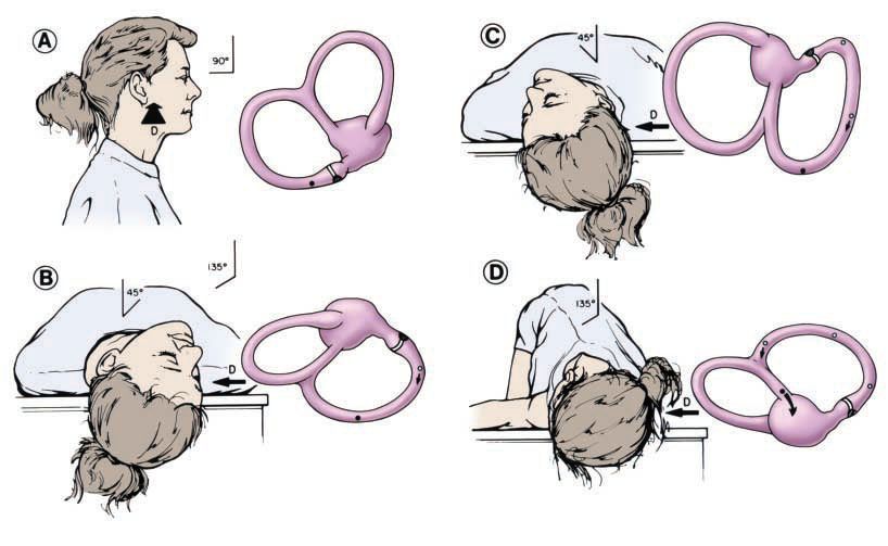
INDEX

1. History                                                                       3

2. Anatomy and physiology                                                        9
      2.1 Peripheral Vestibular Anatomy                                          9
      2.2 Vestibular Afferent Physiology                                        12
      2.3 Central Vestibular Anatomy                                            13
      2.4 Vestibulo-ocular Physiology                                           14
      2.1 Peripheral Vestibular Anatomy                                          9
      2.2 Vestibular Afferent Physiology                                        12

3. Clinical features                                                            16
      3.1 Epidemiology                                                          16
      3.2 Symptoms of BPPV                                                      17
      3.3 Causes                                                                17
      3.4 Pathomechanism                                                        19

4. Diagnosis and Treatments                                                     20
      4.1 Posterior Canal BPPV (PC-BPPV)                                        20
      4.2 Horizontal (Lateral) Canal BPPV (HC-BPPV)                             24
      4.3 Anterior Canal BPPV (AC-BPPV)                                         29
      4.4 Rehabilitation                                                        30
      4.5 Surgical treatment                                                    31
      4.1 Posterior Canal BPPV (PC-BPPV)                                        20

5. Results                                                                      32
      Vestibular neuritis: recurrence and incidence of secondary benign
      paroxysmal positional vertigo                                             33
      Comorbidites of BPPV                                                      38
      Double-blind randomized trial on short-term efficacy of Semont maneuver
      for treatment of posterior canal benign paroxysmal positional vertigo     45
      Double-blind randomized trial on short-term efficacy of Gufoni maneuver
      for treatment of lateral canal benign paroxysmal positional vertigo       51
      Lateral canal BPPV with uncertain side: a new therapeutic procedure       56

6. References                                                                   60

Publications and Abstracts of Conference during the PhD program                 65

                                               2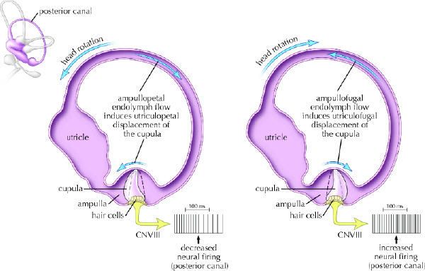
1. History
Benign paroxysmal positional vertigo (BPPV) is the most common vertiginous disorder in the
community; it is characterized by brief recurrent episodes of vertigo triggered by changes in
head position. BPPV is the most common etiology of recurrent vertigo and is caused by
abnormal stimulation of the cupula by free-floating otoliths (canalolithiasis) or otoliths that
have adhered to the cupula (cupulolithiasis) within any of the three semicircular canals. The
cardinal symptom is sudden vertigo induced by a change in head position: turning over in bed,
lying down in bed, looking up, stooping, or any sudden change in head position. There is a
wide spectrum of severity. Mild symptoms are inconsistent positional vertigo. Moderate
symptoms are frequent positional attacks with disequilibrium between. When severe, vertigo
is provoked by most head movements, giving an impression of continuous vertigo. The
symptoms can last for days, weeks, months, or years, or be recurrent over many years.

In the medical literature the first descriptions of positionally induced vertigo are attributed to
Adler [2] and later Barany [3], who believed it was a disorder of the otolith organs.
Barany elicited vertigo in a 27-year-old woman by turning her head from side to side in a
supine position and noted “…the attacks only appeared when she lay on her right side. When
she did this, there appeared a strong rotatory nystagmus to the right. The attack lasted about
thirty seconds and was accompanied by violent vertigo and nausea. If, immediately after the
cessation of these symptoms, the head was again turned to the right, no attack occurred, and
in order to evoke a new attack in this way, the patient had to lie for some time on her back or
on her left side…”
In 1952 Margaret Dix (1911–1981) and Charles Hallpike (1900–1979) [4] at Queen Square
Hospital, based on 100 patients, presented a symptomatological definition and a provocative
positional test for what they called “positional nystagmus of the benign positional type.” For
symptoms they note: “The story given by the patient is characteristically that the giddiness
comes on when he lies down in bed or when he turns over in bed, or when such a position is
taken up during the day; for instance lying down beneath a car or in throwing the head
backward to paint a ceiling.” Their diagnostic test: “ ...the patient is first seated upon the
couch with the head turned to one side and the gaze fixed firmly on the examiner’s forehead.
The examiner then grasps the patient’s forehead firmly between his hands and briskly pushes
the patient back into the critical position [30 degrees below the level of the couch and turned
some 30 to 45 degrees to one side]. The reaction which results calls for some detailed

                                                      3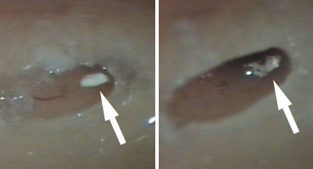
description.” As did Barany they noted a torsional nystagmus with the upper pole of the eye
beating (fast phase) toward the ground and that it “fatigued” on retesting. Additionally, they
observed a response latency of approximately 5 seconds, a crescendo and decline of
nystagmus, and a reversal of the nystagmus as the patient sits up. To eliminate the possibility
that the response could be induced by vascular occlusion from rotation of the neck they tested
patients on an apparatus which avoided it. The same response occurred.
In Britain Hallpike was a pioneer of temporal bone histology. The right temporal bone of 40-
year-old woman with “positional nystagmus of the benign positional type. . .to the right with
the right ear undermost” was examined. In the macula of the utricle, the otolithic membrane
was absent. They concluded: “The general picture is one of chronic tissue changes resulting
either frominfection or trauma. . .” and “We are thus directed to the conclusion that the
lesion is a peripheral one and in the labyrinth towards which, when undermost, the nystagmus
is directed”. Hallpike provided further evidence for a peripheral cause by abolishing
symptoms in two patients with a chemical labyrinthectomy of an acoustically dead ear [5] and
in one patient by an eight nerve section [6]. Both Barany and Dix and Hallpike concluded that
“positional nystagmus of the benign positional type” was caused by disorder of the utricular
macula.
By the early 19th century the bony and some membranous structure of the inner ear were
anatomically well described but their functions unproven. Common notions were the
following. The cochlea was responsible for mediating the nature and pitch of sound; the
saccule and utricle were for perception of loudness, and the semicircular canals for
transmission of bone-conducted sound and perception ofsound direction [7]. Marie-Jean
Flourens (1794–1867) was a professor of comparative anatomy in Paris, and in 1824 he
published his experimental results on pigeon semicircular canals [8]: “If the membranous
ducts are injured, a painful sensitivity to tones is observed, accompanied by abrupt and
violent movements of the head. . .. If the horizontal canals are severed, the animal turns on its
vertical axis; if the posterior vertical canal is severed the animal rolls over backward, and if
the anterior vertical canal is severed the animal falls forward. . ..” Flourens concluded that
the semicircular canals inhibited motion (“forces moderatrices”) and influenced direction of
motion, rather than having a role in balance. Flourens’ work had been largely ignored but was
known to Prosper Meniere and acknowledged in his final paper in 1861 [9]. According to
Adam Politzer (1835–1920) in his “History of Otology” [10] “the realization that the
vestibular and semicircular canal structures are not organs of sound perception, that sound

                                                     4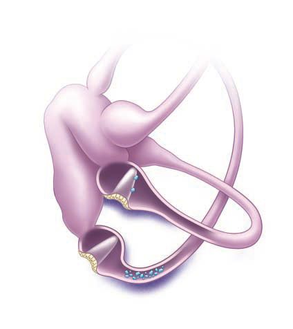
perception is transmitted solely through the cochlea, is the single most important result of
Flourens’ experiments”.
However it was another sixty years until a more sophisticated understanding of semicircular
canal functions and their generated nystagmus was achieved by Julius Ewald (1855–1921)
who was later Professor of Physiology at the University of Strassburg (now Strasbourg). In
pigeons, he cannulated each semicircular canal and applied negative and positive pressures
and observed the directions and intensity of the induced nystagmus [11]. The two major
findings have become known as Ewalds’ Laws: (1) the direction of the induced nystagmus is
in the plane of the canal being stimulated, and (2) in the horizontal canal an ampullopetal
(towards the vestibule) movement of endolymph causes the greatest response where as in the
posterior and superior canals an ampullofugal (away from the vestibule) endolymph
movement causes the greatest response. At the time the differences were perplexing, as
expressed by a writer in 1920 [12]: “It is, however, difficult to imagine how the same
endolymph current can be stimulating for the one endorgan and hindering for the other”.
Thirty years later the advent of the electron microscope allowed a more detailed view of inner
ear ultrastructure.
In 1954 Wersall [13] showed that each vestibular sensory cell has one kinocilium and many
stereocilia. The finding of morphological polarization of kinocilia on vestibular sensor cells
[14, 15] explained Ewald’s paradox. In horizontal canal cristae the kinocilium is on the
vestibule side of the stereocilia; in the posterior and superior canals the kinocilium is on the
canal side of the stereocilia.
In the 1960s, experiments in cats [16] clarified the relationship between canal receptors and
extraocular muscles. Each receptor is connected to one ipsilateral and one contralateral
muscle. The second order neurones are either excitatory (to the agonist muscles) or inhibitory
(to the antagonist muscles).
In 1962 Harold Schuknecht (1917–1996) at Harvard University in Boston [17] proposed that
BPPV “might be caused by detached utricular otoconia, acting upon the cupula of the
posterior semicircular canal. Although at that time there were no confirming human
pathological studies, the concept seemed plausible from a purely theoretical point of view.”
In 1969 Schuknecht [18, 19] confirmed finding basophilic staining masses attached to the
posterior canal cupula in patients who had had BPPV symptoms. He called this cupulolithiasis
(heavy cupula) and assumed the masses were detached utricular otoliths which were removed
by decalcification in preparation. This was supported by Gacek’s report of five patients where
the selective resection of the posterior ampullary nerve abolished BPPV symptoms [20].

                                                     5
Cupulothiasis became the dominant theory for nearly thirty years, although it did not explain
the variable and often long latency and fatiguability of the nystagmus. It was the impetus for
two early specific treatments. Previously “treatment” had been by Cawthorne’s exercises in
which the patient was instructed to repeat continually any movement which caused the vertigo
until it ceased, on the assumption that central adaption was occurring [21].
Based on the cupulolithiasis theory Brandt and Daroff [22] devised an inpatient treatment
where subjects lay down to the provocative side, sat up for thirty seconds, and then lay to the
other side every three hours. After seven to ten days, 61 of 67 subjects were free of
symptoms. The assumed aim was detachment of the particle from the posterior canal cupula.
In France Semont (a physiotherapist) and Sterkers [23, 24] modified this to a logical
physician-controlled treatment they called the Liberatory maneuver, now known as the
Semont maneuver. The patient is lain down to the side of the symptomatic ear, facing down.
When the nystagmus ceases, the patient is moved rapidly through 90 degrees to the opposite
side (where the symptomatic ear becomes uppermost). Either immediately or up to 15 seconds
later the patient experiences vertigo and has nystagmus identical to the symptomatic side. The
technique was little known outside France.
In attempting to explain the latency and fatiguability of BPPV nystagmus, Hall et al. [25] (at
the University of London, Ontario) and later Epley [26] (a solo private practice otologist in
Portland, Oregon) made models of the semicircular canals and proposed that they were better
explained by free-floating particles in the posterior canal, which Epley called canalithiasis.
Also at the University of London, Ontario, Parnes and McClure, in attempting a surgical
posterior canal occlusion, observed and photographed free otoconia in the endolymphatic
compartment [27]. Based on his models Epley proposed a controlled set of head movements
he called the canalith repositioning procedure (CRP) [28]. Epley had presented this as an
instruction course at the American Academy of Otolaryngology, Head and Neck Surgery
meetings since 1980 and endured considerable derision because he used a heavy massage
vibrator over the mastoid process [29].
After seeing canaliths at operation, Parnes [30] described an almost identical particle
repositioning maneuver (PRM) (often known as the Modified Epley maneuver) whose main
difference is its slower pace.
BPPV (85% posterior canal) is now recognized as the most common cause of vertigo in
adults. It is estimated that 2.4% of people experience at least episode in their life [31]. 9% of
residents in a home for the elderly were found to have BBPV [32]. The onset is most
commonly between the fifth and seventh decades. It is the most common cause of vertigo
6after a head injury [33, 34]. An episode of vestibular neuritis [35] and a period of bed rest [36]
are common antecedents. Omission of a simple clinical test can result in patients undergoing
unnecessary, expensive investigations [37]. Previously “nontypical” forms of positionally
induced nystagmus were assumed to always have a central cause. While performing CRPS,
Epley observed a sudden change of “typical” torsional posterior canal nystagmus to horizontal
direction-changing nystagmus and deduced the nystagmus even the superior canal [38].
Without clinical proof Epley predicted the logical treatment for horizontal canal BPPV would
be a 360 degree horizontal plane rotation away from the symptomatic ear.
In 1985 McClure [39] had published the electronystagmographic (ENG) traces of seven
subjects who had intense positional vertigo and direction-changing horizontal nystagmus
when supine. The fast phase was towards the undermost ear (geotropic). McClure suspected a
“viscous plug” in the horizontal canal which was causing a piston effect on the horizontal
canal receptor. As discovered by Ewald, an ampullopetal (towards the vestibule) cupula
deflection is known to cause the most intense nystagmus and vertigo. Horizontal canal BPPV
was then reported by others [40–43] and its particularly intense vertigo confirmed. Early
repositioning attempts failed [41]. A 270 degree “barbecue” rotation was trialled [44].
These simple horizontal repositioning techniques remain the usual way of treating the
horizontal variant of BPPV. Occasionally the most intense nystagmus is away (apogeotropic)
from the undermost ear, implying a particle or particles attached to the cupula, or close to it,
on its canal or utricular side [43, 45–47]. The cupula becomes “heavy” and is ampullofugal
when the symptomatic ear is undermost and ampullopetal when it is uppermost. It can be
difficult to ascertain which is the symptomatic ear, but it is likely to be the undermost ear
which initiates the least nystagmus . Horizontal canal BPPV comprises approximately 15% in
most series. As for posterior canal it can occur de novo, after mild head injury or by “canal
conversion” during posterior canal repositioning [45, 46]. It is likely that patients with
horizontal canal BPPV inadvertently treat themselves by rolling over in their sleep, if it is in
the desirable direction. It they turn in the “wrong” direction they trigger and awake with
vertigo.
Although Brandt et al. [48] in 1994 had alluded to “the rare anterior [superior] canal BPPV,
the spontaneous symptoms occur when the affected ear is uppermost”, the first detailed
description of superior canal BPPV is usually attributed to Herdman and Tusa [49] who
documented two patients whose positionally induced nystagmus was accompanied by
downbeat and torsional nystagmus likely to be caused by a superior canal receptor and which
ceased after repositioning treatment, implying it was rare form of BPPV. Subsequently
7superior canal BPPV was recognized and reported by others [50–56] in whose series it
accounts for approximately 1% of all BPPV diagnoses. In a review [52] of 50 consecutive
patients with positionally induced nystagmus, 75% had a central cause: multiple system
atrophy, cerebellar degeneration, and other miscellaneous causes with immediate onset of
downbeat nystagmus on a Dix Hallpike test. In 25% (“idiopathic”) a Dix Hallpike test or a
head-hanging test elicited downbeat nystagmus with a short latency. In half the subjects a
torsional nystagmus could be seen through Frenzel glasses, but in one it was only discernible
by video imaging. Aw et al. [54] studied forty-four patients whose BPPV had not responded
to conventional repositioning, using 3-dimensional research coils and a 2-axis wholebody
rotator. Seven had downbeat nystagmus with a small torsional component, and all responded
to a “head-overheels” forward rotation in the plane of the superior canal. Differences in the
ampullary segments of the posterior and superior canals most likely explain why superior
canal BPPV downbeat nystagmus can be triggered by a Dix Hallpike test to either side and for
its small (or absent) torsional component. In most cases the symptomatic ear is the uppermost
ear .
82. Anatomy and physiology
2.1 Peripheral Vestibular Anatomy
Within the petrous portion of each temporal bone lies the membranous vestibular labyrinth
(Figure 1). Each labyrinth contains 5 neural structures that detect head acceleration: 3
semicircular canals and 2 otolith organs (Figure 2).
Fig. 1: Spatial orientation of the semicircular canals. Note how the posterior canal on 1 side is in the same plane as the
contralateral superior canal. Both lateral canals are in the same plane, 30º above the horizontal.
The 3 semicircular canals (SCC) (lateral, posterior, and anterior) respond to angular
acceleration and are orthogonal with respect to each other. Alignment of the SCCs in the
temporal bone is such that each canal has a contralateral coplanar mate. The lateral canals
form a coplanar pair, whereas the posterior and contralateral anterior SCC form coplanar
pairs. The anterior aspect of the lateral SCC is inclined 30 degrees upward from a plane
9connecting the external auditory canal to the lateral canthus. The posterior and anterior SCCs
are inclined about 92 and 90 degrees from the plane of the lateral SCC . Because the SCCs are
not precisely orthogonal with earth vertical or earth horizontal, angular rotation of the head
stimulates each canal to varying degrees.
Fig. 2: Anatomy of the vestibular labyrinth. Structures include the utricle (Utr.), sacculus, anterior (or superior)
semicircular canal (Sup.), posterior semicircular canal (Post.), and the lateral semicircular canal (Lat.). Note the superior
vestibular nerve innervating the anterior and lateral semicircular canals as well as the utricle. The inferior vestibular nerve
innervates the posterior semicircular canal and the saccule. The cell bodies of the vestibular nerves are located in Scarpa’s
ganglion (Gangl. Scarpae).
The SCCs are filled with endolymph that has a density slightly greater than that of water.
Endolymph contains a high concentration of potassium, with a lower concentration of sodium,
and moves freely within each canal in response to the direction of the angular head rotation.
The SCCs enlarge at one end to form the ampulla. Within the ampulla lies the cupula, a
gelatinous barrier that houses the sensory hair cells (Figure 3A). The kinocilia and stereocilia
of the hair cells are seated in the crista ampullaris (Figure 3B). Deflection of the stereocilia
caused by motion of the endolymph results in an opening (or closing) of the transduction
channels of hair cells, which changes the membrane potential of the hair cells. Deflection of
the stereocilia toward the single kinocilia in each hair cell leads to excitation (depolarization),
and deflection of the stereocilia away from the kinocilia leads to inhibition
(hyperpolarization).
10Fig. 3 A: The semicircular canals enlarge at one end to form the
ampulla. The cupula of the ampulla is a flexible barrier that
partitions the canal. The crista ampullaris contains the sensory
hair cells. The hair cells generate action potentials in response to
cupular deflection.
Fig. 3 B: Cross-section of crista ampullaris showing kinocilia and
stereocilia of hair cells projecting into the cupula. Deflection of
the stereocilia towards the kinocilia causes excitation; deflection
in the opposite direction causes inhibition.
Fig. 4: Otoconia are embedded in a gelatinous matrix and
provide an inertial mass. Linear acceleration shifts the gelatinous
matrix and excites or inhibits the vestibular afferents depending
on the direction in which the stereocilia are deflected.
Hair cells are oriented in the lateral SCC so that endolymph motion toward the ampulla causes
excitation. In contrast, hair cells of the vertical SCCs (posterior and anterior) are oriented so
that depolarization occurs when endolymph moves away from the ampulla. Each of the SCCs
responds best to motion in its own plane, with coplanar pairs exhibiting a push-pull dynamic.
For example, as the head is turned to the right, the hair cells in the right lateral SCC are
excited, whereas the hair cells in the left lateral SCC are inhibited.
The brain detects the direction of head movement by comparing input from the coplanar
labyrinthine mates.
The saccule and utricle make up the otolith organs of the membranous labyrinth. Sensory
hair cells project into a gelatinous material that has calcium carbonate crystals (otoconia)
embedded in it, which provide the otolith organs with an inertial mass (Figure 4). The utricle
and the saccule have central regions known as the striola, dividing the otolith organs into 2
parts. The kinocilia of the utricular hair cells are oriented toward their striola, whereas the
11kinocilia of the saccular haircells are oriented away from their striola. Motion toward the
kinocilia causes excitation. Utricular excitation occurs during horizontal linear acceleration
or static head tilt, and saccular excitation occurs during vertical linear acceleration.
2.2 Vestibular Afferent Physiology
In primates, primary vestibular afferents of the healthy vestibular system have a resting firing
rate that is typically 70 to 100 spikes per second. The discharge regularity (determined by the
spacing of the interspike intervals between action potentials of vestibular nerve afferents
provides a useful marker for the information carried by these afferents. The coefficient of
variation (standard deviation/mean discharge) of the interspike interval provides a useful
measurement for classifying afferents into irregularly and regularly discharging groups. The
information carried by irregular and regular afferents varies over the spectral range of
frequency and acceleration that encompasses natural head movements. Generally, irregular
afferents are more sensitive to rotations during large head accelerations than regular afferents
are. The increased sensitivity of the irregular afferents may be more critical for the rapid
detection of head movements as well as initiation of the VOR. The regular afferents, in
contrast, provide a signal that is proportional to head velocity over a wide spectral range. In
addition, the regular afferents may be the primary source of input to the VOR for steadystate
responses to sinusoidal rotations because temporarily silencing the irregular afferents has no
affect on the VOR during low-frequency and small head accelerations.
Fig. 5: Schematic drawing of the physiology of the left posterior semicircular canal.
In the image on the right, note the excitatory response (increased neural firing) with utriculofugal cupular displacement.
The same excitatory response would occur in the superior (anterior) canal with utriculofugal cupular displacement,
whereas the opposite (inhibitory) response would occur with utriculofugal cupular displacement in the lateral canal. The
same rules would apply to the image on the left. CNVIII = vestibular nerve, ms = millisecond.
12The cells bodies of vestibular nerve afferents are located in the superior or inferior divisions
of Scarpa’s ganglia, which lie within the internal auditory canal near the emergence of the
vestibular nerve into the cerebellopontine angle. From the vestibular labyrinth, the afferent
information travels ipsilateral in 1 of 2 branches of the vestibular nerve. The superior
vestibular nerve innervates the lateral and anterior SCC as well as the utricle. The inferior
vestibular nerve innervates the posterior SCC and the saccule. It is estimated that between
15,000 to around 25,000 vestibular nerve fibers exist in humans. Variation of nerve fiber
counts among studies appears to be a function of age, although rate of decline of the number
of afferent fibers also appears to be variable. The branches of the vestibular nerve travel
together into the pontomedullary junction where they bifurcate. Primary vestibular afferents
in the superior division of the vestibular nerve include axons that synapse in the superior and
medial vestibular nuclei or the uvula, nodulus, flocculus, or fastigial nucleus of the
cerebellum. Primary vestibular afferents from the inferior branch synapse with neurons in
either the medial, lateral, or inferior vestibular nuclei, which, along with the superior
vestibular nuclei and other subnuclei, comprise the vestibular nuclear complex.
2.3 Central Vestibular Anatomy
Secondary vestibular afferents have been identified as relaying signals from the vestibular
nuclei to the extraocular motor nuclei, the spinal cord, or the flocculus of the cerebellum.
Central vestibular neurons differ in terms of the inputs they receive from regular and irregular
afferents. Those central vestibular neurons that project to the extraocular motor nuclei receive
a majority of their monosynaptic inputs from regular afferents, whereas those that project to
the spinal cord receive a majority of their inputs from irregular afferents. Those central
vestibular neurons projecting to the flocculus of the cerebellum receive relatively equal
contributions from regular and irregular afferents.
Many vestibular reflexes are controlled by processes that exist primarily within the brain
stem. Tracing techniques, however, have identified extensive connections between the
vestibular nuclei and the reticular formation, thalamus, and cerebellum. Vestibular pathways
appear to terminate in a unique cortical area. In studies of primates, fibers terminating in the
junction of the parietal and insular lobes have been identified and considered the location for a
vestibular cortex.
13Recent evidence in studies of humans using functional magnetic resonance imaging appears
to confirm the parietal and insular regions as the cortical location for processing vestibular
information [57]. Connections with the vestibular cortex, thalamus, and reticular formation
enable the vestibular system to contribute to the integration of arousal and conscious
awareness of the body and to discriminate between movement of self and the environment.
The cerebellar connections help maintain calibration of the VOR, contribute to posture during
static and dynamic activities, and influence the coordination of limb movements.
2.4 Vestibulo-ocular Physiology
The ability of the VOR to elicit rapid compensatory eye movements that maintain stability of
images on the fovea depends on relatively simple patterns of connectivity in the central
vestibular pathways. In its most basic form, the pathways controlling the VOR can be
described as a 3-neuron arc. In the case of the lateral SCC, primary vestibular afferents from
the lateral SCC synapse in the ipsilateral medial and ventrolateral vestibular nuclei. Some of
the secondary vestibular neurons receiving innervation from the ipsilateral labyrinth have
axons that decussate and synapse in the contralateral abducens nucleus, whereas others ascend
ipsilaterally to the oculomotor nucleus. Motoneurons from the abducens nucleus and the
medial rectus subdivision of the oculomotor nucleus then synapse at the neuromuscular
junction of the lateral rectus and medial rectus muscles, respectively. Similar patterns of
connectivity exist for the anterior and posterior SCC and the eye muscles that receive
innervations from them (Table 1).
Tab. 1: Innervation Pattern of Excitatory Input From the Semicircular Canals
Primary Afferent Secondary Neuron Extraocular Motoneuron Muscle
Right oculomotor nucleus Right medial rectus
Lateral (right) Medial vestibular nucleus
Left abducens nucleus Left lateral rectus
Left inferior oblique
Anterior (right) Lateral vestibular nucleus Left oculomotor nucleus
Right superior rectus
Left trochlear nucleus Left Right superior oblique
Posterior (right) Medial vestibular nucleus
oculomotor nucleus Left inferior rectus
14Fig. 6:Muscle insertions of the left eye.
The 6 extraocular muscles insert into the sclera and can be
considered as complementary pairs. The medial and lateral
rectus muscles rotate the eyes horizontally, the superior and
inferior rectus muscles principally rotate the eyes vertically,
and the superior and inferior oblique muscles rotate the eyes
torsionally with some vertical component. By convention,
the torsional rotation is noted as it relates to the superior
poles of the eyes. The superior oblique muscle rotates the
eye downward and toward the nose [intorsion], whereas the
inferior oblique muscle rotates the eye upward and away
from the nose [extorsion]. The superior oblique muscle
travels through the fibrous trochlea, which attaches to the
anteromedial superior wall of the orbit.
The VOR has been tested across multiple frequencies and velocities and shows velocity-
dependent nonlinearities, which may correlate with unique afferent physiology. The gain of
the VOR remains constant (linear) across multiple frequencies of sinusoidal rotations, with
peak velocities of3. Clinical features
3.1 Epidemiology
BPPV is the most common disorder of the peripheral vestibular system. Mizukoshi and
colleagues estimated the incidence to be 10.7 to 17.3 per 100 000 per year in Japan, although
this is likely to be an underestimate because most cases of BPPV resolve spontaneously
within months [60].
Several studies have suggested a higher incidence in women, but in younger patients and
those with posttraumatic BPPV the incidence may be equal between men and women. The
age of onset is most commonly between the fifth and seventh decades of life.
BPPV is more likely to involve the right ear, a factor that may be related to the habit of
sleeping on the right side in the general population [61].
BPPV most often involves a single semicircular canal, usually posterior (60-90%), but may
involve both posterior and lateral canals in the same inner ear. Posterior canal BPPV may
convert to lateral canal BPPV following repositioning manoeuvres. Head trauma is the most
common cause of simultaneous bilateral posterior canal BPPV.
Tab. 2
Incidence 95,8/100000/year
Prevalence 131,6/100000/year
Man 37,2
W/M 1,7/1
Women 62,8
Age Range 6-89 years Average 57 years
Unilateral 72%
Posterior canal BPPV 79,4%
Bilateral 7,4%
Geotropic 80%
Lateral canal BPPV 16,9%
Apogeotorpic 20%
Atypic form 3,7%
Caruso et all 1995
163.2 Symptoms of BPPV
Patients describe sudden, severe attacks of either horizontal or vertical vertigo, or a
combination of both, precipitated by certain head positions and movements. Patients typically
develop vertigo when getting out of bed, rolling over in bed, tilting their head back, for
example to look up shelves, or bending forward, for example when fastening their shoes.
Patients can often identify the affected ear by stating the direction of movement that
precipitates the majority of the attacks (e.g., when rolling over in bed to the right, but not the
left, precipitates dizziness, this indicates right ear involvement).
The attacks of vertigo typically last fewer than 30 seconds, however, some patients
overestimate the duration by several minutes. Reasons for this discrepancy may include the
fear associated with the intense vertigo along with the nausea and disequilibrium that may
follow the attack. The vertigo attacks occur in spells; patients have several attacks a week
(23%) or during the course of 1 day (52%). In addition to vertigo, many patients complain of
lightheadedness, nausea, imbalance and, in severe cases, sensitivity to all directions of head
movement.
Many patients also become extremely anxious for 2 main reasons. Some fear that the
symptoms may represent some kind of sinister underlying disorder such as a brain tumour.
For others, the symptoms can be so unsettling that they go to great lengths to avoid the
particular movements that bring on the vertigo. For this reason, some may not even realize
that the condition has resolved, as it so often does over time without any treatment at all.
BPPV can be described as selflimited, recurrent or chronic. As the name implies, BPPV is
most often a benign condition, however, in certain situations it may become dangerous. For
example, a painter looking up from the top of a ladder may suddenly become vertiginous and
lose his or her balance, risking a bad fall. The same would hold true for underwater divers
who might get very disoriented from acute vertigo. Heavy machinery operators should use
great caution especially if their job involves significant head movement. Most people can
safely drive their car as long as they are careful not to tip their head back when checking their
blind spot.
3.3 Causes
The cause of BPPV is mostly unknown (idiopathic). In view of the high prevalence of BPPV
in middle-aged women, hormonal factors may play a role in the development of BPPV. In a
recent study, bone mineral density score was decreased in both women and men with
17idiopathic BPPV compared with that in normal controls without a history of dizziness [59].
The prevalence rates of osteopenia and osteoporosis were also found to be higher in both
women and men with BPPV than in normal controls. Furthermore, in women aged ≥45 years,
the lowest T-scores were also decreased in the recurrent group, compared with those in the de
novo group. These findings suggest the involvement of deranged calcium metabolism in
idiopathic BPPV and a significant association between osteopenia/osteoporosis and idiopathic
BPPV. Otoconia are deposits of calcium carbonate in the form of composite calcite crystals,
and bone contains 99% of the calcium found in the body. Decreased estrogen levels may
disturb the internal structure of the otoconia or their interconnections and attachments to the
gelatinous matrix. Alternatively, an increase in the concentration of free calcium in the
endolymph due to increased calcium resorption may reduce the capacity to dissolve the
dislodged otoconia.
BPPV may develop secondary to various disorders that damage the inner ear and detach the
otolith from the utricular macule. Head trauma causing mechanical damage to the ear is the
most common cause of BPPV. Patients rarely develop BPPV after mastoid surgery or if they
engage in a persistent head-tilt position, such as among barbers or dentists. Compared with
the idiopathic form, traumatic BPPV exhibits several distinctive characteristics, including a
higher incidence of bilaterality, involvement of multiple canals on the same side, equal
occurrence among women and men, a younger and more even age distribution, more difficult
to treat, and frequent recurrences.
In addition, BPPV may develop secondary to any of the inner ear diseases (e.g., vestibular
neuritis, Meniere’s disease) that give rise to degeneration and detachment of the otoconia, but
do not totally impair semicircular canal function [63]. BPPV appears to be more frequent
(9.8%) in vestibular neuritis patients than in the general population, consistently affecting the
posterior canal of the same ear. BPPV occurrence after vestibular neuritis predominantly
affects patients who did not fully recover from the disease. BPPV after vestibular neuritis
appears to be more difficult to treat than idiopathic BPPV.
The incidence of BPPV is also known to be higher in patients who suffer from migraine, even
though the exact mechanism remains to be elucidated [63, 64] BPPV has been reported to
occur in association with giant-cell arteritis, diabetes, and hyperuricemia [ 65-68].
183.4 Pathomechanism
BPPV can theoretically affect each of the 3 semicircular canals, although superior canal
involvement is exceedingly rare.
The detached otolith debris could be either attached to the cupula (cupulolithiasis) or may be
free-floating in the semicircular canals (canalolithiasis) (Figure 7). Pathological studies have
shown that both of these conditions exist (Figure 8).
The otolithic debris deflects the cupula and gives rise to a spinning sensation via a direct
gravitational effect on the cupula or by inducing endolymph flow during head motion in the
direction of gravity. According to the cupulolithiasis theory, a cupular deposit (heavy cupula)
would induce a gravitational effect on the crista. However, the action of free-floating debris is
the currently accepted pathophysiologic mechanism of typical BPPV. According to the
canalolithiasis theory, the free-floating particles move under the influence of gravity when
changing the position of the canal in the earth-vertical plane. The hydrodynamic drag of the
particles induces endolymphatic flow, resulting in cupular displacement and leading to the
observed typical responses.
Fig. 8: Sequential computer-regenerated photographs taken from
an intra-operative video of a fenestrated posterior semicircular
canal. Note the single white conglomerate mass within the
membranous duct (arrow) (left). Note how the mass has
fragmented into tiny particles 2–3 minutes later, after the
membranous duct has been probed (right).
Fig. 7: Left inner ear.
Depiction of canalithiasis of the posterior canal and
cupulolithiasis of the lateral canal.
194. Diagnosis and Treatments
Each type of BPPV is diagnosed by observing the patterns of nystagmus induced during
positioning maneuvers that have been designed to move only the involved canal in the
direction of maximal gravity. However, accurate observations of the nystagmus require the
fixation to be removed during the maneuvers.
BPPV is usually a self-remitting disorder and may resolve as time goes on without specific
treatment. According to a report on the natural course of untreated BPPV, most HC-BPPVs
resolve within 16±19 days and PC-BPPVs within 39±47 days of their onset.26 However, a
correct diagnosis and proper repositioning maneuvers may allow a rapid and simple cure for
the BPPV [69].
4.1 Posterior Canal BPPV (PC-BPPV)
4.1.1 Diagnosis
In PC-BPPV, the positioning nystagmus is typically induced by Dix-Hallpike maneuvers in
the direction of the involved canal (Figure 9). During the Dix-Hallpike maneuver, it is thought
that the free-floating otolithic debris (canalolithiasis) in the posterior canal moves away from
the cupula and stimulates the posterior canal by inducing ampullofugal flow of the endolymph
(Ewald’s first law). Excitation of the posterior canal in turn activates the ipsilateral superior
oblique and contralateral inferior rectus muscles, which results in tonic downward deviation
of the eyes with a torsion in the direction of the uppermost ear. Accordingly, the resultant
nystagmus would be upbeating and torsional, with the upper pole of the eyes beating toward
the lowermost ear.
Figure 9: Posterior canal BPPV in a left ear showing Dix Hallpike test, inner ear, and receptor connections to the
extraocular muscles.
20The nystagmus usually develops with a brief latency of several seconds, resolves within 1
minute (usually within 30 seconds), and its direction is reversed on sitting. The nystagmus
diminishes (i.e., it fatigues) with repeated examinations. The Dix-Hallpike maneuver has been
considered the gold standard for diagnosing PC-BPPV. However, this maneuver should be
performed with caution in patients with a history of neck surgery, cervical radiculopathy, and
vascular dissection syndrome, since it requires rotation and extension of the neck during the
positioning.
4.1.2 Treatments
The most popular methods for treating PC-BPPV are Semont’s liberatory and Epley’s
maneuvers. These maneuvers employ stepwise changes in head position (Fig. 6) to flush
freefloating otolithic debris out of the semicircular canals and back into the utricle.
Liberatory manoeuvre
In 1988, Semont and colleagues37 described the “liberatory manoeuvre” (Fig. 10) based on
the cupulolithiasis theory. It was believed that this series of rapid changes of head position
freed deposits that were attached to the cupula. The manoeuvre begins with the patient in the
sitting position and the head turned away from the affected side. The patient is then quickly
put into a position lying on his or her side, toward the affected side, with his or her head
turned upward. After about 5 minutes, the patient is quickly moved back through the sitting
position to the opposite position lying on his or her side with his or her head now facing
downward. The patient remains in this second position for 5–10 minutes before slowly being
brought back to the sitting position. In their series of 711 patients, Semont and colleagues37
found an 84% response rate after 1 procedure and a 93% response rate after a second
procedure 1 week later [70]. Several other case series have had response rates of 52%–90%
[71-73] with recurrence rates of up to 29%. There has been no difference in efficacy shown
between the liberatory manoeuvre and particle repositioning manoeuvre, which is described in
the following section, in randomized studies by Herdman and colleagues[72] and Cohen and
Jerabek [74]. The liberatory manoeuvre is effective, but is cumbersome with elderly and
obese patients, and shows no increased efficacy compared with the simple particle
repositioning manoeuvre.
21Fig. 10: Liberatory manoeuvre of Semont (right ear). The top panel shows the effect of the manoeuvre on the labyrinth as
viewed from the front and the induced movement of the canaliths (from blue to black). This manoeuvre relies on inertia, so
that the transition from position 2 to 3 must be made very quickly.
Particle repositioning manoeuvre
Although he had been teaching his technique for many years, it was not until 1992 that Epley
published his first report on the “canalith repositioning procedure” (CRP) [75]. This highly
successful “Epley manoeuvre” is performed with the patient sedated. Mechanical skull
vibration is routinely used and the patient’s head is moved sequentially through 5 separate
positions. Epley postulated that the procedure enabled the otolithic debris to move under the
influence of gravity from the posterior semicircular canal into the utricle.
22Most clinicians today are thought to use a modified version of the CRP. One modified CRP is
the particle repositioning manoeuvre (PRM) which is a 3-position manoeuvre that eliminates
the need for sedation and mastoid vibration [76-77] (Fig. 11).
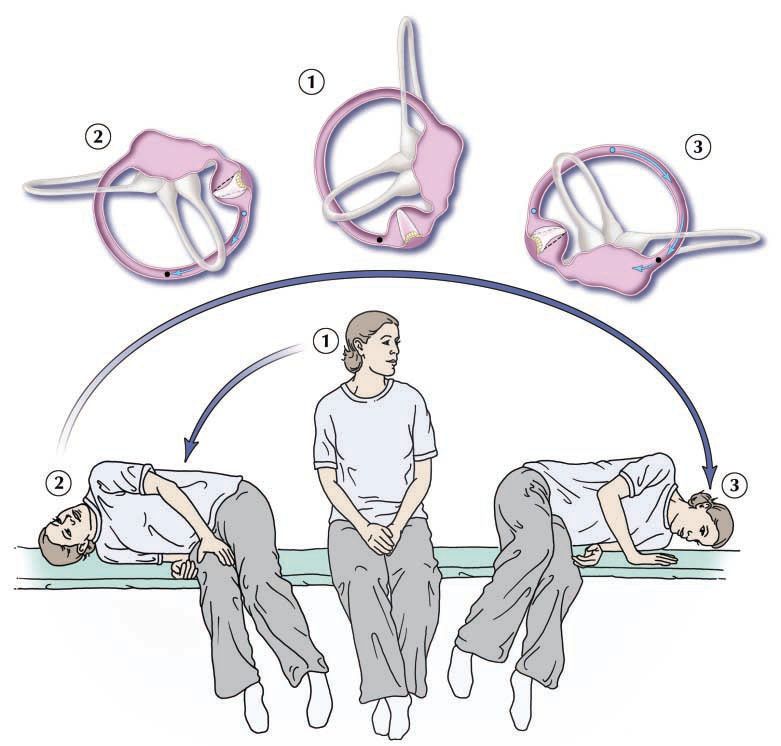
Fig. 11: Particle repositioning manoeuvre (right ear).
Schema of patient and concurrent movement of posterior/superior semicircular canals and utricle. The patient is seated on a
table as viewed from the right side (A). The remaining parts show the sequential head and body positions of a patient lying
down as viewed from the top. Before moving the patient into position B, turn the head 45° to the side being treated (in this
case it would be the right side). Patient in normal Dix–Hallpike head-hanging position (B). Particles gravitate in an
ampullofugal direction and induce utriculofugal cupular displacement and subsequent counter-clockwise rotatory nystagmus.
This position is maintained for 1–2 minutes. The patient’s head is then rotated toward the opposite side with the neck in full
extension through position C and into position D in a steady motion by rolling the patient onto the opposite lateral side. The
change from position B to D should take no longer than 3–5 seconds. Particles continue gravitating in an ampullofugal
direction through the common crus into the utricle. The patient’s eyes are immediately observed for nystagmus. Position D is
maintained for another 1–2 minutes, and then the patient sits back up to position A. Overall, the PRM should take less than 5
minutes to complete. Patients are then typically asked to remain upright for the next 24–48 hours in order to allow the
otoliths to settle, so as to prevent a recurrence of the BPPV.
With proper understanding of inner ear anatomy and the pathophysiology of BPPV, various
appropriately trained health professionals, including family doctors and physiotherapists,
should be able to successfully carry out the PRM in most straightforward cases. Atypical
cases or cases that do not respond to this manoeuvre should be referred to a tertiary care
dizziness clinic.
The results of Epley’s maneuver can be predicted even during the maneuver. When the head
is turned 90º toward the unaffected side after the Dix-Hallpike maneuver, the positioning
23nystagmus develops in the same direction as the maneuver (orthotropic nystagmus) if a clump
of particulate matter moves in the correct direction into the common crus, resulting in a
successful repositioning. However, the direction of the nystagmus would reverse if a heavy
cupula with attached otolithic debris deflects ampullopetally or if the particles move back
toward the cupula, which implies that the repositioning will be unsuccessful [78].
It is difficult to compare studies that use the repositioning manoeuvres, because they vary
considerably in the length of follow-up, number of treatment sessions, number of manoeuvres
per session, the use of sedation and the use of mastoid vibration. The overall response rates
range from 30% to 100%. Most of these studies are case series, but Lynn and colleagues [79]
and Steenerson and Cronin [80] provide good evidence from randomized studies.
Epley’s maneuver is the only recommended method of treating PC-BPPV, with confirmed
evidence level A according to the American Academy of Neurology [81].
4.2 Horizontal (Lateral) Canal BPPV (HC-BPPV)
4.2.1 Diagnosis
HC-BPPV is diagnosed by the supine roll test (the Pagnini-McClure manoeuvre), in which
the head is turned by about 90º to each side while supine. During this maneuver,
horizontalnystagmus may beat toward the ground (geotropic nystagmus) (Figure 12) or
toward the ceiling (apogeotropic nystagmus) (Figure 13). The evoked during positioning in
HC-BPPV usually tends to be more persistent, less fatigability and a shorter latency than in
PC-BPPV.
Determination of the involved side (lateralization) is very important for the proper treatment
of HC-BPPV (Table 3). Since ampullopetal flow of the endolymph evokes a greater response
than ampullofugal flow in the horizontal canal (Ewald’s second law), the induced nystagmus
is stronger when the head is turned toward the affected ear in the geotropic type of HC-BPPV.
In contrast, head turning to the healthy ear generates a stronger nystagmus in apogeotropic
HC-BPPV. Determination of the involved ear is sometimes difficult due to rather symmetrical
responses, especially if the induced nystagmus is not recorded. In these instances, other
findings may provide clues toward determining the affected ear. Subjective sensation can be
helpful: the patient is sometimes able to detect the more uncomfortable side. Caloric test can
show hypoexcitability in the affected ear.
24Figure 12: Horizontal canal BPPV (canalithiasis) in a left ear showing Head Roll test, inner ear, and receptor connections
to the extraocular muscles.
Figure 13: Horizontal canal BPPV (cupulolithiasis) in a left ear showing Head Roll test, inner ear, and receptor
connections to the extraocular muscles.
25Tab. 3. Lateralization of HC-BPPV, based upon the direction and intensity of nystagmus (Ny)
Geotropic nystagmus Apogeotropic nystagmus
Intensity of Ny Stronger side Weaker side
Subjective sensation Affected side Affected side
Pseudospontaneous Ny Healthy side Affected side
Stead Supine Positioning Test Healthy side Affected side
Lying-down Ny Healthy side Affected side
Head-bending Ny Affected side Healthy side
Reversal of initial Ny Possibly occurs Affected side Uncommon
Null point Uncommon, laterality is uncertain Usually present on Affected side
Caloric Hypoexcitability Affected side Affected side
Another sign to diagnose the affected side is the direction of the nystagmus when the patient
is briskly brought from the seated position to supine position (Stead Supine Positioning
Test). When the patient lies supine, having the head flexed 300, the lateral canal is on a
vertical plane; therefore, due to both gravity and the brisk deceleration caused by this
manoeuvre, the otoliths are pushed downwards: when they are in the posterior arm they float
towards the utricle, and when they are near the cupola they float towards the ampulla. The
Stead Supine Positioning Test evokes a nystagmus beating towards the healthy side in the
geotropic forms and towards the affected side in the apogeotropic forms.
In HC-BPPV, nystagmus may be induced by Bow and Lean test: when the patient bows the
head over 900 (bowing nystagmus) and leans the head backward over 450 (leaning nystagmus)
in the sitting position. In up to 80% of HC-BPPV cases, bowing and leaning nystagmus are in
the opposite direction. In geotropic HC-BPPV, bowing nystagmus beats mostly toward the
affected ear (ampullopetal migration of the otoliths), while leaning is directed mostly toward
the healthy ear (ampullofugal displacement of the otoliths). In contrast, bowing nystagmus is
mostly contralesional and leaning nystagmus is usually ipsilesional when observed in
apogeotropic HC- BPPV. Bowing and leaning nystagmus in apogeotropic HC-BPPV are
explained by deflection of the heavy cupula in response to the positional change [82-85].
In apogeotropic HC-BPPV, the induced horizontal nystagmus may disappear when the head is
turned to the affected ear by 10-20º, while supine (null point)[86]. The null point is explained
by alignment of the heavy cupula in the direction of the gravitational vector.
Spontaneous nystagmus, also known as pseudospontaneous nystagmus, is not uncommon in
HC-BPPV. In previous reports, 66-76% of HC-BPPV patients exhibited spontaneous
nystagmus [87]. The spontaneous nystagmus in HC-BPPV may be related to the anatomical
26position of the horizontal semicircular canal, which is inclined 30º backwards from the
horizontal plane. Accordingly, the gravitational force may affect the otolithic debris inside the
canal or the heavy cupula, even when in the upright sitting position. For the same reason,
pseudospontaneous nystagmus disappears when the patient’s head is bent forwards by about
30º. In this position, since the horizontal canal is aligned with respect to the earth horizontal
plane, the effect of gravity is negated. However, pseudospontaneous nystagmus should be
differentiated from continuous nystagmus with sustained vertigo resulting from so-called
canalith jam and negative endolymph pressure between the plug and the cupula [88].
In BPPV, secondary (spontaneous reversal) of the initial positioning nystagmus rarely
occurs without further position changes. In geotropic HC-BPPV, the initial geotropic
nystagmus occasionally reverses spontaneously its direction when the head is turned toward
the lesion side, and the induced nystagmus is intense [89]. Short-term adaptation of the
vestibulo-ocular reflex seems to be the main mechanism underlying this spontaneous reversal
of the initial positioning nystagmus.
4.2.2 Treatments
Geotropic HC-BPPV
Rotations of 270º or 360º around the yaw axis (the so-called barbecue maneuver) toward
the unaffected ear are popular methods for the treatment of geotropic HC-BPPV [90]. These
maneuvers consist of sequential head turning of 90º toward the healthy side while supine
(Figure 14). With these maneuvers, the free-floating otoconial debris migrates in the
ampullofugal direction, finally entering the utricle through the nonampullated end of the
horizontal canal.
Lying with the healthy ear downward for approximately 12 hours (forced prolonged
position) can be employed, especially in patients suffering from severe symptoms who cannot
perform sequential position changes [92, 96].
The Gufoni maneuver is another alternative [93, 94]. After being seated on an examination
couch, the patient lies down on the healthy lateral side with a quick lateral movement and is
maintained in this position for 1-2 minutes until resolution of the evoked nystagmus. A quick
45º rotation of the head toward the floor is then performed, with the patient maintaining this
position for another 2 minutes, followed by a slow return back to the starting position. A
major advantage of the Gufoni maneuver is its simplicity.
27Figure 14: “Barbecue” repositioning for horizontal canal BPPV in a left ear.
Apogeotropic HC-BPV
Apogeotropic HC-BPPV is attributed to either cupulolithiasis or canalolithiasis within the
anterior arm of the horizontal semicircular canal. In apogeotropic HC-BPPV, the therapeutic
goal should be to detach the otolithic debris from the cupula or shift the debris from the
anterior into the posterior arm of the horizontal canal [95].
If the otolithic debris is attached at the utricular side of cupula, its detachment should result in
immediate resolution of the positional vertigo and nystagmus. In the case of adhesion from
the canal side of the cupula or free-floating particles in the anterior arm, detachment and
shifting of the otolithic debris into the posterior arm would give rise to a transition into
geotropic HC-BPPV [96]. Therapeutic head-shaking in the horizontal plane, a modified
Semont maneuver, and the Gufoni method have been proposed as treatment regimens for
apogeotropic HC-BPPV [95].
The aim of head-shaking is to detach the otolithic debris from the cupula, irrespective of the
side to which it is attached, using alternate accelerating and decelerating power.
The modified Semont maneuver comprises the following three steps: 1) the patient is brought
briskly into a side-lying position with the affected ear downward; 2) the patient’s head is
28turned 45º downward, with this position being maintained for 2-3 min; and 3) the patient
resumes the original sitting position. This maneuver was initially designed to dislodge the
debris attached to the utricular side of the cupula.
In the Gufoni maneuver for apogeotropic HC-BPPV, the patient sits with the head directed
straight ahead and then quickly moves into a side-lying position on the affected side,
remaining in this position for 1 or 2 more minutes after the end of apogeotropic nystagmus.
The head is then turned 45º upward very quickly and is kept in this position for 2 minutes,
followed by a slow return to the sitting position. The Gufoni maneuver was designed to
remove the otolithic debris from the anterior arm of the horizontal semicircular canal near the
cupula.
4.3 Anterior Canal BPPV (AC-BPPV)
4.3.1 Diagnosis
BPPV rarely involves the anterior semicircular canal, and AC-BPPV exhibits several
characteristics that contrast with those of PC-BPPV. In AC-BPPV the Dix-Hallpike maneuver
on either side may evoke downbeat nystagmus with an ipsitorsional (upper poles of the eyes
beating toward the involved ear) component (Figure 15). Furthermore, the torsional
nystagmus in AC-BPPV may not be evident, as it is in PC-BPPV.
Figure 15: Superior canal BPPV in a left ear showing Dix Hallpike test, inner ear, and receptor connections to the
extraocular muscles.
4.3.2 Treatments
Various repositioning maneuvers have also been advanced to treat AC-BPPV. In the reverse
Epley maneuver, the patient submits to the same sequence of positional changes after the Dix-
Hallpike maneuver on the side of the healthy ear. Modified repositioning maneuvers and
forced prolonged position have also been adopted in treating this particular BPPV [99, 100].
29Li maneuver [101] where the patient is moved rapidly from a supine (midline) head-hanging
position to a face-down position at the opposite end of the couch (Figure 16).
Figure 16: The Li manoeuvre for superior canal BPV in either ear (left ear).
4.4 Rehabilitation
Irrespective of the involved canals, the Brandt-Daroff exercise may be attempted when the
repositioning maneuvers fail or if patients cannot tolerate the repositioning maneuvers (Figure
17). The exercise may be repeated at liberty until resolution of the symptoms.
With respect to PC-BPPV, vestibular rehabilitation demonstrates superior treatment outcomes
compared with placebo [102]. However, vestibular rehabilitation is less effective than canalith
repositioning procedure in producing complete symptom resolution. There are as yet
insufficient data concerning the response of HCBPPV to vestibular rehabilitation.
Figure 17: Brandt-Daroff exercise. Patients are instructed to rapidly lie on their side, sit up, lie on the opposite side, and
then again sit up. Each position should be maintained for at least 30 seconds. These exercises are repeated serially 5-10
times a day until resolution of the symptoms.
30You can also read