Product Information 2020 - English - NORAS MRI products
←
→
Page content transcription
If your browser does not render page correctly, please read the page content below
Product Information 2020
English
Technical changes reserved

113238

Issue EN
Rev02
October 2019

2
In the picture from left to right : Manuel Noras, Daniel Gareis, Hubert Noras
Dear Valued Customer,
NORAS has been present internationally for over 30 years and has built a solid reputation due to
its customer-oriented service. Besides our products, our slogan „we build your vision“ reflects
exactly what we offer our customers: We are a competent partner with the planning and realiza-
tion of your individual ideas and concepts.
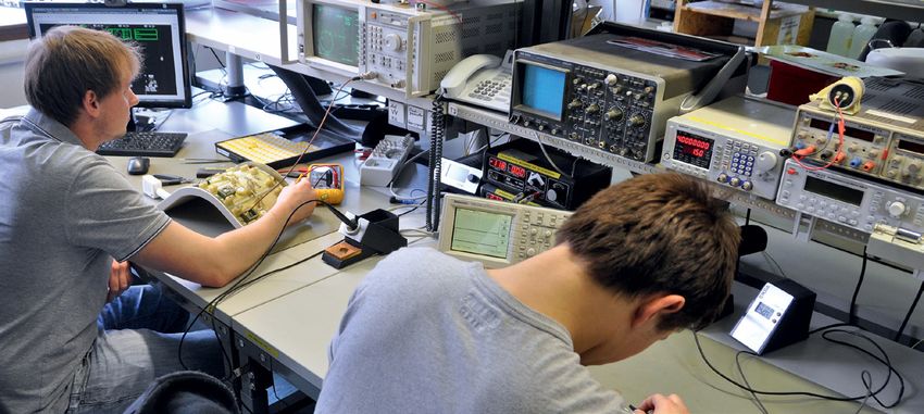
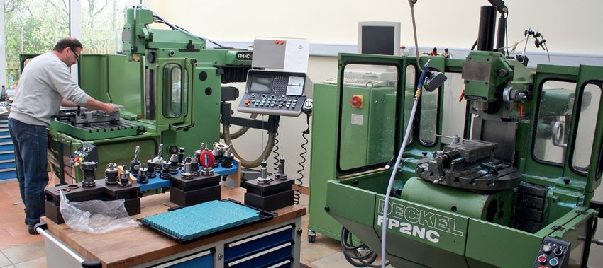
Our development and production is taking place mainly in Germany. The final assembly and final
inspection is done manually by skilled employees, which provides an extra quality advantage.
Within this catalog, you will find a summary of our products and the necessary order information.
You will also learn more about our Research & Development area and customer-specific produc-
tions.
We look forward to working with you in the future and to fulfill your vision.
Best Regards,
Management of NORAS MRI products GmbH
Hubert Noras Manuel Noras Daniel Gareis
3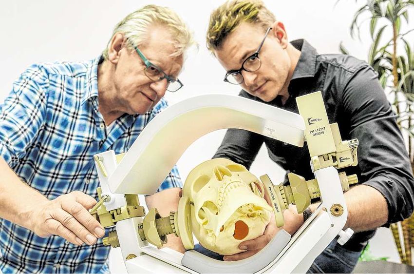
Company History
1986 the business sector MRI products.
Founding of the NORAS Roentgen- und Medi-
zintechnik GmbH. 2006
Initially, the company worked on improving Development of a Patient Rest for MR breast
the MR imaging of surface coils. NORAS beca- examination and breast biopsy.
me well known to MRI users thanks to its in-
house development of an adjustable spine coil 2007
in cooperation with SIEMENS and the Institute Development of an 1.5T MR Breast Coil for inte-
for Imaging Diagnostic in Wuerzburg, Germa- gration with the Patient Rest.
ny (“Institut für bildgebende Diagnostik - Dr. Development of a positioning unit and fixation
Wolfgang Keil”). This was followed by the de- device for MR biopsy with the GE 8Ch Breast
velopment of 40 different surface coils. Coil.
Development of the first adjustable spine coil.
2008
1990 Focus on the core competence MRI products
NORAS MRI products GmbH Establishment of a branch office in Suhl, Ger- and therefore shutdown of the Suhl branch.
many. Complete rebranding to NORAS MRI products
Leibnizstraße 4 GmbH.
97204 Höchberg 1993 Development of a 3T Breast Coil for integration
Germany General agent for MRI Devices Corporation, with the Patient Rest.
USA.
Tel.: +49 (0) 9 31-2 9927-0 2009
Fax: +49 (0) 9 31-2 99 27-20 1995 NORAS becomes vendor for the Siemens He-
Start of developing a fixation unit and positio- althcare AG. Development of a transfer soluti-
mri@noras.de ning system for MR-guided biopsy, which was on for the Philips Neurosurgery concept. Adap-
www.noras.de patented 1996 under DE19626286C2. tion of the OR Head Coil to Philips 3T systems.
1998 2010
Development and production of a flexible OR Development of an improved OR Head Holder.
head coil for the 0.2T Vertical Field Concerto Development of a 4Ch Dental Coil.
from SIEMENS. Adaption of the OR Head Coil to Philips 1.5T
systems. Hiring of a field representative for the
2001 U.S. market.
Production of an OR Head Holder for integrati-
on with a SIEMENS head coil. 2011/2012
Development of a 4Ch Orbit-Array and an 8Ch
2002 Elbow-Array.
MRI Devices Europe GmbH (later Invivo) was Development of a new version of the OR Head
established, for which Hubert NORAS was the Holder, MR guided prostate biopsy devices.
managing director until 2005, besides admi-
nistrating his own companies. 2013/2014
Development of the 16Ch Multipurpose-Array.
2003 Development of the 16Ch Uni-Belt and Uni-Lift
Development and production of an OR Head for diagnostics, biopsy and therapy of the pro-
Holder for SIEMENS, Erlangen, Germany, with state and other internal organs.
an interface to Brainsuite® from the BrainLab
AG, Munich, Germany. 2015/2016
Development of the Endocavity 2-Channel Coil
2004 and advanced USA market entry.
Development of an 8Ch OR Head Coil 1.5T, to- Development of the Mandibula 15-Channel
gether with RAPID Biomedical GmbH in Rimpar Dental Coil as well as project start of HYPMED.
near Wuerzburg, Germany, for integration with
the OR Head H older. 2017
Relocation to a new office, including produc- Development of the Breast Biopsy 7-Channel
tion facilities, in Hoechberg near Wuerzburg, Coil BI 7.
Germany.
2018
2005 Development of the OR Head Holder LUCY.
Development of an 8Ch (2x4) CPC coil, rotatab-
le in 2 axes for parallel imaging.
Internal restructuring and establishment of
4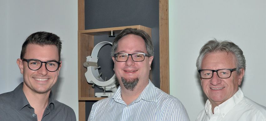
in summary
MaMMA DIAGNOSTICS AND BIOPSY
Biopsy Methods . . . . . . . . . . . . . . . . . . . . . . . . . . . . . . . . . . . . . . . . . . . . . . . . . . . . . . . . . . . . . . . . . . . . . . . . . . . . . . . . . . . . . . . . . . . . . . 6
Cranio-caudal Fixation Units . . . . . . . . . . . . . . . . . . . . . . . . . . . . . . . . . . . . . . . . . . . . . . . . . . . . . . . . . . . . . . . . . . . . . . . . . . . . . . . . . . . . . 7
Breast BI 7 MR Coil Set 1.5T & 3T . . . . . . . . . . . . . . . . . . . . . . . . . . . . . . . . . . . . . . . . . . . . . . . . . . . . . . . . . . . . . . . . . . . . . . . . . . . . . . . . . . . 8
Breat Biopsy and Fixation Unit Sets . . . . . . . . . . . . . . . . . . . . . . . . . . . . . . . . . . . . . . . . . . . . . . . . . . . . . . . . . . . . . . . . . . . . . . . . . . 9
Optional Components for Post&Pillar Biopsy System . . . . . . . . . . . . . . . . . . . . . . . . . . . . . . . . . . . . . . . . . . . . . . . . . . . . . . . . . . . . . . 9
Optional Components for Grid Biopsy System . . . . . . . . . . . . . . . . . . . . . . . . . . . . . . . . . . . . . . . . . . . . . . . . . . . . . . . . . . . . . . . . . . 10
Optional Components for cranio-caudal Fixation Unit . . . . . . . . . . . . . . . . . . . . . . . . . . . . . . . . . . . . . . . . . . . . . . . . . . . . . . . . . . . . 11
Breast Biopsy 4-Channel Coil BI 4 . . . . . . . . . . . . . . . . . . . . . . . . . . . . . . . . . . . . . . . . . . . . . . . . . . . . . . . . . . . . . . . . . . . . . . . . . . . . . . . . . 12
Breast Biopsy and Fixation Unit Sets . . . . . . . . . . . . . . . . . . . . . . . . . . . . . . . . . . . . . . . . . . . . . . . . . . . . . . . . . . . . . . . . . . . . . . . . . 13
Optional Components for Post&Pillar Biopsy System . . . . . . . . . . . . . . . . . . . . . . . . . . . . . . . . . . . . . . . . . . . . . . . . . . . . . . . . . . . . . 13
Optional Components for Grid Biopsy System . . . . . . . . . . . . . . . . . . . . . . . . . . . . . . . . . . . . . . . . . . . . . . . . . . . . . . . . . . . . . . . . . . 15
Optional Components for cranio-caudal Fixation Unit . . . . . . . . . . . . . . . . . . . . . . . . . . . . . . . . . . . . . . . . . . . . . . . . . . . . . . . . . . . . 16
Sterile Soft Tissue Instruments for Biopsy . . . . . . . . . . . . . . . . . . . . . . . . . . . . . . . . . . . . . . . . . . . . . . . . . . . . . . . . . . . . . . . . . . . . . . . . . . . 17
Manual coordinate calculation for the Breast BI 7 MR Coil Set . . . . . . . . . . . . . . . . . . . . . . . . . . . . . . . . . . . . . . . . . . . . . . . . . . . . . . . . . . . . . 18
Manual coordinate calculation for the Post & Pillar Biopsy System . . . . . . . . . . . . . . . . . . . . . . . . . . . . . . . . . . . . . . . . . . . . . . . . . . . . 19
Angulated, manual coordinate calculation for the Post & Pillar Biopsy System . . . . . . . . . . . . . . . . . . . . . . . . . . . . . . . . . . . . . . . . . . . 21
Manual coordinate calculation for the Grid Biopsy System . . . . . . . . . . . . . . . . . . . . . . . . . . . . . . . . . . . . . . . . . . . . . . . . . . . . . . . . . 23
Prostate biopsy and therapy
Uni-Lift Prostate Intervention Device . . . . . . . . . . . . . . . . . . . . . . . . . . . . . . . . . . . . . . . . . . . . . . . . . . . . . . . . . . . . . . . . . . . . . . . . . . . . . . 24
Universal Diagnostics and Intervention
Uni-Belt 16-Channel Diagnostic & Intervention Coil . . . . . . . . . . . . . . . . . . . . . . . . . . . . . . . . . . . . . . . . . . . . . . . . . . . . . . . . . . . . . . . . . . . . 25
dedicated MR receiving coils
Variety 16-Channel Multipurpose Coil 1.5T & 3T . . . . . . . . . . . . . . . . . . . . . . . . . . . . . . . . . . . . . . . . . . . . . . . . . . . . . . . . . . . . . . . . . . . . . . . 26
Typical Applications and Positioning Aids - Orthopedics . . . . . . . . . . . . . . . . . . . . . . . . . . . . . . . . . . . . . . . . . . . . . . . . . . . . . . . . . . . 27
Typical Applications and Positioning Aids - Pediatrics . . . . . . . . . . . . . . . . . . . . . . . . . . . . . . . . . . . . . . . . . . . . . . . . . . . . . . . . . . . . . 29
Available Coil Packages . . . . . . . . . . . . . . . . . . . . . . . . . . . . . . . . . . . . . . . . . . . . . . . . . . . . . . . . . . . . . . . . . . . . . . . . . . . . . . . . . . 32
Optional Components for the Variety 16-Channel Multipurpose Coil . . . . . . . . . . . . . . . . . . . . . . . . . . . . . . . . . . . . . . . . . . . . . . . . . . 33
Mandibula 15-Channel Dental Coil . . . . . . . . . . . . . . . . . . . . . . . . . . . . . . . . . . . . . . . . . . . . . . . . . . . . . . . . . . . . . . . . . . . . . . . . . . . . . . . . 34
Neurosurgery
Intraoperative Neurosurgery solution from NORAS . . . . . . . . . . . . . . . . . . . . . . . . . . . . . . . . . . . . . . . . . . . . . . . . . . . . . . . . . . . . . . . . . . . . 35
OR Head Holder LUCY & OR Head Coil 1.5T / 3T . . . . . . . . . . . . . . . . . . . . . . . . . . . . . . . . . . . . . . . . . . . . . . . . . . . . . . . . . . . . . . . . . 36
Optional Components for NORAS OR Head Holder LUCY . . . . . . . . . . . . . . . . . . . . . . . . . . . . . . . . . . . . . . . . . . . . . . . . . . . . . . . . . . 39
OR Head Holder FLEXIBILITY & OR Head Coil 1.5T / 3T . . . . . . . . . . . . . . . . . . . . . . . . . . . . . . . . . . . . . . . . . . . . . . . . . . . . . . . . . . . . . 40
OR Head Holder HEIDBERG & OR Head Coil 1.5T / 3T . . . . . . . . . . . . . . . . . . . . . . . . . . . . . . . . . . . . . . . . . . . . . . . . . . . . . . . . . . . . . . 43
Optional Components for OR Head Holder FLEXIBILITY and Upgrade Kit HEIDBERG . . . . . . . . . . . . . . . . . . . . . . . . . . . . . . . . . . . . . . . 44
Development of the Noras OR Head Holder . . . . . . . . . . . . . . . . . . . . . . . . . . . . . . . . . . . . . . . . . . . . . . . . . . . . . . . . . . . . . . . . . . . . 45
Additional content
Press Reports and Studies . . . . . . . . . . . . . . . . . . . . . . . . . . . . . . . . . . . . . . . . . . . . . . . . . . . . . . . . . . . . . . . . . . . . . . . . . . . . . . . . . . . . . . 49
NORAS Corporate philosophy . . . . . . . . . . . . . . . . . . . . . . . . . . . . . . . . . . . . . . . . . . . . . . . . . . . . . . . . . . . . . . . . . . . . . . . . . . . . . . . . . . . 67
NORAS Certificates . . . . . . . . . . . . . . . . . . . . . . . . . . . . . . . . . . . . . . . . . . . . . . . . . . . . . . . . . . . . . . . . . . . . . . . . . . . . . . . . . . . . . . . . . . . 68
5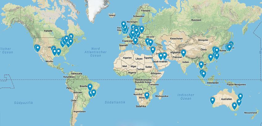
Biopsy Methods
       MAMMA

                           Choose your preferred Breast Biopsy Method
                           NORAS offers two different biopsy methods, each with its own advantages. Both methods are available for our Breast Biopsy Coils BI 4 and BI 7 and
                           support MR-guided wire placement for 12, 14, 16, and 18 Gage, as well as Vacuum Gun Biopsies.
The Post & Pillar Method

                                                                                                                                   High accuracy due to accurate coordinate
                                                                                                                               4   adjustment
                                                                                                                      o il B I
                                                                                For Breast Biopsy 4 - C    ha nn e l C
                                                                                                                                   Angular needle guidance 15 ° and 30 ° pos-
                                                                                                                                   terior and anterior

                                                                                                                                   Sterile disposable needle guide sleeves are
                                                                                                                                   available*

                                        For Breast BI 7 MR Coil Set                                                                                         * distributed by NORAS
The Grid Method

                                                                                                      BI   4
                                                                                              el Coil
                                                                For Breast Biopsy 4 - Chann

                                                                                                                                   Easy to use biopsy method

                                                                                                                                   Multiple biopsies in one process

                                                                                                                                   Sterile disposable grids are available*

                                                  For Breast BI 7 MR Coil Set                                                                               * distributed by NORAS

                           Both methods are compatible with the following Vacuum Guns:

                           C.R. Bard Vacora™                    Suros Surgical ATEC™                              Devicor Mammotome™                 SenoRX Encor MRI™

                           6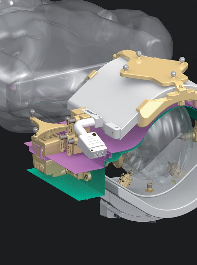
Cranio-caudal Fixation Units

                                                                                                                                                                         MAMMA
With the NORAS Cranio-caudal Fixation Units you can optionally immobilize/fixate one or both breasts at the same time. Using this device, one can
save up to 50% of scanning time, while reducing non-desired motion artifacts.

                                                                                                                                                           l Coil BI 4
                                                                                                                                                          nn e
                                                                                                                                                      ha
                                                                                                                                                 -C
                                                                                                                                                      4
                                                                                                                                                 sy
                                                                                                                                            op
                                                                                                                          For Brea   s t Bi

                                                                                                                                                    ilS et
                                                                                                                                                 Co

                                                                                                                                                  R
                                                                                                                                       I   7M
                                                                                                                          For Breast B

                                                                                                                                                                 7
Breast BI 7 MR Coil Set 1.5T & 3T NORAS product solutions for
MAMMA
Exclusively for the new Siemens MRI scanner generation
Vida and Sola
Please contact your Siemens
The Noras Breast BI 7 MR Coil Set 1.5T & 3T is an innovative solution designed to representative for more information
offer flexible use to meet the needs of the procedure to be performed in your
clinic.
Using the same set of comfort cushions, it can be combined with the Breast 18
You can obtain all components
imaging coil from Siemens.
The rigid coil frame forms a high element-density, 7-channel „phased array” con- Breast Biopsy and Fixation Unit di-
figuration for high resolution imaging. One sided medial and up to two-sided rectly from NORAS. We will be plea-
lateral biopsy access is possible. sed to give you personal advice.
Patented lighting system
integrated
Post&Pillar and Grid Biopsy System
The Post & Pillar biopsy system is designed for targeted needle
guidance, which can be moved both left-right and anterior-pos-
terior.
Additionally, it supports angulated biopsies. As an alternative,
disposable biopsy grids* can be used to perform biopsies also at
numerous lesions at the same time.
Disposable adapters* for all compatible biopsy needles and vacu-
um guns are available directly from NORAS.
* distributed by NORAS
Fixation Units: medio-lateral and cranio-caudal
Specially developed fixation units for medio-lateral or also cranio-
caudal fixation of the breast are available as an option to avoid
motion artifacts and reduce scan time.
8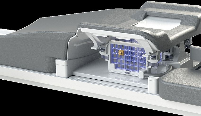
Breat Biopsy and Fixation Unit Sets

                                                                              MAMMA
for Breast BI 7 MR Coil Set 1.5T & 3T                       part number

Breast Biopsy Set for Breast BI 7                           on request

Cranio-caudal fixation Breast BI 7                          on request

Optional Components for Post&Pillar Biopsy System
for Breast BI 7 MR Coil Set 1.5T & 3T
                                                            part number

Post&Pillar Needle Guide Pillar Breast BI 7: autoclavable   118621

Post&Pillar Needle Guide Breast BI 7: autoclavable          118654

Post&Pillar Marker Breast BI 7: autoclavable                118946

Horizontal Slat Plate Breast BI 7 medial: autoclavable      118649

Horizontal Slat Plate Breast BI 7 lateral: autoclavable     118622

                                                                          9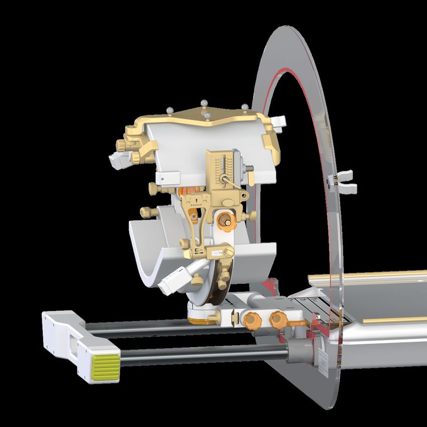
part number
MAMMA

                                                                             18G 118952
                                                                             16G 118953                     18G   16G
        Post&Pillar Needle Guide Sleeves disposable BI 7*: sterile; 5-pack
                                                                             14G 118954
                                                                             12G 118955
        * distributed by NORAS                                                                              14G   12G

                                                                             C.R. Bard Vacora      118956
        Post&Pillar Needle Guide Sleeves disposable BI 7*: for use with MR   Suros Surgical ATEC   118957
        compatible Vacuum Guns ; sterile; 5-pack                             SenoRX EnCor 10G 118958
        * distributed by NORAS
                                                                             SenoRX EnCor 7G    118950

        Optional Components for Grid Biopsy System
        for Breast BI 7 MR Coil Set 1.5T & 3T
                                                                             part number

         Grid Biopsy unit disposable medial BI 7*: sterile; 5-pack           118979
        * distributed by NORAS

         Grid Biopsy unit disposable lateral BI 7*: sterile; 5-pack          118980
         * distributed by NORAS

         Grid Marker Block: autoclavable                                     111251

                                                                             18G 112660
                                                                             16G 112143                     18G   16G
         Grid Needle Blocks disposable*: sterile
                                                                             14G 112659
                                                                             12G 112731
        * distributed by NORAS                                                                              14G   12G

         Breast Elevation Plate Breast BI 7                                  118761

        10
Optional Components for cranio-caudal Fixation Unit
MAMMA
for Breast BI 7 MR Coil Set 1.5T & 3T
part number
Cranio-caudal fixation unit BI 7: single element 118719
CC-320-Plus Cushion Breast 118193
11Breast Biopsy 4-Channel Coil BI 4 NORAS product solutions for
MAMMA
Exclusively for Siemens MRI Scanner
Please contact your Siemens
representative for more information
The Patient Rest can be used for diagnostic
imaging with the 4Ch coil and also with the
optional Siemens 16Ch coil. This solution is
only available through Siemens.
SIEMENS 16-Channel Diagnostic Coil
The NORAS Breast Biopsy 4-Channel Coil BI 4 was developed for the
Siemens TIM system and serves with its 4Ch array coil for diagnostics
as well as biopsy removal. The design of the whole setup is very open
to ensure optimal access to the breast for interventions. Optionally,
you can use the 16Ch coil* for optimized imaging (spectroscopy pos-
sible) with the same Patient Rest.
* distribution via Siemens Healthineers
The Breast Biopsy Kit consists of a complete Post&Pillar positioning
unit and two disposable grids for medial, lateral and cranio-caudal
access. The biopsy set is compatible with the coordination software
from Siemens, and therefore cannot be rotated by 360°.
* distributed by NORAS
The original components are exclusively
available through NORAS or through autho-
rized re-sellers.
Case
The Breast Biopsy Kit consists of a steam ste-
rilizable fixation unit including Post&Pillar po-
sitioning unit.
The sterile pieces* that are necessary for the
biopsies are not part of the delivery content
but can be ordered separately.
Part of the biopsy kit is a 12G non-sterile trai-
ning needle, including sleeve and cube, as well
as two training grids (medial/CC and lateral).
* distributed by NORAS
12Breast Biopsy and Fixation Unit Sets
MAMMA
for Breast Biopsy 4-Channel Coil BI 4
part number
Breast Biopsy Set Post&Pillar: autoclavable and reusable 118941
Breast Biopsy Set Grid disposable: autoclavable and reusable; with
disposable grids in sterile packaging* 118943
* distributed by NORAS
Breast Biopsy Set Grid reusable: autoclavable and reusable 118944
Cranio-caudal Fixation Unit CC-320-PLUS 118341
Optional Components for Post&Pillar Biopsy System
for Breast Biopsy 4-Channel Coil BI 4
part number
Post&Pillar positioning unit with telescope bar: autoclavable and
reusable
111259
Post&Pillar Needle Guide: autoclavable and reusable; able to be angu-
lated by 15° and 30°
111451
Post&Pillar marker: autoclavable and reusable; (remove marker liquid
before autoclaving and refill afterwards)
111300
13part number
MAMMA
Post&Pillar Needle Guide Breast BI 7: autoclavable 118654
Post&Pillar Marker Breast BI 7: autoclavable 118946
18G 112654
16G 112597
Post&Pillar needle guide sleeves*: disposable in sterile packaging
14G 112655
12G 112653
* distributed by NORAS 16G 18G 14G 12G
18G 118952
16G 118953 18G 16G
Post&Pillar Needle Guide Sleeves disposable BI 7*: sterile; 5-pack
14G 118954
12G 118955
* distributed by NORAS 14G 12G
Base unit fixation plate: can be used laterally, medially and cranio-cau-
dally and is used as adapter for slat plates and grids
111242
Base plate 111292
Slat plate horizontal: medial and cc 111212
Slat plate horizontal: lateral 111301
C.R. Bard Vacora 118956
Post&Pillar Needle Guide Sleeves disposable BI 7*: for use with MR Suros Surgical ATEC 118957
compatible Vacuum Guns; sterile; 5-pack SenoRX EnCor 10G 118958
SenoRX EnCor 7G 118950
* distributed by NORAS
14part number
MAMMA
C.R. Bard Vacora MR10062-VAC
Adapter Vacuum Guns for Post&Pillar System: Suros Surgical ATEC MR10062-AT
autoclavable ; 2-pack SenoRX EnCor 10G MR10062-SE
SenoRX EnCor 7G 113315
Guidance bracket for Invivo disposable needle guidance MR10062-MHA
Disposable needle guidance (Multi Hub Assembly): not for 7G 113692
Adapter Vacuum Guns for Post&Pillar System Devicor Mammotome:
autoclavable
MR10062-ET2
Optional Components for Grid Biopsy System
for Breast Biopsy 4-Channel Coil BI 4
part number
Grid Marker Block: autoclavable 111251
Grid biopsy unit: lateral, medial, CC; autoclavable and reusable 111252
Grid biopsy unit height adjustable: lateral; autoclavable and reusable 117143
Grid biopsy unit height adjustable*: lateral; disposable in sterile
packaging; packaging consists of 5 grids 112235
* distributed by NORAS
15part number
MAMMA
Grid biopsy unit height adjustable*: medial, CC; disposable in sterile
packaging; packaging consists of 5 grids 112238
* distributed by NORAS
18G 112660
18G 16G
16G 112143
Grid needle blocks*: disposable in sterile packaging
14G 112659
12G 112731 14G 12G
* distributed by NORAS
Optional Components for cranio-caudal Fixation Unit
for Breast Biopsy 4-Channel Coil BI 4
part number
Cranio-caudal fixation unit CC-320-PLUS: single element 118086
CC-320-Plus Cushion Breast 118193
16Sterile Soft Tissue Instruments for Biopsy
MAMMA
ClipLoc Soft Tissue Marker* system includes the ClipLoc, coaxial introducer and advancing stylet.
1.5T; 18G; 100mm MR10507
1.5T; 18G; 130mm MR10508
1.5T; 18G; 150mm MR10509
3T; 18G; 150mm 112586
Coaxial Needle* coaxial needles can be used as a coaxial guide for biopsy guns;
avail-able in different lenghts and gauge sizes.
3T; 12G; 111mm 112595
3T; 16G; 111mm 112596
1.5T; 12G; 61mm 112587
1.5T; 12G; 91mm 112588
1.5T; 12G; 111mm 112590
1.5T; 12G; 126mm 112589
1.5T; 14G; 111mm 112592
1.5T; 16G; 111mm 112594
Semi-Automatic Biopsy Gun*
14G; 100mm 112535
14G; 150mm 112537
18G; 150mm 112541
Single/Double Hook Wire puncture needle with a localization wire; flexible material, reduces
Localization Needle* the risk of breakage or unwanted dislocation; available in single and
double hook.
Double Hook; 18G; 100mm MR24700
Double Hook; 18G; 150mm MR24701
FlexiLoc* like single hook wire but repositionable in the beginning.
18G; 100mm MR24702
18G; 150mm MR24703
3T; FlexiLoc Set* consisting of articles 112596 (coax needle) and
MR24703 (FlexiLoc).
112585
* distributed by NORAS
NORAS Forceps part number
MR suitable, non-magnetic forceps* for the removal of tissue samp-
le, 4.5“ MR00015
* distributed by NORAS
17MAMMA
Manual coordinate calculation for the Breast
BI 7 MR Coil Set
On the following pages you will find instructions for the manual coordinate calculation of the BI 7 Post & Pillar
and Grid Biopsy Method, which is located on the user interface of your Siemens MR-system.
Besides the manual calculation of coordinates, additional solutions for software-supported coordinate calcu-
lation are offered by third-party providers.
Please contact NORAS or your Siemens representative directly if you have any questions regarding compatibi-
lity and application.
18Manual coordinate calculation for the Post &
MAMMA
Pillar Biopsy System
1 2
Perform a measurement with transversal slices and look for Start the „distance“ tool (menu, right center → „Extras“ → „di-
the slice on which one you can see the marker. stance“ (ruler)).
3 4
Mark that line with a left-click and select the marked line with
a right-click. In the context-menu, select „copy“. By doing so,
Draw a line, straight through the pointer. the line is copied and can be pasted into other slices.
5 6
Select the „distance“ tool again and measure the distance
between the lesion and the reference line that you inserted
in step 5. Please note these measurements on a separate piece
of paper.
Scroll through the other slices until you find the lesion that In this example the Post & Pillar positioning unit has to be mo-
you want to perform a biopsy on. Right-click on this slice und ved 31 mm from the starting position to anterior (because the
select „Paste“. patient is lying in the prone position).
19MAMMA
7 8
In order to determine the shift from head to feet (Post & Pillar
positioning unit to the left or to the right), view the slice po-
sition of the marker and lesion at the transversal slices. The The lesion you have chosen in step 5 is located in our example
marker from step 1 is located on slice-position („SP“=Slice Po- on slice position H14.9. Hence, the difference is 14.9 mm – 0.5
sition) H0.5, therefore 0.5mm towards the head from the laser mm = 14.4 mm. In order to reach the lesion, you have to move
positioning. the Post & Pillar positioning unit by 14mm towards the head.
9 10
Move the Post&Pillar unit with the marker according to the cal-
culated coordinates (in our example 31mm downwards (ante- Perform a control scan in order to check that the marker is
rior) and 14 mm towards the head). located in front of the lesion.
11 12
se the „distance” tool again in order to measure the pun-
U
cture depth from the surface of the skin to the middle of
the lesion (in this case 31 mm). Subtract an offset, depending
on the biopsy system (normally about 5-10 mm), in order to
position the cannula lies in front of the lesion. Now puncture Perform a control scan in order to check that the end of the
the breast with the trocar using this offset-corrected depth needle is positioned in front of
(starting at skin surface). Afterwards pull the needle out of the lesion.
the trocar and insert the plastic-bar into the cannula instead.
The plastic-bar causes fewer artifacts than the needle and pre- The further biopsy may then be carried out by
vents blood back-flow into the cannula. authorized personnel.
20Angulated, manual coordinate calculation for
MAMMA
the Post & Pillar Biopsy System
Perform the steps 1. to 10. from the user manual for the straight, manual calculation of coordinates.
If you notice during the straight biopsy that, for example, one of the bars of the grid is in the way of the biopsy, or if you prefer to continue with an
angulated biopsy for any other reason, you can also work with an angulation of 15 or 30 degrees toward the top or the bottom.
13 14
ow, an angle of 15 degrees (in this example) has to be dra-
N
wn to the original marker line. To do so, one needs to use the
tool, which you find on the right hand side under „Extras” → Draw the first line, in this example from right to left, through
„Angle“. the marker toward the lesion.
15 16
The second line has to have appropriate length toward the
Draw the second line with an angle of 15 degrees from right left so it lasts at least underneath the pivot point of the Post &
to left. This can also be done slightly underneath the first line. Pillar marker. You can also extend that line afterwards by pul-
Please be aware that in this example, where we create the ang- ling its right endpoint. The new Marker design shows the pivot
le counterclockwise, we will prepare an angle of 165 degrees. point as a black cross inside the marker.
17 18
Now, go ahead and move the second line (which forms an
angle of 15 degrees with the first line) so that it has its left Choose the „distance” tool und draw a new line perpendicular
endpoint in the center of the lesion. To do that, left-click the to the first line through the marker at the height of the pivot
second line, hold down the left mouse key and move the line point, marked by the black cross inside the marker. It might
with the mouse. help to start this line a little below the line through the marker.
21MAMMA
19
Now move this new line, till the one ends reaches the referen-
ce line through the marker and shorten it, so that is will touch
the 15 degree line on the other end. You line now shows on the
distance you need to move our needle guide in order to use a
15 degree angulation. In this example: 15mm up (posterior).
20 21
After movement (here 15 mm posterior) and angulation of the
marker (here 15 degrees toward anterior, hence toward the After the puncture with the trocar, perform another control
floor), perform a control scan in order to check that the marker scan in order to check that the end of the cannula lies right in
points at the lesion. Now, determine similarly to step 11 the front of the lesion.
puncture depth from the skin surface to the center of the The further biopsy may then be carried out by autho-
lesion (here 30.5 mm without offset). rized personnel.
22MAMMA
Manual coordinate calculation for the Grid Biopsy
System
In the following the Grid localization process with medial/lateral access
while using sagittal imaging slices is b
riefly described.
The grid pattern itself is used to locate the position of the lesion.
The grid plate is mounted on the biopsy unit for the fixation of the breast. The pattern of this grid is visible on the
sagittal MR-image at the edge of the breast. Therefore the grid position lying directly in line with the lesion can be
identified manually. A needle block is inserted into that grid position.
The lesion is marked by using the imaging tools on the scanner user interface, e.g. a circle or a cross. This marking
is copied and pasted into the parallel sagittal slice, which shows the grid pattern. Now it can be easily seen, where
the needle block and the needle need to be positioned. Additionally, a MR-visible marker block for better orienta-
tion on the grid can be used (in this imaging example, the marker is sitting in the middle of the grid).
With the knowledge of the number and thickness (plus a potential gap between slices) of the sagittal imaging
slices from the surface of the grid to the lesion, the penetration depth can be easily calculated.
23Uni-Lift Prostate Intervention Device
for MR-guided interventions of the prostate
Uni-Lift Prostate Intervention Device 118330
Prostate
The Uni-Lift is designed as an arc in which the legs are mounted
on brackets. The arc is adapted to the MR table and to the bore
size, thereby preventing abutting of the legs while providing
the maximum available space for intervention.
The supine position allows transperineal access for biopsy and
therapy of the prostate.
As an application example a biopsy grid for transperineal access
is shown.
For MR-guided biopsies images can be taken by the specifically
designed flex coil Uni-Belt or ordinary flex coils.
The Uni-Lift Prostate Interventions Device from NORAS is compa- Transperineal Prostate Biopsy
tible to the 70 cm Bore MR Systems (Siemens MAGNETOM Aera
1.5T, Skyra 3T & Espree 1,5T / GE Optima 450W 1.5T & Discovery For the transperineal access, the use of reusable sterilizable
750W 3T). grids made of skin-friendly PEEK is provided.
Height-adjustable grid biopsy unit for transperineal access.
*NORAS is currently working on compatible solutions for
other manufacturers of MR scanners. Kindly contact us in
case you have any questions regarding the compatibility of
the NORAS Uni-Lift Prostate Intervention Device for your
scanner.
24Uni-Belt 16-Channel Diagnostic
& Intervention Coil Uni-Belt 16-Channel Diagnostics
for diagnostic imaging and MR-guided interventions of and Intervention Coil 1.5T for SIE- On request
MENS Scanner
the prostate and other internal organs
INTERVEN-
in development
TION
The flexible 120 cm (47.24 in) long and 32 cm (12.6 in) wide
coil is designed so that it can wrap around both hips, ab-
domen and chest. The coil size is designed for optimal
penetration depth to the body center. Due to a fixation
system using two belts, the coil can be positi-
oned very close and stable to the desired anatomy.
The coil is designed for diagnostic images of the pro-
state or internal organs (liver, kidney, etc), as well as
MR-guided interventions, which can be performed th-
rough integrated biopsy windows inside the coil plane.
A biopsy unit with angulation possibility was specifically developed
for the Uni-Belt coil. It can be easily attached inside the coils’ biopsy
windows. Biopsies of prostate, liver, kidney, spinal cord etc. can be
performed through the steam sterilizable and reusable biopsy unit
made out of PEEK material.
The advantage of the NORAS Uni-Belt is given by the arrangement
of its single elements combined with the complete covering of the
examination area. Futhermore, a better adapting to the anatomy
has positive effects on the result of measurement.
25Variety 16-Channel Multipurpose Coil 1.5T & 3T
The Variety 16-Channel Multipurpose Coil enables high-resolution MR imaging in orthope-
dics and pediatrics.
Variety 16-Channel Multipurpose Coil 1.5T 117235
Variety 16-Channel Multipurpose Coil 3T 117234
Variety long 16-Channel Multipurpose Coil 3T 119722
The „Variety“ Coil is a 16-channel multipurpose array, which has been developed for high flexibility during examination of challenging anatomic
regions. A good example for such areas is the imaging of musculoskeletal areas.
VARIETY
High signal quality based on a design with 8+8 array elements
with high coil element density
High resolution examinations of even small body regions with
reduced scan durations
High speed image acquisition due to the possibility of accele-
rated imaging in all directions
Application for diagnosis in orthopedics, pediatrics and in
veterinary medicine
Enables motion studies of all joints
Enables mounting very close to anatomical region of interest
through dedicated positioning aids (optimal image quality)
• Compatibility with Siemens coil portfolio
NEW: Siemens Coil File available* • No need to remove the spine coil anymore
* Only applies for Siemens Skyra 3T and Aera 1.5T customers
Each half of the coil is based on an 8-channel array, which can be used separately as a surface array for coverage of various body areas. The indivi-
dual coil elements in each array have a small diameter of approximately 7 cm. This design results in a high signal quality, which allows for highest
achievable resolutions.
Based on the high mechanical FLEXIBILITY and on the small size of its array elements the “Variety“ covers a much larger field of applications for high
resolution imaging in comparison to standard flexcoils. Through the use of Velcro® strips and dedicated positioning aids the “Variety“ Array can be
adjusted very well to curved anatomical regions.
The Variety 16-Channel Multipurpose Coil from NORAS MRI products is compatible to all Siemens Tim® systems. NORAS is currently working on
compatible solutions for other manufacturers of MR-scanners. Kindly contact us in case you have any questions regarding the compatibility of the
NORAS 16-Channel Multipurpose Array for your scanner.
26Typical Applications and Positioning Aids - Orthopedics
The 16 channels of the compact Variety Array Coil are specially designed to enable a deep pe-
netration of tissue with a large field of view.
Hand and Wrist
VARIETY
The size of the coil halves enables imaging of the whole hand and wrist.
A special positioning aid, in which the bottom coil half can be placed, creates a sta-
ble and flat surface to put the hand on.
If needed, the second coil half can be put on top of the hand.
Alternatively, the hand can be positioned “standing up“ using our elbow positio-
ning aid (see illustration below right).
Elbow
For smaller elbows (e.g. kids) a single coil half can be used to be wrapped around the
elbow. This coil half can then be fixated using Velcro® strips.
For bigger elbows, the size adjustable positioning aid can be used.
27Shoulder
A special sloped positioning device allows comfortable support on the lower coil
half.
For fixation of the upper coil half, you can use e.g. sandbags.
VARIETY
Knee, Foot and Ankle
The stageless adjustment of the positioning device allows for proper support of pa-
tients with a body size of up to 2.1 m and for knees with high or low volume.
The flexible design also allows imaging in flexed and stretched position. Further-
more, an optional supporting element for imaging in flexed position is available.
We advise to use always the positioning aids in combination to enable support and
less motion of the patient.
28Typical Applications and Positioning Aids - Pediatrics
Pediatrics small (0 - 6 months)
VARIETY
Pediatrics medium (6 - 36 months)
Pediatrics large (4 - 8 years)
29VARIETY
Hand at 3T (Siemens Skyra) Hand at 3T (Siemens Skyra) Wrist at 3T (Siemens Skyra)
Sequence parameters: 0.34 x 0.34 x 2 mm³; Sequence parameters: 0.29 x 0.29 x 1.5 mm³; Sequence parameters: 0.23 x 0.23 x 3 mm³;
TR=3000ms; TE=58ms; TA=2:21; TR=34ms; TE=20ms; TA=2:44; TR=600ms; TE=14ms; TA=1:47;
PAT-Factor: 2; FOV: 220 x 220. PAT-Factor: 3; FOV: 220 x 220. PAT-Factor: 2; FOV: 90 x 90.
Shoulder at 3T (Siemens Skyra) Shoulder at 3T (Siemens Skyra) Knee at 3T (Siemens Skyra)
Sequence parameters: 0.36 x 0.36 x 3 mm³; Sequence parameters: 0.41 x 0.41 x 3.2 mm³; Sequence parameters: 0.36 x 0.36 x 2.5
TR=600ms; TE=24ms; TA=3:34 min; TR=2500ms; TE=34ms; TA=2:40 min; mm³; TR=525ms; TE=11ms; TA=2:26 min;
PAT-Factor: 2; FOV: 160 x 160. PAT-Factor: 2; FOV: 150 x 150. FOV: 150 x 150.
Hip at 3T (Siemens Skyra) Hip at 3T (Siemens Skyra) Jaw joint at 3T (Siemens Skyra)
Sequence parameters: 0.46 x 0.46 x 2 mm³; Sequence parameters: 0.43 x 0.43 x 1.2 mm³; Sequence parameters: 0.23 x 0.23 x 2 mm³;
TR=600ms; TE=11ms; TA=3:48 min; TR=12.6ms; TE=5.1ms; TA=4:50 min; TR=3000ms; TE=95ms; TA=1:42 min;
FOV: 119 x 119. PAT-Factor: 3; FOV: 199 x 220. FOV: 120 x 120.
30VARIETY
Elbow at 3T (Siemens Skyra) Elbow at 3T (Siemens Skyra) Foot at 3T (Siemens Skyra)
Sequence parameters: 0.5 x 0.5 x 2 mm³; Sequence parameters: 0.6 x 0.6 x 3 mm³; Sequence parameters: 0.31 x 0.31 x 2 mm³;
TR=729ms; TE=10ms; TA=2:54 min; TR=3000ms; TE=41ms; TA=4:33; TR=650ms; TE=11ms; TA=2:01 min;
FOV: 89 x 260. PAT-Factor: 2; FOV: 200 x 200. PAT-Factor: 2; FOV: 120 x 120.
Knee at 3T (Siemens Skyra) Ankle at 3T (Siemens Skyra) Ankle at 3T (Siemens Skyra)
Sequence parameters: 0.31 x 0.31 x 3 mm³; Sequence parameters: 0.33 x 0.33 x 2 mm³; Sequence parameters: 0.39 x 0.39 x 3 mm³;
TR=2500ms; TE=24ms; TA=2:27 min; TR=31ms; TE=18ms; TA=2:53 min; TR=3100ms; TE=34ms; TA=2:47 min;
PAT-Factor: 2; FOV: 140 x 140. PAT-Factor: 3; FOV: 150 x 150. PAT-Factor: 2; FOV: 150 x 150.
Child at 3T (Siemens Skyra) Child at 3T (Siemens Skyra) Child at 3T (Siemens Skyra)
Sequence parameters: 0.35 x 0.35 x 4 mm³; Sequence parameters: 0.31 x 0.31 x 3.5 mm³; Sequence parameters: 0.34 x 0.34 x 3 mm³;
TR=2410ms; TE=95ms; TA=5:39 min; TR=600ms; TE=20ms; TA=5:46 min; TR=633ms; TE=23ms; TA=7:32 min;
PAT-Factor: 2; FOV: 181 x 223. PAT-Factor: 2; FOV: 181 x 200. PAT-Factor: 2; FOV: 165 x 220.
31Technical Specifications
Coverage Wrap Diameter
Variety 16-Channel Multipurpose Coil (one coil half)
20 cm x 22 cm 5.5-8 cm
VARIETY
Variety 16-Channel Multipurpose Coil (both coil halves)
20 cm x 44 cm 11-17 cm
Available Coil Packages
part number
Variety 16-Channel Multipurpose Coil 1.5T,
Cable Length: 100 cm
• Variety 16-Channel Multipurpose Coil 1.5T 2 pc.
• Variety Clip 2 pc. 117235
• Variety Positioning Aid Multipurpose 2 pc.
• Velcro® Fastener 300 4 pc.
• Velcro® Fastener 500 4 pc.
• Velcro® Fastener 500 back-to-back 4 pc.
Variety 16-Channel Multipurpose Coil 3T,
Cable Length: 100 cm
• Variety 16-Channel Multipurpose Coil 3T 2 pc.
• Variety Clip 2 pc. 117234
• Variety Positioning Aid Multipurpose 2 pc.
• Velcro® Fastener 300 4 pc.
• Velcro® Fastener 500 4 pc.
• Velcro® Fastener 500 back-to-back 4 pc.
Variety lang 6-Channel Multipurpose Coil 3T,
Cable Length: 150 cm
• Variety lang 16-Channel Multipurpose Coil 3T 2 pc.
• Variety Clip 2 pc. 119722
• Variety Positioning Aid Multipurpose 2 pc.
• Velcro® Fastener 300 4 pc.
• Velcro® Fastener 500 4 pc.
• Velcro® Fastener 500 back-to-back 4 pc
32Optional Components for the Variety 16-Channel Multipurpose Coil
part number
Variety Positioning Aid Set Orthopedics
118353
(incl. Positioning Aids Elbow, Shoulder and Knee/Foot/Ankle)
Variety Positioning Aid Set Pediatrics
118354
(incl. Positioning Aids Pediatrics small, medium and large)
VARIETY
Variety Positioning Aid Multipurpose 118398
Variety Positioning Aid Elbow 118468
Variety Positioning Aid Shoulder 118413
Variety Positioning Aid Knee, Foot and Ankle 118463
Variety Positioning Aid Pediatrics small 118465
Variety Positioning Aid Pediatrics medium 118467
Variety Positioning Aid Pediatrics large 118412
Variety Positioning Aid Inlay 118415
Variety Lagerungshilfe Pädiatrie Kopf 118417
33Mandibula 15-Channel Dental Coil Mandibula 15-Channel Dental
Coil 1,5T for SIEMENS Scanner on request
High-Resolution Imaging for Diagnostics in Orthodontics
Mandibula 15-Channel Dental
Coil 3T for SIEMENS Scanner 118871
Our Mandibula 15-Channel Dental Coil is a multi-element receive array and
positioning system for three-dimensional (3D) high-resolution dental and
maxillomandibular MRI images. Also it ensures maximum patient comfort
due to its design and accessories like a patient rest pillow, an open-mouth
fixation mechanism and a both direction mirror.
Typical applications:
• Orthodontia (instead of x-ray)
• Diagnostics of the jaw joint
DENTAL
• Imaging of periodontium and dental pulp
• Implantology
• Imaging of nerves (foramen mentale)
• Bone density (compacta, spongiosa)
• Forensics (age estimation through wisdom tooth)
• Maxifacially imaging (e.g. search for tumors)
• Larynx
• Inflammation
Transversal view
Advantages:
High resolution dedicated MR imaging in dental area
Highest resolution movement studies of the jaw joint
Coil element underneath chin can be moved in and out for motion
studies, e.g. on the jaw joint
Easy to position and adjustable for each patient
Excellent patient comfortability (especially for children during exami-
nation)
Reduced scan times with higher image quality as compared to stan-
dard coils
Optional mirror attachable for claustrophobic patients
Minimized unwanted signal distortions (vibration, movement etc.)
Sagittal view
Panoramic teeth view
34Intraoperative Neuro-
surgery solution from
NORAS
As early as 2005, NORAS MRI products GmbH presented the
first MR compatible head holder with receiving coils and was
thus the pioneer in the field of intraoperative MRI imaging in
the field of neurosurgery. With the innovative head holder
NORAS was able to quickly establish a leading position in
Neuro
neurosurgery solutions.
Based on a creative and respectful cooperation with leading
neurosurgeons from Germany, Switzerland, the US and Ca-
nada NORAS has created the improved head holders FLEXI-
BILITY and HEIDBERG in 2013.
Developed in close cooperation, the head holder HEIDBERG
became a strong alternative to older versions of NORAS
head holders. Both head holder systems convince besides
excellent image quality with a unique sterile concept and an
optimized workflow.
The significant developments are based on requirements,
ideas and challenges that leading neurosurgeons experien-
ce in their everyday work.
The increased reliability and high availability of the Head
Holders FLEXIBILITY and HEIDBERG are proven in e.g. yearly
more than 200 successfully completed cases in Europe using
these advanced systems for MR-based neurosurgery.
Thanks to ongoing enhancements and innovations, NORAS
belongs to the drivers of intraoperative MR imaging.
This was proven again in 2018: The new LUCY head holder
combines the outstanding attributes of years of experien-
ce in the field of MRI with new applications thanks to its CT
compatibility. With constant improvements and new de-
velopments the NORAS MRI products Ltd is a reliable part-
ner for MR-supported neurosurgery.
35OR Head Holder LUCY & OR Head Coil 1.5T / 3T
MR- and X-Ray compatible for multimodal applications
for Siemens Magnetom Aera 1.5T & Skyra 3T/
Maquet MAGNUS OR Table & Alphamaquet
Plus *
FDA: Pending
LUCY is an innovative neurosurgery solution that combines the
functions of a head holder for secure fixation of the skull during
interventions and a MR coil for intra-operative imaging.
The head holder is compatible with the established 8-channel
NORAS head coil setup. Thanks to the radiotranslucent material,
the OR Head Holder LUCY (without the NORAS OR Head Coils)
can also be used in the X-ray environment – CT and angiogra-
phie – for intra-operative imaging.
Besides tumor resections, “deep brain stimulations” (DBS) and
minimally invasive pituitary surgery, the system also supports
awake craniotomies.
Neuro
The NORAS OR Head Holder LUCY and the NORAS OR Head Coil
1.5T / 3T can be used with the MR systems Siemens Magnetom
Aera / Skyra together with the Combi Dockable Table. Due to
the compatibility to the new transferboard from Maquet our
solution can be combined with nexaris Angio-MR-CT, the Artis
pheno Angiography System and with the Somatom CT Sliding
Gantry Systems.
* NORAS is currently working on compatibility solutions for other MRI scanner and surgical table manufacturers.
Please contact us if you have any questions about the compatibility of the OR head holder LUCY and the
head coil of NORAS.
The design provides a comfortable positioning of the patient in prone, supine or lateral position on the
OR table.
The head frame of the NORAS OR Head Holder allows exact adaptation to the patient‘s head size. For
optimal access to the area of intervention, the head frame can be pivoted, rotated, tilted and adjusted
in height.
A secure attachment of the patient‘s head is ensured by the three-point fixation. With the help of the
integrated force indicator, the required clamping force can be checked exactly.
The entire system is height-adjustable to achieve optimal patient positioning in 70 cm bores (Siemens Magnetom
Aera & Skyra) as well as in Siemens CT scanners. In addition, the entire structure can be
moved along the head-foot-axis and the horizontal axis.
The NORAS 2x4 Ch coil concept, with the ability to position the both coils close to the head, provides excellent
image quality.
The OR Head Holder LUCY can be used in combination with nexaris Angio-MR-CT, Artis pheno Angiography System
and Somatom CT Sliding Gantry Systems.
LUCY is compatible with various surgical equipment (e.g. GREENBERG™, VarioGuide™, LEYLA retractors) and supports the
navigation system of Brainlab.
Thanks to a unique sterile concept, neither cables nor coils need to be sterilized!
36Overview
Collision check with
bore gauge
Brainlab AIR Support
Optional components
Excellent access to the
intervention area
Flexible patient
positioning
No sterilization of the
coils and cables neces-
Neuro
sary
8-channel coil setup 1.5T / 3T
Integrated force
indicator 3-point fixation
Radiotranslucent Removable and height
material adjustable lower coil
element
378-Channel Coil setup 1.5T / 3T
Coil system
Achieve maximum image quality and perfectly homo-
geneous illumination with the NORAS 8-Channel Coil
system!
The two-part construction of the coil system allows the
removal of the coil halves in the preparation phase and
during the intervention, which allows excellent access
to the ROI (region of interest).
The lower coil can be placed close to the patient‘s head
due to the height adjustment possibility!
Neuro
height adjustable lower coil
Sterile workspace: protection
Sterile concept
and time saving
Thanks to a unique sterile concept, neither cables
nor coils need to be sterilized!
The sterile work area is delimited with two surgical di-
sposable drapes. Thus, coils and cables as well as most
of the components of the head-holder can be used in
a non-sterile environment.
The newly designed clamping mechanism ensures
easy handling for optimum surgical drape protection!
383-point fixation with integra-
Fixation
ted force indicator
Choose the right clamping force for a secure position of
the patient‘s head!
The integrated force indicator on the rear end of the
one-point fixation allows you to precisely control the
tension before and during the procedure.
Optional Components for NORAS OR Head Holder LUCY
Neuro
part number
All Purpose Arch Set LUCY: The All Purpose Arch can be attached in
different angles to the toothing of the clamps in head-foot direction.
If mounted in foot direction, the scalp can be sewed onto the arch or 119672
mounted with rubber bands or hooks. If mounted in the front, the
arch serves as a hand rest. Additionally GREENBERG™ retractors can be
mounted onto the arch by using various GREENBERG™ retractors.
VarioGuide™ Adapter LUCY: With this adapter, the BrainLab VarioGui-
de™ Biopsy Arm can be mounted on the left or right side of the Head 119671
Holder.
GREENBERG™ Retractor Adapter: This additional adapter can be atta-
ched to the All Purpose Arch and serves as an adapter of several GREEN-
BERG™ and Budde® Halo retractors. It can be flexibly mounted on the 114295
All Purpose Arch.
Leyla Retractor Socket: The socket serves as mount of the various
LEYLA® Retractors. The top part can be rotated horizontally in various
angles. The bottom part is inserted into the dove tail of the Clamps (p/n 113243
11937) or of the GREENBERG™ Retractor Adapter and locked tightly.
39OR Head Holder FLEXIBILITY & OR Head Coil 1.5T / 3T
established Neurosurgery Solution for intraoperative MRI
for Siemens Magnetom Aera 1.5T & Skyra 3T/
Philips Ingenia 1.5T & 3T/
Maquet Magnus OR Table & Alphamaquet
Plus / Trumpf -TruSystem 7500
Neuro
FLEXIBILITY is an established Neurosurgery Solution for intraoperative MRI. The
head holder ensures safe immobilization of the head during an open skull surgery
while also supporting the NORAS OR head coil system, already convinced with its
excellent image quality.
The Head Holder & OR Head Coils are available for Siemens and Philips 1.5T and 3T
MR systems.
The NORAS OR Head Holder FLEXIBILITY allows supine, prone, lateral positioning of the patient and supports awake cra-
niotomies, DBS and minimally invasive pituitary surgery:
FLEXIBILITY is is also suitable for pediatric applications.
The Head Holder is designed as a three-point fixation in the lateral level. Furthermore, the surgeon has the option of using
up to seven fixation points.
For optimum access to the area of intervention, the head frame can be tilted, swiveled and rotated.
The NORAS OR Head Holder FLEXIBILITY is height-adjustable, which enables excellent patient positioning in 70 cm bore
systems Philips Ingenia and Siemens MAGNETOM Aera & Skyra.
There is the possibility to move the setup along the head-feet-direction, which is very convenient when positioning the
patient on the OR table.
The NORAS OR Head Coils provide very good field homogeneity and excellent image quality.
Due to a proven sterile concept, there is no need to sterilize the coils and cables.
The Head Holder is compatible to various operating tools (e.g. GREENBERG™, VarioGuide™, LEYLA-Retractors) and sup-
ports the navigation systems of Brainlab and Medtronic.
40Brainlab AIR Support
Overview
Fixation with up to
No sterilization seven points possible
of the coils and
cables necessary
8-channel coil setup 1.5T / 3T
Removable lower
coil element
Flexible patient positioning
Neuro
NORAS MRI products has developed a “Sterile Concept” The sterile area is located between the two dra-
Sterile Concept
for the NORAS OR Head Holder and Head Coils. This was pes. Just the lower drape, in the picture turquoi-
done in cooperation with the Neurosurgical University se, is getting perforated (the screws of the clamps
Hospital of the University Clinical Center Heidelberg, penetrate the lower drape). The second drape, in
the Neurosurgical Clinic of the University of Ulm at the picture violet, is not being penetrated at all.
BKH Guenzburg and the Neurosurgery Center of the The coils and cables can stay non-sterile.
ASKLEPIOS Clinic North – Hamburg/Heidberg.
Due to this concept, there is no need to sterilize the co-
ils and cables.
During the development process, NORAS emphasized
that the coils and cables as well as most components
of the Head Holder can be used in an unsterile environ-
ment. The basic concept is to create a sterile chamber
in between the patient’s head and the top coil with two
sterile OR drapes.
A compatible version of the reference star and head
holder adapter has been developed in collaboration
with BrainLab AG. The reference star can now be tightly
attached to the head holder without perforating the
sterile OR drapes.
This sterile concept is offered to all existing customers
as an upgrade kit HEIDBERG.
Left: Setup with All Purpose Arch, Multifunctional
Bar System, VarioGuide™ Adapter and BrainLab
Drape Link Reference Star.
Right: Setup with two All Purpose Archs, VarioGui-
de™ Adapter and BrainLab Drape Link Reference
Star.
41Flexible Pinning Possibilities
Fixation Concept
There are now three mounting points for height-adjus-
table bottom pins. For the upper pins, a combination
of one 2-point fixation and a 1-point fixation or two
2-point fixations can be mounted on each side of the
head frame. This enables flexible pinning combinations
in consideration of all patient positioning options.
Clamping Device with Force
Indicator
The Clamping Device with Force Indicator allows the
surgeon to apply a defined pinning force during fixati-
on of the patient‘s head. Once the desired force is rea-
ched, the fixation rod is tightened up by a lock lever and
the clamping device can be removed.
Neuro
NORAS OR Head Holder FLEXIBILITY in the application
42OR Head Holder HEIDBERG & OR Head Coil 1.5T / 3T
Upgrade-Kit for our existing customers
The Head Holder HEIDBERG is a further development of the older
NORAS OR Head Holder versions for our existing customers. With
the improved HEIDBERG model, several product features of our es-
tablished neurosurgery solution FLEXIBILITY are available.
The head holder ensures a secure stabilization of the head during
the neurosurgical interventions. Thanks to the NORAS 8-channel
OR Head Coils, excellent image quality can be achieved.
We recommend all customers and users of older NORAS
Head Holder versions – SI7000, SI7000-MQ, SI7300 or
113056 or 113066 (version 4) – to upgrade their systems
with the HEIDBERG upgrade kit.
The NORAS OR Head Holder HEIDBERG allows flexible and comfortable patient positioning and supports awake cranioto-
mies, DBS and minimally invasive pituitary surgery.
The OR Head Holder is suitable for use in pediatrics.
The Head Holder is designed as a three-point fixation in the lateral level. Furthermore, the surgeon has the option of using
up to seven fixation points.
For optimum access to the area of intervention, the head frame can be swiveled and rotated.
The NORAS OR Head Coils provide very good field homogeneity and excellent image quality.
The OR Head Holder HEIDBERG is easy to clean. Furthermore, none of the cables nor any of the coils needs to be sterili-
zed.
The Head Holder is compatible to various operating tools (e.g. GREENBERG™, VarioGuide™, LEYLA-Retractors) and sup-
ports the navigation systems of Brainlab and Medtronic.
43Optional Components for OR Head Holder FLEXIBILITY and Upgrade
Kit HEIDBERG
part number
Multifunctional Bar System: Through the modular setup of toothed
joint parts and bars, the bar system can be adjusted ideally. GREEN-
BERG™ retractors can be mounted onto the bars and Budde® Halo re-
tractors can be clamped onto the joint parts. The ending of the joint
113217
parts is composed of a LEYLA Retractor Socket, which can be turned
and locked vertically in various angles.
VarioGuide™ Adapter: With this adapter, the BrainLab VarioGuide™ 111815
Biopsy Arm can be mounted on the left or right side of the Head Holder.
Neuro
All Purpose Arch: The All Purpose Arch can be attached in different
angles to the toothing of the clamps in head-foot direction.
If mounted in foot direction, the scalp can be sewed onto the arch or
113220
mounted with rubber bands or hooks. If mounted in the front, the
arch serves as a hand rest. Additionally GREENBERG™ retractors can be
mounted onto the arch by using various GREENBERG™ retractors.
GREENBERG™ Retractor Adapter: This additional adapter can be atta-
ched to the All Purpose Arch and serves as an adapter of several GREEN- 114295
BERG™ and Budde® Halo retractors. It can be flexibly mounted on the
All Purpose Arch.
LEYLA Retractor Socket: The socket serves as mount of the various
LEYLA® Retractors. The top part can be rotated horizontally in various
angles. The bottom part is inserted into the dove tail of the Clamps (p/n 113243
11937) or of the GREENBERG™ Retractor Adapter and locked tightly.
Pediatric Set: The Pediatric Set has been developed in order to fulfill
the requirements of pediatric demands. It includes a vacuum cushion,
which effectively supports the child‘s head.
Another component is a fixation force gauge with a hysteresis free cera-
114804
mic spring, which provides accuracy between 0N to 60N. A set of 6 tita-
nium pins with small tips is included.
44Historical Overview
Development of the Noras OR Head Holder
Head Holder SI7000 // Head Holder SI7000-MQ
for Trumpf Miyabi Shell for Maquet bearing 1180-71A0 and 6042-01C0
The NORAS Head Holder SI7000 has been developed for the use with the Trumpf OR table Jupiter in combination with the Siemens Neuro Miyabi
Shell for Siemens 1.5T and 3T MR systems (Espree, Symphony, Avanto, Verio, Trio). The NORAS Head Holder SI7000-MQ has been developed for the
use with the Maquet bearing 1180-71A0 and 6042-01C0 for Philips Achieva 1.5T and 3T MR systems.
Neuro
Head Holder SI7300
For Siemens Magnetom Espree, Symphony and Verio with Rotating Table
The NORAS Head Holder SI7300 has been exclusively developed for the Siemens MAGNETOM systems with rotating table. This combination allows
lateral left or right patient positioning. Numerous variations of surgeries are possible, e.g. awake craniotomies in lateral position are feasible. Besides
the standard components, further special components are available for the SI7300.
45Historical Overview
Head Holder 113056 // Head Holder 113066 (Version 4)
for Trumpf Miyabi Shell for Maquet bearing 1180-71A0 and 6042-01C0
The NORAS Head Holder 113056 has been developed for the use with the Trumpf OR table Jupiter in combination with the Siemens Neuro Miyabi
Shell for Siemens 1.5T and 3T MR systems (Espree, Symphony, Avanto, Verio, Trio). The NORAS Head Holder 113066 has been developed for use with
the Maquet bearing 1180-71A0 and 6042-01C0 for Philips Achieva 1.5T and 3T MR systems.
In contrast to the previous Head Holders SI7000 and SI7000-MQ, a new sterile concept has been realized. During the development process, NORAS
emphasized that the coils and cables as well as most components of the Head Holder can be used in an unsterile environment. The basic concept
is to create a sterile chamber in between the patient’s head and the top coil with two sterile OR drapes. For this reason, only one top array coil is
required. Furthermore, the bottom array coil can now be removed for better access during the intervention.
Neuro
Locations of the NORAS Head Holder worldwide
This map shows the locations of existing clients of our neurosurgery-solutions allover the world (Status July 2018).
46You can also read


