An autoinhibitory clamp of actin assembly constrains and directs synaptic endocytosis - eLife
←
→
Page content transcription
If your browser does not render page correctly, please read the page content below
RESEARCH ARTICLE

                                     An autoinhibitory clamp of actin assembly
                                     constrains and directs synaptic
                                     endocytosis
                                     Steven J Del Signore1*, Charlotte F Kelley1, Emily M Messelaar1, Tania Lemos1,
                                     Michelle F Marchan1, Biljana Ermanoska1, Markus Mund2, Thomas G Fai3,
                                     Marko Kaksonen2, Avital Adah Rodal1*
                                     1
                                      Department of Biology, Brandeis University, Walltham, United States; 2Department
                                     of Biochemistry and NCCR Chemical Biology, University of Geneva, Geneva,
                                     Switzerland; 3Department of Mathematics, Brandeis University, Waltham, United
                                     States

                                     Abstract Synaptic membrane-remodeling events such as endocytosis require force-generating
                                     actin assembly. The endocytic machinery that regulates these actin and membrane dynamics
                                     localizes at high concentrations to large areas of the presynaptic membrane, but actin assembly
                                     and productive endocytosis are far more restricted in space and time. Here we describe a
                                     mechanism whereby autoinhibition clamps the presynaptic endocytic machinery to limit actin
                                     assembly to discrete functional events. We found that collective interactions between the
                                     Drosophila endocytic proteins Nwk/FCHSD2, Dap160/intersectin, and WASp relieve Nwk
                                     autoinhibition and promote robust membrane-coupled actin assembly in vitro. Using automated
                                     particle tracking to quantify synaptic actin dynamics in vivo, we discovered that Nwk-Dap160
                                     interactions constrain spurious assembly of WASp-dependent actin structures. These interactions
                                     also promote synaptic endocytosis, suggesting that autoinhibition both clamps and primes the
                                     synaptic endocytic machinery, thereby constraining actin assembly to drive productive membrane
*For correspondence:
                                     remodeling in response to physiological cues.
sdelsig@gmail.com (SJDS);
arodal@brandeis.edu (AAR)

Competing interests: The
authors declare that no              Introduction
competing interests exist.
                                     At neuronal presynaptic terminals, actin assembly affects many physiological processes including
Funding: See page 24                 synapse morphogenesis, traffic of numerous vesicular cargoes, and synaptic vesicle endocytosis,
Received: 21 April 2021              organization, and mobility (Dillon and Goda, 2005; Nelson et al., 2013; Papandréou and Leterrier,
Accepted: 21 June 2021               2018). However, the molecular mechanisms that control F-actin dynamics in space and time at pre-
Published: 29 July 2021              synaptic membranes are largely unknown. Presynaptic terminals maintain constitutively high local
                                     concentrations of actin-associated endocytic regulatory proteins at synaptic membranes
Reviewing editor: Suzanne R
                                     (Reshetniak et al., 2020; Wilhelm et al., 2014), yet only a small fraction of this protein pool is likely
Pfeffer, Stanford University
School of Medicine, United
                                     to be active at any point in time (in response to vesicle release) and space (at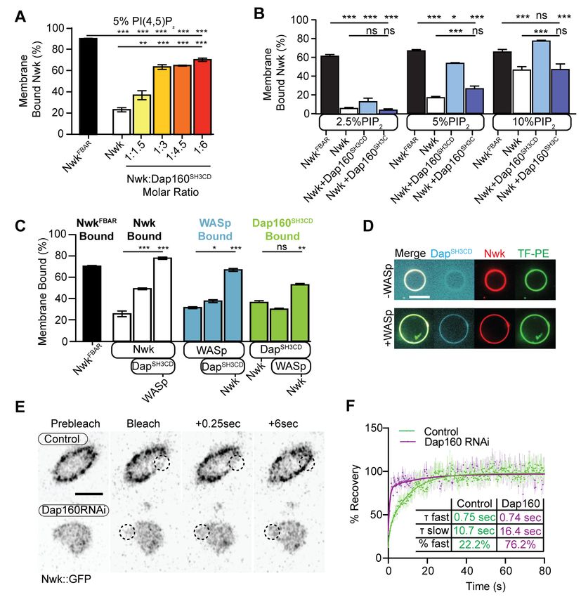
Research article                                                                                      Cell Biology Neuroscience

                                    eLife digest Neurons constantly talk to each other by sending chemical signals across the tiny
                                    gap, or ‘synapse’, that separates two cells. While inside the emitting cell, these molecules are safely
                                    packaged into small, membrane-bound vessels. Upon the right signal, the vesicles fuse with the
                                    external membrane of the neuron and spill their contents outside, for the receiving cell to take up
                                    and decode.
                                        The emitting cell must then replenish its vesicle supply at the synapse through a recycling
                                    mechanism known as endocytosis. To do so, it uses dynamically assembling rod-like ‘actin’ filaments,
                                    which work in concert with many other proteins to pull in patches of membrane as new vesicles. The
                                    proteins that control endocytosis and actin assembly abound at neuronal synapses, and, when
                                    mutated, are linked to many neurological diseases. Unlike other cell types, neurons appear to ‘pre-
                                    deploy’ these actin-assembly proteins to synaptic membranes, but to keep them inactive under
                                    normal conditions. How neurons control the way this machinery is recruited and activated remains
                                    unknown.
                                        To investigate this question, Del Signore et al. conducted two sets of studies. First, they exposed
                                    actin to several different purified proteins in initial ‘test tube’ experiments. This revealed that,
                                    depending on the conditions, a group of endocytosis proteins could prevent or promote actin
                                    assembly: assembly occurred only if the proteins were associated with membranes. Next, Del
                                    Signore et al. mutated these proteins in fruit fly larvae, and performed live cell microscopy to
                                    determine their impact on actin assembly and endocytosis.
                                        Consistent with the test tube findings, endocytosis mutants had more actin assembly overall,
                                    implying that the proteins were required to prevent random actin assembly. However, the same
                                    mutants had reduced levels of endocytosis, suggesting that the proteins were also necessary for
                                    productive actin assembly. Together, these experiments suggest that, much like a mousetrap holds
                                    itself poised ready to spring, some endocytic proteins play a dual role to restrain actin assembly
                                    when and where it is not needed, and to promote it at sites of endocytosis.
                                        These results shed new light on how neurons might build and maintain effective, working
                                    synapses. Del Signore et al. hope that this knowledge may help to better understand and combat
                                    neurological diseases, such as Alzheimer’s, which are linked to impaired membrane traffic and cell
                                    signalling.

                                    mammalian homolog FCHSD2 regulates endocytosis and endocytic traffic in mammalian cells
                                    (Almeida-Souza et al., 2018; Xiao and Schmid, 2020; Xiao et al., 2018). Nwk/FCHSD2 proteins
                                    couple two activities: membrane remodeling and WASp-dependent actin polymerization (Almeida-
                                    Souza et al., 2018; Rodal et al., 2008; Stanishneva-Konovalova et al., 2016). Intramolecular auto-
                                    inhibitory interactions between the Nwk F-BAR and its two SH3 domains mutually inhibit both Nwk
                                    membrane binding and activation of WASp (Stanishneva-Konovalova et al., 2016). Unlike other
                                    F-BAR-SH3 proteins, which are completely released from autoinhibition upon membrane binding
                                    (Guerrier et al., 2009; Meinecke et al., 2013; Rao et al., 2010), the SH3b domain of Nwk continues
                                    to restrict SH3a-mediated WASp activation even after Nwk binds membranes (Stanishneva-
                                    Konovalova et al., 2016). This suggests that autoinhibition allows Nwk-WASp to remain inactive
                                    even after recruitment to the membrane, thus keeping the endocytic machinery in a primed but inac-
                                    tive state. We hypothesized that additional binding partners of NwkSH3b may be required to fully
                                    activate membrane remodeling at discrete times and locations at the synapse.
                                       An excellent candidate for release of Nwk autoinhibition at synapses is the endocytic adaptor
                                    intersectin (Dap160 in Drosophila). Intersectin interacts with numerous endocytic proteins to regulate
                                    endocytosis in mammalian cells (Henne et al., 2010; Okamoto et al., 1999; Praefcke et al., 2004;
                                    Pucharcos et al., 2000; Schmid et al., 2006; Sengar et al., 1999; Teckchandani et al., 2012) and
                                    has been implicated in several steps of the synaptic vesicle cycle (Evergren et al., 2007;
                                    Gerth et al., 2017; Jäpel et al., 2020; Pechstein et al., 2010; Pechstein et al., 2015). Of particular
                                    note, intersectin recruits the Nwk homolog FCHSD2 to sites of endocytosis (Almeida-Souza et al.,
                                    2018), though it is not yet known how this affects FCHSD2 autoinhibition. In Drosophila, Dap160
                                    interacts with WASp, Nwk, and other membrane-remodeling proteins via its four SH3 domains

Del Signore et al. eLife 2021;10:e69597. DOI: https://doi.org/10.7554/eLife.69597                                                   2 of 31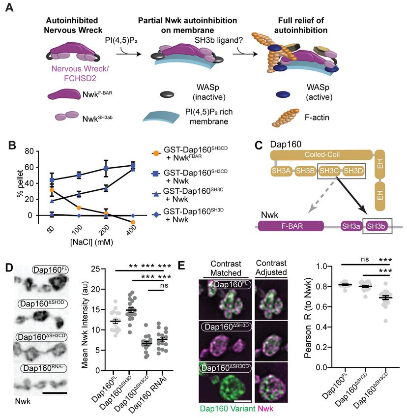
Research article                                                                                      Cell Biology Neuroscience

                                    (SH3AD), and regulates the levels and localization of many of these proteins, including Nwk
                                    (Koh et al., 2004; Marie et al., 2004; Roos and Kelly, 1998). Further, dap160 mutant phenotypes
                                    overlap with those of Nwk and WASp mutants, including impaired synaptic vesicle cycling and syn-
                                    aptic overgrowth (Coyle et al., 2004; Khuong et al., 2010; Koh et al., 2004; Marie et al.,
                                    2004; Winther et al., 2013). Finally, intersectin and Dap160 shift localization from synaptic vesicle
                                    pools to the plasma membrane in response to synaptic activity (Evergren et al., 2007; Gerth et al.,
                                    2017; Winther et al., 2015), suggesting that Dap160 may provide the spatiotemporal link between
                                    salient physiological triggers and Nwk/WASp activation.
                                        The high concentration and broad membrane distribution of inactive endocytic proteins
                                    (Reshetniak et al., 2020; Wilhelm et al., 2014) make it difficult to characterize the molecular
                                    dynamics of synaptic endocytosis (in contrast to non-neuronal cells; Kaksonen and Roux, 2018). To
                                    overcome this barrier, we quantified discrete actin assembly events at the Drosophila NMJ as a
                                    proxy for productive endocytosis, as actin assembly is both a primary target of the endocytic appara-
                                    tus under investigation and is required for synaptic vesicle endocytosis in all forms, including at the
                                    Drosophila NMJ (Kononenko et al., 2014; Wang et al., 2010; Wu et al., 2016). This synapse is an
                                    ideal system to investigate the molecular dynamics of the endocytic machinery due to its large size,
                                    ease of genetic manipulation, and accessibility to live and super-resolution imaging. Here we com-
                                    bine in vitro biochemical approaches with quantitative imaging at the NMJ to define the interactions
                                    among Dap160, Nwk, and WASp that relieve autoinhibition. These interactions drive robust mem-
                                    brane-associated actin assembly in vitro, regulate the frequency and dynamics of synaptic actin
                                    structures in vivo, and are functionally required for normal endocytosis at the NMJ.

                                    Results
                                    Actin assembles in discrete dynamic patches despite broad distribution
                                    of presynaptic membrane-cytoskeleton-remodeling machinery
                                    While the importance of actin in synaptic endocytosis is clear (Kononenko et al., 2014; Wang et al.,
                                    2010; Wu et al., 2016), until now there has been no quantitative analysis of individual actin-depen-
                                    dent membrane-remodeling events at synapses. To better understand presynaptic F-actin dynamics
                                    and to identify sites where the cytoskeleton- and membrane-remodeling machinery is active, we
                                    quantified individual F-actin assembly events by spinning disc confocal microscopy of NMJs presyn-
                                    aptically expressing fluorescent actin probes. To control for developmental variation, all experiments
                                    were performed on late third-instar larvae (~96–120 hr after egg laying) on muscle 6/7 NMJs at
                                    abdominal segments 3–4, since the development and physiology of these synapses are well charac-
                                    terized (Harris and Littleton, 2015). To control for variation in size between neurons, we normalized
                                                                                         patch frequencies by the synapse area measured
                                                                                         and presented data per 10 mm2, which is approx-
                                                                                         imately the size of a synaptic bouton in this sys-
                                                                                         tem. We performed these experiments under
                                                                                         resting conditions, where vesicle release is spon-
                                                                                         taneous at a rate of ~5–6 vesicles/10 mm2/min
                                                                                         (Akbergenova et al., 2018; Melom et al.,
                                                                                         2013), presumably requiring a similar rate of
                                                                                         compensatory endocytosis (Sabeva et al.,
                                                                                         2017).
                                                                                            We first compared the dynamics of three
                                                                                         actin markers: GFP::actin, GFP-tagged moesin
                                                                                         F-actin-binding domain (GMA), and Lifeact::
                                                                                         Ruby. The predominant structures labeled by
                                                                                         these markers were transient patches at the pre-
                                                                                         synaptic membrane (Video 1, Figure 1A, Fig-
                                                                                         ure 1—figure supplement 1A), as has been
                                    Video 1. Dynamics of actin patches labeled by        previously observed (Nunes et al., 2006;
                                    complementary reporters.                             Pawson et al., 2008; Piccioli and Littleton,
                                    https://elifesciences.org/articles/69597#video1      2014). We then quantified individual actin patch

Del Signore et al. eLife 2021;10:e69597. DOI: https://doi.org/10.7554/eLife.69597                                                   3 of 31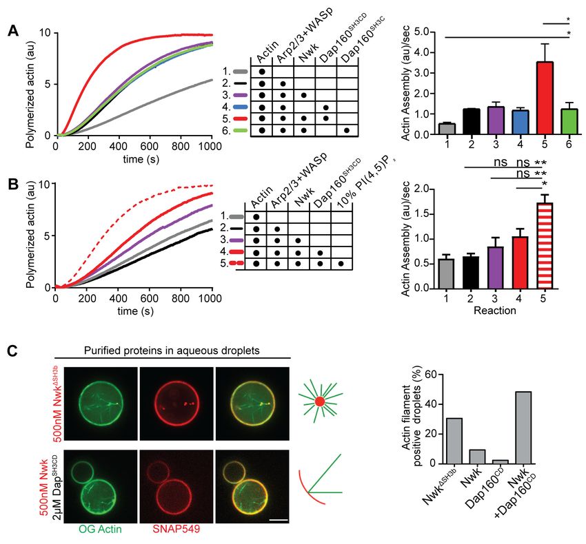
Research article                                                                                                   Cell Biology Neuroscience

Figure 1. Synaptic actin patches are dynamic WASp-dependent structures. (A) Representative maximum intensity projections (MaxIPs) of single
spinning disc confocal microscopy time points, showing C155-Gal4-driven actin probes GFP::actin, GMA, and Lifeact::Ruby. (B–D) Automatic detection
and analysis of movies acquired at 0.25 Hz of F-actin patch intensity amplitude (B), frequency (C), and duration distribution (D) show similar dynamics for
different reporters. (E, F) Single plane Airyscan image of a live muscle 6/7 neuromuscular junction (NMJ) expressing Lifeact::Ruby (magenta) and Arp3::
GFP (green). Actin patches colocalize extensively with Arp3::GFP. (F) Quantification of colocalization by Pearson’s coefficient. Arp3 colocalizes with
Lifeact significantly more than BRP::GFP, a similarly punctate and membrane-associated negative control. Graph shows mean ± sem; n represents
NMJs. (G–I) Patch assembly requires the Arp2/3 activator WASp. GMA patch dynamics in control and WASp mutant animals imaged at 0.25 Hz. (G)
MaxIPs of single spinning disc confocal microscopy time points, showing pan-neuronally expressed GMA localization in control and wsp1 mutant
muscle 6/7 NMJs. (H) Quantification of patch frequency. Graph shows mean ± sem; n represents NMJs. (I) Quantification of patch-duration distribution.
Bins are 20 s; X-axis values represent bin centers. n represents patches. Scale bars in (A) and (G) are 5 mm, and scale bar in (E) is 2.5 mm. Associated
with Figure 1—figure supplement 1, Figure 1—figure supplement 2, and Video 1.
The online version of this article includes the following source data and figure supplement(s) for figure 1:
Source data 1. Source data for Figure 1 and associated figure supplements.
Figure supplement 1. Additional characterization of actin patches.
Figure supplement 2. Actin dynamics analysis is robust to tracking parameters.

                                     dynamics using automated particle tracking and quantification (Berro and Pollard, 2014;
                                     Tinevez et al., 2017), which captured on the order of 30–50% of visible actin structures (see
                                     ’Materials and methods’, Figure 1—figure supplement 2, and Figure 6—figure supplement 1 for
                                     more details on optimization and validation of actin particle analysis). We first imaged at 0.25 Hz
                                     and measured an average of 1.2 GMA patches/10 mm2/min, exhibiting a mean duration of 48.0
                                     s ± 45.6 s, with an average relative amplitude of 68 ± 32% ((Imax-Imin)/Imean) (Figure 1B–D).

Del Signore et al. eLife 2021;10:e69597. DOI: https://doi.org/10.7554/eLife.69597                                                                  4 of 31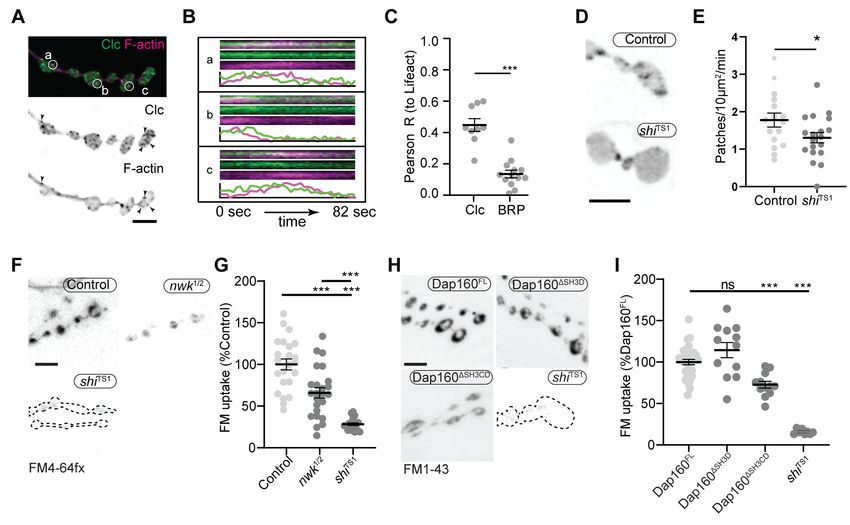
Research article                                                                                               Cell Biology Neuroscience

Figure 2. Periactive zone proteins accumulate broadly across the NMJ. (A) The periactive zone (PAZ) proteins Nwk (magenta) and Dap160 (green)
accumulate in a micron-scale mesh adjacent to active zones (AZ) (Bruchpilot, blue). Image shows maximum intensity projection (MaxIP) of a structured
illumination microscopy (SIM) Z-stack. (B) Surface projection (top) and medial optical section (bottom) SIM images of live-imaged endogenous Nwk::
GFP showing abundant and specific membrane recruitment, similar to fixed imaging. (C–F) PAZ proteins partially colocalize with actin patches. Optical
slices of SIM micrographs showing F-actin (labeled with GMA) localization with presynaptically expressed WASp::Myc and Nwk (C) or Dap160 (E). (D, F)
Quantification of colocalization between GMA and WASp::Myc, and Nwk (D) or Dap160 (F). (D, F) Quantification (Pearson correlation coefficient R) of
colocalization between the indicated pairs of proteins. Graphs show mean ± sem; n represents neuromuscular junctions (NMJs).
The online version of this article includes the following source data for figure 2:
Source data 1. Source data for Figure 2.

                                    Quantification of GFP::actin and Lifeact::Ruby showed very similar dynamics to GMA, suggesting
                                    that these measurements robustly reflect the underlying actin dynamics and not the specific proper-
                                    ties of a particular probe. We did note a high percentage of patches in the minimum duration bin,
                                    suggesting the existence of even briefer patches. To address this, we also performed imaging at 1
                                    Hz, which could not capture the entire lifetime distribution due to photobleaching but was able to
                                    identify a larger population of short-duration patches (Figure 1—figure supplement 1B) with an
                                    average duration of ~16 +/- 20 s. Given this range of measurements at different sampling frequen-
                                    cies and the efficiency of our automated detection, we estimate that patch frequency is between
                                    2.8 and 10.3 events/10 mm2/min (see ’Materials and methods’ for calculations), on par with the
                                    expected frequency of endocytic events, and with a similar albeit broader distribution of durations
                                    compared to yeast (15 s; Berro and Pollard, 2014) and mammalian cells (~40 s; Taylor et al., 2011).
                                       We next examined the molecular determinants of synaptic actin patch assembly. Patches strongly
                                    co-labeled with Arp3::GFP (Pearson coefficient 0.70), significantly higher than the active zone marker
                                    Bruchpilot (BRP), which served as a punctate and membrane-localized negative control (Figure 1E–

Del Signore et al. eLife 2021;10:e69597. DOI: https://doi.org/10.7554/eLife.69597                                                              5 of 31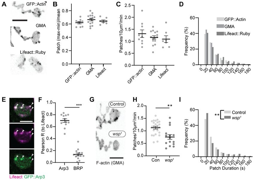
Research article                                                                                                 Cell Biology Neuroscience

Figure 3. Distinct SH3-SH3 and SH3-BAR domain interactions drive Dap160-Nwk association in vitro and at synapses. (A) Model for autoinhibition of
Nwk membrane binding and WASp activation. Neither membrane-bound nor membrane-free Nwk efficiently activates WASp-mediated actin
polymerization, due to persistent SH3b-mediated autoinhibitory interactions, suggesting that an SH3b domain ligand is required for activation. (B)
Dap160SH3CD exhibits electrostatic and hydrophobic interactions with the Nwk F-BAR and SH3 domains, respectively. Glutathione-S-transferase (GST)
fusion proteins were immobilized on glutathione agarose and incubated with the indicated purified proteins. Pellets and supernatants were fractionated
by sodium dodecyl sulphate–polyacrylamide gel electrophoresis (SDS-PAGE), Coomassie stained, and quantified by densitometry. Graphs show the
average ± sem of three independent reactions. [NwkF-BAR] = 1.5 mM, [Nwk] = 0.8 mM, [GST-Dap160SH3CD] = 1.6 mM, [GST-Dap160SH3C/D] = 1.2 mM. (C)
Summary of Dap160SH3CD-NwkSH3ab interactions. Gray and black arrows indicate electrostatic and hydrophobic interactions, respectively. (D, E)
Maximum intensity projection (MaxIP) spinning disc confocal (D) or single Z-plane structured illumination microscopy (SIM) micrographs (E) of muscle 4
neuromuscular junctions (NMJs) expressing C155-GAL4-driven UAS-Dap160 rescue transgene variants in a dap160 null background (dap160D1/Df). Loss
of the Dap160SH3CD domains (Dap160DSH3CD), but not the SH3D domain alone (Dap160DSH3D), decreases the abundance of Nwk (D, right) and Dap160-
Nwk colocalization (E, right) at synapses. Contrast-matched panels in (E) are displayed with the same brightness/contrast. Adjacent panels are contrast-
adjusted per image to facilitate comparison of Nwk-Dap160 colocalization. Graphs show mean ± sem; n represents NMJs. Scale bars in (D) and (E) are 5
mm and 2.5 mm, respectively. Associated with Figure 3—figure supplements 1–2.
The online version of this article includes the following source data and figure supplement(s) for figure 3:
Source data 1. Source data for Figure 3 and associated figure supplements.
Figure supplement 1. Domain specific interactions between Dap160 and Nwk.
Figure supplement 2. Validation of Dap160 transgene rescue and knockdown experiments.

Del Signore et al. eLife 2021;10:e69597. DOI: https://doi.org/10.7554/eLife.69597                                                               6 of 31
Research article                                                                                      Cell Biology Neuroscience

                                    F). These data suggest that actin patches are predominantly composed of branched F-actin, similar
                                    to sites of endocytosis in other cell types (Akamatsu et al., 2020; Collins et al., 2011). To test
                                    whether synaptic actin patches require Arp2/3 activation, we analyzed patch dynamics in larvae lack-
                                    ing the Arp2/3 activator WASp. We compared a genomic mutant (Figure 1G–I), likely hypomorphic
                                    due to maternal contribution (Ben-Yaacov et al., 2001), to presynaptic depletion in neurons
                                    expressing WASp RNAi (Figure 1—figure supplement 1 and C–F). Using both approaches allows
                                    us to distinguish neuron-autonomous from non-autonomous effects of WASP, which is present both
                                    pre- and postsynaptically (Coyle et al., 2004). Both genomic and RNAi manipulations significantly
                                    reduced the number of actin patches, while genomic mutants also skewed the distribution of patch
                                    durations toward both shorter and longer events (Figure 1I). These differences could reflect variable
                                    loss of function between the RNAi and mutant, or identify separable presynaptic autonomous (patch
                                    frequency) vs non-autonomous (patch duration) effects of WASp. Overall, these data clearly indicate
                                    that WASp is autonomously required in neurons to initiate assembly of presynaptic actin patches,
                                    similar to its involvement in endocytosis in yeast, mammalian non-neuronal cells, and in the NMJ
                                    (Hussain et al., 2001; Kessels and Qualmann, 2004; Khuong et al., 2010; Madania et al., 1999).
                                       We next examined the synaptic distribution of two likely WASp regulators, Nwk and Dap160. By
                                    conventional and super-resolution microscopy of neurons in diverse organisms, these and other pre-
                                    synaptic membrane-remodeling proteins localize to a broad membrane domain surrounding active
                                    zones, termed the periactive zone (PAZ) (Coyle et al., 2004; Denker et al., 2011; Gerth et al.,
                                    2017; Koh et al., 2004; Marie et al., 2004; Sone et al., 2000). Consistent with these prior descrip-
                                    tions, we observed by structured illumination microscopy (SIM) that the PAZ proteins Nwk and
                                    Dap160 localize to a membrane-proximal mesh that surrounds active zones, which were labeled with
                                    BRP (Figure 2A). We observed similar results by live imaging of an endogenously tagged Nwk pro-
                                    tein by SIM, which revealed most proteins to be close to the plasma membrane (Figure 2B). We
                                    then compared the localization of PAZ proteins to F-actin patches at the NMJ. As expected, actin
                                    patches were much sparser than the endocytic machinery, and GMA-labeled patches only partially
                                    overlapped with each of the presynaptic WASp, Nwk, and Dap160 (Figure 2C–F; Pearson’s coeffi-
                                    cients of 0.38, 0.38, and 0.36, respectively). These data confirm that, in sharp contrast to the actin
                                    regulatory machinery, which localizes broadly across the PAZ, actin assembly itself is much sparser
                                    both spatially and temporally at the NMJ. This raises the question of how PAZ machinery might itself
                                    be locally regulated to promote the formation of productive synaptic actin assemblies.

                                    Multiple interaction interfaces between Dap160 and Nwk regulate Nwk
                                    autoinhibition
                                    The hypothesis that PAZ protein-mediated actin assembly might be locally activated is particularly
                                    interesting given that we and others have previously shown that autoinhibition of both Nwk and its
                                    mammalian homolog FCHSD2 suppresses both WASp activation and membrane binding (see
                                    Figure 3A for summary model; Almeida-Souza et al., 2018; Rodal et al., 2008; Stanishneva-
                                    Konovalova et al., 2016). These results suggest that transient or localized relief of autoinhibition
                                    could explain how the PAZ controls actin assembly. To determine if and how the candidate activator
                                    Dap160 might relieve Nwk autoinhibition, we first mapped their specific interaction domains using
                                    glutathione-S-transferase (GST) pulldown assays and found that purified Dap160 SH3C-containing
                                    protein fragments (SH3C, SH3CD, or SH3ABCD) directly interact with NwkSH3b, while SH3D alone
                                    does not (Figure 3B, Figure 3—figure supplement 1; see Figure 3—figure supplement 2A for
                                    details of constructs used). Unexpectedly, Dap160 SH3C, SH3D, and SH3CD domain fragments also,
                                    each, interact with the isolated Nwk F-BAR domain (Figure 3—figure supplement 1B). We next
                                    determined how Dap160 interacts with NwkF-BAR compared to a Nwk fragment containing the
                                    F-BAR and both SH3 domains. Dap160-NwkF-BAR interactions were progressively eliminated by
                                    increasing salt, suggesting they are mediated by electrostatic interactions. By contrast,
                                    Dap160SH3CD-Nwk interactions were maintained (Figure 3B, Figure 3—figure supplement 1C), sug-
                                    gesting that the SH3-SH3 interaction is mediated primarily by hydrophobic interactions, consistent
                                    with their mammalian homologs (Almeida-Souza et al., 2018; see summary of interactions in
                                    Figure 3C). Finally, we found that truncation of Dap160SH3CD decreased the levels of Nwk in synap-
                                    tic boutons similarly to Dap160 knockdown (Figure 3D, Figure 3—figure supplement 2B–C).
                                    Dap160DSH3CD also exhibited reduced colocalization with Nwk compared to wild-type Dap160
                                    (Figure 3E, Figure 3—figure supplement 2C), further supporting an in vivo requirement for this

Del Signore et al. eLife 2021;10:e69597. DOI: https://doi.org/10.7554/eLife.69597                                                  7 of 31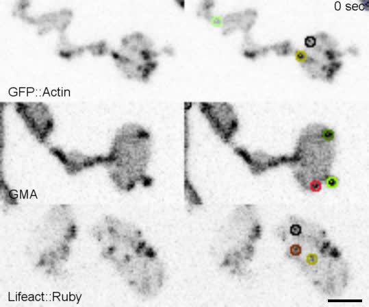
Research article Cell Biology Neuroscience
interaction. Notably, truncation of Dap160SH3D did not exhibit a phenotype in these assays despite
lower levels of expression (Figure 3—figure supplement 2B), suggesting that additional factors
absent from our in vitro assays may collaborate to regulate Nwk in vivo.
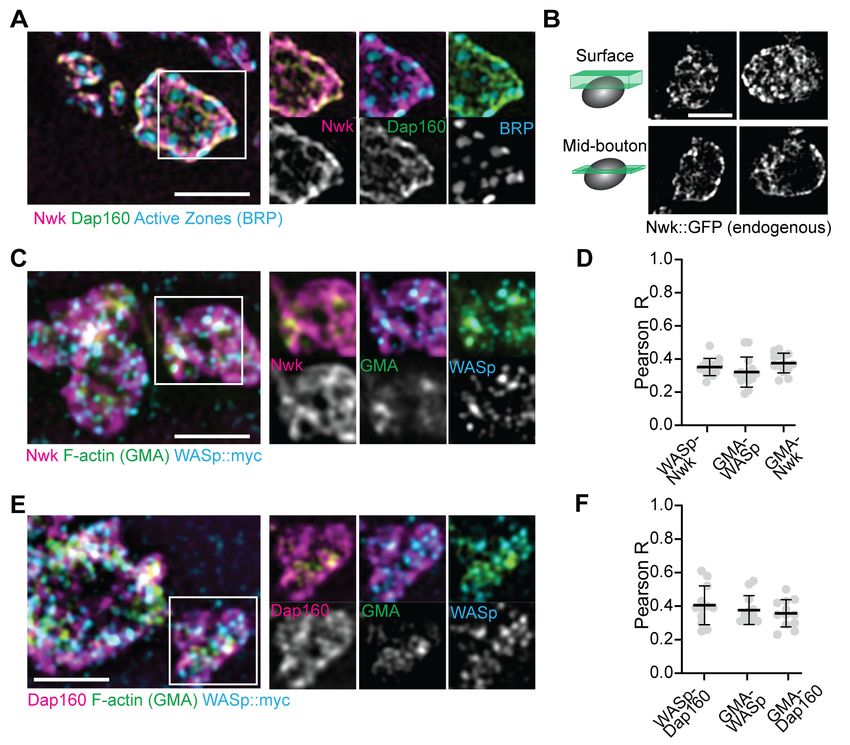
Dap160SH3CD and membranes relieve inhibition of Nwk-WASp-Arp2/3
actin assembly in vitro
We have previously shown that Nwk only weakly activates WASp-dependent actin assembly in vitro,
due to Nwk autoinhibition (Stanishneva-Konovalova et al., 2016). To test whether Dap160SH3CD
Figure 4. Nwk, Dap160, and PI(4,5)P2 potentiate WASp-mediated actin assembly at membranes. (A, B) Pyrene-actin assembly assay (2.5 mM actin, 5%
pyrene-labeled). Curves are representative single experiments demonstrating actin assembly kinetics; graphs represent rates calculated from the linear
range of assembly from at least two independent experiments. (A) The combination (red trace) of Nwk and Dap160SH3CD enhances WASp-Arp2/3-
mediated actin assembly. Either alone (magenta and blue traces) has no effect on WASp activity. [Nwk] = 500 nM, [Dap160] = 2 mM, [WASp] = 50 nM,
[Arp2/3] = 50 nM. (B) PI(4,5)P2 enhances Nwk-Dap160 activation of WASp-mediated actin assembly. Nwk alone or in combination with 10% PI(4,5)P2
liposomes fails to activate WASp, while the addition of Dap160SH3CD and PI(4,5)P2 synergistically enhances WASp-mediated actin assembly. [Nwk] = 100
nM, [Dap160] = 500 nM, [WASp] = 50 nM, [Arp2/3] = 50 nM. (C) Single slices from spinning disc confocal micrographs of water-droplet actin assembly
assay: SNAP-labeled Nwk constructs (red) and Oregon Green actin (green) were mixed with the indicated proteins in an aqueous solution and
emulsified in 97.5% 1,2-diphytanoyl-sn-glycero-3-phosphocholine (DPHPC), 2.5% PI(4,5)P2 in decane. Both deregulated NwkDSH3b and
Nwk + Dap160SH3CD promote F-actin assembly in droplets. However, while Nwk - Dap160SH3CD derived F-actin associates with the lipid interface, de-
regulated NwkDSH3b promotes actin assembly from asters that do not associate with membranes. [Nwk1-xxx] = 500 nM, [Dap160] = 2 mM, [WASp] = 50
nM, [Arp2/3] = 50 nM. Graph indicates percentage of droplets with observable actin filament assembly. Scale bar in (C) is 10 mm.
The online version of this article includes the following source data for figure 4:
Source data 1. Source data for Figure 4.
Del Signore et al. eLife 2021;10:e69597. DOI: https://doi.org/10.7554/eLife.69597 8 of 31Research article Cell Biology Neuroscience
might relieve Nwk autoinhibition, we performed pyrene-actin assembly assays (Figure 4). At moder-
ate Nwk-Dap160 concentrations (500 nM and 2 mm, respectively), Nwk and Dap160SH3CD signifi-
cantly enhanced the rate of WASp-Arp2/3-mediated actin assembly compared to Nwk plus WASp
alone (Figure 4A). This effect is through Nwk, as Dap160SH3CD had no effect on WASp-Arp2/3 in the
absence of Nwk. Further, Dap160 enhancement of Nwk-WASp actin assembly required the
Dap160SH3D domain, further showing that the specific Dap160SH3D-NwkF-BAR interaction relieves
functional Nwk autoinhibition in vitro. Thus, multiple Nwk-Dap160 interactions work together to
relieve autoinhibition of Nwk.
To generate salient physiological force, actin assembly must be coupled to membranes, and neg-
atively charged lipids are an important ligand for both Nwk and WASp. Thus, we next tested
whether addition of PI(4,5)P2-rich liposomes modified actin assembly by Nwk, Dap160, and WASp
(Figure 4B). Indeed, PI(4,5)P2-containing liposomes synergistically activated WASp-mediated actin
assembly in concert with Dap160 and Nwk. By contrast, neither Nwk, PI(4,5)P2, nor Nwk + PI(4,5)P2
on their own were sufficient to activate WASp above baseline (Figure 4B). Since PI(4,5)P2 is also
insufficient to robustly activate either WASp or Nwk under these conditions (Stanishneva-
Konovalova et al., 2016), our data suggest that WASp activation reflects coordinated relief of Nwk
autoinhibition by both Dap160 and membranes. To further explore the coupling between lipid asso-
ciation and actin assembly, we conducted F-actin assembly assays in a droplet assay, in which pro-
tein-containing aqueous droplets are surrounded by a lipid interface, with lipid head groups facing
the aqueous phase (Figure 4C). In this assay, we found that coordinated interactions among Nwk,
Dap160, and WASp directed actin assembly to the lipid interface. By contrast, substitution of Nwk
lacking its autoinhibitory and Dap160-interacting SH3b domain (NwkDSH3b) caused actin to assemble
as free-floating asters (Figure 4C). We have previously found that expression of a similarly deregu-
lated fragment (Nwk1-631) at the NMJ led to diffuse actin filament assembly throughout the synapse
(Stanishneva-Konovalova et al., 2016). Together, these data suggest that NwkSH3b has a dual role
in maintaining autoinhibition via Nwk-F-BAR interactions and permitting actin assembly at specific
synaptic locations via Dap160-mediated activation.
Dap160 and WASp relieve Nwk autoinhibition and promote its
membrane association
Our actin assembly data suggest that membrane recruitment is a critical regulator of the Nwk-
Dap160-WASp complex (Figure 4B–C). To test whether Nwk-Dap160 interactions directly regulate
membrane recruitment, we performed liposome cosedimentation assays. We found that
Dap160SH3CD enhanced Nwk membrane binding in a dose-dependent fashion (Figure 5A). This
effect depended on membrane charge, as Dap160SH3CD significantly enhanced Nwk membrane
binding at both 5 and 10%, but not at 2.5% PI(4,5)P2 (Figure 5B). Only at 10% PI(4,5)P2 did
Dap160SH3CD promote Nwk membrane binding to the same extent as the completely uninhibited
NwkFBAR domain alone, suggesting that membrane charge and intermolecular interactions with
Dap160 together tune Nwk membrane recruitment. Indeed, this effect required the full
Dap160SH3CD-NwkSH3b interaction: Dap160SH3C alone was unable to promote membrane binding by
Nwk, and Dap160SH3CD did not enhance membrane binding of Nwk lacking its Dap160-interacting
SH3b domain (Figure 5—figure supplement 1A). These data further support the hypothesis that
Dap160SH3CD relieves NwkSH3b-mediated autoinhibition.
As we found that Dap160SH3CD is insufficient to fully activate membrane binding by Nwk at inter-
mediate phosphoinositide concentrations (Figure 5A), we asked whether WASp could further
enhance Nwk membrane recruitment. Indeed, the addition of Dap160SH3CD and WASp together
enhanced Nwk membrane association to the level of the isolated F-BAR domain (Figure 5C). More-
over, coordinated binding of all three components resulted in significantly enriched membrane asso-
ciation of both WASp and Dap160 (Figure 5C). We directly observed the coordinated recruitment of
Nwk and Dap160 in the presence of WASp using fluorescently labeled proteins on GUVs
(Figure 5D). Consistent with the direct Dap160-NwkSH3b interaction, we found that deletion of the
NwkSH3b domain abolished both the Dap160SH3CD-dependent increase and the coordinated recruit-
ment of WASp and Dap160 (Figure 5—figure supplement 1A). Notably, addition of Dap160 and
WASp did not change the nature of membrane deformations generated by Nwk (scalloped and
pinched membranes; Becalska et al., 2013), suggesting that Dap160 and WASp together potentiate
Del Signore et al. eLife 2021;10:e69597. DOI: https://doi.org/10.7554/eLife.69597 9 of 31Research article Cell Biology Neuroscience
Figure 5. Dap160SH3CD and WASp promote Nwk membrane association. (A–C) Liposome cosedimentation assays between the indicated purified
proteins and liposomes composed of [mol% = DOPC/DOPE/DOPS/PI(4,5)P2 = 80-x/15/5/x], with x representing PI(4,5)P2 concentration as noted.
Quantification from Coomassie-stained gels represents the mean fraction of total protein that cosedimented with the liposome pellet ± sem. (A) 1:3
Nwk:Dap160SH3CD saturates enhancement of Nwk membrane association at 5% PI(4,5)P2, but not to the level of the isolated Nwk F-BAR alone (NwkF-
BAR
, black bar). [NwkF-BAR] = 3 mM, [Nwk] = 1.125 mM, [Dap160SH3CD] = 1.7–6.8 mM. (B) Dap160SH3CD (but not Dap160SH3C) enhances Nwk association
with membranes at a range of PI(4,5)P2 concentrations. Maximum binding (comparable to NwkF-BAR) occurs only at 5–10% PI(4,5)P2 concentrations.
[Nwk1-xxx] = 2 mM, [Dap160] = 6 mM. (C) Nwk, WASp, and Dap160SH3CD mutually enhance membrane recruitment. Addition of Dap160SH3CD and WASp
additively enhances Nwk membrane association, while Dap160SH3CD and WASp show maximum recruitment to 10% PI(4,5)P2 liposomes in the presence
of both other proteins. [Nwk] = 1 mM, [WASp] = 1 mM, [Dap160SH3CD] = 3 mM. (D) Giant unilamellar vesicle (GUV) decoration assay, with 10% PI(4,5)P2
GUVs labeled withResearch article Cell Biology Neuroscience
rather than alter the inherent activity of Nwk (Figure 5—figure supplement 1D). These data indicate
that Dap160-Nwk SH3-mediated interactions potentiate Nwk association with membranes in vitro.
Finally, to test whether Dap160 promotes Nwk membrane association in vivo, we examined the
dynamics of Nwk at the synapse in the presence and absence of Dap160. Knockdown of Dap160 by
RNAi (Figure 5E, Figure 3—figure supplement 2D) led to a striking loss of endogenously tagged
Nwk::GFP from synaptic membranes (note strong peripheral labeling in control bouton cross-sec-
tions; Figure 5E). Further, Dap160 knockdown significantly increased the rate of recovery of Nwk::
GFP after photobleaching, consistent with a shift in localization from membrane-bound to cytosolic
(Figure 5F). These data suggest that the Dap160SH3CD-Nwk interaction promotes Nwk membrane
association in vivo. Taken together, our data indicate that multiple coordinated interactions between
Nwk, WASp, Dap160SH3CD, and membranes are required to relieve Nwk autoinhibition, allowing for
tight control of membrane-coupled actin assembly in the PAZ.
Dap160-Nwk interactions regulate synaptic F-actin patch dynamics
To determine how these mechanisms direct WASp-mediated actin assembly at the synapse, we mea-
sured actin dynamics in nwk (Figure 6A–C, Video 2) and dap160 domain (Figure 6D–F) mutant
NMJs. We predicted two possible but non-exclusive functions based on the dual roles that we found
Figure 6. Loss of the Dap160-Nwk interaction disrupts actin patch dynamics at synapses in vivo. (A, D) Maximum
intensity projections (MaxIPs) of live spinning disc confocal micrographs of presynaptically expressed GMA in
muscle 6/7 neuromuscular junctions (NMJs) of the indicated genotypes, imaged at 1 Hz. Graphs quantify patch
frequency (B, E) and distribution of patch durations (C, F). Loss of nwk (A–C) or of the Nwk-interacting
Dap160SH3CD domain (D–F) increases the frequency of actin patch assembly. In both cases, there is no change in
the distribution of patch durations (C, F). Scale bars in (A, D) are 5 mm. Associated with Figure 6—figure
supplements 1–2, Video 2.
The online version of this article includes the following source data and figure supplement(s) for figure 6:
Source data 1. Source data for Figure 6 and associated figure supplements.
Figure supplement 1. Analysis of actin dynamics is robust to tracking parameters at 1 Hz imaging.
Figure supplement 2. Validation of actin particle analysis in nwk mutants.
Del Signore et al. eLife 2021;10:e69597. DOI: https://doi.org/10.7554/eLife.69597 11 of 31Research article Cell Biology Neuroscience
for the Nwk-Dap160-WASp module in vitro: if
Nwk and Dap160 are primary activators of
WASp, then loss-of-function mutants are likely to
diminish patch frequency, duration, or intensity.
Importantly, multiple WASp activators exist in
the synaptic endocytic machinery (e.g., Cip4 and
Snx9; Almeida-Souza et al., 2018; Gallop et al.,
2013; Nahm et al., 2010; Ukken et al., 2016),
and therefore, these could make significant con-
Video 2. Loss of nwk increases the frequency of brief
tributions to WASp activation in addition to
actin patches. Nwk. Conversely, if an important function of
https://elifesciences.org/articles/69597#video2 autoinhibition is to ‘clamp’ actin assembly at the
synapse, we expected that loss of Nwk and/or
Dap160 would lead to spurious actin assembly
events by these other WASp regulators. We
found that both nwk and Dap160DSH3CD mutants significantly increased patch frequency (Figure 6B,
E, Figure 6—figure supplement 1), supporting a clamp function for these proteins. We did not
detect a difference in the distribution of patch lifetimes, suggesting that it is the frequency of events,
and not their duration per se, that changes (Figure 6C,F).
We also analyzed actin dynamics using a complementary approach in which we measured the nor-
malized intensity variation (coefficient of variation, CoV) over time across the entire NMJ. Interest-
ingly, the magnitude of variation was significantly higher in nwk mutants (Figure 6—figure
supplement 2A–B), but the area of the NMJ that was highly variant was similar between genotypes,
suggesting that actin assembly is more dynamic in time in these mutants, rather than more extensive
in space (Figure 6—figure supplement 2C). We validated this analysis for its sensitivity in detecting
changes in event frequency by analyzing synthetic data (Figure 6—figure supplement 2D, see
’Materials and methods’ for details). The modeled data suggest that the difference in CoV that we
measured between Control and nwk is consistent with a 43% increase in patch frequency, which is
slightly higher than our measurement by particle tracking (28%; Figure 6A). This complementary
analysis does not rely on particle tracking and makes no assumptions about the nature of actin
dynamics, and is consistent with our particle-based metrics. Thus, we conclude that these pheno-
types are robust to the method of analysis used.
Nwk and Dap160SH3CD are required for normal synaptic vesicle
endocytosis
We next investigated the physiological function of actin patches in vivo. Considering that patch mor-
phology, frequency, and duration resembled endocytic dynamics, we first compared actin patches
with the endocytic adaptors Clc and AP2a. Like other endocytic proteins, both presynaptically
expressed Clc::GFP and endogenously tagged AP2a::GFP were primarily enriched at the plasma
membrane relative to the cytoplasm (Figure 7—figure supplement 1A) and covered a large area
fraction of the membrane, similar to other endocytic proteins (Figure 2). In addition to diffuse signal,
both probes localized to short- and long-lived puncta, a subset of which dynamically colocalized
with actin patches (Figure 7A–C, Figure 7—figure supplement 1B, Video 3). A significant propor-
tion of endogenously labeled AP2 at the NMJ is likely associated with the closely apposed postsyn-
aptic membrane, which accounts for its slightly lower correlation coefficient with Lifeact::Ruby.
Considering that the rates of exo/endocytosis at this synapse at rest are relatively low (see above),
these observations suggest that like other PAZ endocytic proteins, a large pool of membrane-local-
ized clathrin coat and adaptor proteins are not actively engaged in endocytosis. Despite these cav-
eats, we found that actin significantly colocalized with both Clc (Figure 7C) and AP2 (Figure 7—
figure supplement 1C), consistent with a role in endocytosis for these actin-enriched sites. To more
rigorously and functionally test the hypothesis that actin patches are endocytic, we acutely disrupted
endocytic dynamics using the temperature-sensitive dominant-negative dynamin/shiTS1 allele. When
imaged under restrictive conditions, shi disruption decreased the frequency of actin patch dynamics
(Figure 7D–E). Together, these data suggest that a significant fraction of presynaptic actin patches
are associated with endocytosis.
Del Signore et al. eLife 2021;10:e69597. DOI: https://doi.org/10.7554/eLife.69597 12 of 31Research article Cell Biology Neuroscience
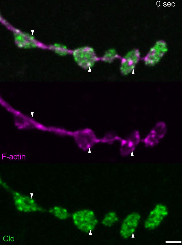
Figure 7. Actin patches and the Nwk-Dap160 interaction are associated with synaptic endocytosis. (A, B) Sum intensity projection (A) and
representative kymographs (B) of spinning disc confocal timelapse of presynaptically expressed Lifeact::Ruby and clc::GFP. (A) Sum projection of 41
frames (82 s) highlights overlapping intensities of clc and Lifeact (circles and arrowheads). Circles indicate locations of kymographs in panel (B). (B)
Kymographs of clc and Lifeact signals. Kymographs span the full duration of the movie from left (0 s) to right (82 s). Intensity profiles were normalized
per channel from the minimum to the maximum value of each profile. (C) Quantification of colocalization between Lifeact::Ruby and Clc::GFP.
Presynaptically expressed Lifeact::Ruby was co-expressed with either presynaptically expressed Clc::GFP or a BRP::GFP knockin and imaged in 3D
stacks by Airyscan. Bruchpilot (BRP) control is the same dataset as in Figure 1F (all data were acquired contemporaneously). (D, E) Normal patch
assembly requires dynamin activity. (D) Maximum intensity projections (MaxIPs) of single spinning disc confocal microscopy time points, showing pan-
neuronally expressed GFP::actin in control and shiTS1 mutant muscle 6/7 neuromuscular junctions (NMJs), imaged at 1 Hz, at the restrictive temperature
of 31oC under stimulating conditions to drive the terminal shiTS1 phenotype (45 mM KCl, 2 mM CaCl2). Graph shows mean ± sem. n represents NMJs.
(E) Quantification of patch frequency. (F–I) FM dye uptake assays at muscle 6/7 NMJs following 5 min of 90 mM potassium stimulation at 36˚C. (F, H)
MaxIPs of spinning disc confocal micrographs of FM dye uptake assays. Dotted lines highlight synapse outline in shiTS1 NMJs. (F, G) nwk mutants
exhibit partially defective FM4-64fx dye uptake relative to shiTS1 mutants. (H, I) Loss of Dap160-Nwk interactions in a Dap160DSH3CD truncation (but not
Dap160DSH3D) exhibits partially defective FM1-43 dye uptake relative to shiTS1, similar to nwk mutants. Graphs show mean ± sem; n represents NMJs.
Scale bars are 2.5 mm (A, B) or 5 mm (D, F, H). Associated with Figure 7—figure supplements 1–3, Video 3.
The online version of this article includes the following source data and figure supplement(s) for figure 7:
Source data 1. Source data for Figure 7 and associated figure supplements.
Figure supplement 1. Colocalization of actin with AP2a and Clathrin light chain.
Figure supplement 2. All Dap160 transgenes rescue dap160 satellite bouton phenotype.
Figure supplement 3. nwk and dap160 domain mutants do not disrupt FM dye unloading.
We next tested the physiological requirement of the Nwk and Dap160SH3CD interaction. As both
Nwk and Dap160 are implicated in the endocytic trafficking of synaptic growth-promoting bone
morphogenetic protein (BMP) receptors (O’Connor-Giles et al., 2008; Rodal et al., 2008), we
tested whether the Dap160-Nwk interaction was required for normal synaptic growth, which we
assayed by counting satellite boutons, a hallmark phenotype of both null mutants. Surprisingly, we
found that both Dap160DSH3D and Dap160DSH3CD truncations rescued satellite bouton numbers to
Del Signore et al. eLife 2021;10:e69597. DOI: https://doi.org/10.7554/eLife.69597 13 of 31Research article Cell Biology Neuroscience
wild-type levels (Figure 7—figure supplement
2). These data indicate that synaptic vesicle and
growth factor endocytosis are mechanistically
separable, and suggest that actin dynamics phe-
notypes in the Dap160DSH3CD mutant are not
associated with synaptic growth regulation. We
next examined synaptic vesicle endocytosis and
recycling by FM dye uptake. nwk1/2 null mutants
led to a 34% decrease in FM4-64fx uptake com-
pared to controls (Figure 7F–G), an intermediate
phenotype compared to dominant negative
dynamin in shiTS1 mutants (72% decrease).
dap160 null mutants have been previously shown
to exhibit an endocytosis defect (Koh et al.,
2004; Marie et al., 2004), so we next tested
whether the interaction between Dap160 and
Nwk is required to support normal endocytosis.
Indeed, we found that expression
of Dap160DSH3CD in dap160 null mutants also sig-
nificantly diminished FM dye uptake to a similar
extent as loss of nwk (27% reduction; Figure 7H–
I). By contrast, loss of the Dap160SH3D domain
alone caused no defects in FM uptake, consistent
with the lack of effect of this mutation on Nwk
Video 3. Clc-GFP and Lifeact::Ruby partially colocalize.
accumulation and localization (Figure 3D–E), sug-
https://elifesciences.org/articles/69597#video3
gesting that this interaction, though required in
vitro, may be compensated by additional factors
in vivo. Both nwk and Dap160DSH3CD mutants
unloaded FM dye to the same extent as controls, suggesting that diminished endocytosis is a direct
phenotype, and not secondary to exocytic deficits (Figure 7—figure supplement 3). Importantly,
these data indicate that spurious actin assembly events in nwk and dap160 mutants are likely to be
unproductive for normal endocytosis. Overall, our data support the hypothesis that normal synaptic
actin patches represent active endocytic events and indicate that Dap160-Nwk regulation of actin
patch dynamics is functionally required for synaptic vesicle endocytosis.
Discussion
Here we have identified a mechanism by which autoinhibition clamps the presynaptic endocytic
machinery to regulate the dynamics of discrete synaptic actin assembly events and the efficiency of
synaptic endocytosis. Our data suggest a model in which specific interactions among Nwk, Dap160,
and WASp function in two ways to potentiate membrane-associated actin dynamics. (1) Persistent
autoinhibition of Nwk allows for stable binding of inactive PAZ machinery to presynaptic membranes
to constrain spurious actin assembly events. (2) Coordinated relief of Nwk autoinhibition by Dap160
and WASp robustly activates F-actin assembly and ensures that actin assembles into structures that
are likely to productively remodel membranes. This provides a mechanism by which synapses can
use the micron-scale PAZ organization of endocytic machinery as a regulated reservoir to efficiently
generate 50- to 100-nm-scale endocytic events, in response to physiological cues such as synaptic
transmission.
Predominant presynaptic actin structures resemble endocytic patches
Here we provide the first quantitative analysis of the composition and dynamics of individual presyn-
aptic F-actin structures. Numerous studies have examined actin dynamics at the level of entire synap-
ses or qualitatively described dynamics of discrete actin structures (Bloom et al., 2003;
Colicos et al., 2001; Nunes et al., 2006; Piccioli and Littleton, 2014; Sankaranarayanan et al.,
2003; Zhao et al., 2013), and identified diverse roles for actin, including synaptic vesicle endocytosis
(Holt et al., 2003; Kononenko et al., 2014; Soykan et al., 2017; Watanabe et al., 2013; Wu et al.,
Del Signore et al. eLife 2021;10:e69597. DOI: https://doi.org/10.7554/eLife.69597 14 of 31Research article Cell Biology Neuroscience
2016; Zhao et al., 2013), synaptic vesicle organization and mobilization (Guzman et al., 2019;
Lee et al., 2012; Marra et al., 2012; Owe et al., 2009; Sakaba and Neher, 2003; Wolf et al.,
2015), active zone organization and function (Pilo Boyl et al., 2007; Morales et al., 2000;
Wagh et al., 2015; Waites et al., 2011; Wang et al., 1999), and receptor-mediated endocytosis
(Kim et al., 2019; Rodal et al., 2008). Bulk analyses, which do not separate individual dynamic actin
structures in space and time, are limited in their ability to discern how the regulation and dynamics
of actin contribute to these distinct functions. We leveraged our ability to extract data describing
individual structures to find that synaptic actin predominantly assembled into discrete Arp2/3-associ-
ated patches, and identified points of control over their dynamics. Specifically, we found that loss of
endocytic proteins differentially affected the frequency and kinetics of individual actin patches, which
correlate with functional deficits in endocytosis.
The link between the actin structures that we observed and endocytic events is supported by sev-
eral lines of evidence: the morphology and duration of synaptic actin patches are similar to WASp/
Arp2/3-dependent endocytic actin dynamics in yeast (16 s; Berro and Pollard, 2014) and somewhat
briefer than in cultured mammalian cells (~40 s; Grassart et al., 2014; Merrifield et al., 2004;
Taylor et al., 2011). Given the measured time constant for endocytosis (~14 s; Poskanzer et al.,
2006) and clathrin dependence of vesicle cycling in this synapse (Heerssen et al., 2008), these val-
ues support the hypothesis that synaptic actin patches are likely sites of clathrin-mediated endocyto-
sis. Further, the frequency of patch assembly, which we measured in resting synapses, approaches
the rate of spontaneous synaptic vesicle release at this synapse (Figure 1—figure supplement 1B,
Figure 1—figure supplement 2A) (~5–6/10 mm2/min; Melom et al., 2013; Akbergenova et al.,
2018). Further, actin patches colocalize partially with endocytic adaptors, and their assembly is sensi-
tive to disruption of endocytosis (Figure 7). Finally, we found that the same endocytic proteins and
protein interactions that regulate endocytosis at this synapse also alter the dynamics of actin
patches.
Technical challenges due to the high density of endocytic proteins and synaptic vesicle cargoes,
and the difficulty of conducting sparse single vesicle measurements at this synapse (compared to
neurons in culture; Chanaday and Kavalali, 2018; Peng et al., 2012), make it difficult to directly link
the dynamics of actin structures to specific membrane or cargo internalization events. However, the
frequency of the events captured by our approach makes it unlikely that they represent rare F-actin-
dependent events at this synapse, such as those that control macropinocytosis or new bouton
growth (Khuong et al., 2010; Kim et al., 2019; Piccioli and Littleton, 2014), and more likely that
they represent bona fide endocytic events. Thus, while we do not rule out other biological functions
for a subset of patches, together our data indicate that a significant and measurable fraction of syn-
aptic actin patches are associated with endocytosis.
Autoinhibition clamps PAZ membrane-remodeling machinery at
synapses
Many endocytic proteins accumulate across broad membrane domains at the Drosophila NMJ and
other synapses (Gerth et al., 2017; Guichet et al., 2002; Koh et al., 2007; Roos and Kelly, 1998;
Verstreken et al., 2008, Verstreken et al., 2003). Our data indicate that much of this membrane-
remodeling machinery is likely held in an inactive state at the presynaptic membrane: Nwk and
Dap160 accumulate in a micron-scale membrane domain (Figure 2), and their loss increases the fre-
quency of short-lived actin patches (Figure 6). These data suggest that these PAZ proteins are held
in a partially autoinhibited state at the membrane in vivo, consistent with our prior in vitro observa-
tions (Stanishneva-Konovalova et al., 2016). This is further consistent with the broad distribution of
Clc and AP2 to the membrane (Figure 7, Figure 7—figure supplement 1). Given the comparatively
low rate of endocytosis expected at rest at this synapse, this suggests that most Clc and AP2 puncta
at the synapse are either not stabilized to form productive endocytic sites (Aguet et al., 2013) or
associated with some non-endocytic functions (Gimber et al., 2015).
The fact that loss of Nwk increases the frequency of patches while decreasing FM uptake sug-
gests that the actin structures assembled in the nwk mutant are unproductive for synaptic vesicle
endocytosis. These spurious patches could reflect non-specific actin assembly, perhaps due to
unmasking of the Nwk ligand PI(4,5)P2 at the membrane and/or inappropriate activation of alterna-
tive WASp-dependent actin assembly pathways. Indeed, additional WASp activators such as Snx9
and Cip4/Toca-1 may play accessory roles in endocytic actin assembly (Almeida-Souza et al., 2018;
Del Signore et al. eLife 2021;10:e69597. DOI: https://doi.org/10.7554/eLife.69597 15 of 31Research article Cell Biology Neuroscience
Gallop et al., 2013), consistent with our finding that loss of presynaptic WASp leads to a decrease
in the total number of patches (Figure 1G–I, Figure 1—figure supplement 1C–E). Our data indicate
that at the synapse, where endocytic machinery accumulates at high concentrations (Wilhelm et al.,
2014) and recruitment appears uncoupled from activation, these layers of autoregulation are critical
to constrain actin assembly generally.
Our findings on autoinhibition and clamping connect two prevailing models of the organization
and function of the synaptic endocytosis machinery—preassembly and delivery. In the first model,
preassembly of clathrin and accessory proteins is hypothesized to ensure fast endocytosis in
response to synaptic vesicle fusion (Hua et al., 2011; Mueller et al., 2004; Wienisch and Klingauf,
2006). Here, Nwk autoinhibition provides a mechanism to assemble an inactive, yet poised endo-
cytic apparatus. In the second model, endocytic machinery associates with the synaptic vesicle pool,
providing a ready source or buffer of proteins that can be released to the plasma membrane upon
calcium signaling or vesicle fusion (Bai et al., 2010; Denker et al., 2011; Gerth et al., 2017;
Winther et al., 2015). Because Dap160/intersectin can shuttle between the synaptic vesicle pool
and the plasma membrane, is itself subject to autoregulation (Gerth et al., 2017), and can regulate
other endocytic proteins (e.g., dynamin, Nwk), it could serve as a single activator that couples the
preassembly and delivery models.
Coordinated relief of autoinhibition directs membrane-associated actin
dynamics
Our in vitro data show that beyond functioning as a clamp, Nwk and Dap160 collaboratively activate
WASp to promote robust actin assembly. Together with the defects we observed in vivo for actin
dynamics and FM dye uptake, these data suggest that Dap160-Nwk-WASp interactions could serve
as a coincidence detection mechanism to relieve autoinhibition of Nwk and promote productive
actin assembly with other WASp activators. Coincidence detection has been demonstrated in several
systems to control membrane-associated actin assembly (Case et al., 2019; Sun et al., 2017), sug-
gesting that amplification of WASp membrane binding could drive robust actin patch assembly at
synapses. Similarly, in human cells, the interaction between FCHSD2, intersectin, and WASp pro-
motes actin assembly and endocytic maturation (Almeida-Souza et al., 2018) or initiation
(Xiao et al., 2018). The Dap160-Nwk module could act by directing and/or organizing actin assem-
bly specifically at endocytic events, akin to the membrane-directed actin assembly we observed in
vitro (Figure 5D), and/or ensure that it is sufficiently robust for productive membrane remodeling
(Akamatsu et al., 2020). Direct support for these models will require new analytical or imaging
approaches to directly visualize the coupling of membranes and actin to the endocytic machinery, in
order to distinguish spurious (due to unclamping) vs bona fide but underpowered endocytic actin
assembly events.
Physiological implications of autoregulatory mechanisms in the
periactive zone
Our data suggest that the endocytic machinery can be deployed as clamped, primed, or activated
complexes at different locations at the synapse. The next critical step will be to determine the mech-
anisms that control switching between these states. Many potential mechanisms that link calcium-
dependent exocytosis and endocytosis could activate actin assembly. These include direct effects of
calcium on the endocytic machinery (Maritzen and Haucke, 2018), the accumulation of synaptic ves-
icle cargoes (Cousin, 2017), stoichiometry-dependent changes in protein interactions or activities
(Case et al., 2019), changes in membrane mechanics (Anantharam et al., 2010; Dai et al., 1997;
Roux et al., 2010), and changes in membrane charge/mode of membrane binding (Kelley et al.,
2015). One intriguing possibility is that these mechanisms might enable an endocytic PAZ to rapidly
switch between different modes of endocytosis (e.g., ultrafast, conventional, or bulk) in response to
a wide range of synaptic activity patterns (Gan and Watanabe, 2018). These endocytic regulatory
mechanisms could also be locally poised to regulate, respond, or adapt to the specific release prop-
erties of nearby active zones (Akbergenova et al., 2018; Melom et al., 2013; Dickman et al.,
2006), and serve as novel points of control over synaptic plasticity and homeostasis.
Del Signore et al. eLife 2021;10:e69597. DOI: https://doi.org/10.7554/eLife.69597 16 of 31You can also read