SARS-COV-2 PORTRAYED AGAINST HIV: CONTRARY VIRAL STRATEGIES IN SIMILAR DISGUISE - PREPRINTS.ORG
←
→
Page content transcription
If your browser does not render page correctly, please read the page content below

Preprints (www.preprints.org) | NOT PEER-REVIEWED | Posted: 8 April 2021                   doi:10.20944/preprints202104.0193.v2

                   Review

                   SARS-CoV-2 portrayed against HIV: contrary viral
                   strategies in similar disguise
                   Ralf Duerr 1,*, Keaton M. Crosse 2, Ana Mayela Valero Jimenez 3 & Meike Dittmann 4
                       Department of Microbiology, New York University School of Medicine, New York, NY, USA;
                     1-4

                     1 Ralf.Duerr@nyulangone.org
                     2 Keaton.Crosse@nyulangone.org

                     3 AnaMayela.ValeroJimenez@nyulangone.org

                     4 Meike.Dittmann@nyulangone.org

                     * Correspondence: Ralf.Duerr@nyulangone.org; Tel.: +1-212-263-4159

                     Abstract: SARS-CoV-2 and HIV are zoonotic viruses that rapidly reached pandemic scale causing
                     global losses and fear. The COVID-19 and AIDS pandemics ignited massive efforts worldwide to
                     develop antiviral strategies and characterize viral architectures, biological and immunological
                     properties, and clinical outcomes. Although both viruses have a comparable appearance as
                     enveloped viruses with positive-stranded RNA and envelope spikes mediating cellular entry, the
                     entry process, downstream biological and immunological pathways, clinical outcomes, and disease
                     courses are strikingly different. This review provides a systemic comparison of both viruses’
                     structural and functional characteristics delineating their distinct strategies for efficient spread.

                     Keywords: SARS-CoV-2; HIV; zoonotic viruses; COVID-19 and AIDS pandemics; viral entry

                   1. Introduction
                        SARS-CoV-2 and HIV each rapidly became and continue to be considerable global health
                   concerns. Unparalleled scientific efforts enabled characterization of these viruses and their resulting
                   diseases in record time, which has led to rapid development of public health measures and antiviral
                   strategies. Both viruses have elementary similarities, being enveloped viruses with a positive (+)
                   single-stranded (ss) RNA genome. Consequently, preventive and therapeutic approaches that were
                   studied or established for HIV have been tested against SARS-CoV-2 including vaccination strategies,
                   reverse vaccinology and monoclonal antibodies (mAbs), and investigational or approved anti-HIV
                   drugs [1-3]. While more data keeps unfolding, we know that despite overall similarities, both viruses
                   have key differences spanning structural and functional characteristics, cellular tropism, induced
                   immune responses, clinical outcome, and responsiveness to vaccines and treatments (Table 1). For
                   SARS-CoV-2, emergency use authorizations of vaccines have been achieved to prevent severe
                   coronavirus disease COVID-19; however, treatment options remain scarce. For HIV, more than 45
                   antiretroviral drugs are available to manage chronic disease and delay AIDS; however, we still lack
                   an efficient vaccine. This review will illustrate the fundamental similarities and differences of SARS-
                   CoV-2 and HIV to further our understanding of their biological and clinical characteristics and thus
                   support the development of antiviral strategies against current and future viral outbreaks. Since HIV-
                   1 is responsible for >95% of global HIV infections and HIV-2 has remained largely restricted to
                   Western Africa [4], this review focuses on HIV-1.

            © 2021 by the author(s). Distributed under a Creative Commons CC BY license.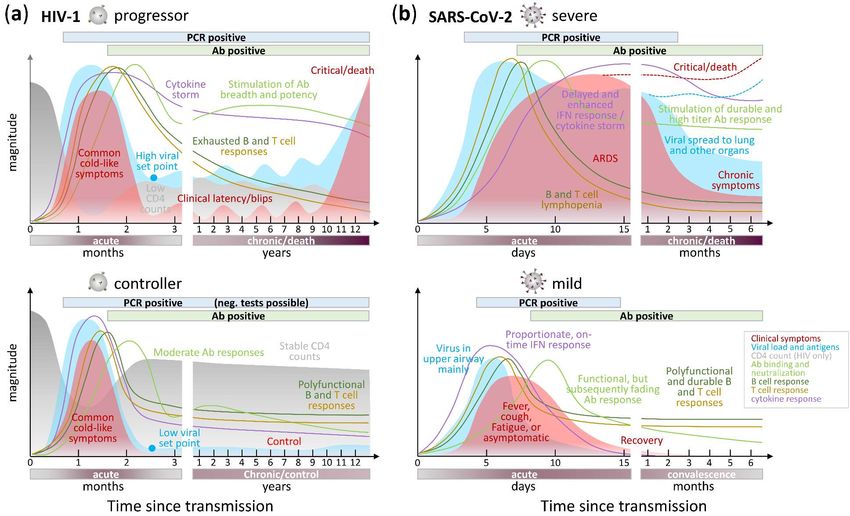
Preprints (www.preprints.org) | NOT PEER-REVIEWED | Posted: 8 April 2021                      doi:10.20944/preprints202104.0193.v2

                                                                                                                                                                         2 of 67

                                              Table 1. Comparison of key features between SARS-CoV-2 and HIV-1 and their associated diseases.

                                              HIV-1                                                        SARS-CoV-2                                                 Refs
                                                                                   Demographic features
          geographic origin                   West-Central Africa (Cameroon, DR Congo)                     China (Wuhan)                                              [5], [6]
          first recorded case                 HIV-1: 1959 (DR Congo)                                       SARS-CoV-2: Nov. 17, 2019 (China)                          [7,8],
                                                                                                                                                                      [9,10]
                                              AIDS: 1981 (USA)                                             COVID-19: Dec. 31, 2019 (China)
          est. time of origin/cross-species   1920s                                                        October/November 2019                                      [7,11],
          transmission                                                                                                                                                [12,13]
          animal source                       non-human primates                                           primary host: bats, intermediate hosts: pangolins or yet   [5], [14-16]
                                                                                                           undefined
          active cases                        38 Mio   b                                                   21 Mio a                                                   [17], [18]
          cases since pandemic start                   b
                                              76 Mio (1.7 Mio new infections in 2019)                      126 Mio    a                                               [17], [18]
          deaths since pandemic start                  b
                                              33 Mio (0.7 Mio in 2019)                                     2.8 Mio   a                                                [17], [18]
                                                                                       Viral features
          Baltimore virus classification      Group VI                                                     Group IV                                                   [19,20]
          Virus family                        Retroviridae                                                 Coronaviridae
          virus diameter                      100-150 nm                                                   60-140 nm                                                  [21],
                                                                                                                                                                      [6,22]
          number of Spikes per virus          7-14                                                         15-40                                                      [23],
                                                                                                                                                                      [22,24]
          Spike size (height x width)         12 x 15 nm                                                   20 x 13 nm                                                 [25],      [26-
                                                                                                                                                                      28]
          Spike amino acids                   856                                                          1273                                                       [29], [30]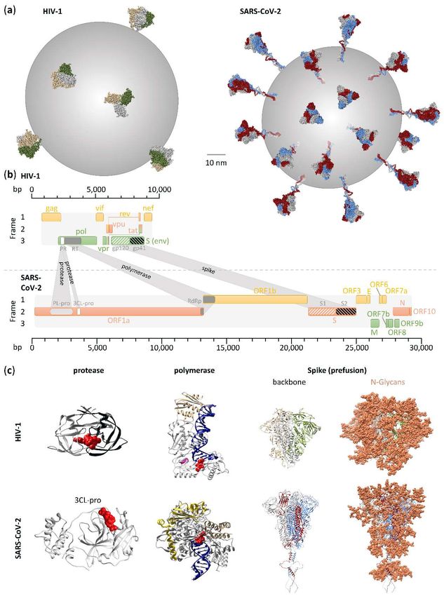
Preprints (www.preprints.org) | NOT PEER-REVIEWED | Posted: 8 April 2021                         doi:10.20944/preprints202104.0193.v2

                                                                                                                                                                        3 of 67

          potential   N-glyco    sites    per    31 (HxB2)                                                  22 (Wuhan-Hu-1)                                          [29], [30]
          Spike monomer
          Spike proteolytic cleavage sites       1                                                          2                                                        [31], [32]
          capsid                                 Conical (many hexagons and 12 pentagons of subunits)       helical                                                  [33],
                                                                                                                                                                     [34,35]
          genome                                 (+)ssRNA, diploid                                          (+)ssRNA, haploid                                        [36], [37]
                                                 dsDNA genome intermediate
          genome size                            9.7 kb (one of the smallest viral genomes)                 29.7 kb (one of the largest viral genomes)               [29], [30]
          evolution rate                         proviral DNA: 4 x 10 E-3 per base per cell (1 mutation     1 x 10 E-3 per base per year (2 mutations per month)     [38,39],
                                                 every 250 base pairs)                                                                                               [12]
                                                 virus in plasma: 2 - 17 x 10 E-3 per base per year
          within host diversity (in the
Preprints (www.preprints.org) | NOT PEER-REVIEWED | Posted: 8 April 2021             doi:10.20944/preprints202104.0193.v2

                                                                                                                                                                     4 of 67

                                        bnAbs require high rates of somatic hypermutation         Potent nAbs do not require high rates of somatic
                                                                                                  hypermutation (SHM), but SHM fosters breadth, potency,
                                                                                                  and resilience to viral escape
          cellular response             impaired B cell, T cell and macrophage/monocyte           impaired B cell, T cell and macrophage/monocyte                 [57],     [58-
                                        responses                                                 responses                                                       62]
          cytokine response             delayed and enhanced anti-inflammatory response,          delayed and enhanced anti-inflammatory response,                [63,64],
                                        impaired IFN response in progressive cases                impaired IFN response in severe cases                           [65-69]
                                                                  Disease features, treatment, and vaccines
          clinical symptoms             AIDS                                                      COVID-19                                                        [48,70,71],
                                        1) initially mild, common cold-like symptoms, Zhu 2020    1) respiratory infection (fever, cough, sore throat, fatigue,   [72-75]
                                        NEJM                                                      loss of smell)
                                        2) acquired immune deficiency and opportunistic           2) systemic dissemination throughout the body (blood
                                        infections and malignancies                               vessels, nervous system, inner organs)
          type of infection             chronic (HIV-1 integrates as provirus into host genome)   acute
          duration of infection         life-long                                                 1-2 months (mild)
                                                                                                  2-9 months (severe) & possible chronic complications
          primary site of infection     lymphatic system of gut and reproductive system           respiratory system
          primary mode of infection     sexual transmission                                       droplet infection of airways
          treatment                     >45 FDA-approved drugs, strong viral-suppressive          emergency use authorization of a few drugs, limited             [76],
                                        effect but no cure                                        clinical     benefit   (dexamethasone,   remdesivir,    nAb     [69,77]
                                                                                                  cocktails)
                                        drugs mainly target the polymerase region (reverse
                                        transcriptase, protease, and integrase)
          vaccine                       no vaccine                                                emergency use authorization of a few vaccines, up to 95%        [78,79],
                                                                                                  vaccine efficacy                                                [80-82]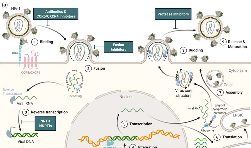
Preprints (www.preprints.org) | NOT PEER-REVIEWED | Posted: 8 April 2021                 doi:10.20944/preprints202104.0193.v2

                                                                                                                                                                    5 of 67

                                        7 vaccine efficacy trials completed, best efficacy: 31%        >200 vaccine trials ongoing
                                        (RV144, 2009)
          correlates of protection      animal models: neutralizing antibodies;                        neutralizing antibodies, supported by cellular responses   [78,83],
                                        human vaccine trial (RV144): ADCC, low plasma anti-                                                                       [84]
                                        Env IgA/IgG, poly-functional B cell responses, non-
                                        neutralizing V2 antibodies
                                                                     a   end of March 2021, b end of 2019.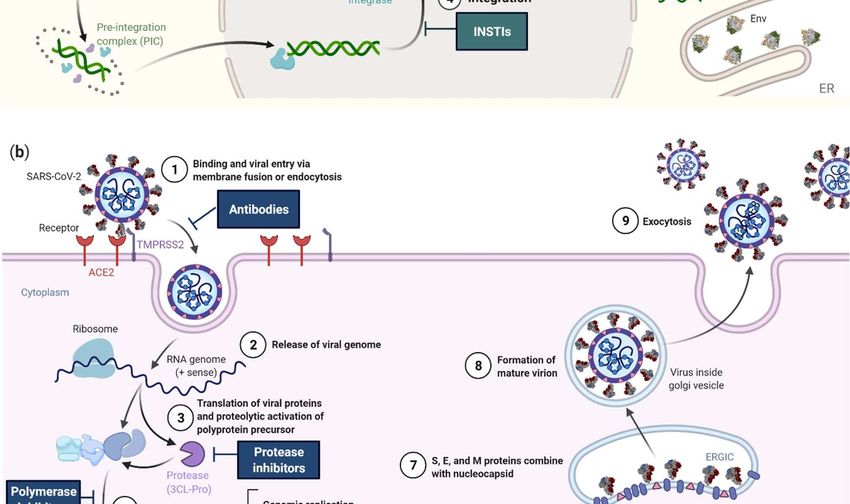
Preprints (www.preprints.org) | NOT PEER-REVIEWED | Posted: 8 April 2021                       doi:10.20944/preprints202104.0193.v2

                                                                                                                        6 of 67

                   2. Methods
                        Structural analyses were done using UCSF Chimera v.1.15rc [85] based on pdb files downloaded
                   from the RCSB protein data bank. Protein structures were generated using pdb files 3j5m or 6wpu
                   (prefusion “closed” HIV-1 Env), S.pdb [86] (prefusion “closed” SARS-CoV-2 Spike trimer), 4l1a (HIV-
                   1 protease in complex with lopinavir), 6wnp (SARS-CoV-2 main protease in complex with
                   Boceprevir), 3v4i/3v81 (HIV-1 reverse transcriptase [RT] in complex with DNA, azathioprine-
                   triphosphate [3v4i; red], and nevirapine [3v81; purple], the latter superimposed after structural
                   overlay of 3v4i and 3v81 RT structures), and 7bv2 (SARS-CoV-2 polymerase RdRp [NSP12] in
                   complex with NSP7, NSP8, template-primer RNA, and Remdesivir triphosphate). Complex entry
                   models were created using structural overlays using MatchMaker in Chimera of pdb structures 6vyb,
                   6m17, and S_ACE2 [86](SARS-CoV-2), or 6met and 5vn3 (HIV-1). Models of fusion intermediates
                   were created based on pdb files 6m3w (SARS-CoV-2) and 2zfc (HIV-1). In silico glycosylation of
                   proteins was done using GlyProt [87], and the composition of oligomannose, hybrid, and complex
                   N-glycans matched with reference literature [88,89]. Viral life cycle schematics were created with
                   BioRender including trimer structures of prefusion “closed” HIV-1 Env (pdb 3j5m) and SARS-CoV-
                   2 Spike (pdb 6vxx) as well as activated, “partially open” HIV-1 Env (pdb 5vn3) and SARS-CoV-2
                   Spike (pdb 6vyb). Viral sequences were downloaded from the Global initiative on sharing all
                   influenza data (GISAID)-EpiCoV and the Los Alamos National Laboratory (LANL) HIV Databases
                   [29,90]. For better comparability, phylogenetics and genetic diversity analyses were done using HIV-
                   1 and SARS-CoV-2 sequences from one entire year, respectively. For SARS-CoV-2, the first year of
                   the pandemic was studied (mid-December 2019 – mid-December 2020), and all full-length sequences
                   with high coverage were downloaded for the studied countries. For HIV-1, we selected a year
                   (January – December) in which a comparable number of HIV-1 unique sequences had been deposited
                   (1.5 log difference) relative to the SARS-CoV-2 data set in the same country. Duplicate HIV-1
                   sequences were removed using the ElimDupes tool from the LANL database. Multiple sequence
                   alignments were performed using Mafft on XSEDE v.7.402 as implemented in the CIPRES Science
                   Gateway v. 3.1 [91]. RAxML maximum likelihood trees were generated using RAxML-HPC v.8 on
                   XSEDE with 1,000 bootstrap replicates on CIPRES. Phylogenetic trees were visualized using FigTree
                   v.1.4.3 [92]. Highlighter plots were created using ten study sequences covering all major branches of
                   the phylogenetic trees against SARS-CoV-2 or HIV-1 reference sequences Wuhan_Hu_1 (EPI ISL
                   402125) or HxB2 (K03455), respectively. Genetic distances were calculated using the ape package
                   (“K80” model without pairwise deletion of sites with missing data) and displayed as heatmaps
                   (upper triangle of all pairwise genetic distances) using the complex heatmap package in program R
                   v.4.0.2 x64 and RStudio v.1.3.959 [93,94]. The longitudinal course of clinical and laboratory
                   parameters was modeled based on available data at the time of the manuscript's completed writing
                   (March 2021) as summarized and cited in the review’s respective sections.

                   3. Operating principles of SARS-CoV-2 and HIV-1
                         Differences in both viruses’ basic functioning extend over their genome organization, repertoire
                   and functioning of viral proteins (see 3.1), and viral life cycle (see 3.2). The latter includes differences
                   in viral entry, specifically their cellular and receptor tropism (see 3.2.1), translation and transcription
                   programs, which, in the case of HIV-1, involve RNA reverse transcription and integration of HIV-1
                   proviral DNA into the host genome (see 3.2.3). Both viruses benefit from their customized annexation
                   of the cellular machinery (see 3.2.3) and have peculiarities in the proteolytic processing of viral
                   proteins (see 3.2.4).

                   3.1. Viral composition
                         Both HIV-1 and SARS-CoV-2 are enveloped viruses with a (+)ssRNA genome and a critical set
                   of structural and functional proteins complemented by a broad spectrum of viral co-factors (Figure
                   1). Despite a ~3-fold difference in genome size, SARS-CoV-2 and HIV-1 virions are similarly sized
                   (~100 nm in diameter). According to the Baltimore virus classification, SARS-CoV-2 belongs to group
                   IV viruses, characterized by (+)ssRNA, whereas HIV-1 is listed among group VI viruses, i.e., ssRNA-
                   RT viruses with (+)sense RNA and a DNA intermediate in the life-cycle [19,20]. More specifically,
                   SARS-CoV-2 belongs to the family of Coronaviridae (realm: Riboviria, order Nidovirales, genus: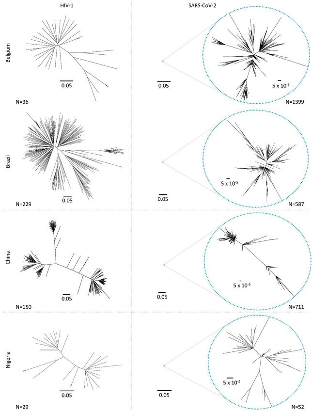
Preprints (www.preprints.org) | NOT PEER-REVIEWED | Posted: 8 April 2021                      doi:10.20944/preprints202104.0193.v2

                                                                                                                       7 of 67

                   Betacoronaviruses, subgenus: Sarbecoviruses, species: Severe acute respiratory syndrome-related
                   coronaviruses), which are the largest known RNA viruses with genomes around 30 kb in size (Figure
                   1, Table 1)[95]. Coronaviruses are named after their crown-like appearance in electron microscopic
                   pictures evoked by their large Spike glycoproteins extending from the roughly spherical virions. HIV-
                   1 belongs to the family of Retroviridae (realm: Riboviria, order: Ortervirales, genus: Lentiviruses)[96].
                   Two copies of HIV-1 RNA are enclosed by a characteristic conical capsid of ~2,000 copies of the viral
                   Gag protein (p24). The capsid is surrounded by a mantle of viral matrix proteins (p17) and a lipid
                   bilayer derived from the infected host cell. The latter is spiked with trimeric viral envelope proteins
                   (Env), the sole viral protein on the HIV-1 surface and the mediator of cellular entry. We frequently
                   refer to this protein also as Spike to align it with SARS-CoV-2 nomenclature. While the SARS-CoV-2
                   Spike number is moderate compared to other enveloped viruses such as influenza, it is even lower
                   for HIV-1 with 14 or fewer Env spikes incorporated into the viral membrane (Figure 1, Table 1)[97].
                   The high plasticity of HIV-1 and SARS-CoV-2, as characteristic for Retroviridae and Coronaviridae,
                   and their potential to tolerate sequence and structural changes without critical loss of function has
                   warranted their zoonotic transmission and ongoing evolutionary success [98,99].
                         The HIV-1 genome codes for nine viral genes (gag, pol, env, vif, vpr, vpu, tat, rev, nef) encoding 19
                   proteins. Besides the structurally and functionally necessary gag, pol, and env genes, the regulatory
                   and accessory tat, rev, nef, vif, vpr, and vpu modulate HIV-1 infection, replication, viral release, and
                   immune recognition [100]. The SARS-CoV-2 genome codes for 14 open-reading frames (ORFs)
                   encoding 29 proteins [101]. The 5’-end of the genome is dominated by the large ORF1a and ORF1b
                   genes comprising more than 2/3 of the entire ~30 kb genome. They encode polyproteins, which, upon
                   translation, are proteolytically processed into 16 non-structural proteins (NSP1–NSP16) that mostly
                   belong to the replicase–transcriptase complex. At the 3′-end of the genome, 13 ORFs are expressed
                   from subgenomic RNAs: besides nine accessory proteins, SARS-CoV-2 encodes four structural
                   proteins, as typical for coronaviruses, i.e., Spike, Envelope (E), Matrix (M), and Nucleocapsid (N), the
                   latter complexing the RNA genome in the absence of a surrounding capsid as known from HIV-1. In
                   contrast to HIV-1, all the three remaining structural SARS-CoV-2 proteins are incorporated into the
                   viral membrane (Spike, E, and M), with Spike mediating viral entry. Besides the canonical ORFs,
                   numerous discontinuous transcription events make the SARS-CoV-2 transcriptome highly complex,
                   including transcripts encoding unknown ORFs with fusion, deletion, and/or frameshift [102].
                         HIV-1 and SARS-CoV-2 encode Spike glycoproteins, comprised of mainly oligomannose N-
                   glycans in HIV-1 and more balanced complex, oligomannose, and hybrid N-glycans in SARS-CoV-2
                   [88,89,103]. The higher number of N-glycans on a smaller Spike protein renders the HIV-1 glycan
                   shield denser than that of SARS-CoV-2, complicating Ab access to critical entry epitopes with
                   consequences for the development of efficient vaccines (Figure 1, Table 1).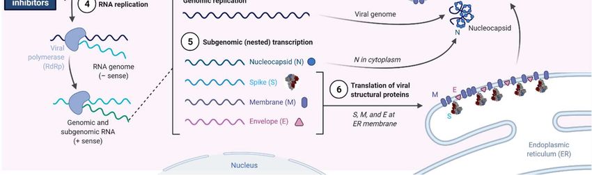
Preprints (www.preprints.org) | NOT PEER-REVIEWED | Posted: 8 April 2021                        doi:10.20944/preprints202104.0193.v2

                                                                                                                          8 of 67

                       Figure 1. Comparison of HIV-1 and SARS-CoV-2 key viral features. (a) HIV-1 and SARS-CoV-2 are
                       enveloped viruses with a diameter of ~100 nm. They are decorated with trimeric Spike proteins that
                       mediate viral entry, yet SARS-CoV-2 Spikes appear in higher numbers than HIV-1 Env (see Table 1).
                       (b) HIV-1 and SARS-CoV-2 possess differently sized, positive-stranded RNA genomes. The HIV-1
                       genome is ~10 kb in size, whereas the SARS-CoV-2 genome spans almost 30 kb. Genomic regions
                       coding for the key functional or structural proteins protease, polymerase, and spike are highlighted
                       in white, gray, and black and white stripes. (c) Three structural and functional proteins highlighted
                       in (b) are also shown as 3D structures (ribbon representation), with bound inhibitors shown in red or
                       purple (sphere representation). Protein subunits are colored differentially. Polymerase-bound
                       RNA/DNA is shown in blue. HIV-1 and SARS-CoV-2 Spike proteins are shown as amino acid
                       backbone structures (left) and glycoproteins (right) with modeled N-glycans (coral; sphere
                       representation).
Preprints (www.preprints.org) | NOT PEER-REVIEWED | Posted: 8 April 2021 doi:10.20944/preprints202104.0193.v2
9 of 67
3.2. Viral replication
Although HIV-1 and SARS-CoV-2 are both enveloped viruses with a (+)ssRNA genome, they
have evolved different strategies to enter their host cells, replicate, and release their progeny (Figure
2). Despite engaging different entry receptors and target cells, HIV-1 and SARS-CoV-2 follow similar
principles of class I glycoprotein-mediated viral fusion and entry (see 3.2.1). However, transcription
and downstream processes are critically different (see 3.2.2). Maybe the most fundamental difference
is that the SARS-CoV-2 life cycle occurs entirely in the cytoplasm, whereas the HIV-1 life cycle
partially occurs in the nucleus. For this reason, HIV-1 replication takes approximately double the
time of SARS-CoV-2 replication. Specifically, HIV-1 reverse transcription was shown to initiate at
around 3 hours post-infection (p.i.), with double-stranded viral cDNA being detectable 2 hours later
[43]. Integration starts 8.5 hours p.i. and all viral transcripts are detectable ~15 hours p.i. The viral
gene expression peak is reached between 20 and 23 hours p.i. with ~0.6% of all transcripts in the cell
demanded by the virus. The release of viral particles stretches over several hours and is initiated at
18 hours p.i. and can continue to 36 hours p.i. in vitro or 60 hours p.i. in vivo (Figure 2a)[43,44]. In
contrast, a SARS-CoV-2 replication cycle takes only ~12 hours in A549 cell and in human airway
epithelial cell cultures (HAEC), and time-of-addition experiments showed that initial translation and
viral replication start simultaneously at between 2-3 hours p.i. (Figure 2b)[47]. The following chapter
presents the life cycles of both viruses with a focus on contrasting these two lifestyles.Preprints (www.preprints.org) | NOT PEER-REVIEWED | Posted: 8 April 2021 doi:10.20944/preprints202104.0193.v2
10 of 67
Figure 2. Replication cycles of HIV-1 and SARS-CoV-2 and major sites of therapeutic intervention.
3CL-pro: 3C-like protease; ACE2: angiotensin-converting enzyme 2; ER: endoplasmatic reticulum;
ERGIC: endoplasmic-reticulum-Golgi intermediate compartment; INSTI: integrase strand transfer
inhibitor; NRTI: nucleoside analog reverse transcriptase inhibitor; NNRTI: non-NRTI; TMPRSS2:
Transmembrane protease serine 2.Preprints (www.preprints.org) | NOT PEER-REVIEWED | Posted: 8 April 2021 doi:10.20944/preprints202104.0193.v2
11 of 67
3.2.1. Viral entry
As the first step of the viral replication cycle, cellular entry is one of the most critical, as it decides
the fate of both the virus and the cell. For this reason, the viral glycoproteins and cellular receptors
that facilitate this process are central targets for vaccines, antibody therapies, and small molecular
drugs (Figure 2). For enveloped viruses, including HIV-1 and SARS-CoV-2, entry begins with an
attachment step to cellular receptors, followed by conformational changes of their receptor-binding
glycoproteins, and is completed with the fusion of viral and host membranes.
Attachment of SARS-CoV-2 and HIV-1 is facilitated by the glycoproteins incorporated within
their viral envelope membranes. The glycoproteins of both SARS-CoV-2 and HIV-1 are trimeric class
I fusion proteins, named Spike or Env (also known as gp160), respectively. Both are composed of an
N-terminal attachment domain (S1 or gp120, respectively) mediating receptor binding and a C-
terminal fusion domain (S2; gp41) consisting of four critical elements enabling viral fusion, i.e., fusion
peptide (FP), heptad repeat A and B (HRA, HRB), and transmembrane domain (Figure 3) [104,105].
To facilitate efficient attachment, both Spike and Env are glycosylated and furin-cleaved during viral
maturation. Glycosylation aids immune evasion and is considerably greater on HIV-1 Env than on
SARS-CoV-2 Spike (Figure 1c). Cleavage of both glycoproteins by the host protease furin generates
non-covalently bound subunits of their respective N- and C-terminal domains, thereby priming each
glycoprotein for engagement with subsequent receptors or host proteases. The cellular receptor for
SARS-CoV-2 is the widely expressed angiotensin-converting enzyme 2 (ACE2)[106], and it binds via
the receptor binding domain (RBD) of Spike (Figure 3)[52,107]. Notably, the additional cell surface
receptor neuropilin-1, which is highly expressed in the respiratory and olfactory epithelium, has been
shown to bind exclusively to the furin-cleaved Spike, potentiating SARS-CoV-2 infectivity in these
tissues [108,109]. Alternatively, SARS-CoV-2 Spike has also been shown to interact with the host cell
receptor CD147 (basigin) to facilitate viral endocytosis [110]. In contrast to SARS-Cov-2, initial
attachment of HIV-1 occurs via binding of gp120 to the cell surface immunoglobin glycoprotein CD4
(Figure 3), which is expressed on subsets of T cell and macrophages [48,111], alleviated by additional
cellular membrane proteins such as integrin α4β7 [112,113].
Upon engagement with their host cell receptors, SARS-CoV-2 Spike and HIV-1 Env undergo
conformational changes to facilitate viral-host membrane fusion. The conformational changes
exhibited by both Spike and Env enable the extension of their hydrophobic fusion peptides, which
are essential for virus-host membrane fusion and subsequent virus entry into the host cytoplasm. The
molecular triggers for these conformational changes are different for both viruses. For SARS-CoV-2,
extension of the fusion peptide within the S2 domain is triggered through cleavage by host cell
proteases at the S2’ site (Figure 3). The canonical entry occurs through membrane fusion directly at
the plasma membrane and involves S2’ cleavage by host protease TMPRSS2 at the cell surface
following ACE2 engagement [114,115]. Alternatively, SARS-CoV-2 can enter via endocytosis and
membrane-fusion-mediated release from endosomes. In support of this second route of entry, the
endosome-localized host protease cathepsin-L has been shown to participate in S2’ cleavage of SARS-
CoV-2 Spike at the endosomal membrane, likely following CD147-mediated endocytosis [110,115].
These alternate mechanisms of SARS-CoV-2 fusion provide the virus with independent and
redundant avenues of entry, which likely contributes to the broad tissue tropism of this virus. In
contrast to SARS-CoV-2, the binding alone of HIV-1 gp120 to its cell surface receptor CD4 is sufficient
for triggering conformational changes. These conformational changes expose and stabilize the
variable loop V3 binding site for co-receptor engagement at the cell surface [111]. CCR5 and CXCR4
can both act as co-receptors for HIV-1. No co-receptors have been identified for SARS-CoV-2. CCR5
or CXCR4 binding to V3 of gp120 induces further conformational changes to gp120, which, after
dissociation of gp120, releases the fusion peptide of bound gp41 for insertion into the host cell
membrane (Figure 3)[116,117]. Subsequent rearrangement of the heptad repeat regions of gp41 bring
the viral and host cell membranes in close proximity for fusion and release of the viral capsid into the
host cell cytoplasm [116]. The released viral capsid of HIV-1 continues to encapsulate the viral
replication components as the pre-integration complex (PIC), until it delivers the HIV-1 dsDNA to
the nucleus. Alternatively, the capsid of SARS-CoV-2 immediately uncoats the viral RNA upon entry
into the cytoplasm.Preprints (www.preprints.org) | NOT PEER-REVIEWED | Posted: 8 April 2021 doi:10.20944/preprints202104.0193.v2
12 of 67
Figure 3. Spike-mediated cellular entry of HIV-1 (top) and SARS-CoV-2 (bottom). Structural model
depicting transition/activation states of viral Spike proteins during viral entry. (1) prefusion “closed”
state, (2) partially “open” state after interaction of Spike proteins with cellular receptors, (3) fusion
intermediates after dissociation of cellular attachment domains gp120 (HIV-1) or S1 (SARS-CoV-2),
which exposes fusion peptides for insertion into the target cell membrane. Schematics of HIV-1 Env
and SARS-CoV-2 Spike coding genomic regions are shown in the middle with domains colored the
same way as shown in the structural models. ACE2: angiotensin-converting enzyme 2, B0AT1:
sodium-dependent neutral amino acid transporter, RBD: receptor-binding domain, FP: fusion
peptide, HR: heptad repeat.Preprints (www.preprints.org) | NOT PEER-REVIEWED | Posted: 8 April 2021 doi:10.20944/preprints202104.0193.v2
13 of 67
3.2.2. Translation, transcription, and reverse transcription
The initial stages of SARS-CoV-2 and HIV-1 replication are remarkably different despite both
viruses starting with positive-stranded RNA genomes. Immediately after uncoating in the cytoplasm,
the SARS-CoV-2 genome acts as mRNA for the translation of two ORFs (Figure 2b). ORF1a and
ORF1b produce polyproteins named pp1a and pp1ab. ORF1a encodes pp1a while the larger pp1ab
is the fusion product of ORF1a and ORF1b, resulting from a -1 ribosome frameshift during translation
[46]. Following their proteolytic cleavage into 16 NSP subunits, these polyproteins comprise the
complete SARS-CoV-2 replicase-transcriptase complex (RTC), responsible for the transcription of the
remaining ORFs as well as the full-length gRNA [46]. Conversely, the HIV-1 genome is reverse-
transcribed into dsDNA during the final stages of entry, a step that is catalyzed by the viral RT
prebound to the viral genome (Figure 2a). Interestingly, this reverse-transcription process is much
more error-prone than the transcription of the SARS-CoV-2 genome, contributing to the
comparatively broad genomic diversity of HIV-1 (Table 1, Figure 4). Still within the HIV-1 capsid in
the form of a PIC, the dsDNA complex is transported to the nucleus, where it is uncoated and released
into the nucleus via the nuclear pore. Within the nucleus, the dsDNA complex is integrated into the
host genome by the viral enzyme Integrase [118-120]. It is only after integration that the HIV-1
genome is transcribed into mRNAs by host enzymes in the nucleus, which are then transported out
and translated in the cytoplasm. HIV-1 achieves productive infection by preferential integration of
its viral genome in intron regions of highly expressed host genes [118,121]. The chronicity of HIV-1
is caused by latent infection of long-lived memory CD4+ T cells and constitutes a major barrier
towards a cure of HIV-1 infection [122]. Latent infection is accomplished by integrating HIV-1 into
transcriptionally silent regions of the genome of quiescent CD4 + T cells [123,124].
The transcription of subgenomic (sg) mRNAs and their subsequent translation also differs
considerably between these two viruses. The SARS-CoV-2 RTC forms at lipid droplet factories within
the cytoplasm [46]. There, the NSP12 RNA-dependent RNA polymerase (RdRp) generates both full-
length negative-strand RNA, which acts as the template for replicating SARS-CoV-2 gRNA and
shorter sgRNAs of the accessory and structural gene-encoding ORFs [46]. Typical of coronaviruses,
transcription regulatory sequences (TRSs) upstream of each of these ORFs prematurely terminate
negative-strand RNA transcription in a process referred to as discontinuous transcription. The
resulting transcripts are a set of structurally polycistronic nested sgRNAs; however, it is assumed
that functionally these transcripts are monocistronic and that only the 5’-most ORF in each sgRNA is
translated [125,126]. Once translated from these sgRNAs, the structural proteins Spike, E, M, and N
package the gRNA to form infectious virions at the ER-to-golgi intermediate compartment for
secretion by exocytosis. In addition to these canonical ORFs encoding structural proteins, recent
ribosomal-profiling has identified 23 translationally active unannotated SARS-CoV-2 ORFs with
currently unknown function [127]. In contrast to SARS-CoV-2, the transcription of the integrated
HIV-1 genome is carried out by cellular polymerases in the nucleus and through means of alternate
splicing gives rise to over 50 viral RNA transcripts [128]. Governed by the cellular export machinery,
only fully spliced RNA transcripts are exported from the nucleus for translation. Hence, the HIV-1
gene products can be separated into early and late based on their necessity for intron retention. The
early viral transcripts rev, tat, and nef are encoded by fully spliced transcripts and consequently, can
be immediately exported for translation, while the remaining genes encoded by partially or unspliced
transcripts rely on sufficient levels of Rev to accumulate. Rev binds HIV-1 intron-containing
transcripts and enables their alternate export to the cytoplasm. The structural Gag and Gag-Pol
polyproteins are translated from the unspliced gRNA and consequently are among the last proteins
translated. Moreover, Gag is translated from the gag gene, while Gag-Pol is a fusion protein generated
by a ribosomal frameshift during translation of the gag gene to the alternate pol reading frame [129].
Once all the HIV-1 structural proteins are translated, virion assembly proceeds at the cell membrane.
Despite their differences in replication strategies outlined above, SARS-CoV-2 and HIV-1 also
share some similarities that may pose universal targets for therapeutic intervention. Each of these
viruses is encapsulated by a replication complex during their replication within the host cytoplasm.
The RNA synthesis of SARS-CoV-2 has been shown to occur in ER-derived double-membrane
vesicles [130], while the reverse-transcription of HIV-1 occurs within the viral capsid/PIC [131]. These
complexes act to segregate immunogenic viral replication intermediates from cytosolic innatePreprints (www.preprints.org) | NOT PEER-REVIEWED | Posted: 8 April 2021 doi:10.20944/preprints202104.0193.v2
14 of 67
immune sensors [132]. Moreover, the translation of the polyproteins of each virus relies on ribosomal
frameshift events. During SARS-CoV-2 replication, this frameshift occurs when translating ORF1a
and ORF1b to give rise to pp1ab, while in HIV-1 replication, a frameshift gives rise to the Gag-Pol
polyprotein. Interestingly, the frameshift efficiency in SARS-CoV-2 is 57% ± 12% giving rise to only
slightly greater expression of pp1a than pp1ab[127], while during HIV-1 replication, the frameshift
efficiency is only 5% which generates 20 times greater expression of Gag than Gag-Pol[133]. The
efficiency of the frameshift during HIV-1 replication is strictly maintained, and any disruption to this
rate is detrimental to virus assembly, genome packaging, and maturation [134-137]. In contrast, the
regulation and implications of the frameshift giving rise to SARS-CoV-2 pp1ab are not as well
characterized, but likely serve similar regulatory purposes for viral protein expression.Preprints (www.preprints.org) | NOT PEER-REVIEWED | Posted: 8 April 2021 doi:10.20944/preprints202104.0193.v2
15 of 67
Figure 4. Phylogenetic diversity of HIV-1 and SARS-CoV-2. Maximum likelihood phylogenetic trees
(RAxML, 1,000 bootstrap replicates) were generated with full length HIV-1 (left) and SARS-CoV-2
(right) genomic sequences from four different countries/continents. For SARS-CoV-2, all available
full-length sequences with high coverage were used, deposited to GISAID within one year since
ignition of the outbreak in mid-December 2019. Comparably, HIV-1 sequences from one entire year
were studied, selected based on comparable case numbers (Preprints (www.preprints.org) | NOT PEER-REVIEWED | Posted: 8 April 2021 doi:10.20944/preprints202104.0193.v2
16 of 67
3.2.3. Virus-host interaction and exploitation of the cellular machinery
Viral pathogens depend on the cellular machinery to propagate. Thus, it is evident that both
HIV-1 and SARS-CoV-2 redirect large parts of the cell-intrinsic biological processes for their
opportunistic use involving several thousands of genes and proteins [43,138-142]. While the complex
network of interactions between these viruses and their host cells has only been disentangled
rudimentarily, the viral strategies of exploiting cellular components and programs appear manifold
in both HIV-1 and SARS-CoV-2 infection. In CD4+ T cells, the primary target of HIV-1, more than 70%
of all expressed genes are modulated in concordance with key steps of HIV-1 viral replication and
more than 50% of the longitudinal variability in the host transcriptome can be explained by
correlations with main phases of the viral life cycle [43]. This leads to a massive change in cellular
physiology with a pronounced early transcriptional shutdown, followed by a progressive, fine-tuned
upregulation of parts of the cellular machinery. These changes support viral processing and
reproduction and are only partly due to the cell’s triggered defense mechanisms. Single-cell
transcriptome studies revealed that HIV-1 targets heterogeneous cells including subpopulations with
low expression of interferon-stimulated genes [121]. Proteomic studies confirmed HIV-1 protein-
mediated surface downregulation of HIV-1 restriction factors such as SERINC3/5 and interference
with CD4+ T cell mitogenesis [139]. A study of ~9000 proteins in primary human CD4 + T cells across
multiple donors revealed 650 HIV-1-dependent changes against the background of T cell activation
[138]. Accessory HIV-1 proteins including Vif, Vpr, Vpu, and Nef played a dominant role accounting
for 46% of the HIV-1-specific proteomic changes in primary T cells.
Similar to HIV-1, SARS-CoV-2 profoundly interacts and modifies the cellular physiology both
on the transcriptional [140-144] and protein level [101,141,145,146]. Host responses to SARS-CoV-2
vary substantially depending on viral load and infection stage as well as age and sex of the host [143].
Coupled to the induction of an antiviral response, the expression of the SARS-CoV-2 receptor and
interferon-responsive gene ACE2 is upregulated by the infected host cell in a viral load-dependent
manner. In contrast, B cell–specific proteins and neutrophil chemokines are elevated in individuals
with lower viral load. Transcriptional levels of the SARS-CoV-2 Spike-processing host protease
TMPRSS2 depend on infection time point and cell type [142,143]. Time series transcriptome profiling
of Calu-3 cells infected in vitro with a clinical SARS-CoV-2 isolate revealed a strong upregulation of
TMPRSS2 mRNA within the very first few hours post infection [142], whereas at later time points,
the levels revert to baseline or even slightly below [142,143]. Males and older individuals exhibit
impaired transcriptional activity affecting trafficking and/or antiviral responses through the reduced
function of cytotoxic T cells, B cells, and natural killer cells [143]. Proteomic studies further showed
that SARS-CoV-2 reshapes cellular translation, splicing, carbon metabolism, protein homeostasis,
and nucleic acid metabolism [145]. In addition to viral proteins, it was shown that SARS-CoV-2 RNA
directly and specifically binds and/or modulates a broad network of human proteins in infected
human cells [147], and host mitochondria serve as an organelle platform for anti-SARS-CoV-2
immunity [148].
3.2.4. Proteolytic processing of viral proteins
A central component of viral replication, including that of SARS-CoV-2 and HIV-1, is the
proteolytic processing of viral proteins, which serves to orchestrate genome replication, assembly
and maturation. This process involves the cleavage of viral proteins mediated by proteases that are
encoded either by the virus or the host. Virally encoded proteases are typically responsible for the
autocatalytic excision from polyproteins in which they reside, and for subsequent proteolytic
processing of the remaining polyprotein components (i.e. 3CL-pro in pp1a/pp1ab or PR in Gag-Pol).
While minimizing coding space within the viral genome, this polyprotein processing also coordinates
synchronized translocation of tethered viral proteins to assembly sites within the host cell [149]. In
addition to processing by viral proteases, viral proteins, including those of SARS-CoV-2 and HIV-1,
are also cleaved by host proteases (i.e. furin), most notably to mediate viral maturation, which primes
progeny virions for efficient entry into new cells. The critical functional role of post-translational viral
protein cleavage proposes this process as an attractive target for antiviral therapeutic development.
Characteristic for viruses with a positive-sense RNA genome, SARS-CoV-2 and HIV-1 both
encode polyproteins that undergo proteolytic processing by virally encoded proteases. However, thePreprints (www.preprints.org) | NOT PEER-REVIEWED | Posted: 8 April 2021 doi:10.20944/preprints202104.0193.v2
17 of 67
replication stage at which the polyprotein proteolytic processing occurs is different between SARS-
CoV-2 and HIV-1, reflecting their alternate replication strategies. For SARS-CoV-2, polyprotein
processing occurs post viral entry and prior to viral replication. The two SARS-CoV-2 polyproteins
pp1a and pp1ab are translated from the incoming positive-sense RNA genome and are proteolytically
processed by two cysteine proteases, papain-like protease (PL-pro) and 3-chromtrypsin-like protease
(3CL-pro, or main protease; M-pro), which reside within NSP3 and NSP5 respectively, and are
released auto-catalytically (Figure 1)[150]. PL-pro catalyzes the cleavage of NSP1-3 and the amino-
terminal of NSP4, while 3CL-pro cleaves the carboxyl terminal of NSP4 as well as the remaining
NSP5-16 [151]. These cleavage events are essential for the subsequent steps of SARS-CoV-2
replication, and consequently, their inhibition through therapeutic intervention is a topic of ongoing
research efforts (Figure 2)[47,152]. In the HIV-1 life cycle, polyprotein processing occurs at later
stages, post viral replication and during viral assembly. Indeed, proteolytic cleavage of HIV-1’s
integral structural and replicative proteins Gag and Gag-Pol typically occurs during virion assembly
and maturation at the cell surface or within the budded virion. Cleavage is performed by the aspartic
HIV-1 protease (PR), itself harbored within the packaged Gag-Pol polyprotein and released auto-
catalytically (Figure 1)[153]. The PR cleavage of Gag generates the main structural proteins MA, CA
and NC which subsequently rearrange to form the mature, infectious particle [154]. The PR cleavage
of Gag-Pol, while also generating the structural proteins, additionally gives rise to the TFP, PR, RT,
RNase H and IN proteins required for initial reverse-transcription and integration upon entry into a
new cell [154]. Incompletely processed HIV-1 polyproteins fail to covert the assembled virus particle
into a mature infectious virion, and HIV-1 PR has consequently been a target of anti-HIV-1 therapies
for numerous years (Figure 2)[154,155].
In addition to polyprotein cleavage by viral proteases, the life cycles of both HIV-1 and SARS-
CoV-2 include proteolytic processing of their receptor-binding glycoproteins, which are performed
by host proteases. These events occur late in the life cycle for both viruses, during a process called
viral maturation. Both viruses utilize the host furin-like proteases for cleavage of their glycosylated
receptor binding proteins within the trans-golgi network during virion assembly. The SARS-CoV-2
Spike protein possesses a furin cleavage site at the S1/S2 junction [156], which has been demonstrated
to be critical for SARS-CoV-2 pathogenicity [114,157-160]. It enhances the binding affinity of Spike to
ACE2 by three orders of magnitude [161], but is not entirely essential for SARS-CoV-2 infection,
possibly by the aforementioned secondary Spike cleavage event which can be mediated by proteases
other than furin [159,160,162]. The HIV-1 Env polyprotein cleavage by furin produces the receptor
binding gp120 and transmembrane gp41 subunits (Figure 3)[163-167]. In contrast to furin-mediated
cleavage of SARS-CoV-2 Spike, the cleavage of HIV-1 Env appears essential for HIV-1 entry and
infection [168-170].
Unlike HIV-1 and most other viruses, SARS-CoV-2 possesses an additional cleavage site within
its receptor-binding glycoprotein spike, named S2’, which facilitates exposure of its fusion peptide
(Figure 3). This second cleavage is mediated by alternate host proteases TMPRSS2 and Cathepsin-L.
While initial discrepancies in findings disputed the contribution of either protease, a recent study has
determined a spatial delineation underpinning their alternate contributions [115]. It is now
understood that both proteases may cleave Spike to facilitate fusion; however, TMPRSS2 acts to
cleave Spike at the cell surface, while Cathepsin-L cleaves Spike within the endosome, which likely
contributes to the tropisms displayed by SARS-CoV-2 [115]. Overall, it is apparent that the maturation
of both viruses employs host proteases and in the case of SARS-CoV-2 also during host cell
recognition, which primes their receptor binding glycoproteins for subsequent entry.
4. Humoral immune responses
Antibody (Ab) immune responses play a central role in protecting the host from viral infections
[171]. Ab-mediated protection is primarily attributed to the Ab binding and neutralization capacity
(see 4.1), complemented by Ab Fc-mediated responses (see 4.2). The Abs’ high specificity to defined
viral epitopes imposes a strong selection pressure, which may favor the selection of viral escape
mutations (see 4.3). In all, the relationship between Abs and viruses uniquely shapes their co-
evolution in virus-infected individuals and entire populations.Preprints (www.preprints.org) | NOT PEER-REVIEWED | Posted: 8 April 2021 doi:10.20944/preprints202104.0193.v2
18 of 67
4.1. Antibody binding and neutralization
In natural HIV-1 infection, HIV-1 specific Abs are elicited within the first weeks of infection.
These include IgM and subsequently class-switched IgG and IgA that are mainly directed against the
immunogenic, highly variable Env gp41 and gp120 regions such as V3 at this time. These early non-
or weakly-neutralizing Abs are narrow, mostly strain-specific, and predisposed to rapid immune
evasion [172-174]. Broadly neutralizing Abs (bnAbs) occur only in a small percentage of HIV-1
infected individuals (~10%), requiring a few years of continuous antigenic stimulation and
maturation, mostly involving high rates of somatic hypermutation [175]. In HIV-1 infected
individuals, the development of bnAbs, or high neutralization levels in general, are not associated
with better clinical outcome/slow progression, but in turn, correlate with severity of disease and high
viral load (Figure 5) [176-178]. Knowledge gained from the tedious process of natural bnAb
development is currently translated into germline-targeting vaccine strategies with sequential
boosting [179]. Similar to SARS-CoV-2, neutralization is considered the lead effector function to
protect from HIV-1 infection, since passive administration of bnAbs in animal models of HIV-1
infection can confer protection against viral challenges [171,180,181]. This strategy is currently tested
in human clinical trials [182,183]. However, while vaccines can induce sufficiently potent bnAbs
against SARS-CoV-2 [184-186], and COVID-19 convalescent individuals acquire protective immunity
through natural infection [187], it has not been possible to induce broadly protective Abs by HIV-1
vaccines [78] and primary HIV-1 infection does not adequately protect from superinfection [188-190].
Ab responses induced in participants of HIV-1 human vaccine trials such as RV144 were mostly non-
or weakly neutralizing, waned rapidly, and/or suffered from rapid viral escape (see 4.3)
[78,83,191,192].
In contrast to HIV-1 infection, natural SARS-CoV-2 infection rapidly induces the generation of
neutralizing Abs (nAbs) encompassing a broad range of heavy chain and light chain V genes [193].
Most individuals develop very similar nAb responses with moderate breadth and plasma
neutralization activity that require only low rates of somatic hypermutation [194]. Prolonged viral
replication in immunocompromised hosts may favor the generation and selection of nAb escape
mutants, which in turn drives Ab affinity maturation and eventually enhanced neutralization breadth
and potency [195]. In humans, previous SARS-CoV-2 infection and anti-spike Ab seropositivity
significantly reduce the risk of SARS-CoV-2 reinfection, [187,196,197], implying that SARS-CoV-2
binding and/or nAbs exert protective effects, which is supported by vaccination outcome analyses
[198-200]. Non-human primate models of SARS-CoV-2 infection corroborated these findings and
suggest that nAbs play the leading role in protection from SARS-CoV-2 infection [84,201]. In rhesus
macaques, Ab-based protection or their therapeutic potential is dose-dependent. Although low Ab
titers were sufficient to protect rhesus macaques from SARS-CoV-2 infection or reinfection, higher
Ab titers were required to achieve a drop in viral load once infected. Furthermore, cellular immunity
contributes to viral control and may compensate waning or insufficient Ab-mediated responses
[84,202] (see 4.1), e.g., CD8+ T cell depletion reduced the protective efficacy of natural immunity
against SARS-CoV-2 reinfection in convalescent animals [84]. The early anti-SARS-CoV-2 binding
and neutralizing Ab response is dominated by IgM, which wanes rapidly. IgA and IgG peak
subsequently, and IgG-mediated neutralizing responses are most durable and persist over months,
mirrored by neutralization half-lives of a few months in serum but up to >8 months in purified IgG
samples [203-206]. The persisting Ab response can be attributed to a maturing humoral immunity
driven by a sustained SARS-CoV-2 antigenic stimulation in the gut of COVID-19 patients [207].
Similar to HIV-1, higher anti-SARS-CoV-2 Ab and neutralization levels are found in severe cases
[54,208-211]. However, COVID-19 survivors exhibit enhanced neutralization potency [208] and a
more balanced Ab maturation pathway [212]. In addition to SARS-CoV-2 Spike, the nucleocapsid
protein (N) and ORFs 3b and 8 are highly immunogenic with implications as serological markers
[213]. Qualitative differences in early Ab profiles point to elevated Ab responses to the N protein in
deceased individuals [214].
Ab immune responses have mainly been studied in the blood, whereas little data exists about
the responses at the local sites of infection such as the respiratory system. Of interest, the mucosal
immune system comprises the largest part of the immune system. On-site production of secretory
IgA (sIgA) by far exceeds all other immunoglobulin isotypes, which renders the mucosa, as site ofPreprints (www.preprints.org) | NOT PEER-REVIEWED | Posted: 8 April 2021 doi:10.20944/preprints202104.0193.v2
19 of 67
viral entry, prepared for the initial wave of adaptive defense [215]. Consequently, anti-SARS-CoV-2
IgG and IgM levels, which mainly transudate from the blood into the mucosa, correlate well between
both compartments. In contrast, IgA was found to be more abundant in the mucosa, particularly early
during disease, which supports the hypothesis that SARS-CoV-2 infection triggers local sIgA
production [216,217].
Combining immunological and epidemiological analyses on seasonal coronaviruses has shown
that infection-blocking immunity wanes rapidly, but disease-reducing immunity is long-lived, which
suggests a model of SARS-CoV-2 transitioning within years to endemicity with mitigated
pathogenicity [218].Preprints (www.preprints.org) | NOT PEER-REVIEWED | Posted: 8 April 2021 doi:10.20944/preprints202104.0193.v2
20 of 67
Figure 5. Courses of natural, untreated HIV-1 and SARS-CoV-2 infection. Estimated models of key clinical, viral, and immune parameters and their longitudinal changes
in representative courses of HIV-1 (a) and SARS-CoV-2 (b) infection. Models of more severe/progressive disease courses are shown on top; mild/slow progressive courses
are shown at the bottom. Features are color-coded according to the legend and key features directly annotated. The curves are estimates based on the state of knowledge
at the time of writing (March 2021), described in more detail in respective sections.Preprints (www.preprints.org) | NOT PEER-REVIEWED | Posted: 8 April 2021 doi:10.20944/preprints202104.0193.v2
21 of 67
4.2. Antibody Fc-mediated functions
Antibody Fc-mediated functions complement Ab neutralization functions and provide a link
between Ab- and cell-based immunity (e.g., NK cells and phagocytes) or soluble effectors (e.g.,
complement) [219,220]. As such, Fc-mediated Ab functions can act hand-in-hand with neutralization
or as an additional line of defense before or after neutralization.
In HIV-1 infection, Fc-mediated effector functions have been studied in detail in recent years,
particularly antibody-dependent cellular cytotoxicity (ADCC) and antibody-dependent cellular
phagocytosis (ADCP) [221,222]. Using a quantitative approach in HIV-1–infected humanized mice
and Simian-HIV (SHIV)-infected rhesus macaques, 25–45% of the total antiviral activity of anti-HIV-
1 mAbs was attributed to Fc-mediated effector functions [223]. In support of that, mAbs with non-
functional Fc-receptors had dramatically decreased capacity to protect animal models from SHIV
infection [224]. Since the isolated depletion of complement binding had no impact on the protective
activity, Fc-mediated cellular responses appear to play the dominant role. Indeed, Fc-mediated
cellular responses such as ADCC and ADCP have been associated with protection from HIV-1 disease
progression and protection from (S)HIV infection in animal models or in a human vaccine trial
[78,221,225,226]. For example, ADCC responses in the presence of low plasma IgA/IgG ratios
correlated with protection from infection in a large human vaccine trial with partial efficacy (RV144)
[83,192,227-229], yet a complete mechanistic explanation remains elusive [78,221]. Furthermore, Fcγ-
phenotyping in vaccinees of the same trial revealed that distinct single-nucleotide polymorphisms
(SNP) in the FCγR2C gene conferred 91% vaccine efficacy against HIV-1 carrying immunodominant
epitopes in Env that experienced vaccine selection pressure. In contrast, individuals with a different
SNP exhibited only 15% vaccine efficacy [230].
Many studies have shown a tight linkage between Ab Fc-mediated effector functions and
neutralization in HIV-1 infections [224,231-235]. A recent study showed that the neutralization
activity of an anti-HIV-1 mAb was potentiated >5,000-fold in vitro when expressing the IgG high-
affinity Fc receptor FCγRI compared to the same mAb without [233]. Moreover, the antisera from
animals immunized with the respective mAb epitope-based vaccine neutralized diverse HIV-1
clades, including more resistant tier-2 viruses, in an FCγRI-dependent manner [233]. Nonetheless,
the mutual impact between Fc-mediated functions and neutralization can vary considerably as it was
shown, for example, that Fc-mediated activity was partially redundant for a very potent bnAb [236],
and differences in antibody binding affinity for HIV-1 and SIV Env uncoupled mAb-mediated ADCC
from neutralization [237]. Fc-mediated functions are influenced by the antigenicity and conformation
of the infecting strain/molecular clone, Ab binding levels, Ab specificity, Ab orientation on the bound
antigen, gp120 shedding, capacity to form multivalent antigen-Ab complexes, degree of
internalization of antigen-Ab complexes, and killer cell receptor ligand expression (e.g., NKG2D)
[221,235,238-241].
In SARS-CoV-2 infection, data on the impact of Fc-mediated effector functions is still unfolding,
but similar to HIV-1, these effector functions appear to be critical [202,242,243]. Studies in non-human
primates demonstrated that Fc-mediated functions correlated with protection from SARS-CoV-2
infection [244]. This was confirmed by studies in mice and hamsters, where nAbs provided better
protection when coupled with Fc-receptor functionality [242,245,246]. Fc-effector functions are
elicited in symptomatic and asymptomatic COVID-19 individuals, but they are elevated in severe
cases [243]. COVID-19 non-survivors had a higher incidence of compromised Fc-receptor binding
and effector functions, implying a crucial role for Ab Fc-effector functions in limiting severe disease
and reducing patient mortality [212]. An in vitro model of ADCC, using full-length Spike proteins
expressed on the surface of a target cell line, and PBMCs from healthy individuals serving as effector
cells, provided additional mechanistic insights. In this model, the ADCC activity of convalescent
plasma decreased only modestly compared to the more pronounced decrease in neutralization
activity. Substantial ADCC activity was maintained in 85% of donors’ plasma up to eight months
post symptom onset and strongly correlated with plasma IgG responses [203]. Notably, three weeks
post-vaccination with an mRNA vaccine, a time point at which vaccine efficacy is estimated to be
>90%, nAb responses are still mostly absent, but anti-SARS-CoV-2 ADCC responses well-developed
[202]. It implies a possible role for Fc-mediated effector functions and other cellular responses inPreprints (www.preprints.org) | NOT PEER-REVIEWED | Posted: 8 April 2021 doi:10.20944/preprints202104.0193.v2
22 of 67
vaccine-mediated protective effects. The collected data so far suggest a vital role for Fc-mediated
effector functions in sustained protection from reinfection and vaccine-induced protection.
4.3. Antibody escape and mutant variants
HIV-1 and SARS-CoV-2 Ab escape is based on similar principles of immune pressure exerted by
Abs on their targeted viral epitopes [247]. However, the strength and timing of the driving immune
forces, and the capacities to evade these forces are very different in both viruses. Important
discriminative factors are the acute nature of SARS-CoV-2 infection, resulting in a small temporal
window of active replication and adaptation, combined with a low mutation rate due to the proof-
reading mechanism of the SARS-CoV-2 polymerase complex. This contrasts with the chronic nature
of HIV-1 infection that allows for a lifelong ongoing viral replication and adaptation with a high
mutation rate in the absence of proof-reading by the HIV-1 polymerase complex. In consequence,
HIV-1 immune escape is a constant factor in almost every HIV-1 infected individual, whereas SARS-
CoV-2 immune escape is rare and seems to occur preferably in immunocompromised individuals
with prolonged viral replication and fostered by treatment with mAbs or convalescent plasma (blood
plasma from a donor who has recovered from COVID-19) (Figure 6)[248-252].Preprints (www.preprints.org) | NOT PEER-REVIEWED | Posted: 8 April 2021 doi:10.20944/preprints202104.0193.v2
23 of 67
Figure 6. (a) Mutational landscape of HIV-1 and SARS-CoV-2. Highlighter plots indicating mutations/mismatches of HIV-1 and SARS-CoV-2 genomes from four studied
countries compared to the references HxB2 (HIV-1, left) and Wuhan-Hu-1 (SARS-CoV-2, right). Base pair mutations are shown as colored tics according to the color codePreprints (www.preprints.org) | NOT PEER-REVIEWED | Posted: 8 April 2021 doi:10.20944/preprints202104.0193.v2
24 of 67
on top. Genome maps are shown at the bottom. Gray diamonds indicate recurrent SARS-CoV-2 mutations occurring in all four countries. Analyses of ten representative
sequences are shown that covered all major branches of the phylogenetic trees in Figure 4. The pairwise genetic distances of the entire set of study sequences per country
are summarized in triangle heatmaps (upper right corner of each panel) with colored ranges from white to yellow, orange, red, pink, and purple according to genetic
distances from low to high (color code indicated on top). The envelope region (env) of HIV-1 and the Spike region (S) of SARS-CoV-2 are indicated by gray bars and arrows.
(b) Variable domains in HIV-1 env (all in gp120) are highlighted in the structural Env trimer model (#6wpu) and the gene map shown below. Variable domains are shown
in sphere representation in the structure, colored in black, and labeled in one monomer. In the gene map, the variable domains are shown in black and labeled. The Env
epitope regions of five major broadly neutralizing antibody classes are indicated by arrows and labeled. (c) Amino acid mutations in globally emerging SARS-CoV-2
variants are shown in a SARS-CoV-2 Spike structure (S.pdb)[86] and a gene map. The mutations are shown in sphere representation and highlighted in one monomer in
black and labeled in the structure. Amino acid replacements are labeled in black and deletions in gray. The mutations in emerging variants of concern are indicated by
circles colored according to the legend to the left; half-circles indicate that mutations occur in only a fraction of variant sequences. The main sites of vulnerability to nAbs
are indicated by gray arrows (RBD and NTD). The two most C-terminal mutations are only shown in the gene map, indicated with a dotted line and labeled in italic, i.e.,
V1176F and M1229I, occurring in P.1 and cluster 5 variants, respectively. Cleavage sites and important amino acid positions, including those of all HIV-1 variable domains
and SARS-CoV-2 NTD and RBD domains, are indicated. CD4bs: CD4 binding site; FP: fusion peptide; MPER: membrane-proximal external region; NTD: N-terminal
domain; RBD: receptor-binding domain; SP: signal peptide; TM: transmembrane domain.You can also read