ZOONOTIC ORIGIN OF SARS-COV-2 - BASHIR MUZAMMIL MUSTAPHA1,2*, ISMAIL RABI ALIYU1, DOGARA MM2, KINJAL BOLIA1, ABHISHEK KUMAR VERMA4, ADAM ...
←
→
Page content transcription
If your browser does not render page correctly, please read the page content below
IOSR Journal of Agriculture and Veterinary Science (IOSR-JAVS)
e-ISSN: 2319-2380, p-ISSN: 2319-2372. Volume 13, Issue 9 Ser. II (September 2020), PP 14-21
www.iosrjournals.org

                                  Zoonotic origin of SARS-CoV-2
      Bashir Muzammil Mustapha1,2*, Ismail Rabi Aliyu1, Dogara MM2, Kinjal
        Bolia1, Abhishek Kumar Verma4, Adam Abdulkadir1, Umar Adamu
                      Hamza3,SaminuAbdullahi Muhammad1
                      1
                       Department of Zoology, Mewar University, Chittorgarh, Rajasthan, India.
                2
                 Department of Biological Sciences, Federal University Dutse, Jigawa State, Nigeria.
                  1
                    Department of Biotechnology, Mewar University, Chittorgarh, Rajasthan, India.
                   1
                     Department of Biochemistry, Mewar University, Chittorgarh, Rajasthan, India.

Abstract
Zoonotic disease is an infectious illness causes by a pathogen that is moved from an animal body to humans.
SARS-CoV-19 is believed to be of zoonotic origin. The virus were detected from throat or rectal swabs of five
domestic cats, one of which had antibodies reactive with SARS CoV. SARS-CoV and MERS are not well adapted
to human, SARS-CoV2 is believed to have evolved from those viruses. These data are consistent with SARS
animal transmission studies which revealed that the respiratory tract of both mice and cats can be
experimentally infected with SARS CoV, although infections in both species remained sub-clinical. SARS-CoV-2
shares 96.2% nucleotide homology with a bat CoV isolated from Rhinolophusaffinisbats. About to 75% of
emerging infectious diseases are found to be zoonotic in nature. Epidemiological and phylogenetic results have
been presented suggesting that SARS evolved from a wild animal host, but no definitive evidence yet exists to
prove this hypothesis. The recently emerged SARS in healthy adults surprised the medical community, but
veterinary coronavirologists have long recognized the potential of CoVs to produce lethal infections in young
animals. Coronaviruses cause a broad range of diseases in both domestic and wild animals, poultry (such as
chickens, turkeys etc.) and rodents ranging from mild to severe enteric, respiratory or systemic disease, as well
as minor colds in humans.
Keywords: Zoonosis, SARS-CoV, SARS-CoV-2, MERS
---------------------------------------------------------------------------------------------------------------------------------------
Date of Submission: 04-09-2020                                                                  Date of Acceptance: 19-09-2020
---------------------------------------------------------------------------------------------------------------------------------------

                                                       I. Introduction
         Zoonotic disease or Zoonosis is derived from greek (Zoon “animal” and nosos “sickness” is an
infectious illness caused by a pathogen (such as Bacterium, Virus, Parasite etc.) that is moved from an animal
body to humans [1].
         Majority of today’s diseases such as Ebola Virus, HIV, and Corona Virus etc. are believed to have been
originated from animal hosts. Of about 1,415 pathogens known to infect humans, 61% were found to be
zoonotic [2]. Most human diseases originated in other animals; however, only diseases that routinely involve
non-human to human transmission, such as rabies, are considered direct zoonosis [3].
         Zoonotic diseases can be transmitted from animals via a number of mechanisms, SARS_COVID19 is
believed to have been brought to humans from animals through hunting and Bush meat.
         Coronavirus disease 2019 (COVID-19) is an infectious disease caused by severe acute respiratory
syndrome coronavirus 2 (SARS-CoV-2). The Human version of this pathogenic illness was identified in
December 2019 in Wuhan, China, and has since spread throughout the globe, resulting in global pandemic [4].
The clinical symptoms of thepathogen include fever, cough, fatigue, inability to breathe smoothly and loss of
smell and taste[5]. Some of these symptoms may develop to Acute Respiratory Distress Syndrome (ARDS),
blood clots etc.[6]. After exposure, the virus may take up five days but it might range from two to fourteen (2-14)
days before it shows manifestation [7].
         The most recent common ancestor of all coronaviruses is estimated to have existed as recently as
8000 BCE, implying long term coevolution with bat and avian species [8]. The most recent origin ofancestry of
alpha-coronavirus line was placed to be around 2400 BCE, ofbeta-coronavirus line at 3300 BCE, of the gamma-
coronavirus line at 2800 BCE, and of the delta-coronavirus line at about 3000 BCE. Bats and birds, as warm-
blooded animals, are considered to be ideal natural reservoir for the coronavirus gene pool (with bats the
reservoir for alpha coronaviruses and beta-coronavirus – and birds the reservoir for gamma-coronaviruses and
delta-coronaviruses). The large number and global range of bat and avian species that host viruses has enabled
extensive evolution and dissemination of coronaviruses.
DOI: 10.9790/2380-1309021421                                     www.iosrjournals.org                                      14 | Page
Zoonotic origin of SARS-CoV-2
The coronaviruses that infect humans were discovered in the 1960s. These viruses were isolated using
two distinct techniques[9] by E.C. Kendall, Malcom Byone, and David Tyrrell working at the Common Cold
Unit in 1960 isolated from a boy a novel common cold virus B814[10]. In 1965, Tyrrell and Byone successfully
cultured the novel virus by passing it through organ culture of human embryonic trachea.The new culturing
technique was introduced to the lab by BertilHoorn[11].The isolated virus when inoculated into volunteers
showed symptoms of cold and was inactivated by ether which indicated it had a lipid envelope.
Coronaviruses (CoVs) are members of the Coronaviridae family, this familyis composed of a group of
enveloped, positive-sensed, single-stranded RNA viruses[12]. The virus contains the largest genome of about 26
to 32 kilo-bases (kb) amongst RNA viruses, they were called“CoVs” because of their crown-like morphology
under high power microscope [12].
On the Basis of the difference in their protein arrangement, CoVs can be grouped into four different
genera (alpha-CoV, betaCoV, gamma-CoV and delta-CoV), whichalso include the beta-CoV genera that
contains most Human Corona Viruses (HCoVs) . Phylogenetic results has shown that bats and rodents are the
gene source of most alpha-CoVs and beta-CoVs, and birds are the main reservoir of gamma-CoVs and delta-
CoVs[12]. For thousands of years, CoVs have constantly crossed species barriers and some have emerged as
important human pathogens[13]. To date, seven human CoVs (HCoVs) are known.
Among them HCoV-229E and HCoV-NL63 are alpha-CoVs. The other five beta-CoVs include HCoV-
OC43, HCoV HKU1, severe acute respiratory syndrome coronavirus (SARS-CoV), Middle East respiratory
syndrome coronavirus (MERS-CoV) and SARS-CoV-2 [14]. HCoV-229E, HCoV-OC43, HCoV-HKU1 and
HCoV-NL63 usually cause moderate symptoms, such as common cold and/or diarrhea [15]. In contrast, SARS-
CoV, MERS-CoV and the newly identified SARS-CoV-2 are very much pathogenic, causing serious lower
respiratory tract infection in patients that have higher vulnerability to develop acute respiratory distress
syndrome (ARDS) and extrapulmonary symptoms.
Table I: Animal coronaviruses: groups, target tissues and types of diseases
Disease/Infected tissue
Enteric
Genetic group Virus Host Respiratory enteric
Other
I Human coronavirus-229E Human X[a] (Upper)
Transmissible gastrointestinal virus Pig X (Upper) x (SI) Viraemia
Porcine respiratory coronavirus Pig X (Upper/Lower)
Porcine epidemic diarrhoea virus Pig x (SI, Colon)
Feline enteric coronavirus Cat x
Feline infectious peritonitis virus Cat X (Upper) x (SI) Systemic
Canine coronavirus Dog x (SI)
Rabbit coronavirus Rabbit Systemic
II Human coronavirus-043 Human X (Upper) BCoV
Mouse hepatitis virus Rat coronavirus Mouse x Liver,CNS
Rat Coronavirus Rat x Eye,SLG
(Sialodocryadenitis)
Haemagglutinating Encephalitis Pig x CNS
Virus
Bovine Coronavirus Cattle x (Upper, lung) x (SI, colon)
III Infectious bronchitis virus Chicken x (Upper) x (no
diarrhea) Oviduct
Turkey coronavirus Turkey x (SI)
IV Severe acute respiratory syndrome Human x (Lung) x? Viraemia?
Civet cat coronavirus Palm Civet x x Subclin?
Raccoon dog coronavirus Raccon Dog ? x Sub clin?
a) no specific information is provided in parentheses, the entire respiratory/gastrointestinal tract is
affected or the specific site of infection is not known
b) bovina coronavirus-like coronavirus from a child (102)
SI Small intestine
? unknown or unreported
DOI: 10.9790/2380-1309021421 www.iosrjournals.org 15 | PageZoonotic origin of SARS-CoV-2
CNS: central nervous system
History of human corona virus
All the four different community-acquired HCoVsthat cause moderate symptoms show great adaptation
to human beings. In other words, both the viruses could be resistant strains of the of ancient HCoV pandemics.
HCoVs that cause higher rate of infections in humans and humans who developed severe HCoV diseases have
been wiped out. For this to occur, HCoVs have to reproduce in humans to sufficient extent to allow the
accumulation of adaptive mutations that counteract host restriction factors. In this sense, the longer the SARS-
CoV-2 outbreak persists and the more people that it infects, the greater chance that it will fully adapt to humans.
If it adapts well, its transmission in humans would be difficult to stop by quarantine or other infection control
measures. For many years, the four community-acquired CoVs circulate in human populations, triggering
common cold in immunocompetent subjects. These viruses do not need an animal reservoir. In contrast, highly
pathogenic SARS-CoV and MERS-CoV have not adapted to humans well and their transmission within humans
cannot be sustained. There is a need to maintain and extend in their zoonotic reservoirs and look for the chance
to spillover to a more susceptible human targets, possibly through one or more intermediate hosts. The
characteristic features of SARS CoV-2 and SARS-CoV/MERS-CoV are almost similar. It is highly
transmissible just like other community-acquired HCoVs, at least for the time being. However,SARS-CoV-2 is
more pathogenic than community-acquired HCoVs and less pathogenic than SARS-CoV or MERS-CoV. It
remains to be seen whether it will adapt fully to humans and circulate within humans without a reservoir or
intermediate animal host[16].
Before we discuss the zoonosis of HCoVs, it will good to talk about the definitions andfeatures of
evolutionary, natural, reservoir, intermediate and amplifying hosts of HCoVs. An animal serves as the
evolutionary host of an HCoV if it sustain a closely related ancestor sharing high genetic similarities at the level
of nucleotide sequence. The ancestral virus is usually well adapted and nonpathogenic in this host. Likewise, a
reservoir host housesHCoV continuously and for a very long term. In both cases, the hosts are naturally infected
and are the natural hosts of HCoV or its parental virus. In contrast, if the HCoV is newly introduced to an
intermediate host right before or around its introduction to humans, it is not well adapted to the new host and is
often pathogenic. This intermediate host can serve as the zoonotic source of human infection and play the role of
an amplifying host by allowing the virus to replicate transiently and then transmitting it to humans to amplify
the scale of human infection. An HCoV can undergo a dead-end infection if it cannot sustain its transmission
within the intermediate host. On the contrary, HCoVs can also adapt to the intermediate host and even establish
long-term endemicity. In this case, the intermediate host becomes a natural reservoir host [16].
History of SARS-CoV
Epidemiological data revealed retrospectively that the index case of SARS had a contact history with
conserved animals [17]. Subsequent investigations indicated that animal traders had a higher prevalence of anti-
SARS CoVIgG compared with that of the general population [17]. Masked palm civets (Pagumalarvata) and a
racoon dog in live animal markets were first identified to carry SARS-CoV-like viruses that are almost identical
to SARS-CoV[18]. This was indirectly supported by the fact that after killing the civets no any further reported
case of CoV. However, it has been reported that masked palm civets from the wild or farms without exposure to
the live animal markets were largely negative for SARS-CoV[19], suggesting that masked palm civets might only
serve as the intermediate amplifying host but not the natural reservoir of SARSCoV. It was noted that, since
80% of the different animals in the markets in Guangzhou have anti-SARS-CoVantibodies[20], the possibilities
that multiple species of small mammals might also serve as intermediate amplifying hosts of SARS-CoV cannot
be excluded.
All of these appear to be dead-end hosts of SARS-CoV. further search for the natural animal host of
SARS-CoV unveiled a closely related bat CoV, termed SARS related RhinolophusbatCoV HKU3 (SARSr-
RhBatCoV HKU3), which was found in Chinese horseshoe bats[21]. These bats are positive for anti-SARS-CoV
antibodies and genome sequence of SARSr-RhBatCoV HKU3 [22]. This and other bat CoVs share 88-92%
nucleotide sequence homology with SARS-CoV. These studies have laid the foundation for the new concept
that bats host emerging human pathogens. Several SARS-like CoVs (SL-CoVs) have also been identified from
bats, but none except for one designated WIV1 can be isolated as live virus [23]. Human angiotensin converting
enzyme 2 (ACE2) is known to be the receptor of SARS-CoV.
Thus far, WIV1 represents the most closely related ancestor of SARS-CoV in bats[24], sharing 95%
nucleotide sequence homology. Albeit the high homology between these two viruses, it is generally believed
that WIV1 is not the immediate parental virus of SARSCoV and bats are not the immediate reservoir host of
SARS-CoV.
DOI: 10.9790/2380-1309021421 www.iosrjournals.org 16 | PageZoonotic origin of SARS-CoV-2
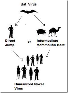
History of SARS-CoV-2
SARS-CoV-2 has about 96.2% similar nucleotide arrangement with a bat CoV isolated from
Rhinolophusaffinisbats[24]. As in the cases of SARS-CoV and MERS-CoV, the sequence divergence between
SARSCoV-2 and bat CoV is too big to assign parental relationship. This shows that, bats might not necessarily
be the immediate reservoir host(s) of SARS CoV-2 unless almost identical bat CoVs are found in future.
Supposedly, the intermediate animal hosts of SARSCoV-2 should comefrom the wildlife species sold and killed
at the Huanan Seafood Wholesale Market, with which many of the initial cases of COVID-19 were associated,
this indicates an assumable animal-to-human transmission event [25]. Several recent studies based on
metagenomic sequencing narrated that a group of endangered small mammals known as pangolins
(Manisjavanica) could also be the host of ancestral beta-CoVs related to SARS-CoV-2[26]. The genomes of
novel pangolin CoV share 85-92% nucleotide sequence homology with SARS CoV-2. However, they are
equally closely related to RaTG13 that has about 90% identity at the level of nucleotide sequence. They are
found in two sub-lineages of SARSCoV-2-like viruses in the phylogenetic tree, one of which has a more similar
receptor binding domain (RBD) with SARS-CoV-2, with 97.4% amino acid sequence identity[26]. In stark
contrast, the RBDs of SARS-CoV-2 and RaTG13 are more divergent, although a higher degree of sequence
homology genome-wide. An earlier analysis on diseased pangolins also reported the detection of viral contigs
from lung samples of the animal, which turn out to be similarly related to SARS-CoV-2[26]. The study adopted
different assembly methods and manual healing to generate a partial genome sequence comprising about 86.3%
of the full length viral genome [27]. The possibility of pangolin being the intermediate animal host of SARS-
CoV-2 cannot be overlooked[26]. However, currently there is insufficient evidence which supports pangolin as
the intermediatehost of SARS-CoV-2 due to the sequence divergence between SARS-CoV-2 and pangolin
SARS-CoV-2-related beta-CoVs. In addition, the distance between SARS-CoV-2 shorter than that between
SARSCoV-2 and pangolin SARS-CoV-2-related beta-CoVs.
The evolutionary pathway of SARS-CoV-2 in bats, pangolins and other mammals remains to be
established. Whereas the highest sequence homology has been found in the RBDs between SARS-CoV-2 and
pangolin, SARS CoV-2-related and beta-CoVs, SARSCoV-2 share the highest genome-wide sequence
homology. It is highly speculative that the high degree of similarity between the RBDs of pangolin SARS-CoV-
2-related beta-CoVs and SARS-CoV-2 is driven by selectivitymediated convergent evolution. A counter-
proposal is in favor of a recombination between a pangolin SARS-CoV-2-related beta-CoV and RaTG13 in the
third wild animal species. As a driving force in evolution, recombination is widespread among betaCoVs [28]. The
jury is still out on the immediate zoonotic origin of SARS-CoV-2.
Wildlife reservoirs
Close to 75% of emerging infectious diseases are of zoonotic origin [1]. Epidemiological and genetic
resultshave been presented suggesting that SARS evolved from aswild animal host, but no definitive evidence
yet exists to prove this hypothesis. Epidemiological observations supporting the theory include the following:
the index patient in Guangxi Province was a wild animal trader, two of seven index patients were restaurant
chefs, food handlers were over-represented in early-onset cases with no contact history and early-onset patients
were more likely to live near agricultural live animal markets [29]. This temporal and spatial clustering of index
cases is consistent with the classical emergence of new agents from animal reservoirs. Based on genetic
analysis, CoVs isolated from two clinically normal wild animal species (Himalayan palm civets, also referred to
as masked palm civets or civet cats [Pagumalarvata] and a raccoon dog (Neyctereutesprocyonoides) from wild
animal markets in Shenzen, the People’s Republic of China, have been assigned as members of the new SARS
CoVgroup [18]. All the animal SARS CoV isolates possessed a 29 nucleotide sequence not found in most human
isolates. Furthermore, the highest IgG antibody titres to SARS CoV were observed in traders of masked palm
civets (72.7%) compared to traders of all live animals (13%) and healthy controls (1.2%) [30]. However, the
reservoir for SARS is still unknown and whether civet cats transmitted SARS CoV to humans or vice versa is
undefined. Nevertheless, these data show that live animal markets (wet markets), which not only exist in the
People’s Republic of China but throughout the world, are likely to have played a vital role in the emergence of
SARS CoV. These live markets are acknowledged as breeding grounds for influenza virus outbreaks such as
that which occurred in Hong Kong in February 2003. The unsanitary crowded conditions, co-mingling of or
close contact among different species of animals and between animals and humans, the carryover of animals and
the introduction of new animals and animalslaughter on the premises, with the dispersion of bloodand secretions
or offal, all foster an environment conducive to the emergence of new zoonotic diseases. In addition, unless
more stringent, but cumbersome virus neutralization tests are conducted to confirm antibody sero-prevalence in
humans and animals, the results using intact SARS CoV or nucleocapsid (N) protein may be suspect. This is
because of the documented antigenic cross-reactivity (enzyme linked immunosorbent assay [ELISA], Western
(blots, immunofluorescence) observed between SARSCoV and animal group I CoVs [31]attributed to the N
protein. These issues hinder definitive analysis of animal reservoirs for SARS. Between December 2003 and
January 2004, several new cases of SARS were re-established in humans in Guangdong Province, the People’s
DOI: 10.9790/2380-1309021421 www.iosrjournals.org 17 | PageZoonotic origin of SARS-CoV-2
Republic of China [32]. In most of these cases there was no link to known risk factors such as civet cats. Other
postulated reservoirs including rats and cats were tested, but no final conclusions were drawn concerning the
origin of this re-emergent case. However, based on sequence data suggesting that the re-emerged SARS strains
were most similar to the civet cat isolates [32], the Government of the country ordered the destruction of large
numbers of civet cats in the wildlife markets in the People’s Republic of China [33].
Further toMetropole Hotel outbreak, a second major outbreak of SARS occurred in 2003 in another
location in Hong Kong at the Amoy Gardens apartments where over 321 people were ultimately infected [34].
This outbreak was clinically more severe and associated with more cases of diarrhoea (73%), higher intensive
care unit admissions (32%) and mortality rates (13%) than the Metropole Hotel outbreak. Environmental factors
(faulty sewage system) were postulated to have contributed to virus spread in the Amoy Gardens via aerosolized
faecal material. However, an alternative hypothesis proposed was that an animal vector, such as roof rats,
infected by the index patient, rapidly spread the disease among the 150 affected households [35]. The authors
further speculated that dual infections of rats with a rat CoV and SARS CoV may have been required to cause a
productive SARS CoV infection in other rats. Indeed, CoV was detected in rodent droppings from the apartment
complex, but since the rodents showed no disease, they were postulated to be mechanical viral vectors [36]. It is
also interesting to note that the virus were detected from throat or rectal swabs of five domestic cats, one of
which had antibodies reactive with SARS CoV[35]. These data are consistent with SARS animal transmission
studies which revealed that the respiratory tract of both mice and cats can be experimentally infected with SARS
CoV, although infections in both species remained sub-clinical. Moreover, both experimentally-infected cats
and ferrets (Mustelofuro) transmitted virus to their contact-exposed cage mates[37]. In the experimental animal
transmission studies performed to date, only cynomologus macaques (Macacacynomologus) and ferrets have
been reported to develop variable disease expression after infection by SARS CoV, with SARS CoV shedding
detected from nasal or pharyngeal swabs [37]. However, in neither species do the clinical signs completely mirror
those of human SARS cases, which include the delayed onset of clinical disease and frequent diarrhea with CoV
shedding in stools [31]. Attempts have been made to experimentally transmit SARS CoV to domestic livestock
and poultry. [38]reported failure to transmit SARS CoV to six-week-old pigs that were seropositive for antibodies
to porcine respiratory CoV (PRCV) (a group I animal CoV). However, the authors detected SARS CoV
ribonucleic acid (RNA) in the blood by reverse transcriptase-polymerase chain reaction (RT PCR) and noted
that the pigs seroconverted with neutralizing antibodies to SARS CoV. This study should be repeated in pigs
seronegative for PRCV or other CoV antibodies because several investigators have noted that antibodies to
PRCV and other animal group I CoVs cross-react with SARS CoV[12]. Whether such preexistingantibodies to
group I CoV interfere with SARS CoVinfection is unclear. Both [38] and [39] also reported lack of SARS CoV
transmission to specific pathogen-free chickens, turkeys, ducks or quail, although again some RT-PCR positive
samples were detected among the exposed poultry.
Picture 1
Evolution SARS-CoV
DOI: 10.9790/2380-1309021421 www.iosrjournals.org 18 | PageZoonotic origin of SARS-CoV-2
The emergence of SARS in healthy adults surprised the medical community, but veterinary
coronavirologists have long recognized the potential of CoVs to produce lethal infections in young animals.
Coronaviruses cause a broad range of diseases in domestic and wild animals, poultry and rodents ranging from
mild to severe enteric, respiratory or systemic disease, as well as minor colds in humans [40]. In livestock and
poultry, CoVs cause mainly localize denteric or respiratory infections, although infectious bronchitis virus
(IBV) of poultry causes both upper respiratory and systemic infections targeting the kidney (interstitial
nephritis) and oviduct (decreased egg production). Coronaviruses are enveloped and possess four major
proteins, i.e. the nucleocapsid (N) protein surrounding the RNA genome and three membrane proteins: the
surface spike (S) glycoprotein, the membrane (M) glycoprotein and the envelope (E) protein [41]. In addition,
some group II CoVs can be distinguished from other CoVs by a surface haemagglutinin (HE), apparent as a
shorter layer of projections on the virion surface compared to the longer S projections. The S protein appears to
be a critical determinant for viral attachment and fusion, cell tropism, species specificity, pathogenicity and
induction of neutralizing antibodies [42]. The CoV genome consists of linear, single-stranded RNA of positive
polarity and ranges from 28 kb-32 kb in length [43]. For CoVs, the large size of the RNA genome, the replication
strategy (nested set of sub-genomic RNAs) and the lack of proof-reading enzymes for RNA replication
(analogous to other RNA viruses), all contribute to the recognized propensity of CoVs to recombine or mutate
and for new strains to emerge. Numerous examples illustrate the emergence of new animal CoV strains or the
mutation of existing strains to produce natural variants or host range mutants. In the late 1970s and into the
1980s, a new group I porcine CoV, the porcine epidemic diarrhea CoV (PEDV), appeared in Europe and rapidly
spread to Asia [44]. The disease resembled TGEV of swine and caused severe diarrhea with major losses of
piglets, before becoming enzootic in swine.
In addition, animalCoVs may acquire new genes via recombination, as demonstrated by the acquisition
of an influenza C-like HE by BCoVor an ancestralCoV[45]. Recombination among CoVs may also generate new
strains with altered tissue or host tropisms. Experimental targetedrecombination between feline and mouse S
protein genesenablesFCoV to infect mice. Recent phylogenetic analysis suggests that SARS CoV may have
evolved from a distant past recombination event between mammalian-like and avian-like parent strains, with the
S gene representing a mammalian (group I)-avian (group III) origin mosaic [46]. This recognition that CoVs can
further evolve in a host population to acquire new tissue tropisms or virulence via mutations or recombination
suggests that similar events may occur if SARS CoV infections persist in humans.
Interspecies transmission of Human Corona Virus
A study in the phylogenetic relation between the CoVs has provided evidence for interspecies
transmission events of HCoVs in the history. When HCoV-OC43 crossed species to infect humans from
domestic livestock around 1890, a pandemic of respiratory infection was recorded [47]. There are no sufficient
explanation to discuss the interspecies transmission of HCoV-229E. Bat alpha-CoVsclosely related to HCoV-
229E have been found. Between them there is an alpaca alpha-CoV. Several lines of evidence support the
transmission of virus from bats to humans directly. First, humans but not alpacas might have contact with bats in
a shared ecological niche. Instead, humans have close contact with alpacas. Second, HCoV229E-related bat
alpha-CoVs are diverse and non-pathogenic in bats, whereas alpaca alpha-CoV caused an outbreak of
respiratory disease in infected animals [49]. Finally, alpaca alpha-CoV has not been found in feral animals. Thus,
the possibility cannot be excluded that alpacas obtain the HCoV-229E-related alpha-CoV from humans. In fact,
bats are the direct source of human pathogenic viruses including rabies virus, Ebola virus, Nipahvirus and
Hendra virus [48]. It is therefore not too surprising that bats might transmit HCoV-229E to humans directly.
Alternatively, whereas bat alpha-CoVs serve as the gene pool of HCoV-229E, alpacas and dromedary camels
might serve as intermediate hosts that transmit viruses to humans, exactly as in the case of MERS-CoV ([48].
II. Conclusion
SARS-CoV-2 is a zoonotic disease that infects human and it is believed to have emerged from two
different viruses that almost the same nucleotide sequence with it, these viruses are Severe Acute Respiratory
Syndrome Corona virus(SARS-CoV) and Middle East Respiratory Syndrome (MERS) which infect cats, there
are several hypothesis which postulate that SARS-CoV-2 has a bush meat reservoir host.
References
[1]. WHO. "Zoonoses". Archived from the original on 3 January 2015. Retrieved 18 December 2014.
[2]. Taylor LH, Latham SM, Woolhouse ME (2001). "Risk factors for human disease emergence". Philosophical Transactions of the
Royal Society B: Biological Sciences. 356 (1411):98989. doi:10.1098/rstb.2001.0888. PMC 1088493. PMID 11516376.
[3]. Marx PA, Apetrei C, Drucker E (October 2004). "AIDS as a zoonosis? Confusion over the origin of the virus and the origin of the
epidemics". Journal of Medical Primatology. 33 (5–6): 220–6. doi:10.1111/j.1600-0684.2004.00078.x. PMID 15525322
[4]. Hui DS, I Azhar E, Madani TA, Ntoumi F, Kock R, Dar O, et al. (February 2020). "The continuing 2019-nCoV epidemic threat of
novel coronaviruses to global health - The latest 2019 novel coronavirus outbreak in Wuhan, China". International Journal of
Infectious Diseases. 91: 264–266. doi:10.1016/j.ijid.2020.01.009. PMC 7128332. PMID 31953166.
DOI: 10.9790/2380-1309021421 www.iosrjournals.org 19 | PageZoonotic origin of SARS-CoV-2
[5]. Hopkins C. "Loss of sense of smell as marker of COVID-19 infection". Ear, Nose and Throat surgery body of United Kingdom.
Retrieved 28 March 2020.
[6]. Bikdeli B, Madhavan MV, Jimenez D, Chuich T, Dreyfus I, Driggin E, et al. (April 2020). "COVID-19 and Thrombotic or
Thromboembolic Disease: Implications for Prevention, Antithrombotic Therapy, and Follow-up". Journal of the American College
of Cardiology. doi:10.1016/j.jacc.2020.04.031. PMID 32311448.
[7]. Velavan TP, Meyer CG (March 2020). "The COVID-19 epidemic". Tropical Medicine &
InternationalHealth. 25 (3):278280. doi:10.1111/tmi.13383. PMC 7169770. PMID 32052514.
[8]. Wertheim JO, Chu DK, Peiris JS, Kosakovsky Pond SL, Poon LL (June 2013). "A case for the ancient origin of
coronaviruses". Journal of Virology. 87 (12):703945. doi:10.1128/JVI.03273-12. PMC 3676139. PMID 23596293.
[9]. Monto AS (1984). "Coronaviruses". In Evans AS (ed.). Viral Infections of Humans. Viral Infections of Humans: Epidemiology and
Control. Springer US. pp. 151–165. doi:10.1007/978-1-4684-4727-9_7. ISBN 978-1-4684-4727-9.
[10]. Kendall EJ, Bynoe ML, Tyrrell DA (July 1962). "Virus isolations from common colds occurring in a residential school". British
Medical Journal. 2 (5297): 82–6. doi:10.1136/bmj.2.5297.82. PMC 1925312. PMID 14455113.
[11]. Tyrrell DA, Fielder M (2002). Cold Wars: The Fight Against the Common Cold. Oxford University Press. pp. 93–95. ISBN 978-0-
19-263285-2
[12]. Sun Z.F. &Meng X.J. (2004). – Antigenic cross-reactivity between the nucleocapsid protein of severe acute respiratory syndrome
(SARS) coronavirus and polyclonal antisera of antigenic group I animal coronaviruses: implication for SARS diagnosis. J. clin.
Microbiol.,42, 2351-2352.
[13]. Zhou P, Yang XL, Wang XG, Hu B, Zhang L, Zhang W, et al. A pneumonia outbreak associated with a new coronavirus of
probable bat origin. Nature. 2020.
[14]. Cui J, Li F, Shi ZL. Origin and evolution of pathogenic coronaviruses. Nat Rev Microbiol. 2019; 17: 181-92.
[15]. Woo PC, Lau SK, Tsoi HW, Huang Y, Poon RW, Chu CM, et al. Clinical and molecular epidemiological features of coronavirus
HKU1-associated community-acquired pneumonia. J Infect Dis. 2005; 192: 1898-907.
[16]. Zi-Wei Ye, Shuofeng Yuan, Kit-San Yuen, Sin-Yee Fung, Chi-Ping Chan, and Dong-Yan Jin (2020) “Zoonotic origins of human
coronaviruses” Int. J. Biol. Sci.
[17]. Centers for Disease C, Prevention. Prevalence of IgG antibody to SARS-associated coronavirus in animal traders--Guangdong
Province, China, 2003. MMWR Morb Mortal Wkly Rep. 2003; 52: 986-7.
[18]. Guan Y, Zheng BJ, He YQ, Liu XL, Zhuang ZX, Cheung CL, et al. Isolation and characterization of viruses related to the SARS
coronavirus from animals in southern China. Science. 2003; 302: 276-8.
[19]. Poon LL, Chu DK, Chan KH, Wong OK, Ellis TM, Leung YH, et al. Identification of a novel coronavirus in bats. J Virol. 2005; 79:
2001-9.
[20]. Tu C, Crameri G, Kong X, Chen J, Sun Y, Yu M, et al. Antibodies to SARS coronavirus in civets. Emerg Infect Dis. 2004; 10:
2244-8.
[21]. Lau SK, Woo PC, Li KS, Huang Y, Tsoi HW, Wong BH, et al. Severe acute respiratory syndrome coronavirus-like virus in Chinese
horseshoe bats. ProcNatlAcadSci U S A. 2005; 102: 14040-5.
[22]. Li W, Shi Z, Yu M, Ren W, Smith C, Epstein JH, et al. Bats are natural reservoirs of SARS-like coronaviruses. Science. 2005; 310:
676-9
[23]. Hu B, Ge X, Wang LF, Shi Z. Bat origin of human coronaviruses. Virol J. 2015; 12: 221.
[24]. Ge XY, Li JL, Yang XL, Chmura AA, Zhu G, Epstein JH, et al. Isolation and characterization of a bat SARS-like coronavirus that
uses the ACE2 receptor. Nature. 2013; 503: 535-8.
[25]. Huang C, Wang Y, Li X, Ren L, Zhao J, Hu Y, et al. Clinical features of patients infected with 2019 novel coronavirus in Wuhan,
China. Lancet. 2020
[26]. Lam T, Shum M, Zhu H, Tong Y, Ni X, Liao Y, et al. Identification of 2019-nCoV related coronaviruses in Malayan pangolins in
southern China. BIORXIV. 2020;doi: 10.1101/2020.02.13.945485
[27]. Liu P, Chen W, Chen JP. Viral Metagenomics Revealed Sendai Virus and Coronavirus Infection of Malayan Pangolins
(Manisjavanica). Viruses. 2019; 11.
[28]. Hon CC, Lam TY, Shi ZL, Drummond AJ, Yip CW, Zeng F, et al. Evidence of the recombinant origin of a bat severe acute
respiratory syndrome (SARS)-like coronavirus and its implications on the direct ancestor of SARS coronavirus. J Virol. 2008; 82:
1819-26.
[29]. Xu R.-H., He J.-F., Evans M.R., Peng G.-W., Field H.E., Yu D.-W., Lee C.-K., Luo H.-M., Lin W.-S., Lin P., Li L.-H., Liang W.-J.,
Lin J.-Y. &Schnur A. (2004). – Epidemiologic clues to SARS origin in China. Emerg. infect. Dis. (serial on the internet). Website:
http://www.cdc.gov/ncidod/EID/ vol10no6/03-0852.htm.
[30]. Centers for Disease Control (2003). – Prevalence of IgG antibody to SARS-associated coronavirus in animal traders: Guangdong
Province, China, 2003. MMWR, 52, 986-987.
[31]. Ksiazek T.G., Erdman D., Goldsmith C.S., Zaki S.R., Peret T., Emery S., Tong S., Urbani C., Comer J.A., Lim W., Rollin P.E.,
Dowell S.F., Ling A.E., Humphrey C.D., Shieh W.J.,
Guarner J., Paddock C.D., Rota P., Fields B., DeRisi J., Yang J.Y., Cox N., Hughes J.M., LeDuc J.W., Bellini W.J., Anderson L.J.
& Group S.W. (2003). – A novel coronavirus associated with severe acute respiratory syndrome. N. Eng. J. Med., 348, 1953 1966.
[32]. Normile D. (2004). – Viral DNA match spurs China’s civet roundup. Science, 303, 292.
[33]. Watts J. (2004). – China culls wild animals to prevent new SARS threat. Lancet, 363, 134.
[34]. Chim S.S.C., Tsui S.K.W., Chan K.C.A., Au T.C.C., Hung E.C.W., Tong T.K., Chiu R.W.K., Ng E.K.O., Chan P.K.S., Chu C.M.,
Sung J.J.Y., Tam J.S., Fung K.P., Waye M.M.Y., Lee C.Y. & Yuen K.Y. (2003). – Genomic characterisation of the severe acute
respiratory syndrome coronavirus of Amoy Gardens outbreak in Hong Kong. Lancet, 362, 1807-1808.
[35]. Ng S.K.C. (2003). – Possible role of an animal vector in the SARS outbreak at Amoy Gardens. Lancet, 362, 570-572.
[36]. Hong Kong Department of Health (2003). – Outbreak of severe acute respiratory syndrome (SARS) at Amoy Gardens, Kowloon
Bay, Hong Kong: main findings of the investigation. Website: http://www.info.gov.hk/info/ap/pdf/amoy_e.pdf/
[37]. Martina B.E., Haagmans B.L., Kuiken T., Fouchier R.A., Rimmelzwaan G.F., Van Amerongen G., Peiris J.S., Lim W. &Osterhaus
A.D. (2003). – Virology: SARS virus infection of cats and ferrets. Nature, 425, 915
[38]. Weingartl H.M., Copps J., Drebot M.A., Marszal P., Smith G., Gren J., Andova M., Pasick J., Kitching P. &Czub M. (2004). –
Susceptibility of pigs and chickens to SARS coronavirus. Emerg. infect. Dis., 10, 179-184.
[39]. Swayne D.E., Suarez D.L., Spackman E., Tumpey T.M., Beck J.R., Erdman D., Rollin P.E. &Ksiazek T.G. (2004). – Domestic
poultry and SARS coronavirus, Southern China. Emerg. infect. Dis., 10, 914-916.
[40]. Rota P.A., Oberte M.S., Monroe S.S., Nix W.A., Campagnoll R., Icenogle J.P., Penaranda S., Bankamp B., Maher K., Chen M.-H.,
Tong S., Tamin A., Lowe L., Frace M., DeRisi J.L., Chen Q., Wang D., Erdman D.D., Peret T.C.T., Burns C., Ksiazek T.G., Rollin
P.E., Sanchez A., Liffick S., Holloway B., Limor J., McCaustland K., Olsen-Rassmussen M., Fouchier R.A., Gunther S.,
DOI: 10.9790/2380-1309021421 www.iosrjournals.org 20 | PageZoonotic origin of SARS-CoV-2
OsterhausA.D., Drosten C., Pallansch M.A., Anderson L.J. & Bellini W.J. (2003). – Characterization of a novel coronavirus
associated with severe acute respiratory syndrome. Science, 300, 1394-1399
[41]. Lai M.M. & Cavanagh D. (1997). – The molecular biology of coronaviruses. Adv. Virus Res., 48, 1-100.
[42]. Navas S., Seo S.H., Chua M.M., Das Sarma J., Hingley S.T., Lavi E. & Weiss S.R. (2001). – Role of the spike protein in murine
coronavirus induced hepatitis: an in vivo study using targeted RNA recombination. Adv. exp. med. Biol., 494, 139-144.
[43]. Lai MM, Cavanagh D. The molecular biology of coronaviruses. Adv Virus Res. 1997; 48: 1-100.
[44]. Pensaert M.B. (1999). – Porcine epidemic diarrhea. In Diseases of swine (B. Straw, ed.), 8th Ed. Iowa State Press, Ames, 179-185.
[45]. Brian D., Hogue B. &Kienzle T. (1995). – The coronavirus hemagglutinin esterase glycoprotein. In The coronaviridae (S. Siddell,
ed.). Plenum Press, New York, 165-179.
[46]. Stavrinides J. &Guttman D.S. (2004). – Mosaic evolution of the severe acute respiratory syndrome coronavirus. J. Virol., 78, 76-82
[47]. Vijgen L, Keyaerts E, Moes E, Thoelen I, Wollants E, Lemey P, et al. Complete genomic sequence of human coronavirus OC43:
molecular clock analysis suggests a relatively recent zoonotic coronavirus transmission event. J Virol. 2005; 79: 1595-604
[48]. Sabir JS, Lam TT, Ahmed MM, Li L, Shen Y, Abo-Aba SE, et al. Co-circulation of three camel coronavirus species and
recombination of MERS-CoVs in Saudi Arabia. Science. 2016; 351: 81-4.
[49]. Corman VM, Baldwin HJ, Tateno AF, Zerbinati RM, Annan A, Owusu M, et al. Evidence for an Ancestral Association of Human
Coronavirus 229E with Bats. J Virol. 2015; 89: 11858-70.
Bashir Muzammil Mustapha, et. al. “Zoonotic origin of SARS-CoV-2.” IOSR Journal of
Agriculture and Veterinary Science (IOSR-JAVS), 13(9), 2020, pp. 14-21.
DOI: 10.9790/2380-1309021421 www.iosrjournals.org 21 | PageYou can also read