INSIGHTS INTO SARS-COV-2 PERSISTENCE AND ITS RELEVANCE - MDPI
←
→
Page content transcription
If your browser does not render page correctly, please read the page content below
viruses
Review
Insights into SARS-CoV-2 Persistence and Its Relevance
Belete A. Desimmie * , Yonas Y. Raru, Hesham M. Awadh, Peimei He, Samson Teka and Kara S. Willenburg *

                                          Department of Internal Medicine, Marshall University Joan C. Edwards School of Medicine,
                                          Huntington, WV 25701, USA; raru@marshall.edu (Y.Y.R.); awadh@marshall.edu (H.M.A.);
                                          hep@marshall.edu (P.H.); teka@marshall.edu (S.T.)
                                          * Correspondence: desimmie@marshall.edu (B.A.D.); willenburg@marshall.edu (K.S.W.);
                                            Tel.: +1-(304)-691-1000 (K.S.W.)

                                          Abstract: Severe acute respiratory syndrome coronavirus 2 (SARS-CoV-2), the causative agent of
                                          coronavirus disease 2019 (COVID-19), continues to wreak havoc, threatening the public health
                                          services and imposing economic collapse worldwide. Tailoring public health responses to the SARS-
                                          CoV-2 pandemic depends on understanding the mechanism of viral replication, disease pathogenesis,
                                          accurately identifying acute infections, and mapping the spreading risk of hotspots across the globe.
                                          However, effective identification and isolation of persons with asymptomatic and mild SARS-CoV-2
                                          infections remain the major obstacles to efforts in controlling the SARS-CoV-2 spread and hence the
                                          pandemic. Understanding the mechanism of persistent viral shedding, reinfection, and the post-acute
                                          sequalae of SARS-CoV-2 infection (PASC) is crucial in our efforts to combat the pandemic and provide
                                          better care and rehabilitation to survivors. Here, we present a living literature review (January 2020
                                          through 15 March 2021) on SARS-CoV-2 viral persistence, reinfection, and PASC. We also highlight
                                          potential areas of research to uncover putative links between viral persistence, intra-host evolution,

                                   host immune status, and protective immunity to guide and direct future basic science and clinical
                                          research priorities.
Citation: Desimmie, B.A.; Raru, Y.Y.;
Awadh, H.M.; He, P.; Teka, S.;
                                          Keywords: coronaviruses; SARS-CoV-2; COVID-19; viral persistence; reinfection; long COVID; PASC
Willenburg, K.S. Insights into
SARS-CoV-2 Persistence and Its
Relevance. Viruses 2021, 13, 1025.
https://doi.org/10.3390/v13061025
                                          1. Introduction
Academic Editors: Mary Kearney and              In December 2019, SARS-CoV-2, the etiology of COVID-19, first emerged in Wuhan,
Bill Sugden                               China, and rapidly seeded multiple outbreaks across the globe. COVID-19 is a major
                                          pandemic with unprecedented public health burden and death toll worldwide. As of 19
Received: 26 April 2021                   April 2021, the SARS-CoV-2 pandemic has afflicted more than 141 million individuals
Accepted: 25 May 2021                     worldwide, and has led to confirmed deaths of over 3 million people from 223 countries
Published: 29 May 2021
                                          and territories [1].
                                                Of the seven members of Coronaviridae family known to infect humans, SARS-CoV-2 is
Publisher’s Note: MDPI stays neutral
                                          the third outbreak in less than two decades resulting in a major pandemic [2–6]. The other
with regard to jurisdictional claims in
                                          two major coronavirus acute respiratory disease outbreaks are SARS, caused by SARS-
published maps and institutional affil-
                                          CoV (2002–2003), and Middle East respiratory syndrome (MERS), caused by MERS-CoV
iations.
                                          (emerged in 2012) [2]. SARS-CoV-2 is genetically closely related to SARS-CoV [2,6–9]; both
                                          exhibit an age-related increase in disease severity and mortality [10]. Whereas SARS-CoV-2
                                          is associated with a significantly less crude case fatality rate (0.25–5%) [11], SARS-CoV and
                                          MERS-CoV were associated with high case fatality rates of ~10% and 34%, respectively [12].
Copyright: © 2021 by the authors.         The other endemic human coronaviruses—HKU1, NL63, OC43, and 229E—cause primarily
Licensee MDPI, Basel, Switzerland.
                                          common cold and contribute to 15% to 29% of common cold cases annually [13].
This article is an open access article
                                                The speed of the scientific advances in understanding the biology of SARS-CoV-2
distributed under the terms and
                                          and the pathophysiology of COVID-19 as well as the development of effective vaccines
conditions of the Creative Commons
                                          will remain one of the greatest achievements of the human race. Within weeks, scientists
Attribution (CC BY) license (https://
                                          were able to describe the clinical syndrome [3–5], identify SARS-CoV-2 as the causative
creativecommons.org/licenses/by/
                                          agent [5,6], develop diagnostic tests [14–16], sequence the complete genome of the virus
4.0/).

Viruses 2021, 13, 1025. https://doi.org/10.3390/v13061025                                                   https://www.mdpi.com/journal/viruses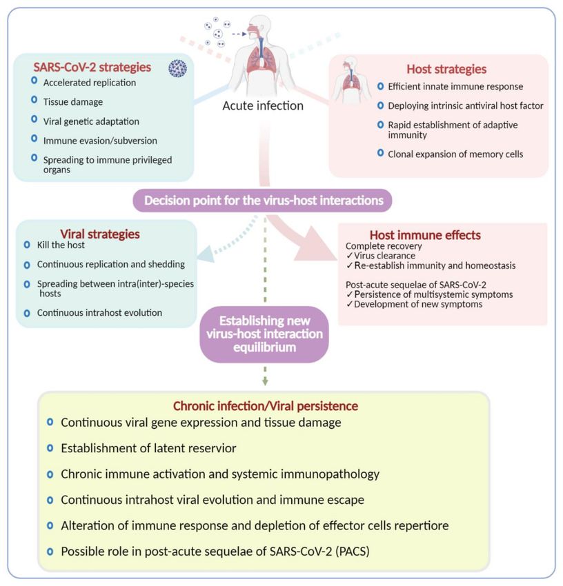
2021, 13, 1025 2 of 25
Viruses 2021, 13, 1025 2 of 23
were able to describe the clinical syndrome [3–5], identify SARS-CoV-2 as the causative
agent [5,6], develop diagnostic tests [14–16], sequence the complete genome of the virus
isolated from clinical
isolatedsamples [5], and
from clinical start developing
samples several
[5], and start vaccines
developing candidates.
several vaccines Fast
candidates. Fast
forward, in lessforward,
than oneinyear, we were able to successfully develop several efficacious
less than one year, we were able to successfully develop several efficacious
vaccines; a fewvaccines;
of them are now
a few ofbeing
them used to vaccinate
are now being usedthetogeneral public
vaccinate [17,18]. In
the general this [17,18]. In this
public
review, we explore the available evidence on key virological, immunological, and clinical
review, we explore the available evidence on key virological, immunological, and clinical
characteristics of SARS-CoV-2ofinfection
characteristics SARS-CoV-2withinfection
the emphasis on emphasis
with the viral persistence, reinfec-
on viral persistence, reinfection,
tion, and PASC.and
WePASC.
highlight areas that warrant further investigation for the development
We highlight areas that warrant further investigation for the development of
of improved therapeutic
improved and prevention
therapeutic andinterventions.
prevention interventions.
2. SARS-CoV-22.and Its Tropism
SARS-CoV-2 and Its Tropism
Coronaviruses Coronaviruses
are large, enveloped, positive-sense,
are large, single-strandedsingle-stranded
enveloped, positive-sense, RNA (+ssRNA)RNA (+ssRNA)
viruses that cause various
viruses that diseases in mammals
cause various diseases(Figure 1a). Human
in mammals (Figurecoronaviruses
1a). Human such coronaviruses such
as SARS-CoV-2asshareSARS-CoV-2 share
a significant a significant
number numberwith
of key features of key features
other familywith other of
members family members
the Nidovirales of the Nidovirales
order, and primarily order, andrespiratory
cause primarily tract
causeinfections
respiratory[2].tract
Theseinfections
key fea- [2]. These key
features of
tures of coronaviruses coronaviruses
include (a) a highlyinclude (a) a highly
conserved conserved
genomic genomic
organization organization
that has a that has a
large replicase gene on the 5 0 end encoding for several proteins with enzymatic activities,
large replicase gene on the 5′ end encoding for several proteins with enzymatic activities,
which is upstreamwhich is upstream
of the structuralofandthe accessory
structuralgenes
and accessory genes
(Figure 1b); (b) (Figure
they all 1b); (b) they all efficiently
efficiently
use ribosomalto
use ribosomal frameshifting frameshifting
express theirtononstructural
express their proteins
nonstructural
(nsps),proteins (nsps), polyproteins
polyproteins
(Figure
(Figure 1b), which 1b), which
are essential forare
in essential for in vivo
vivo replication; (c) replication;
as a member(c)ofasthea member
nidovirusesof the nidoviruses
(nidus is Latin for “nest”), their downstream genes are expressed
(nidus is Latin for “nest”), their downstream genes are expressed by synthesis of 3′ nested by synthesis of 30 nested
subgenomic mRNAs subgenomic
(FiguremRNAs
1b). (Figure 1b).
(a)
(b)
Figure 1. Cont.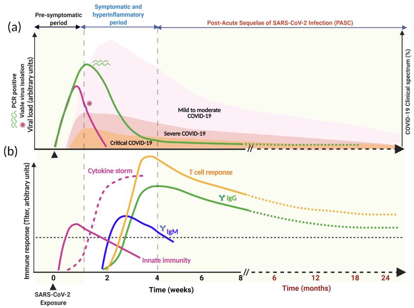
2021, 13, 1025 3 of 25
Viruses 2021, 13, 1025 3 of 23
(c)
Figure 1. SARS-CoV-2
Figure 1. SARS-CoV-2 genome organization
genome organization and replication
and replication cycle. (a) Thecycle. (a) The SARS-CoV-2
SARS-CoV-2 virion struc-
virion structure. While the viral
ture. While the viral membrane and envelope proteins ensure its genomic RNA incorporation and
membrane and envelope proteins ensure its genomic RNA incorporation and assembly, the trimeric spike (S) protein
assembly, the trimeric spike (S) protein provides specificity and high-affinity binding for its receptor
provides specificity and high-affinity binding for its receptor to enter into target cells. The positive-sense, single-stranded
to enter into target cells. The positive-sense, single-stranded RNA genome (+ssRNA) is encapsidated
RNA genome by(+ssRNA) is encapsidated
the nucleocapsid. by the nucleocapsid.
(b) Schematic depiction of (b) Schematic genome
SARS-CoV-2 depictionarchitecture
of SARS-CoV-2and genome
the poly-architecture
and the poly-(proteins)
(proteins) it encodes. (c) Schematics of SARS-CoV-2 replication: (1) SARS-CoV-2 virions bind to ACE2, its
it encodes. (c) Schematics of SARS-CoV-2 replication: (1) SARS-CoV-2 virions bind to
cellular receptor,
ACE2,and the typereceptor,
its cellular 2 transmembrane
and the type serine protease (TMPRSS2),
2 transmembrane a host factor
serine protease that promotes
(TMPRSS2), viral particles
a host factor
that promotes
entry and fusion viral particles
at the plasma membrane entry and fusion at
or endosomes. (2)the
In plasma membrane
the cytosol, or endosomes.
the incoming gRNA will(2) In
be the
released and
cytosol, the incoming gRNA will be released and subjected to immediate translation
subjected to immediate translation of the ORF1a and ORF1b open reading frame resulting in the polyproteins (pp1a of the ORF1a
and pp1ab),andwhichORF1b open reading
are further frame resulting
proteolytically processedin the polyproteins
to the components (pp1a andreplicase
of the pp1ab), which are further
complex. (3) The replicase
proteolytically processed to the components of the replicase complex. (3) The replicase
complex is an assemblage of multiple nonstructural proteins (nsps) that orchestrate transcription of the viral RNAs complex is and viral
an assemblage of multiple nonstructural proteins (nsps) that orchestrate transcription of the viral
replication. Concordantly, the virus compels the host cell to create the viral replication organelles, including the perinuclear
RNAs and viral replication. Concordantly, the virus compels the host cell to create the viral replica-
double-membrane vesicles (DMV), the convoluted membranes, and small, open double-membrane spherules (DMS). These
tion organelles, including the perinuclear double-membrane vesicles (DMV), the convoluted mem-
structures provide
branes, aand protective microenvironment
small, open double-membrane for spherules
the viral gRNA
(DMS).and subgenomic
These structuresmRNA
provideduring replication and
a protective
transcription. (4,5) The newly synthesized
microenvironment for the viralsurface
gRNA and structural proteins
subgenomic translocate
mRNA duringtoreplication
the ER membrane and transit through
and transcription.
the ER-to-Golgi
(4,5) intermediate compartment
The newly synthesized (ERGIC),
surface which
structural then assembles
proteins translocateand
toencapsidates
the ER membrane the ribonucleocapsid.
and transit (6,7)
through
Ultimately, the progeny the virions
ER-to-Golgi intermediate
bud into the lumencompartment
of the secretory (ERGIC),
vesicleswhich then
and will be assembles
released byand encapsi-
exocytosis.
dates the ribonucleocapsid. (6,7) Ultimately, the progeny virions bud into the lumen of the secretory
vesicles and will2.1. SARS-CoV-2
be released Origin
by exocytosis.
Human coronaviruses have an approximately 30 kb RNA genome and a diameter
2.1. SARS-CoV-2 Originfrom 50 to 150 nm with a distinctive crown-like appearance of their glycoprotein
ranging
spike (S) protein
Human coronaviruses have (Figure 1a) [19,20]. These
an approximately 30 kbRNARNAviruses
genome undergo genetic recombination,
and a diameter
ranging from 50deletions,
to 150 nm mutations, and othercrown-like
with a distinctive forms of variation
appearance allowing
of theirthem to adapt to infect new
glycoprotein
hosts and spread within the same species independent of their
spike (S) protein (Figure 1a) [19,20]. These RNA viruses undergo genetic recombination, natural reservoir. SARS-
deletions, mutations, and other forms of variation allowing them to adapt to infect new level across
CoV-2 emerged in late 2019 and is ~80% and ~96% identical at the nucleotide
its genome
hosts and spread withsame
within the SARS-CoV
speciesand the bat coronavirus,
independent RaTG13,
of their natural respectively
reservoir. SARS-[3,9]. Structural
and biochemical characterization of the genome revealed that the
CoV-2 emerged in late 2019 and is ~80% and ~96% identical at the nucleotide level across receptor-binding domain
(RBD) of the SARS-CoV-2 S protein is the most variable region
its genome with SARS-CoV and the bat coronavirus, RaTG13, respectively [3,9]. Structural with distinctive gain-
and biochemicalof-function mutations
characterization to bind
of the genometo the cognate
revealed cellular
that receptor, angiotensin-converting
the receptor-binding do-
enzyme 2 (ACE2), with high affinity [3,5,9,21,22]. Furthermore,
main (RBD) of the SARS-CoV-2 S protein is the most variable region with distinctive it was
gain-suggested that
SARS-CoV-2 seemingly adapted through natural selection and genetic recombination in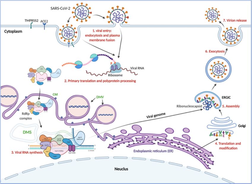
Viruses 2021, 13, 1025                                                                                           4 of 23

                         humans or other intermediate hosts such as the pangolin with high homology to the ACE2
                         receptor [3,6,9,23,24] before it emerged as a major pandemic in late 2019 in Wuhan, China.
                               SARS-CoV, MERS-CoV, and SARS-CoV-2 originated likely from bats and transmitted
                         to human through intermediate hosts: SARS-CoV in 2002/2003 emerged through adap-
                         tation in palm Civets in a wildlife market in Guangdong, China [25], MERS-CoV in 2012
                         transmitted to human from dromedary camels in the Arabian Peninsula [26], but the defini-
                         tive intermediate host for SARS-CoV-2 is unknown, and remains a source of controversy.
                         Several groups reported that horseshoe bats as likely origin and imported pangolins as
                         possible intermediate host for SARS-CoV-2. However, according to a comparative genomic
                         analysis [27], none of these highly identical (90–96% identity at nucleotide level) coron-
                         aviruses that were found in these animals appear to be a direct progenitor of SARS-CoV-2,
                         indicating that further adaptation in humans or other intermediate hosts must have taken
                         place preceding the emergence of the COVID-19 pandemic [9].

                         2.2. SARS-CoV-2 Tropism
                               SARS-CoV-2 infects the target cell by binding to its cognate cell surface receptor, the
                         ACE2 protein, using the RBD of the S protein (Figure 1c) [3,5,9,21,22,28–32]. The S proteins
                         of SARS-CoV and SARS-CoV-2 are highly homologous and structurally similar with only
                         minor differences, and they both use ACE2 to enter into target cells [3,21,28,30,31,33]. ACE2
                         is a type I transmembrane protein, which is ubiquitously expressed in endothelial and
                         most epithelial cells of different organs, including in the high expressers such as the lungs,
                         heart, kidneys, and small intestine [29,34–39]. It is regulator of the potent vasoconstrictor
                         angiotensin maturation and hence offsets the vasomotor effect of ACE on the cardiovascular
                         system [36].
                               Akin to all coronaviruses, the S protein of SARS-CoV-2 mediates two essential virus-
                         host interactions in tandem during virus entry. First, it binds to the primordial and
                         abundant sugar moiety with its N-terminus region, and second, it engages with high-
                         affinity and selectivity to its protein receptor, ACE-2, to initiate fusion of the viral and
                         host cell membranes via its C-terminal region (Figure 1c) [40]. These cascading events are
                         believed to give coronaviruses an evolutionary advantage to easily adapt and expand their
                         host ranges. SARS-CoV-2 virions entry into the cells is then orchestrated by proteolytic
                         cleavage and activation of the ACE2-bound S protein by the type 2 transmembrane serine
                         protease (TMPRSS2) on the cell surface [28,29,41–43]. After entry, through a series of highly
                         regulated steps and virus–host interactions, the virus completes its replication cycle as
                         described in Figure 1 [44]. Thus, a coordinated expression of ACE2 and TMPRSS2 is critical
                         to augment viral entry and infectivity. Hou et al. [45], using a reverse genetics approach
                         by single-cell RNA sequencing and in situ RNA mapping, showed an expression gradient
                         of ACE2 in the normal airway epithelia, with the highest expression being in the nasal
                         epithelial cells and lesser expression in the lower airway epithelial cells, terminating in
                         significantly minimal ACE2 level in the alveolar pneumocytes. Interestingly, the expression
                         of TMPRSS2 was not significantly different between the upper and lower airways’ epithe-
                         lia [45,46]. However, the expression gradient of ACE2 and hence the very low frequency
                         of dual ACE2+ /TMPRSS2+ cells in the lower respiratory tract and alveoli likely translates
                         to the early and greater SARS-CoV-2 replication in the upper airway than in the lower
                         airway, and thus to the clinical course of the disease [47]. Germane to this scenario is that
                         the SARS-CoV-2-induced high-IFN inflammatory microenvironment upregulates ACE2
                         expression without significantly affecting TMPRSS2 expression [45,46,48]. This may poten-
                         tially increase the frequency of virus-susceptible dual ACE2+ /TMPRSS2+ cells in the lower
                         respiratory tract, although there is significant interhost variability in SARS-CoV-2 disease
                         severity [45,46]. In fact, the degree of IFN-induced expression of ACE2 correlates with
                         organ tropism, symptom onset, and severity of the SARS-CoV-2 illness [29,38,45,46,48].
                               During the early period of the pandemic, it was speculated that there is a notable
                         upregulation of ACE2 by the potent antihypertensive drugs, ACE inhibitors may have
                         contributed to the disproportionately increased susceptibility of patients with hyperten-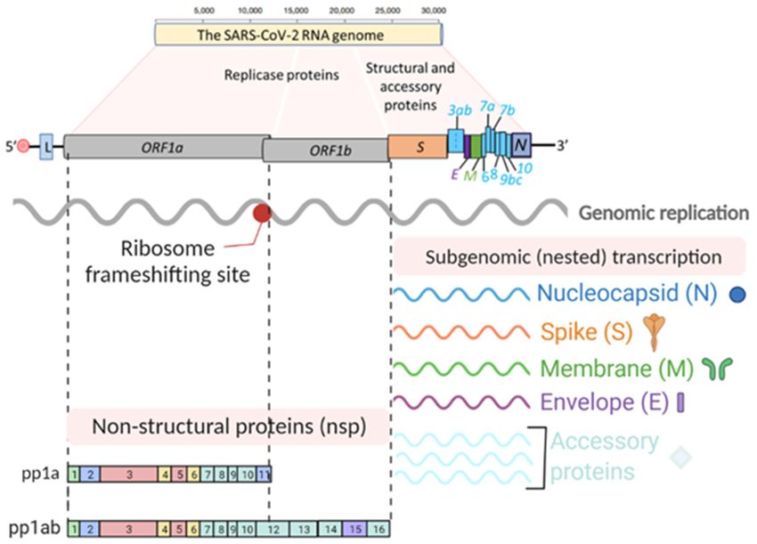
Viruses 2021, 13, 1025                                                                                           5 of 23

                         sion, heart diseases, and diabetes mellitus who are taking these drugs to SARS-CoV-2
                         infection and risk of developing a more severe disease. However, several studies have
                         found no meaningful association between taking these drugs and risk of SARS-CoV-2
                         infection, disease severity, or mortality due to COVID-19-related illnesses [49–53]. Thus,
                         the variability of SARS-CoV-2 susceptibility of the airway epithelial cells and disease
                         course cannot fully be explained by ACE2 and TMPRSS2 expression levels, begging fur-
                         ther in-depth analysis of the virus–host interactions and identification of major determi-
                         nants. Indeed, one of the hallmarks of SARS-CoV-2 infection is that the virus hijacks a
                         number of cellular factors and dampens the host immune response to facilitate its repli-
                         cation. After cell entry, SARS-CoV-2 compels the host cells to support its replication by
                         co-opting different cellular factors and machineries. The molecular basis of acute SARS-
                         CoV-2 infection, virus–host interactions, and immunopathogenesis has been extensively
                         studied [4,5,21,28–32,34,37,38,43,45–48,54–67] and has led to the development of select
                         therapeutics (see WHO guideline [68]) and successful vaccines [17,18].

                         3. COVID-19 Pathogenesis
                               SARS-CoV-2 is primarily a respiratory virus, and is transmitted by respiratory droplets,
                         aerosols, and direct/indirect contacts. It can also infect other cell types and has been de-
                         tected in a wide range of organs and tissues, notably the intestinal epithelium, liver,
                         kidneys, brain, pancreas, eye, and immune cells [39,61] (Figure 2a). Such broader organ
                         tropism is dictated by the expression of ACE2. The portal of entry is believed to be the na-
                         sopharyngeal or conjunctival epithelial cells that express high levels of ACE2 [29,45,57,69].
                         Although it is not clear whether the virus gains access to the terminal airways and alveoli
                         by aspiration of the nasopharyngeal content, systemic dissemination, or by progressively
                         moving distally on the bronchial tree mucosa, some viruses infect the alveolar epithelial
                         cells and lung tissue-resident macrophages [55,69] (Figure 2b). Unlike SARS and MERS,
                         the majority of individuals with SARS-CoV-2 remain asymptomatic or develop only mild
                         symptoms [10,55,70,71] (Figure 3a). This is largely because of the clearance or control of
                         the infection by the innate immune response effectors and virus-specific T cells without in-
                         tensified tissue damage [60] (Figure 3b). The major pathophysiological feature and leading
                         cause of death in COVID-19 patients is acute viral pneumonia characterized by bilateral
                         ground glass opacities on imaging studies and histopathological features such as diffuse
                         and organizing alveolar damage, proinflammatory infiltration, microvascular thrombosis,
                         and persistence of SARS-CoV-2 in the respiratory tract [19,66,72,73].
                               A subgroup of patients with certain risk factors listed in Table 1 are associated with
                         severity and increased fatality of COVID-19 [59,74–76]. The proxy for inflammation-
                         induced multiorgan injuries is the presence of markedly elevated levels of proinflamma-
                         tory biomarkers such as C-reactive protein (CRP), ferritin, IL-1, and IL-6 [74,77], and as
                         such an unrestrained host immune response misfires to induce severe immunopatholog-
                         ical changes, and hence multiorgan failure [19,38,46,55,62,66,69,72,78–84]. Su et al. [79]
                         carried out an integrated clinical and multi-omics analysis on a large cohort of patients
                         to understand the role of the hyperinflammatory state on the heterogeneity of severe
                         COVID-19, and they described a distinct profile of proinflammatory cytokines associated
                         with a loss of metabolites in severe COVID-19 compared to mild/moderate COVID-19.
                         Such atypical immunophenotype signatures and stressed proinflammatory environments
                         in severe SARS-CoV-2 infection were also observed by other research groups [65,78,79].
                         However, understanding the molecular crosstalk among direct viral cytopathicity, host
                         immune dysregulation, and SARS-CoV-2-induced hypercoagulable states in SARS-CoV-2
                         viral sepsis and severe COVID-19 warrants further investigations.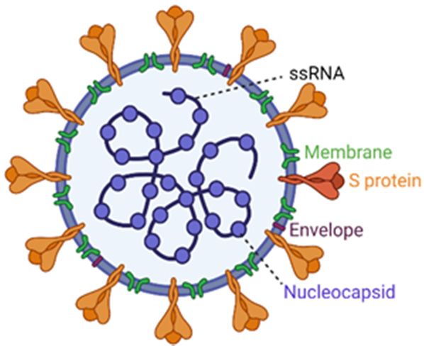
Viruses 2021, 13, 1025 6 of 23
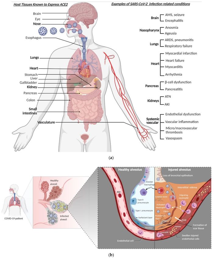
Figure 2. SARS-CoV-2 tropism, clinical presentation, and pathophysiology of alveolar damage. (a) A wide range of clinical
and laboratory abnormalities can be observed in SARS-CoV-2 infection. In severe COVID-19 cases, the pathophysiology
of the disease involves accelerated viral replication, cytokine storm, hyperinflammatory state, systemic endotheliitis, and
hypercoagulability with secondary organ dysfunction (often pulmonary, cardiovascular, renal, or hepatic). (b) When
SARS-CoV-2 makes it to the lower airway and infects type II pneumocytes (right side), the accelerated replication of the
virus induces host cell death by pyroptosis and release of proinflammatory and damage-associated molecules, further
activating and recruiting proinflammatory immune cells, including neutrophils, lymphocytes, and monocytes to the site of
infection. In a defective or misfiring immune response, there will be an overproduction of proinflammatory and profibrotic
cytokines, resulting in diffuse alveolar damage with fibrin-rich hyaline membrane deposition and loss of the gas exchange
function of the alveoli, and hence development of hypoxic respiratory failure. Furthermore, the resultant cytokine storm
leads to multi-organ damage. However, in a healthy immune response, virus-specific immune cells will be recruited and
eliminate the infected cells before the virus spreads to ultimately minimize the alveolar damage (the left side). AMS denotes
altered mental status, ARDS denotes acute respiratory distress syndrome, AKI denotes acute kidney injury, and ATN
denotes acute tubular necrosis.Viruses 2021, 13, 1025 7 of 23
Figure 3. Time course of SARS-CoV-2 infection, clinical spectrum, and immune response. (a) Kinetics of viral load and
clinical spectrum. (b) A schematic representation of immune response following SARS-CoV-2 exposure. The innate immune
response appears shortly after exposure (purple line) accompanied by cytokine storm (purple dashed line), followed by
adaptive immunity development after the first week post-infection. Seroconversion and T cell response occur during the
second week after symptoms onset. While IgM peaks early and decays faster, IgG titers and T cell response detected
around day 10 and their peak and height are likely to be influenced by disease severity and virus load. The level of
antibody protection from reinfection, the duration of the total humoral immune response above this protective threshold
(dashed black horizontal line), and the rate of decline from mild- or severe-infection-induced antibodies is not known for
SARS-CoV-2 during the PASC period.
Table 1. Risk factors for severe COVID-19 and worse outcomes.
Factors 1 References
Male sex [59,74,75]
Advanced age [59,74,75,85]
Chronic respiratory diseases (asthma, COPD, ILD) [75,86]
Major cardiovascular disease (CAD, CHF, CMP) [74,75,85]
Hypertension [74,75,85]
Diabetes mellitus [74,75,85,87]
Obesity (body mass index ≥ 30) [75,88]
Chronic liver disease [75,89]
Chronic kidney disease [75,90]
Cancers [75,91,92]
Immunocompromised state from solid-organ transplantation [75,93]
Immunosuppressive therapies, Rituximab, and JAK inhibitors [94,95]
1Abbreviations: COPD, chronic obstructive pulmonary disease; ILD, interstitial lung diseases; CAD, coronary
artery disease; CHF, congestive heart failure; CMP, cardiomyopathy; JAK, Janus kinase.
Molecular Pathology in Severe COVID-19
Acute respiratory distress syndrome (ARDS) in severe COVID-19 is characterized by
loss of the epithelial–endothelial barrier integrity, capillary fluid leakage, diffuse alveolarViruses 2021, 13, 1025 8 of 23
damage, hyaline membrane deposition, and unrestricted recruitment of inflammatory
effectors [19,66] (Figure 2b). Although the central role of lung pathologies as manifested by
severe pneumonia and ARDS is the hallmark of severe COVID-19, extrapulmonary patholo-
gies have also been consistently reported [19,96] (Figure 2a). The cytokine storm associated
with severe SARS-CoV-2 infection has ripple effects across the body contributing to the viral
sepsis, multiorgan failure, and even death [97]. SARS-CoV-2 hijacks the cellular machiner-
ies to facilitate its propagation and to induce a hyperinflammatory state [98]. The severity
of SARS-CoV-2 infection is fueled by the dysregulation of the host immune response
primarily by inhibiting type I interferon (IFN) response in acutely infected cells [65,78,83].
This hyperinflammatory state is marked by an accelerated viral replication associ-
ated with greater tissue damage by pyroptosis and coagulopathy, triggering a cascade
of systemic immune response disequilibrium and a shift in the dynamics of host genes
expression [63,78,82,84,96,97]. A recent analysis of the lung proteome showed a unique set
of pathways that are required for SARS-CoV-2 replication in infected cells [96]. The analysis
also identified important virus–host interactions that inhibit the function of host innate
immunity-related genes. As discussed in Figure 1, the translation of nested subgenomic
RNAs of SARS-CoV-2 requires capping, and the cap-recognizing host factor eukaryotic
translation initiation factor 4E (EIF4E) is upregulated selectively in SARS-CoV-2-infected
lung tissue. Furthermore, several genes involved in the antiviral innate immune response
pathways, including the stress granule related factor Ras GTPase-activating protein-binding
protein 1 (G3BP1), the mitochondrial protein translocase of the inner membrane 10 (TIM10),
transcription regulators, ubiquitination pathway components, and the proinflammatory
cytokine receptor interleukin 17 receptor A (IL17RA), were dysregulated in SARS-CoV-2-
infected lungs [64,96].
The virus employs these host factors and pathways either directly or indirectly to
promote its replication, which further facilitates the recruitment of proinflammatory cells
and activation of the profibrotic pathways to incur severe tissue damage. For instance,
Nie et al. [96] conducted an unbiased proteomic atlas using tissues obtained from different
organs (lungs, kidneys, heart, testis, spleen, liver, and thyroid) at autopsy of COVID-19
cases. Of the 11,394 proteins they analyzed, 5336 involved in the host immune response,
coagulation regulation, angiogenesis, and profibrotic processes were significantly perturbed
during acute SARS-CoV-2 infection with an organ-specific niche, suggesting the existence
of crosstalk among multiple organ systems during the exuberant immune response and
accompanying tissue hypoxia, a phenomenon that was also described by others [78].
However, the heterogeneity of the host immune response, immunopathology, and clinical
spectrum of SARS-CoV-2 infections in individuals with comparable viral loads remains
a vexing question. For example, children and young adults with SARS-CoV-2 tend not
to develop severe disease irrespective of viral load [71]. Alternatively, an intriguing
imbalance between virus infectivity and the host immune response was demonstrated
in a twin publication by Casanova and colleagues [76,99]; the authors uncovered over-
representation of individuals with inborn errors in the type I IFN pathway in severe COVID-
19 patients compared to either mild COVID-19 patients or healthy donors [76]. What is
even more startling is that in the accompanying publication, the authors demonstrated
that the presence of anti-type I IFN autoantibodies, presumably virus-induced, is linked
with disease severity [99]. Although severe inflammation with aberrant immune activation
is not unexpected in severe SARS-CoV-2 infection, these discoveries represent important
inroads for future studies.
4. SARS-CoV-2 and Persistent Viral Shedding
4.1. Viral Dynamics and Duration of Infectiousness
Quantitative studies of SARS-CoV-2 viral load [47,59,100–107], dynamics
[47,100,103–106,108], shedding [47,103,106,107], and viable virion isolation [47,103,109–112]
have provided the following understandings into the pathogenesis of COVID-19: (i) the
average incubation period (time from infection to symptom development) for SARS-CoV-2Viruses 2021, 13, 1025 9 of 23
is ~5 days (range 2–14 days), which is shorter than that for SARS and MERS [113–116].
(ii) Unlike SARS-CoV and MERS-CoV, SARS-CoV-2 viral load, and kinetics of shedding,
is much higher in the upper than in the lower respiratory tract [101,108]. It is reported
that the positivity rate was in fact higher for lower respiratory tract samples than it was
for the nasopharyngeal swab [117]; however, the authors pointed out that their data
could not be correlated with clinical symptoms or disease course. The demonstration of
a strong concordance between nasopharyngeal swab and bronchoalveolar lavage (BAL)
samples for SARS-CoV-2 RT-PCR test positivity established nasopharyngeal swab as the
cost-effective and least invasive diagnostic method for SARS-CoV-2 [118,119]. (iii) The
quick decline in the viral load in individuals with SARS-CoV-2 makes isolation and other
prevention interventions challenging and less effective [116]. (iv) COVID-19 has a broad
clinical spectrum (mild, moderate, severe, and critical COVID-19), and yet a great majority
of individuals with SARS-CoV-2 are asymptomatic, and are as infectious as the symp-
tomatic ones [116,120–123] (Figure 3a). A new report based on the analytical model and
meta-analysis of select reports suggested that about 50% of new SARS-CoV-2 infections
were contributed by exposure to either asymptomatic or presymptomatic individuals with
SARS-CoV-2 [116]. This further emphasizes that successful control of the pandemic re-
quires multipronged approaches including identification and isolation of asymptomatic
and symptomatic cases, universal wearing of face masks, social distancing, contact tracing,
and widespread use of therapeutics and/or vaccines.
Several studies reported that on average the duration of viral particle shedding
from the upper airway is ~17 days, the lower respiratory tract ~15 days, feces ~13 days,
and blood ~17 days (reviewed in ref. [124]). The longest durations reported to date for
detection of viral RNA were 83 days, 59 days, 126 days, and 60 days in the upper airway,
the lower respiratory tract, feces, and blood, respectively [124,125]. Not surprisingly,
SARS-CoV and MERS-CoV RNA were also detected from different samples such as upper
respiratory tract, lower respiratory tract, serum, and stool weeks to months after recovery
from the acute illness [126–132]. Despite prolonged detection of viral RNA, viable virus
isolation was successful only in the first 8 days after symptom onset and the success of
isolation of viable virions from respiratory samples directly correlates with the initial viral
load [47,110,111,133] (Figure 3a). Because of the precipitous decline in viral load after
the second week post-infection, transmission of SARS-CoV-2 was infrequent after the
first 10 days of illness. Even though viral RNA was detected after a prolonged period in
the blood samples, no replication-competent virus was isolated from PCR-positive blood
samples [33], viable virus was isolated from fecal samples [134]. Nonetheless, fecal–oral
route transmission of SARS-CoV-2 is yet to be demonstrated.
4.2. The Relevance of Prolonged SARS-CoV-2 Viral Shedding
4.2.1. Role of SARS-CoV-2 Organotropism and Immune Privilege in Viral Persistence
Viruses are highly sophisticated molecular machines that can undergo intra-host
evolution to develop effective strategies to overcome host immunity and establish chronic
infection by replicating continuously, establishing latent reservoir, or integrating into the
host cell genome. If the immune system cannot eliminate the virus, the chronic immune
activation and/or the cytopathic effect of the virus will continue until the infection resolves
or kills the host. Certain viruses chronically persist by establishing metastable virus–
host immune response interaction equilibrium. For example, viruses such as the human
immunodeficiency virus (HIV), hepatitis B virus (HBV), and hepatitis C virus (HCV)
compel the host cell to persist in latent state and evade the host antiviral immunity [135].
Other viruses like Ebola virus taught us that persistence in immune-privileged sites can also
lead to transmission to a new host via an unexpected and different route [136]. Whether
SARS-CoV-2 can establish chronic infection or persist in immune-privileged anatomic
sanctuary sites remains to be demonstrated.
SARS-CoV-2 infection is associated with accelerated replication and high viral load
during the acute phase, which is followed by a rapid decline after the first week [47]. InViruses 2021, 13, 1025 10 of 23
some instances, the peak of the viral titer in the respiratory tract occurs during the presymp-
tomatic period [137]. Interestingly, analysis of autopsy samples from severe COVID-19
patients revealed that viral RNA can be detected until the time of death, suggesting that
prolonged shedding of virus may indicate a grave outcome [85]. SARS-CoV-2 nucleic
acids persistence during convalescence and the risk of infectiousness remain controversial.
The lingering detection of SARS-CoV-2 RNA in different clinical specimens of convales-
cent patients could be due to slow replication, reactivation of latent virus, or residual
nonreplicating genetic material; however, it needs to be systematically explored to defini-
tively determine its relevance. Uncovering the transition of the disequilibrium among
the exuberant antiviral immune response, rapid SARS-CoV-2 replication during the acute
phase, and severe immunopathology to reset the virus–host interaction and establish per-
sistent infection, if any, will be an interesting avenue of basic science and clinical research
(Figure 4).
Figure 4. Outcomes of SARS-CoV-2 infection. During the acute infection, the host and the virus compete for dominance to
tip the balance in their favor, resulting in different outcomes as indicated in the highlighted boxes. If the host strategies
prevail to achieve complete recovery, the virus will be cleared. Another outcome that is poorly understood and yet getting
traction is the multisystemic sequelae of SARS-CoV-2 infection called PASC. On the other hand, if the virus wins the battle,
a variety of outcomes are expected, as indicated in the illustration. Viral persistence in a minority of patients, particularly in
immunocompromised patients, has now been well documented; however, the underlying mechanism is unknown. There
is a possibility of establishing metastable equilibrium between the host and SARS-CoV-2 to facilitate persistence and/or
chronic infection.Viruses 2021, 13, 1025 11 of 23
Viruses infect specific cell types and persist with little to no viral gene expression by
sabotaging the cellular gene expression machineries to subvert sterilizing immunity. They
can also establish persistent infection in immune-privileged tissues and evade constant
immune surveillance. The respiratory tract epithelial cells and alveolar macrophages are
the primary targets for SARS-CoV-2 replication. However, the cellular and organ tropism
of SARS-CoV-2 is expanding [39,61], and whether some of these SARS-CoV-2-targeted
immune-privileged sites can serve as sites of viral persistence remains an area of future
investigation.
4.2.2. Mechanism of SARS-CoV-2 Evasion and Subversion of Host Immunity
SARS-CoV-2 has a limited array of genes with approximately 14 open reading frames
to encode 29 different proteins [138] (Figure 1b). Gordon et al. [138] mapped the SARS-
CoV-2 interactome and uncovered the role of most of the viral proteins, which manipulate
the host cell biological processes and factors. Some exhibit specialized and niche-specific
roles to evade or subvert the immune response. For instance, Nsp9, Nsp13, Nsp15, Orf3a,
Orf9b, and Orf9c were identified to interfere with the IFN pathway and proinflammatory
cytokine expressions; others disrupt the adaptive immune response, gene expression
and protein translation machineries [138]. Another large-scale multilevel study showed
that SARS-CoV-2 disrupts different cellular functions, including transcriptome, proteome,
ubiquitinome, and phosphoproteome [139]. Identifying and targeting the vulnerability
hotspots of SARS-CoV-2 and mechanisms of immune evasion are promising research areas
for further research and development of antiviral agents.
4.2.3. Viral Persistence in Immunocompetent Hosts
Although understanding the kinetics and dynamics of viral shedding in relation
to infectivity is critical in implementing infection prevention and control strategies, it
has been a futile endeavor to demonstrate a correlation between prolonged detection of
viral RNA and isolation of viable virus from clinical samples [47,111,140]. A recent meta-
analysis identified a correlation between prolonged SARS-CoV-2 shedding as determined
by RT-PCR and the increased risk of developing severe disease [141]. When the authors
stratified the pooled median duration of respiratory RNA shedding by disease severity,
they found that the median duration of nasopharyngeal viral RNA shedding was 19.8 days
among severely ill patients and was 17.2 days in mild-to-moderate COVID-19 cases [141].
To define the SARS-CoV-2 transmission window, Sai et al. [142] carried out a COVID-19
pathogenesis and transmissibility study using a hamster SARS-CoV-2 infection model
and found a strong correlation between transmission and isolation of viable virus in
cell cultures. Interestingly, they found no correlation between prolonged detection of
viral RNA from the nasopharyngeal swab and transmissibility, further corroborating that
infectivity declines precipitously and prolonged detection of viral RNA does not necessarily
mean that the individual is infectious. The authors also demonstrated the existence of an
inverse correlation between infectiousness and development of neutralizing antibodies, an
observation corroborated by others in recovered COVID-19 patients [143] as well as in a
nonhuman primate model [144,145]. The median duration of infectiousness of the wild-
type strain was determined to be approximately 8 days post onset of symptoms [47,111,143],
and using probit analysis, van Kampen et al. [143] showed that the probability of detecting
viable virus from respiratory specimens is ≤5% when the duration of symptoms is more
than two weeks. To this end, there is only one report of successfully isolated infectious
virus from the nasopharyngeal swab 18 days after symptoms onset [140], confirming that
infectiousness drops dramatically after the second week. It appears that detection of
subgenomic viral RNA is a poor predictor of infectiousness and often outlasts the detection
of replication-competent virus, and the prolonged detection of viral RNA could be due to
the suboptimal immunological clearance of SARS-CoV-2 infection [47,111,146].
High SARS-CoV-2 viral load, duration of symptoms less than 7 days, absence of
neutralizing antibodies, and host immunocompromization were proposed as potentialViruses 2021, 13, 1025 12 of 23
predictors of prolonged shedding of infectious virus from respiratory tract. However,
only high viral load >7 Log10 RNA copies/mL in respiratory samples and absence of
SARS-CoV-2-neutralizing antibodies were found to be independent predictors of infec-
tiousness [47,111,143]. These studies were based on data gathered from hospitalized severe
COVID-19 patients. Knowing that the great majority of individuals with SARS-CoV-2 are
either asymptomatic or exhibit only mild disease symptoms, a proper and evidence-driven
public health risk stratification and intervention cannot be employed using these reports
only. To address this issue, in a recent retrospective analysis using data of nonhospitalized
patients from 282 transmission clusters, Marks et al. [147] confirmed that high viral load is
indeed a major predictor of infectiousness of index cases. The author further suggested
that, irrespective of the clinical spectrum of COVID-19, determination of viral load and
reinforcement of contact tracing and quarantine policy will remain crucial in devising
and revising the pandemic control policies. It is also important to understand why some,
but not all, COVID-19 patients continue to shed viral particles for a prolonged period, as
cost-effective stratification and intervention strategies are the cornerstone of our public
health effort in controlling the pandemic.
4.2.4. Viral Persistence in Immunocompromised Hosts
A recent analysis of the literature on the outcomes of SARS-CoV-2 infection in indi-
viduals with a wide range of conditions found that lung and hematologic malignancies,
solid-organ transplantation, and primary immunodeficiencies put the patient at higher
risk for (i) nosocomial acquisition of SARS-CoV-2, (ii) development of severe COVID-19,
and (iii) COVID-19-related death compared with immunocompetent individuals [148]. On
the other hand, those on biologics and immunosuppressive therapy appear to not be at
increased risk of developing severe COVID-19 [92,148,149], an evidence further supporting
the role of unrestrained immunity in disease severity and the potential role of inflamma-
tion modulators in treating COVID-19, including steroids [150]. In sub-Saharan Africa
where the burden of another viral illness, HIV/AIDS, is paramount, the concern was if
HIV-SARS-CoV-2 co-infection results in greater severity and higher mortality of COVID-19.
In fact, a few large-scale retrospective analyses indicated that people living with HIV are
at higher risk of developing and dying from severe COVID-19 [151–153]. Whether this
holds true, it is important to carry out studies by comparing controlled versus uncontrolled
HIV infection and by analyzing the role of other comorbidities, which are likely to bias
the outcome of any meaningful interactions between the two pathogens in co-infected
individuals [154,155].
Bridging these knowledge gaps will allow us to understand the pathophysiology of
SARS-CoV-2 infection in immunocompromised individuals and develop patient-tailored
interventions. Furthermore, this allows us to explore the interplay between preexisting
immunosuppression and SARS-CoV-2 infection, with an emphasis on COVID-19 clinical
course and outcome, identification of atypical manifestations, and the effect of immuno-
suppressants on SARS-CoV-2 replication in this special group of at-risk population. The
few case reports suggest that there is a high likelihood of viral persistence, reactivation, or
re-infection in immunocompromised patients, which also warrants further exploring.
As discussed above, prolonged shedding and recurrent detection of viral RNA by RT-
PCR from the clinical samples of asymptomatic patients with SARS-CoV-2 as well as from
symptomatic patients have been well documented. However, there is very little success
in isolating infectious virus after 1–2 weeks of symptom onset in immunocompetent
patients [124]. Interestingly, there are several case reports demonstrating isolation of
infectious viral particles from immunocompromised patients for weeks to months. Some of
them presented with relapse of the symptoms [156–161]. Others required multiple courses
of treatment with remdesivir [162], and ultimately succeeded to clear the virus [156,159,161].
The longest duration reported so far to isolate viable virus from nasopharyngeal swab was
~8 months, which was from a non-Hodgkin’s lymphoma patient [161]. One can speculate
that the selective pressure in an immunocompromised patient is likely to be different fromViruses 2021, 13, 1025 13 of 23
that in immunocompetent individuals. However, it is difficult to extrapolate and generalize
based on the few case reports discussed here on SARS-CoV-2 persistence and prolonged
shedding of infectious virus, for the following reasons: (i) the phylogenetic analyses of
these limited case reports were not carefully designed to explore factors that influence the
risk of SARS-CoV-2 persistence or reinfection; (ii) these case reports involve an atypical
disease course of SARS-CoV-2 infections since the majority of infections are classically
acute in nature, arguing against the establishment of sustained infections and thus limiting
generalization based on the reported intra-host viral genetic diversity in these cases; (iii) the
rate of SARS-CoV-2 evolution is slow [163], an extra Achilles heel to the diversification
of SARS-CoV-2 viral quasispecies. However, because the SARS-CoV-2 pandemic is a
widespread infection, it is possible that the host immune pressure could shape SARS-CoV-
2 evolution by contributing to genetic diversity and selection, and thus contributing to
phenotypic changes and establishment of chronic infections. This dimension of SARS-
CoV-2 infection warrants large-scale studies designed to understand the underpinning
mechanism by defining temporospatial resolution of SARS-CoV-2 persistence to help
develop potential patient-tailored therapeutics and preventive strategies. In light of the
accumulating and emerging evidence on the effect of the new SARS-CoV-2 variants on
efficacy of vaccines, their transmissibility, and severity of the disease [164], it would be
also interesting to investigate the relationship between intra-host viral evolution and the
mechanism of immune evasion in different patient groups in the context of viral persistence,
infectiousness, and reinfection.
5. SARS-CoV-2 Reinfection
SARS-CoV-2 reinfection is rare, but there are a few case reports of reinfection that were
confirmed by whole-genome sequencing and phylogenetic analysis [165–173]. It is also
important to note that these reports are likely to represent a small fraction of reinfections.
The corollary of pathogen invasion is that the host attempts to control the primary infection
and generates protective immune memory to combat future re-emergence/reinfection,
the basis of vaccination strategies. Likewise, we expect that the majority of individuals
who recovered from primary SARS-CoV-2 infection developed protective B cell and T cell
immunity, even if the longevity of protective immune memory is unclear [174]. Indeed,
Lumley et al. [174] analyzed longitudinal cohort data to determine the incidence of SARS-
CoV-2 reinfection in healthcare workers, and they were able to uncover the relationship
between seropositivity during primary infection and incidence of reinfection over a six-
month period of follow up during convalescence. The authors found a strong inverse
association between the presence of high anti-spike IgG titer and reinfection, reinforcing
the observations in earlier reports by other researchers [144,145,175,176]. Irrespective of
the detection of subgenomic viral RNA in the nasopharyngeal swab in select patients and
animal models, the presence of an anamnestic immune response after primary infection
provides potent protection against symptomatic SARS-CoV-2 reinfection [177]; a critical
insight into the durability of immunotherapies and vaccine protection as well as the timing
of booster doses.
The heterogeneity of the COVID-19 clinical spectrum and host antiviral immune
responses coupled with the intrinsic viral evasion mechanism(s) highlight the need to iden-
tify factors that predict not only disease severity but also factors that drive the generation
of new viral variants. SARS-CoV-2 continues to spread, and the effects of evolutionary
pressure are revealing viral adaptation and escape variants, which are highly infectious and
potentially more fatal. Some of the emerging mutants are summarized here [178]. As new
variants continue to emerge with mutations in the S protein, the growing concern will be
their gain-of-function to evade protective immunity afforded by the currently available vac-
cines and leading to a widespread reinfection. Harrington et al. [179] reported the first case
of reinfection by one of the most virulent variants, VOC-202012/01, after approximately
8 months post-primary infection, causing severe COVID-19 during his second infection.
Moreover, the recent phase 1b/2 trial that was conducted in South Africa assessed theViruses 2021, 13, 1025 14 of 23
efficacy of the AstraZeneca ChAdOx1 nCoV-19 vaccine (AZD1222) against the original
D614G and the B.1.351 variants (see Table 2), and the vaccine did not protect against the
B.1.351 variant infection, suggesting that reinfection by emerging variants is conceivable in
immune individuals [180]. The number of new SARS-CoV-2 variants is expanding, and the
variants listed in Table 2 are of significant public health consequences in regard to their
potential impact on reinfection rate, the efficacy of currently available vaccines, and the
design of newer versions of vaccine, if needed, in the future.
Table 2. Emergent SARS-CoV-2 variants of high consequence.
Name of Variants
Key S Protein Substitutions Origin References
(Pangolin Lineage 1 )
Current dominant variant D614G 2 Europe [181]
∆69/70, ∆144Y, N501Y A570D,
B.1.1.7 UK [182]
P681H
B.1.351 K417N, E484K, N501Y South Africa [183,184]
B.1.427/B.1.429 S13I, W152C, L452R USA (California) [185]
B.1.526 T95I, D253G, E484K USA (New York) [186]
P.1 K417T, E484K, N501Y Japan, Brazil [187,188]
1 SARS-CoV-2 Pangolin lineages nomenclature was proposed by Rambaut et al. [189]; 2 One of the first mutations
identified, which since April 2020 has become the dominant variant. All the other variants listed in the Table share
the D614G mutation, which is believed to help them spread more quickly than viruses without this mutation [181].
As these and other reports of reinfection surface, understanding and integrating the
correlates of protective immunity and intra-host evolution as determined by genomic
surveillance need further studies to document and help control the pandemic both at
local and global levels. Robust data on the duration of protective immunity and viral
shedding, the implication of reinfection and infectiousness of the new variants, the role of
antibody-dependent enhancement with the new variants in the setting of prior vaccination,
and the efficacy of the currently available vaccines against the emerging SARS-CoV-2
variants are all crucial for informed decision-making and policy revision. Because of the
dynamic nature of the SARS-CoV-2 spread, the expansion of the ongoing population-based
genomic surveillance initiatives in various countries will be beneficial since reinfection
and reintroduction will accelerate the molecular clock of the emergence of more virulent
and/or resistant variants worldwide, which could potentially affect the efficacy of the
current effective vaccines developed using the wild-type strain.
6. The Post-Acute Sequalae of SARS-CoV-2 Infection (PASC)
The SARS-CoV-2/COVID-19 pandemic has rapidly engulfed the world with nearly
120 million confirmed infections and over 2.6 million associated fatalities as of 15 March
2021 [1]. Compelling evidence is amassing and we are now beginning to understand the
aftermath of SARS-CoV-2 infection referred to as “Long COVID”, which is experienced
by ~10% of SARS-CoV-2 infection survivors [190,191]. Although there is no consensus on
the definition of “Long COVID”, most of the reports defined the syndrome as a cluster
of symptoms that persisted for more than 4 weeks following a confirmed or suspected
SARS-CoV-2 infection [191]; others used 2 months as a clinical cut off to define “Long
COVID” [192,193].
Recently, the NIH renamed “Long COVID’ as post-acute sequalae of SARS-CoV-2
infection (PASC) and launched a comprehensive research initiative to understand the
sequalae of SARS-CoV-2 infection [194] (Figure 3). PASC is a syndrome in COVID-19
survivors that present with a plethora of symptoms, including cough, dyspnea, fatigue,
myalgia, dysautonomia manifested as postural orthostatic tachycardia syndrome, gastroin-
testinal symptoms, dermatological, or neurological symptoms [191–193,195]; symptoms
should either persist beyond 4 weeks after their onset or newly develop as sequelae of
COVID-19. To this end, the NIH multipronged initiative on PASC is the first of its kind,
which will hopefully provide critical data on the natural history of PASC at a popula-Viruses 2021, 13, 1025 15 of 23
tion level and will shed light on the potential long-term interventions to be employed in
managing COVID-19 survivors.
Long-term complications are not unexpected given the broader organotropism of
SARS-CoV-2 and severity of the tissue injuries [61]. However, the pathophysiology and
natural course of PASC is unclear, meriting further studies. Some potential mechanisms
may include (i) the cytokine storm, (ii) the immune misfiring and virus-triggered autoim-
munity, (iii) the overwhelming systemic endotheliitis resulting in chronic microangiopathy
and thromboembolic events, (iv) persistent viral RNA shedding triggering chronic im-
mune activation, and (v) finally, the possible role of viral persistence, reinfection, and
reintroduction of new variants [192,196]. All of these may contribute to PASC pathogenesis
either individually or in combination. Therefore, it is vital to recognize the magnitude,
complexities, and heterogeneities of COVID-19 illnesses and to continue to support better
research to develop long-term multidisciplinary care and rehabilitation for COVID-19
patients during convalescence.
7. Conclusions
The unprecedented nature of the COVID-19 pandemic has demanded an urgent and
unmatched productivity by all stakeholders, and the scientific community has met the call
with a historic proficiency to develop effective vaccines in less than one year. SARS-CoV-2
is the third outbreak in the last two decades, and it exhibits some differences compared to
its predecessors SARS-CoV and MERS-CoV. This review summarizes the rapidly evolving
important discoveries on SARS-CoV-2 with an emphasis on the mechanism and relevance of
viral persistence and PASC. We also highlight potential areas of research on viral persistence
and PASC. Addressing these issues will provide a robust framework to fulfill the unmet
needs in our efforts to control this pandemic and enable readiness for any future pandemics.
Author Contributions: B.A.D. prepared the manuscript. All authors (B.A.D., Y.Y.R., H.M.A., P.H.,
S.T., and K.S.W.) have contributed to the manuscript. All authors have read and agreed to the
published version of the manuscript.
Funding: This work was supported by the Department of Internal Medicine, Joan C. Edwards School
of Medicine, Marshall University, Huntington, WV 25701, USA.
Acknowledgments: The funders had no role in study design, data collection and analysis, decision to
publish, or preparation of the manuscript. All the figures were created with BioRender.com (accessed
on 8 January 2021).
Conflicts of Interest: The authors declare no conflict of interest.
References
1. WHO. Coronavirus Disease (COVID-19) Pandemic. Available online: https://www.who.int/emergencies/diseases/novel-
coronavirus-2019 (accessed on 19 April 2021).
2. Coronaviridae Study Group of the International Committee on Taxonomy of Viruses. The species Severe acute respiratory
syndrome-related coronavirus: Classifying 2019-nCoV and naming it SARS-CoV-2. Nat. Microbiol. 2020, 5, 536–544. [CrossRef]
3. Zhou, P.; Yang, X.L.; Wang, X.G.; Hu, B.; Zhang, L.; Zhang, W.; Si, H.R.; Zhu, Y.; Li, B.; Huang, C.L.; et al. A pneumonia outbreak
associated with a new coronavirus of probable bat origin. Nature 2020, 579, 270–273. [CrossRef]
4. Zhu, N.; Zhang, D.; Wang, W.; Li, X.; Yang, B.; Song, J.; Zhao, X.; Huang, B.; Shi, W.; Lu, R.; et al. A Novel Coronavirus from
Patients with Pneumonia in China, 2019. N. Engl. J. Med. 2020, 382, 727–733. [CrossRef]
5. Wu, F.; Zhao, S.; Yu, B.; Chen, Y.M.; Wang, W.; Song, Z.G.; Hu, Y.; Tao, Z.W.; Tian, J.H.; Pei, Y.Y.; et al. A new coronavirus
associated with human respiratory disease in China. Nature 2020, 579, 265–269. [CrossRef] [PubMed]
6. Lu, R.; Zhao, X.; Li, J.; Niu, P.; Yang, B.; Wu, H.; Wang, W.; Song, H.; Huang, B.; Zhu, N.; et al. Genomic characterisation and
epidemiology of 2019 novel coronavirus: Implications for virus origins and receptor binding. Lancet 2020, 395, 565–574. [CrossRef]
7. Peiris, J.S.; Lai, S.T.; Poon, L.L.; Guan, Y.; Yam, L.Y.; Lim, W.; Nicholls, J.; Yee, W.K.; Yan, W.W.; Cheung, M.T.; et al. Coronavirus as
a possible cause of severe acute respiratory syndrome. Lancet 2003, 361, 1319–1325. [CrossRef]
8. Wu, A.; Peng, Y.; Huang, B.; Ding, X.; Wang, X.; Niu, P.; Meng, J.; Zhu, Z.; Zhang, Z.; Wang, J.; et al. Genome Composition and
Divergence of the Novel Coronavirus (2019-nCoV) Originating in China. Cell Host Microbe 2020, 27, 325–328. [CrossRef]
9. Andersen, K.G.; Rambaut, A.; Lipkin, W.I.; Holmes, E.C.; Garry, R.F. The proximal origin of SARS-CoV-2. Nat. Med. 2020, 26,
450–452. [CrossRef] [PubMed]You can also read