Common Diseases of Urban Wildlife - BIRDS
←
→
Page content transcription
If your browser does not render page correctly, please read the page content below
The Australian Registry of Wildlife Health is
 committed to the preservation of Australia's
        biodiversity through increased
   understanding of the interaction among
    animals, the environment, and disease
                causing agents.

Common
Diseases of
Urban Wildlife
BIRDS

Dr Karrie Rose, Dr Hannah Bender & Jane
H ll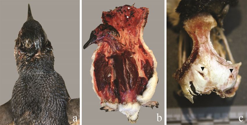
Production of this document was made possible by: Wildlife Rescue and Rehabilitation – an
Australian Government initiative

Cite this document as: Hall, J. and Rose, K. 2021 Common Diseases of Urban Wildlife: Birds.
Taronga Conservation Society Australia, Sydney.

All Images are subject to Copyright©

The information and materials contained in this section of the site are subject to copyright and are
for individual educational use only. Authorisation should be sought from the Registry for any other
use of these materials.

The views expressed in this document are those of the authors, and not necessarily of their
organisations. The Registry makes every effort to verify the information contained within this
document, but the accuracy and completeness of the information cannot be guaranteed. The
reader assumes all risk in using information provided. This document contains images of sick and
dead wildlife. These images are included for the sole purpose of improving wildlife care and
welfare. If you have any concerns regarding information contained in this document, please
contact the Registry directly, arwh@taronga.org.au.

                                                                                         1|Page
Contents
1     List of images                                                           4
2     Introduction                                                             6
3     Parasitic Disease                                                        6
    3.1     Ectoparasites                                                      6
          Knemidocoptes intermedius                                            6
    3.2     Endoparasites                                                      8
                    Throat worm: Cheilospirura (Xenocordon) gymnorhinis        8
                    Gapeworm: Syngamus trachea                                 9
                    Trematodes (flatworms)                                     9
                    Nematodes (roundworms)                                     9
                    Cestodes (tapeworms)                                      11
                    Protozoa (single-celled parasites)                        11
          3.2.6.1     Toxoplasma gondii                                       12
          3.2.6.2     Trichomoniasis                                          13
          3.2.6.3     Coccidia                                                13
                    Protozoan blood parasites                                 14
4     Bacterial Disease                                                       16
    4.1     Escherichia spp.                                                  16
    4.2     Yersiniosis                                                       17
    4.3     Necrotic Enteritis                                                17
    4.4     Chlamydiosis                                                      19
5     Viral Disease                                                           20
    5.1     Circovirus: Psittacine Beak & Feather Disease (PBFD)              20
    5.2     Poxvirus                                                          23
    5.3     Adenovirus                                                        24
    5.4     Avian Herpesvirus                                                 24
6     Fungal Disease                                                          25
    6.1     Aspergillosis                                                     25
    6.2     Candidiasis                                                       26
    6.3     Macrorhabdus ornithogaster                                        27
    6.4     Mycotoxins                                                        27
7     Nutritional Disease                                                     28
    7.1     Nutritional Osteodystrophy                                        28
    7.2     Thiamine Deficiency of Red Wattlebirds                            28
8     Toxicity                                                                29
    8.1     Botulism                                                          29

                                                                          2|Page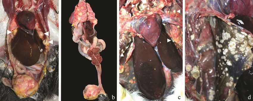
8.2      Oil Toxicity                                      30
    8.3      Lead Toxicity                                     31
    8.4      Organophosphate Toxicity                          31
9     Traumatic Injury                                         32
    9.1      Shock                                             32
    9.2      Skeletal Injury                                   33
    9.3      Soft Tissue Injury                                34
           Scalping injuries                                   34
           Exertional myopathy                                 34
           Pododermatitis (bumblefoot)                         34
           Bite wounds                                         34
    9.4      Central Nervous System Injury                     35
10         Diseases of Unconfirmed Aetiology                   36
    10.1     Clenched Claw Syndrome of Rainbow Lorikeets       36
    10.2     Lorikeet Paralysis Syndrome                       36
    10.3     Black and White Bird Disease                      36
11         Species mentioned in text                           38
12         References                                          40

                                                           3|Page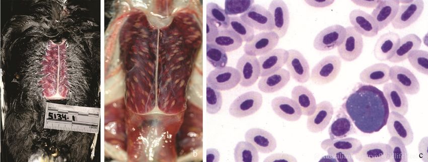
1 List of images

Figure 1 Progressively severe proliferative lower limb lesions from Knemidocoptes sp., pied
currawong (a-c), Knemidocoptes sp. mites from a skin scrape stained with methylene blue (d). ......... 7

Figure 2 Progressively severe proliferative lower limb lesions from Knemidocoptes sp., satin
bowerbird. (Images courtesy of South Penrith Veterinary Clinic and Carol Probets). ........................... 7

Figure 3 Harpyrhynchus rosellasinus cysts over the face, head and neck of a musk lorikeet (a), and
similar lesions over the face, neck and wings of a scaly-breasted lorikeet (b), also presumably caused
by Harpyrhynchus sp. mites. ................................................................................................................... 7

Figure 4 Hippoboscid fly.......................................................................................................................... 8

Figure 5 Cheilospirura (Xenocordon) gymnorhinis, oropharynx, a) Pied Butcherbird, b) Australian
Magpie .................................................................................................................................................... 8

Figure 6 Gapeworm (Syngamus trachea) in the trachea of an Australian Magpie. These worms
(arrow) have formed a thick raft along the entire length of the trachea in this bird. When removed,
the worms are red and have the typical forked appearance (b). ........................................................... 9

Figure 7 a) captive long-billed corella with a heavy burden of intestinal ascarids, and b) painted
button quail with severe oral capillariasis presented as thick yellow oral plaques occluding the palate
and throat. ............................................................................................................................................ 10

Figure 8 a) Ascarid ovum, b) strongyle-type ovum, c) cestode ovum, and d) capillaria ovum with
coccidial oocyst in bird faeces.............................................................................................................. 10

Figure 9 Cestode parasites, dorsal neck, spotted harrier ..................................................................... 11

Figure 10 Trichomonads in an oral smear and stained with Diff Quick. You can see the anterior
flagella (arrow) ...................................................................................................................................... 13

Figure 11 Coccidia oocysts in faeces from a) tawny frogmouth, and b) little penguin. ....................... 14

Figure 12 Lesions due to Leucocytozoon sp. in the pectoral muscle of a pied currawong before (a)
and after (b) the skin is reflected, and Leucocytozoon sp. schizont in a routine blood smear of a pied
currawong (c). ....................................................................................................................................... 15

Figure 13 Haemoproteus sp. in erythrocytes on blood smears from a little penguin (a) and an
Australian magpie (b) ............................................................................................................................ 15

Figure 14 (a) Enlarged liver extending beyond the keel (arrow) of a blue-faced parrot finch with
severe Plasmodium sp. infection. (b) Blood smears from a blue-faced parrot finch showing
Plasmodium sp. in erythrocytes. (c) Intraerythrocytic Plasmodium sp. (arrows) seen on spleen
impression of a little penguin ............................................................................................................... 16

Figure 15 Microfilaria, peripheral blood smear, barking owl ............................................................... 16

                                                                                                                                           4|Page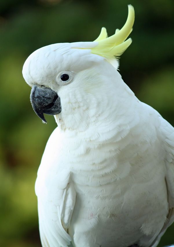
Figure 16 Rainbow lorikeet with multifocal yellow caseous lesions caused by enteropathogenic
Escherichia coli (EPEC)........................................................................................................................... 17

Figure 17 Necrotic segments in the small intestine (arrows) of a Rainbow Lorikeet ........................... 18

Figure 18 King parrot liver with scattered, randomly distributed, coalescing areas of cavitation and
yellow-brown discolouration (necrosis) positive for Chlamydia psittaci by PCR. ................................ 20

Figure 19 Sulphur-crested cockatoo with moderate (a) and severe (b) clinical signs of Psittacine beak
and feather disease (Images courtesy of Dr John Martin), and a Major Mitchell's cockatoo with mild
crest feather lesions caused by infection with PBFD. ........................................................................... 21

Figure 20 Rainbow lorikeet with Psittacine Beak & Feather Disease (Circovirus) showing stunted tail
and wing feathers including missing primary wing feathers (a and b), and deformed calamus of
pulled feathers (c) (Images courtesy of Dr Lydia Tong). ....................................................................... 22

Figure 21 Juvenile White-bellied sea eagle with feather damage to both wing and tail feathers and
prominent lesions of the feather calamus (insert). Images courtesy of Dr L Tong. ............................. 22

Figure 22 Pox lesions on the face, commissures of the beak, and feet of an adult Australian Magpie
(Image courtesy of Dr Bryn Lynar, Pittwater Animal Hospital) ............................................................. 23

Figure 23 a,b) Pied currawong, multiple white-yellow granulomas, lungs (arrows), and massively
expanding bursa, (c,d) Australasian gannet with characteristic fluffy white-green lesions, air sacs and
lungs caused by Aspergillus fumigatus. ................................................................................................ 26

Figure 24 Conidiophores with associated spores of Aspergillus fumigatus. ........................................ 26

Figure 25 Budding yeasts and hyphae of Candida albicans stained with Diff Quik .............................. 27

Figure 26 Macrorhabdus ornithogaster in the proventriculus of a Zebra Finch stained using a) Brown
& Brenn Gram stain, b) H&E stain, and c) PAS stain ............................................................................. 27

Figure 27 a) Australian white ibis, soft beak, malnutrition b) sulphur-crested cockatoo, skeletal
deformities including feet and keel - sunflower seed diet, c) semi captive guineafowl, malnutrition
and keel deformity (arrow), crop parasites (asterisk) .......................................................................... 28

Figure 28 A chestnut teal (a) with mild neck and leg paresis, and a mixed breed mallard (b) with
severe paresis from an outbreak of botilism in the same lagoon. ....................................................... 30

Figure 29 Wandering albatross with marked degeneration of the leg musculature............................ 34

Figure 30 Little penguins showing a) minimal external wounds from bite marks, b) puncture wounds
and severe cranial haemorrhage and fracture, c) penetrating bite wounds over skull damaging brain
and introducing feathers as a foreign body .......................................................................................... 35

Figure 31 Australian magpie with Black and White Bird Disease exhibiting a) paralysis of wings and
limbs, b) unable to right itself when placed on back though alert, and c) profound weakness and
paresis ................................................................................................................................................... 37

                                                                                                                                            5|Page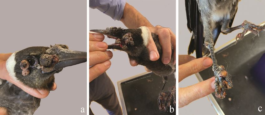
2 Introduction
A variety of diseases have been recognised within free ranging Australian birds. The purpose of this
document is to review the diseases that occur often within particular species or taxonomic groups of
birds. We hope that this information assists with the timely recognition of common parasites,
microbes, intoxicants and injuries to accelerate the appropriate care and welfare of wild animals.
Throughout the text we offer advice towards achieving a diagnosis. As best-practice wildlife
treatments change rapidly over time, treatment of birds in a rehabilitation situation should be made
in consultation with a veterinary professional.

 A notifiable disease is one that must be reported to agricultural authorities. If you suspect or can
 confirm that an animal is showing symptoms of one of the diseases listed as reportable, you must
 report it to:
     • your local vet or
     • Wildlife Health Australia’s state coordinator,
          wildlifehealthaustralia.com.au/AboutUs/ContactDetails.aspx
     • your state or territory's department of primary industries or agriculture by phoning the
          Emergency Animal Disease Watch Hotline on 1800 675 888.

3 Parasitic Disease
For additional information see also: Atkinson C.T., Thomas N.J., Hunter D.B. (2009) Parasitic Diseases
of Wild Birds. Wiley-Blackwell, Ames, Iowa.

3.1 Ectoparasites

 Zoonotic: May be vectors for zoonotic pathogens
 Species records: All
 Similar presentation to: viruses (e.g. pox virus), fungal dermatopathies
Birds can be parasitised by ticks, lice, mites, fleas and hippoboscid flies. Heavy burdens of
ectoparasites are often a reflection of a debilitated bird that is insufficiently grooming. Biting
ectoparasites can transmit blood parasites, and can contribute to anaemia when present in large
numbers.

Ixodes holocyclus, the paralysis tick, is found occasionally on birds. Anecdotal reports of tick paralysis
in birds have been documented. Three species of the genus Ornithodorus are known in Australia, O.
macmillani on wild birds, O. capensis on sea birds and O. gurneyi primarily on kangaroos (Barker &
Walker, 2014).

Lice commonly infest wild birds, but rarely cause disease, such as anaemia. Heavy infestations may be
treated with topical antiparasitic powders and by regularly changing substrate or enclosure materials
such as towels for birds undergoing rehabilitation.

Knemidocoptes intermedius infestations are recorded as causing severe debility in forest ravens, pied
currawongs, and superb lyrebirds (Holz, et al., 2005). Infestations are associated with significant
epidermal proliferation, primarily involving the skin of the lower portions of the legs. Mites can be
demonstrated by microscopic examination of scrapings of the thickened skin. Oral or subcutaneous
administration of Ivermectin-like drugs will control Knemidocoptes sp. infestation, but more severe

                                                                                              6|Page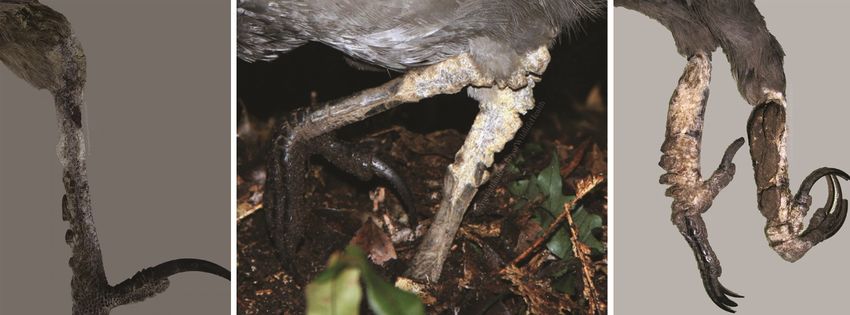
infestations are often associated with foot deformation and general debility, which may not be
amenable to treatment.

Figure 1 Progressively severe proliferative lower limb lesions from Knemidocoptes sp., pied currawong (a-c). Knemidocoptes
sp. mites from a skin scrape stained with methylene blue (d).

Figure 2 Progressively severe proliferative lower limb lesions from Knemidocoptes sp., satin bowerbird. (Images courtesy of
South Penrith Veterinary Clinic and Carol Probets).

A similar mite, presumed to be from the
Harpyrhynchus genus has been
described in Rainbow lorikeets, scaly-
breasted      lorikeets,      red-collared
lorikeets, musk lorikeets, and some
finches. The presentation of this mite is
very different from those above and
appear as firm, raised, yellow
subcutaneous skin nodules or cysts on
the head, wings, and body. These lesions
contain mites which are visible
microscopically if material from a cyst is
squashed between two glass slides, or in
a formalin fixed biopsy processed
routinely for histology. If you see
                                                    Figure 3 Harpyrhynchus rosellasinus cysts over the face, head and neck
affected birds, consider collecting a               of a musk lorikeet (a), and similar lesions over the face, neck and wings
sample into ethanol to enable more                  of a scaly-breasted lorikeet (b), also presumably caused by
precise identification of these parasites.          Harpyrhynchus sp. mites.

                                                                                                                7|Page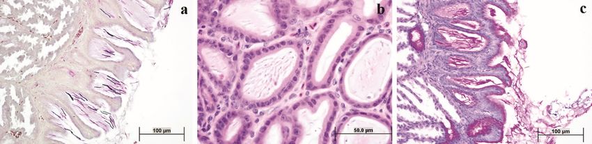
We have a lot to learn about the identity and ecology of these organisms.

Hippobosca are a genus of fly that occurs commonly within
the plumage of pigeons and birds of prey, especially tawny
frogmouths, and owls. Hippoboscid flies can bite and are
capable of acting as a vector in the transmission of disease.
These flies will infest and bite humans, but do not seem to
remain on human hosts for longer than 12-24 hours.

Sternostoma tracheacolum is the tracheal and air sac mite
of Gouldian finches. Heavy burdens of these mites are
capable of causing coughing, sneezing, and open mouth
breathing. Disease associated with this parasite is most
common in captive finches. A diagnosis can often be
achieved by reflecting the neck feathers and shining a bright
light across the trachea, or via microscopic examination of Figure 4 Hippoboscid fly
respiratory discharges. Ivermectin-like drugs can be used to
treat affected birds.

3.2 Endoparasites

        Throat worm: Cheilospirura (Xenocordon) gymnorhinis

  Zoonotic: No
  Species records: Australian Magpie
  Similar presentation to: gapeworm, trichomoniasis, capillariasis

Cheilospirura (Xenocordon) gymnorhinis, also referred to as throat worm, has been described in
juvenile magpies (De Chaneet & Robertson, 1983). The same, or similar, parasite also occurs in the
oral cavity and pharynx of pied currawongs, pied butcherbirds, magpie larks, black-faced cuckoo
shrikes, painted firetail finches, and rufous whistlers.

        Figure 5 Cheilospirura (Xenocordon) gymnorhinis, oropharynx, a) Pied Butcherbird, b) Australian Magpie

C. gymnorhinis burrows its head into the mucosa of the oral cavity and pharynx. The host then
responds by creating a fibrous nodule around the parasite. Although small numbers of parasites result

                                                                                                          8|Page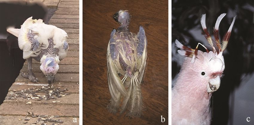
in self-limiting infections, large numbers can impair prehension of food, or partially obstruct the glottis
causing malnutrition or respiratory symptoms respectively. Repeated manual removal of the parasites
is recommended and this is assisted by application of moxidectin directly to the nematodes.
Euthanasia may be a humane option for severely debilitated young birds with heavy burdens of C.
gymnorhinis.

         Gapeworm: Syngamus trachea

  Zoonotic: No
  Species records: domestic poultry, Australian magpie, metallic starling, Australian raven, pied
  currawong, variegated fairy-wren
  Similar presentation to: throat worm, oral capillariasis, trichomoniasis
Syngamus trachea (gapeworm), should not be confused with C. gymnorhinis (throat worm; see
above), although similar in presentation, the symptoms and treatment are different. S. trachea are
small, red, worms that typically infect domestic poultry. S. trachea has been found in the trachea of
a variety of native birds and occasionally magpies that die suddenly are found to have complete
tracheal obstruction with masses of these parasites. The male parasites are very small, and they stay
in copulation with the much larger females, creating a forked appearance.

Figure 6 Gapeworm (Syngamus trachea) in the trachea of an Australian Magpie. These worms (arrow) have formed a thick
raft along the entire length of the trachea in this bird. When removed, the worms are red and have the typical forked
appearance (b).

         Trematodes (flatworms)

Austrobilharzia spp. are schistosome parasites that have been reported from the nasal cavity of
various duck species (Blair & Ottesen, 1979), and from blood vessels of silver gulls and various other
waterbirds (Appleton, 1983). Most commonly reported in silver gulls, A. terrigalensis uses the whelk
Velacumantus australis as its intermediate host, and can cause contact dermatitis or “swimmers itch”
in people (Appleton, 1983). Austrobilharzia sp. infection is most often an incidental finding in birds.

Mawsonotrema eudyptulae and Renicola sp. are trematodes of little penguins that can be found in
the liver and bile ducts (Obendorf & McColl, 1980; Harrigan, 1991). M. eudyptulae were found to be
highly pathogenic and contributed to little penguin deaths in Victoria (Harrigan, 1991).

         Nematodes (roundworms)

Gastrointestinal nematodiasis is usually an incidental finding in wild birds and parasite burdens are
generally mild. Captive birds, however, may experience excessive parasite burdens that can contribute
to debility.

                                                                                                         9|Page
Figure 7 a) captive long-billed corella with a heavy burden of intestinal ascarids, and b) painted button quail with severe oral
capillariasis presented as thick yellow oral plaques occluding the palate and throat.
Capillaria spp. are often evident within serpiginous tracts created as the nematode burrows through
the mucosa and lamina propria of the oesophagus, proventriculus and liver. Eggs may also be present
in faeces and appear as football shaped organisms with a thick shell and distinct bipolar plugs.
Capillariasis occurs primarily in captive birds. Infected birds may suffer from extensive hyperplasia of
the oesophageal mucosa and marked inflammation surrounding the parasites in the mucosa and
lamina propria. Emaciation, dehydration, weight loss, diarrhoea, regurgitation, anaemia and oral
necrotic plaques (Figure 9) can result from these infections. Capillaria sp. infections in wild birds are
primarily an incidental finding during post mortem examination.
Figure 8 a) Ascarid ovum, b) strongyle-type ovum, c) cestode ovum, and d) capillaria ovum with coccidial oocyst in bird faeces
Contracaecum spp. are nematodes that parasitise the oesophagus and proventriculus of piscivorous
birds. Small numbers of parasites pose no threat to the host. There are reports of large parasite
burdens of C. spiculigerum being associated with proventricular ulceration, haemorrhage, emaciation
and death in little penguins in Victoria (Harrigan, 1991).
10 | P a g eDispharynx species are nematode parasites that burrows into the mucosa of the crop, oesophagus or
proventriculus in water birds, finches and birds of prey. Heavy infections can be associated with poor
condition, diarrhoea, undigested seed in the faeces or death. Small numbers of organisms are easily
tolerated, whereas larger numbers of organisms are often accompanied by inflammation, mucosal
thickening and secondary infections.
Serratospiculum sp. reside within the airsacs of birds of prey, and infection is most common in falcons
and Australian Hobbies. Square-tailed kites have also been identified as possible hosts. Two species
are known in Australia: S. guttatum from Falco longipennis and F. peregrinus and S. tendo from F.
peregrinus. Clinical respiratory disease has been described in Australian birds infected with
Serratospiculum sp. that are subject to stress or concurrent disease. Diagnosis requires endoscopic
examination of the air sacs. Ivermectin-like drugs can be used to treat affected birds.
Oxysprirura spp. are nematode parasites that can be found within the conjunctiva and nictitating
membrane of a number of species. Infection with this parasite is usually asymptomatic, but may be
associated with conjunctivitis in a small proportion of infected birds (Pass, 1993).
Angiostrongylus cantonensis, the rat lungworm, has been found to cause neurological dysfunction
associated with eosinophilic or non-suppurative encephalomyelitis in yellow-tailed black cockatoos,
and more commonly in tawny frogmouths (Montali, et al., 2004; Monks, et al., 2005; Ma, et al.,
2013). Infection in tawny frogmouths is now a common occurrence and seems to have a seasonal
prevalence. Birds become infected with the parasite by eating infected snails and slugs, the
intermediate hosts. The parasites migrate through the intestinal wall and find their way to the spinal
cord, where they ascend to the brain. Affected tawny frogmouths present unable to fly, with various
levels of weakness/paresis and although they appear alert, are unable to right themselves if placed
on their back. Spinal damage progresses to central nervous signs. Diagnosis of the infection can be
very difficult, since birds do not usually develop eosinophilia in blood. Cerebrospinal fluid taps
collected from infected animals are also often non-suppurative rather than eosinophilic, making it
difficult to differentiate angiostrongylosis from viral or protozoal infection. NSW Department of
Primary Industries now offer PCR to aid diagnosis via CSF ante mortem or central nervous tissue post
mortem. Treatment of the infection in birds is also difficult. The parasite’s cuticle retains many
antigens and killing the worms can result in release of antigens with subsequent severe host immune
response. Concurrent antiparasitic and anti-inflammatory
treatment may halt parasite progression, but damage to the
host’s central nervous system is unlikely to regress.
Cestodes (tapeworms)
Birds are parasitised by many species of cestode. None of these
is considered to be highly pathogenic in free-living birds. It is
possible, however, that large burdens of cestodes will add to
the debility of a captive or compromised bird. If necessary,
cestodiasis is treated with praziquantel. The treatment is
usually repeated 10 days after the first dosage.
Protozoa (single-celled parasites)
A wide variety of protozoa has been reported within the
gastrointestinal tract, cardiovascular system, musculature and
Figure 9 Cestode parasites, dorsal neck,
renal tissues of free-flying birds. The following discussions spotted harrier
regarding protozoa are limited to those protozoal infections
known to be clinically significant.
11 | P a g eSpironucleus-like (formerly Hexamita) organisms have been associated with numerous outbreaks and
individual cases of emaciation, diarrhoea and fatal enteritis in Australian king parrots (Philbey, et al.,
2002). Similar parasites have been identified in emaciated, wild sulphur crested cockatoos from
western NSW (Registry unpublished). These birds become emaciated and have very thin walled
intestinal tracts, often filled with fetid brown fluid. The intestinal tissues of affected birds seem to
decompose very rapidly making it difficult to identify organisms on histologic examination.
Microscopic examination of saline wet-mount preparations of intestinal scrapings can be used to
demonstrate the organism during gross post mortem examination.
Gastrointestinal Giardia spp. infections have been documented in a variety of wild and aviary birds in
Australia. Giardia spp. have been recovered from the intestinal lumen of straw-necked ibis in Western
Australia, and a sulphur-crested cockatoo in Victoria (Forshaw, et al., 1992; Gallagher, et al., 1995).
Giardiasis in captive young budgerigars can result in decreased growth rates, dehydration, and
diarrhoea (Filippich, et al., 1998). Diagnosis of giardiasis is based upon direct microscopic examination
of faeces or intestinal content. Giardia sp. trophozoites are pear shaped, binucleate, and have eight
flagella. A cyst form, with four nuclei is occasionally shed in the faeces. Infection can be diagnosed
with PCR, Wrights or modified acid-fast stained faecal samples or intestinal scrapings. Treatment of
budgerigars with metronidazole decreased shedding of these protozoa in the faeces (Filippich, et al.,
1998). Treatment for giardiasis is the same as for trichomoniasis. Careful attention to hygiene will
prevent clinical infection in most captive birds.
Cryptosporidia spp. have been observed within the intestinal and proventricular brush border of wild
Pacific black ducks, red-tailed black cockatoo, various captive finches and mannikins, Australian
magpie, rock parrot, scaly-breasted lorikeet, southern boobook, and regent honeyeater (Registry). The
significance of this parasite as an avian pathogen is poorly understood.
3.2.6.1 Toxoplasma gondii
Zoonotic: Yes
Species records: pied currawong, tawny frogmouth, satin bowerbird, little penguin, regent
bowerbird, red-whiskered bulbul, crimson rosella
Similar presentation to: septicaemia, Angiostrongylus cantonensis infection
Toxoplasmosis is a potentially fatal disease in native and aviary birds, caused by the single celled
parasite Toxoplasma gondii (Wendte, et al., 2011; Dubey, 2002). Cats are the definitive hots of this
parasite and excrete oocytes in their faeces. Birds become infected when they ingest these oocytes,
or when they ingest cysts in the tissues of small birds or mammals. Birds with toxoplasmosis are
depressed, fluffed, debilitated or are found dead. Gross post mortem findings consist of pulmonary
oedema, pulmonary congestion, and pale foci within the liver, spleen and intestinal mucosa. Histologic
examination of fixed tissues reveals pulmonary oedema and congestion, fibrin within the distal
airways, and non-suppurative inflammation or necrosis within the liver, spleen, brain, skeletal muscle,
ventriculus, adrenal gland, or intestine. Numerous protozoa, morphologically consistent with T. gondii
may be observed in avian tissues. Definitive diagnosis of T. gondii infection can be established in native
birds using PCR or immunohistochemistry (Hartley & Dubey, 1990; Dubey, 2002).
Protozoal cysts resembling those of T. gondii are observed in the absence of inflammation during
routine histologic examination of nervous tissue of a variety of native birds, especially the tawny
frogmouth. These cysts are consistent morphologically with T. gondii and they appear to be a common
incidental finding. Clinical disease occurs when these cyst walls break down and liberate the internal
zoites, which incite the host’s immune response causing tissue damage.
12 | P a g e3.2.6.2 Trichomoniasis
Zoonotic: No
Species records: Peaceful dove, little penguin, rock (feral) pigeon, southern boobook, painted
button-quail, brown goshawk, budgerigar, peregrine falcon, bar-shouldered dove, powerful owl,
Australian bustard, barking owl, superb lyrebird, rainbow lorikeet, crested pigeons, spotted dove,
Australian hobby falcon, black-shouldered kite, little eagle, pied currawong, Australian raven,
Australian magpie, channel-billed cuckoo, tawny frogmouth (Park, 2011).
Similar presentation to: oral Capillaria species, throat worm
Oral trichomoniasis has been observed in debilitated free ranging birds, but is most common in captive
wildlife that are undergoing treatment for various injuries. Trichomonads are common commensal
agents within the avian alimentary tract and can be present in faeces, oral mucous or crop secretions.
Trichomonads are ovoid single celled parasites (protozoa) that have four anterior flagella and an
undulating membrane. These organisms are spread through either direct or indirect contact,
including; contaminated food or water sources, consumption of infected prey, or during feeding of
young. The factors that predispose a bird to develop trichomoniasis are unknown.
Caseous oral plaques are created when the organisms cause
tissue necrosis. Lesions are often subject to secondary
bacterial infection. Weight loss, depression, anorexia,
vomiting and difficulty swallowing are common clinical signs,
and difficulty breathing and asphyxiation may occur in severe
cases where plaques block the airway. A diagnosis of
trichomoniasis is best made by examining a wet mount
preparation of the caseous debris (Figure 3). Flagellates can
be seen moving within the wet preparations under light
microscopy. The organisms are much more difficult (often
impossible) to see within cytologic and histologic
preparations of affected tissues.
Figure 10
3 Trichomonads
Trichomonadsininan
anoral
oralsmear
smearand
and
stained with Diff Quick. You can see the Treatment of trichomoniasis includes debridement of the
anterior flagella (arrow) caseous plaques, supportive care and administration of
antiprotozoal agents.
3.2.6.3 Coccidia
Eimerian and isosporan coccidial oocysts are commonly identified within the faeces of healthy captive
and free-flying birds. Disease associated with coccidial infection in free ranging birds is rare. Coccidia
may cause necrotising enteritis in young captive birds of a variety of species. Microscopic examination
of faecal wet preparations and flotations is recommended to monitor coccidia burdens in birds
maintained in aviaries. When large numbers of faecal oocysts are detected or if oocysts accompany
diarrhoea, treatment is advisable.
Renal coccidiosis is a common incidental finding within little penguins, Australasian gannets, and
short-tailed shearwater (mutton bird). Limey disease is the term used to describe clinically apparent
renal coccidiosis in nestling short-tailed shearwater. Chicks with limey disease are thin and have urate
and faecal soiling of the pericloacal feathers (Munday, et al., 1971). Renal enlargement and multiple
pale foci throughout the kidney are evident on gross post mortem examination. The ureter and cloaca
may also be distended with urates. Microscopic examination of the affected renal tissue reveals
inflammation within the interstitium surrounding large collecting ducts. Coccidial oocysts are often
13 | P a g eevident within multiloculated granulomas within the collecting duct mucosa and in the surrounding
interstitium.
Figure 11 Coccidia oocysts in faeces from a) tawny frogmouth, and b) little penguin.
Caryospora spp. are coccidian parasites that can be found within the intestinal lamina propria and
mucosa of carnivorous birds, but these are generally incidental findings. Caryospora spp. can be found
infecting reptiles, birds and rodents and can have a single host, or two host (predator-prey) lifecycle.
The intestinal forms of Caryospora spp. are characterised by a single sporocyst containing eight
elliptical sporozoites.
Systemic coccidiosis associated with Atoxoplasma (formerly Lankestrella) spp. and Isospora spp. have
been identified within circulating monocytes of various birds within Australia. Some of these include
the regent honeyeater, red wattlebird, brown treecreeper, house sparrow, silvereye, and various
aviary birds (Mackerras & Mackerras, 1960; Yang, et al., 2015). These systemic coccidian parasites
undergo sexual reproduction (gametogony) within the mucosa of the gastrointestinal tract, and
asexual reproduction (schizogony) within extra-intestinal tissues such as the liver and spleen.
Sporozoites of this organism are transported among these sites within mononuclear cells. Sporozoites
are round basophilic organisms that have a small basophilic nucleus. These sporozoites are visible,
usually as individual organisms, within the cytoplasm of mononuclear cells, which have an indented
nucleus.
Protozoan blood parasites
Haemoproteus, Leucocytozoon, Plasmodium, Atoxoplasma, and a Babesia-like organism are genera of
the family Plasmodiidae that are commonly found within the peripheral blood of wild Australian birds.
Each of these organisms is arthropod borne. A bird may be infected with two or more of these
organisms concurrently without any clinical signs. Young or debilitated birds may develop anaemia,
anorexia, and depression as a result of large parasite burdens.
Although most often a common, incidental infection, on occasion, Leucocytozoon megaloschizonts.
can cause clinically significant myopathy in pied currawongs and Australian magpies within the Sydney
region. Infection occurs in juvenile, sub-adult and adult birds of both sexes, at any time of year. Heavy
parasite burdens result in lethargy, weakness and debility. If the breast feathers are parted, pale oval
foci may be evident throughout the pectoral musculature of affected birds. There is no known
treatment for this protozoal infection and birds often die shortly after initial examination. The lifecycle
of this protozoal agent is unknown.
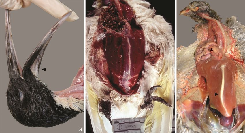
14 | P a g eFigure 12 Lesions due to Leucocytozoon sp. in the pectoral muscle of a pied currawong before (a) and after (b) the skin is
reflected, and Leucocytozoon sp. schizont in a routine blood smear of a pied currawong (c).
Upon post mortem examination of affected birds, discrete, pale oval foci measuring up to 1.5 cm long
and 0.5 cm wide are scattered throughout the skeletal muscles, tongue, myocardium and ventricular
muscularis externa. Histopathologic examination demonstrates that pale foci consist of central
megaloshizonts, surrounded by necrotic muscle and an intense inflammatory response. Haemorrhage,
necrosis, and inflammation are most severe around ruptured megaloschizonts. Pigmented oval
Leucocytozoon gamonts may or may not be evident within blood of affected birds, and are generally
most concentrated in renal tissue.
Haemoproteus columbae were first described from the rock (feral) pigeon in 1890 and Haemoproteus
sp. have since been described from a variety of bird species. The taxonomy of Haemoproteus sp. has
been subject to many restructures as DNA sequencing technology evolves. The vectors for these
parasites are believed to be hippoboscid flies and infection is generally mild and incidental. In severe
infections, birds may develop anaemia, become reluctant to move, anorexic, appear fluffed-up,
become lame, have difficulty breathing, or simply be found dead. Post mortem findings may include
enlarged spleen, liver, kidneys, or gizzard, chocolate-brown coloured organs, and large skeletal muscle
cyst-like lesions similar to those described above for Leucocytozoon infections. Blood films and tissue
impression smears may contain infected blood and skin cells respectively.
Figure 13 Haemoproteus sp. in erythrocytes on blood smears from a little penguin (a) and an Australian magpie (b)
The family Plasmodiidae includes the genus Plasmodium which is responsible for malaria infections.
The vectors for this organism are typically mosquitoes and infection may be incidental, severe or fatal.
Naïve populations are most at risk of severe disease. Birds may present weak, thin, or dead. Gross
findings may include an enlarged, dark brown spleen or liver. Blood films and tissue impression smears
are important for diagnosing avian malaria. Organisms can be difficult to locate and identify within
histological sections.
15 | P a g eFigure 14 (a) Enlarged liver extending beyond the keel (arrow) of a blue-faced parrot finch with severe Plasmodium sp.
infection. (b) Blood smears from a blue-faced parrot finch showing Plasmodium sp. in erythrocytes. (c) Intraerythrocytic
Plasmodium sp. (arrows) seen on spleen impression of a little penguin
Microfilariae are occasionally found during examination of
peripheral blood smears and histological sections of wild
birds. Adult filarial nematodes may reside within the air
sacs, coelomic cavity, subcutaneous tissues, heart, greater
vessels, or lungs, but they are very fine and can be difficult
to spot. Infection is diagnosed during microscopic
examination of peripheral blood smears or buffy coat
smears. Microfilariae are transmitted by haematophagous
arthropods. Microfilarial infections are incidental to the
Figure 15 Microfilaria, peripheral blood smear, host.
barking owl
Trypanosomes are occasionally found within the peripheral blood of native birds (Zidkova, et al.,
2012). These single celled parasites are extracellular flagellates that are transmitted by biting flies or
midges. Trypanosomes are reported most commonly in little penguins, and they do not appear to be
pathogenic (Jones & Woehler, 1989).
4 Bacterial Disease
Sporadic outbreaks of mortality in native birds have been attributed to localised or systemic infection
with E. coli, Salmonella spp., Pasteurella spp., Mycobacterium spp., Erysipelothrix rhusiopathiae,
Listeria monocytogenes, Streptococcus spp., Staphylococcus aureus, Haemophilus spp., Mycoplasma
spp., and Clostridium spp. (Registry). Ideally treatment of bacterial infection is based upon isolation of
the organism within lesions, and antimicrobial sensitivity testing. Treatment without consultation and
confirmation of the infectious agent may lead to ineffective treatment, and antimicrobial resistance.
Some of these organisms are potentially zoonotic. Sound hygiene protocols for birds in rehabilitation
will protect both birds and their rehabilitators.
4.1 Escherichia spp.
Zoonotic: Yes
Species records: various
Similar presentation to: other bacterial pathogens, toxoplasmosis
16 | P a g eWhile Escherichia coli can be considered an opportunistic
bacterium and a pathogen of concern in its own right, there
are several specific strains of E. coli that can cause
considerable disease in both wildlife, and people.
Enteropathogenic E. coli (EPEC) causes diarrhoea and
mortality in both birds and humans globally. While most
reports are from captive birds such as parrots, owls, doves
and pigeons, this disease has been recovered from wild
rainbow lorikeets during a localised unusual mortality event
in Australia (Registry, unpublished) which highlights the
importance of wild birds as a reservoir for human disease
(Sanches, et al., 2017). Transmission is via ingestion of
contaminated food or water, or contact with other infected
animals. EPEC adheres to the intestinal mucosa causing
severe ulceration and watery or haemorrhagic diarrhoea. In
order to identify the specific pathotype, diagnosis is via
traditional culture methods followed by molecular typing or Figure 16 Rainbow lorikeet with multifocal
via PCR. Treatment is complicated by multi-drug resistance yellow caseous lesions caused by
making positive identification with sensitivity testing enteropathogenic Escherichia coli (EPEC)
integral for treatment of infected birds.
Escherichia albertii is very difficult to distinguish from E. coli, is also zoonotic, found in wild and
captive birds, and causes diarrhoeal disease and sudden death in various bird species. Some birds
may act as carriers (Wildlife Health Australia, 2013). Dead birds are often in good body condition but
gross lesions may vary depending on species infected. Infection has been recorded in spotless crake,
chickens, Australian magpie, little corella, galah, rainbow lorikeet, brown-headed honeyeater, grey
fantail and superb fairy-wren from the east coast of Australia, but not in Western Australia or
Tasmania (Wildlife Health Australia, 2013). Transmission is by ingestion of food and water
contaminated by infected faeces. E. albertii is capable of surviving in the environment for some time
under optimal conditions, therefore hygiene of bird feeding and bathing stations is essential.
Diagnosis can be made from culture of faeces or cloacal swabs in live birds, or liver and spleen in
dead birds, on MacConkey agar. PCR is also required to definitively identify E. albertii.
4.2 Yersiniosis
Yersinia pseudotuberculosis infections can result in either acute enteritis and septicaemia (systemic
bacterial infection, most often spread in the circulatory system), or multisystemic abscesses. Some
birds show no symptoms and are simply found dead while others may show chronic diarrhoea,
weakness, ruffled feathers, lameness and progressive weight loss.
4.3 Necrotic Enteritis
Wild rainbow lorikeets, scaly-breasted lorikeets, and king parrots in coastal eastern Australia are
seasonally affected with necrotising enteritis. A variety of organisms, primarily coliforms, have been
isolated within the necrotic intestinal tissue. Clostridium perfringens, Cl. tertium, and Escherichia coli
are most commonly isolated within the intestine and other tissues of birds with necrotic enteritis. It
can be difficult to interpret these findings, as these organisms can be normal residents of the intestinal
tract.
An investigation into the occurrence of necrotic enteritis identified 58 dead rainbow lorikeets, red-
collared lorikeets, and scaly-breasted lorikeets originating from 18 different flocks in eastern Australia
over a ten-year period (McOrist & Reece, 1992). Cl. perfringens was isolated from the intestinal tissues
17 | P a g eof many birds, and beta toxin was demonstrated within the bacterial colonies and within intestinal
content using gas liquid chromatography (McOrist & Reece, 1992).
Carbohydrate overload has been suggested as a means of causing intestinal overgrowth with
Clostridium sp., and subsequent necrotic enteritis (Pass, 1993; McOrist & Reece, 1992; Ferrell & Tell,
2001). Artificial feeding stations established for lorikeets in urban areas which provide only sugar
water, and have questionable hygiene for cleaning surfaces between feeds, are potential sources of
infection. The presence of an underlying viral infection in birds suffering from necrotic enteritis, has
not been thoroughly investigated.
Necrotic enteritis occurs in male and female birds, juvenile animals and adults. Free-ranging birds are
most commonly diagnosed with necrotic enteritis; however, the disease has been observed in captive
lorikeets. Necrotic enteritis is most often observed in July and August.
Necrotic enteritis is identified based upon the clinical signs and microbial culture of faeces. Many birds
with necrotic enteritis are found dead. Post mortem examination and microbial culture of segments
of intestine, liver and other filtering organs are used to establish a diagnosis.
Figure 17 Necrotic segments in the small intestine (arrows) of a Rainbow Lorikeet
Live birds with necrotic enteritis exhibit a variety of clinical signs. Most of these birds are in good body
condition, but are weak, depressed, dehydrated, regurgitate clear fluid, and have soiled vent feathers
as a result of watery diarrhoea. The bird’s abdomen may be palpably distended. Species affected by
necrotic enteritis normally have wet faeces, and the detection of diarrhoea may be challenging.
During the gross post mortem examination of these birds, the intestinal tract is distended by gas or
reddish-brown fluid. A tan inflammatory (diphtheritic) membrane coats the mucosa, or the mucosa is
found to be friable and haemorrhagic. Microscopic examination of intestinal scrapings illustrates large
numbers of bacterial colonies, epithelial and inflammatory cells. Histological examination of formalin
fixed segments of intestine reveals the following: mucosal to transmural necrosis, intense
mononuclear cell infiltration, oedema and congestion throughout the lamina propria and submucosa,
and colonies of bacteria scattered throughout a superficial layer of necrotic debris and fibrinous
exudate.
Although antimicrobial sensitivity testing of bacteria isolated within the intestine of birds with necrotic
enteritis guides therapy, treatment of these birds is rarely successful. Presumably, the birds are
suffering from either enterotoxaemia or septicaemia by the time they demonstrate clinical signs and
reach care.
18 | P a g e4.4 Chlamydiosis
Zoonotic: Yes
Species records: sulphur-crested cockatoos, little corellas, crimson rosellas, king parrots, feral
(rock) doves
Similar presentation to: septicaemia, toxoplasmosis, intoxication, malnutrition
Chlamydophila psittaci is a bacterium of the family Chlamydiaceae. This bacterium is capable of
causing severe disease in free-living birds, aviary birds and humans. When this zoonotic bacterium
infects humans, it is referred to as psittacosis, and may cause serious pneumonia. C. psittaci is endemic
throughout Australia. It is a notifiable disease. Avian infections with this organism are termed
ornithosis, or chlamydiosis. Psittacine and columbiform birds are most susceptible to C. psittaci
infection. Chlamydial disease is most common in rosellas, and to lesser extent lorikeets, cockatoos,
budgerigars, and king parrots, but any bird can be affected and a potential source for human infection.
C. psittaci is transmitted through the faecal-oral route, respiratory secretions or the inhalation of
organisms in aerosolised feather dander or faeces. C. psittaci is highly infectious and a single organism
can cause severe disease in birds and humans. The bacterium can form compact elementary bodies
that can remain infective within the bird, dried faeces or dander for several months. Chlamydiosis
should be considered among the differential diagnoses in any emaciated wild bird, and barrier
methods should be employed to prevent potential spread of infection to other wildlife or humans.
Birds with active chlamydiosis may exhibit a broad range of symptoms associated with either acute or
chronic disease. Many birds will function as asymptomatic carriers of the organism, while others may
suffer severe or fatal infection. Chlamydiosis is most often manifested as respiratory or
gastrointestinal illness. Neurological signs are rare, but a recognised outcome of avian chlamydiosis.
Clinical signs associated with C. psittaci infection include: weight loss, depression, lethargy, anorexia,
diarrhoea, bile stained faeces, ocular or nasal discharge, and dyspnoea (difficult or laboured
breathing). Monocytosis is a common haematological finding in affected birds and on occasion,
elementary bodies may be visible within monocytes, particularly if concentrated in a buffy coat
preparation.
Post mortem findings can be highly variable in birds suffering from chlamydiosis. Some birds may die
acutely with very few morphologic lesions, while some may exhibit splenomegaly and hepatomegaly,
and others may have fibrinous air sacullitis, pericarditis and enteritis.
Definitive diagnosis of chlamydiosis relies upon detection of the organism using PCR. Marked
leucocytosis, monocytosis, and an elevated AST may be suggestive of C. psittaci infection, however,
there is significant variability in the haemogram of birds with chlamydiosis. Antigen can be detected
within conjunctival, nasal, or faecal swabs using antigen capture ELISA tests or direct
immunofluorescence testing. Diagnostic tests based upon antigen capture are highly sensitive, but
may not be highly specific. Some gram negative bacteria will cross react with the antibody used in the
ELISA test, thus, conjunctival and choanal swabs will provide far fewer false positive reactions
compared with faecal swabs. ELISA based antigen capture test kits are commercially available for in-
house identification of Chlamydia sp. antigen. These kits are marketed for the detection of human C.
trachomatis within urine samples, but they are effective in the identification of C. psittaci.
19 | P a g eFigure 18 King parrot liver with scattered, randomly distributed, coalescing areas of cavitation and yellow-brown
discolouration (necrosis) positive for Chlamydia psittaci by PCR.
Post mortem diagnosis of chlamydiosis is usually based on finding multisystemic histiocytic
inflammation on histologic examination and identification of the organism within lesions. Methanol
fixed impression smears of spleen, lung, and liver can be stained using modified Machiavello’s staining
protocols. This protocol can also be used to identify the organism within paraffin embedded tissue,
but organisms are easier to identify within tissue impression smears. Fresh tissues, such as liver, spleen
and lung may be submitted fresh, or after freezing at – 80oC, to a microbiology laboratory for culture,
or swabs from fresh tissues can be tested with antigen capture ELISA tests or PCR.
Immunohistochemical demonstration of the organism is possible in fixed tissues. Always communicate
your concern regarding the possibility of chlamydiosis when submitting samples to a laboratory,
museum, taxidermist or researcher.
5 Viral Disease
5.1 Circovirus: Psittacine Beak & Feather Disease (PBFD)
Zoonotic: No
Species records: sulphur-crested cockatoos, rainbow lorikeets, many others
Similar presentation to: feather mites, fungal infections, feather trauma, nutritional insults
Psittacine circovirus, the causative agent of Psittacine Beak and Feather Disease (PBFD), was listed as
a key threatening process for the survival of 5 endangered species, including the orange-bellied parrot,
in 2004 by the Commonwealth Government.
PBFD is a common disease in wild and aviary psittacines throughout Australia. PBFD has also been
identified in rainbow bee-eater, Gouldian finch, zebra finch, powerful owl, boobook, brown goshawk,
laughing kookaburra, Australian raven, tawny frogmouth, Australian white ibis, and wedge-tailed
eagle (Phalen, 2019). The disease is manifested by lesions in the feathers, beak and occasionally the
claws. Disease transmission may occur via the faecal-oral route, feather dust, regurgitated crop
contents or possibly through the egg. Raptors may become infected though the consumption of
infected parrots (Phalen, 2019).
Clinical signs of infection with psittacine circovirus are highly variable depending on the age and
species of the bird, and the quantity of virus in the infective exposure. The progression of disease is
20 | P a g ealso highly variable, ranging from acute to chronic. Young birds most often exhibit the acute form of
infection. Clinical signs of acute psittacine circovirus infection include diarrhoea, weight loss, anorexia,
depression and either death or residual feather damage. The chronic form of psittacine circovirus
infection in cockatoos begins with loss of the powder keratin in the plumage, and the production of
abnormal down feathers over the hips. Powder down feathers become short and lose the plumaceous
barbs. The loss of powder down feathers results in a dull and dirty look of contour and flight feathers,
and imparts a glossy black appearance to the beak.
Figure 19 Sulphur-crested cockatoo with moderate (a) and severe (b) clinical signs of Psittacine beak and feather disease
(Images courtesy of Dr John Martin), and a Major Mitchell's cockatoo with mild crest feather lesions caused by infection with
PBFD.
When damaged by psittacine circovirus, the beak may become elongated, softened, broken, cracked,
or it may have uneven wear. These changes are most commonly seen in cockatoos in the late stages
of infection. If the germinal epithelium of the beak is exposed by fractures or cracks in the keratin, the
bird will often stop eating due to pain.
Young lorikeets of the genus Trichoglossus that are infected with psittacine circovirus will often
present with the last two to four primary feathers of the wings broken or missing. If pulled from their
follicles, the calamus of the remaining tail feathers and flight feathers will often exhibit characteristic
morphologic lesions. These lorikeets are called “runners” since they are unable to fly, yet healthy
enough to forage and run on the ground. Young lorikeets are identified by their dark brown beaks.
Occasionally, these young lorikeets will have a blotchy yellow pattern on the tail feathers that are
usually green. Beak lesions rarely occur in lorikeets infected with psittacine circovirus.
Feathers damaged by psittacine circovirus are curled, clubbed, easily broken, or they have retained
feather sheaths, haemorrhages within the calamus (shaft), or annular constrictions of the calamus.
Replacement feathers grow slowly, or fail to regrow.
21 | P a g eFigure 20 Rainbow lorikeet with Psittacine Beak & Feather Disease (Circovirus) showing stunted tail and wing feathers
including missing primary wing feathers (a and b), and deformed calamus of pulled feathers (c) (Images courtesy of Dr Lydia
Tong).
Histologic lesions associated with psittacine circovirus occur primarily
in the growing feather, but may also be evident within the follicle.
Necrosis occurs in the germinal layer of the follicular and feather
epithelium, and basophilic cytoplasmic inclusion bodies may be evident
within the epithelium, and in reticuloendothelial cells in the dermis,
feather pulp, and bursa of Fabricius.
Psittacine circovirus infects the thymus and bursa of Fabricius and is
associated with lymphoid necrosis and premature atrophy of these
tissues, resulting in immunosuppression. Birds with psittacine beak and
feather disease often succumb to secondary viral, bacterial or fungal
infections.
The presumptive diagnosis of psittacine beak and feather disease is
based upon gross and microscopic lesions in the feather and feather
follicle.
Psittacine circovirus is very difficult to isolate in culture. Definitive
diagnosis of infection with this virus can be established through
serological testing, which is available through commercial and Figure 21 Juvenile White-bellied sea
eagle with feather damage to both
academic laboratories. Haemagglutination inhibition (HI) testing wing and tail feathers and prominent
detects antibodies to psittacine circovirus in blood, serum and yolk, lesions of the feather calamus (insert).
while haemagglutination (HA) testing detects the virus in faecal Images courtesy of Dr L Tong.
samples or feathers. Birds that suffer from severe psittacine beak and
feather disease may not mount an effective immune response to the virus and their titres measured
via HI tests may not be elevated. Elevated HI titres merely indicate that antibodies have been formed
in response to exposure to psittacine circovirus. Birds with either the acute or chronic form of
circovirus infection often have low titres. Budgerigars, lorikeets, and king parrots, however, usually
have high psittacine circovirus titres and continue to shed the virus.
Immunohistochemistry, PCR, and DNA in-situ hybridisation tests for psittacine circovirus are available
overseas, while PCR, HI and HA are primarily used in Australia. In a live bird, a blood spot on filter
paper and affected feathers can be tested (Phalen, 2012).
22 | P a g eSome species of psittacine can spontaneously recover from the acute form of beak and feather
disease. Rainbow lorikeets, budgerigars, eclectus parrots and king parrots may recover from this
infection with only mild residual feather changes. Birds with the chronic form of psittacine circovirus
infection rarely recover, but they can live for several years. The cause of death most often relates to
secondary infection with other viral, bacterial or fungal agents as a result of immunosuppression.
There is no known cure for PBFD. Nursing care to keep the bird warm and eating will prolong the life
of cockatoos. Lorikeets may spontaneously recover from psittacine beak and feather disease, but can
shed the virus for a prolonged period.
Since there is no effective treatment for birds suffering from PBFD, controlling the spread of the virus
relies of strict hygiene and euthanasia of affected birds. If euthanasia is not under consideration,
affected birds should be maintained under strict quarantine. Viracidal disinfectants used to kill
parvovirus should inactivate psittacine circovirus.
5.2 Poxvirus
Australian magpies, native pigeons and raptors are occasionally clinically affected by poxvirus
infection. Poxvirus is a member of the genus Avipox, which has a worldwide distribution. Poxvirus is
shed in saliva, nasal secretions, faeces and wound exudates or scabs. The virus is stable for several
months in the environment under favourable conditions. Poxvirus is transmitted primarily by
haematophagous arthropods, such as mosquitos; however, other vectors and fighting can also result
in transmission. Infection results in viraemia and then localisation within the skin or mucosa.
Clinical signs of poxvirus infection vary from blistering and small nodules in the skin to large dermal
nodules with markedly hyperplastic epithelium, which may have foci of ulceration. These lesions
primarily occur on the skin of the feet, legs and head, and around the eyes, mouth and cloaca.
Secondary bacterial infection is a common finding in poxvirus lesions. Some birds will recover
spontaneously, while others will become debilitated due to difficulty walking or obtaining food.
Figure 22 Pox lesions on the face, commissures of the beak, and feet of an adult Australian Magpie (Image courtesy of Dr
Bryn Lynar, Pittwater Animal Hospital)
The microscopic lesions associated with poxvirus infection include marked epidermal thickening.
Hyperplastic epithelial cells may contain cytoplasmic vacuoles that house large, eosinophilic inclusion
bodies. These inclusion bodies are called Bollinger bodies.
23 | P a g eYou can also read