Nipah Virus Assays and Animal Models for Vaccine Development - Landscape Analysis, January 2021
←
→
Page content transcription
If your browser does not render page correctly, please read the page content below
Nipah Virus Assays and
Animal Models for Vaccine
Development
Landscape Analysis, January 2021

Author:
Albert Price, IAVI

Secondary authors/reviewers:
Donata Sizemore, IAVI
Thomas Hassell, IAVI
Raúl Gómez Román, CEPI
Johan Holst, CEPI
Paul Kristiansen, CEPI
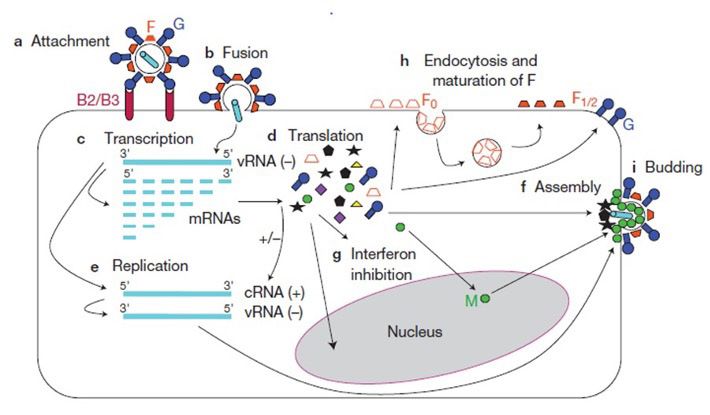
TABLE OF CONTENTS
I. Introduction                                                                4

II. Background                                                                  5

      1. Epidemiology                                                           5

      2. NiV Clinical Features and Pathogenesis in Humans                       9

      3. Diagnosis and Treatment                                              10

      4. NiV Molecular Biology and Structure                                    11

      5. Vaccine Development                                                   13

III. Standardization of Assays and Animal Models                               18

IV. NiV Serological Assays                                                     21

      1. Detection of antigen – specific serum IgG                             21

      2. Detection of serum neutralizing antibodies                           22

V. NiV Animal Models                                                          27

      1. Syrian Golden Hamster                                                29

      2. Ferret                                                               32

      3. African Green Monkey (AGM)                                           34

VI. Conclusions                                                               38

VII. References                                                               40

VIII. Statement of Support                                                    46

      1. Article H.20. Publication And Publicity                              46

2                 Nipah Virus Assays and Animal Models for Vaccine Development, Landscape Analysis, January 2021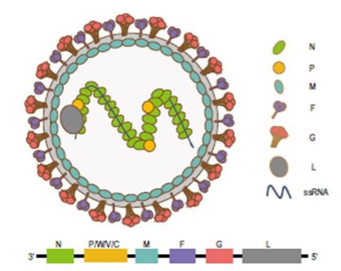
LIST OF FIGURES
Figure 1. Geographic distribution of Pteropid fruit bats
           and Henipavirus outbreaks (Enchery and Horvat, 2017)                5

Figure 2. NiV structure and organization of the 18.2 kB ssRNA
           (-) genome (Sun et al., 2018).                                      11

Figure 3. The Henipavirus infection and replication cycle
           (Aguilar and Lee, 2011).                                           12

LIST OF TABLES
Table 1. NiV Outbreaks by Year and Location*                                   7

Table 2. Differences in Clinical and Epidemiological Characteristics
         Between NiV Malaysia and Bangladesh Outbreaks                        8

Table 3.  NiV Viral-vector Vaccine Candidates Tested in Animals               15

Table 4. NiV Submit Vaccine Candidates Tested in Animals                      16

Table 5. NiV vaccine candidates supported by CEPI                             17

Table 6. Benefits and Potential Challenges of Implementing
          Biological Standards for NiV Vaccine Development                   20

Table 7. Pros and Cons of NiV Serological Assays                             24

Table 8. Serological Assays Used in NiV Pre-Clinical Vaccine Studies         25

Table 9. Serological Assays Used in Other NiV Research Studies               26

Table 10. S
           ummary of clinical signs and pathology in the NiV hamster,
          ferret and AGM challenge models                                    28

Table 11. NiV Hamster Model Challenge Studies                                30

Table 12. NiV Hamster Model Challenge Studies, Continued                      31

Table 13. NiV Ferret Model Challenge Studies                                 33

Table 14. NiV African Green Monkey Model Challenge Studies                   35

Table 15. NiV African Green Monkey Model Challenge Studies, Continued        36

Table 16. Pros and Cons of the Major NiV Animal Challenge Models             37

3                Nipah Virus Assays and Animal Models for Vaccine Development, Landscape Analysis, January 2021
I. INTRODUCTION
Nipah is an emerging, zoonotic viral disease that causes severe neurologic
and respiratory symptoms and has an overall case fatality rate of 59%.
Although relatively rare and currently targeted for development To this end, CEPI is focusing on
currently confined to sporadic of prophylactic vaccines as an developing biological standards,
outbreaks in southern and urgent priority2 and is actively validating assays and supporting
southeast Asia (including Malaysia, supporting efforts toward a the development and refinement of
Singapore, Bangladesh and India), protective NiV vaccine. animal models for three emerging
the extreme virulence, lack of a diseases in its vaccine development
vaccine or effective therapeutic Development of new vaccines portfolio: Nipah, MERS-CoV
options, broad species tropism against any disease is most and Lassa. The purpose of this
and wide geographical distribution efficient when there is Landscape Analysis, supported by
of the Nipah virus’ (NiV) primary standardization of key R&D tools, NIH/NIAID/DMID and prepared for
animal reservoir (Pteropid fruit particularly analytical methods, CEPI, is to analyze the current state
bats) led the World Health reagents and animal models, of NiV assays and animal models
Organization (WHO) to label NiV so that experimental results currently in use within the context
a “Priority Pathogen” for the from different investigators and of NiV biology, epidemiology,
development of effective medical developers can be directly and and vaccine development. This
countermeasures (MCMs), and in confidently compared. CEPI has document will serve both as an
2017 developed a Target Product identified a set of research and internal resource at CEPI to guide
Profile (TPP) for a NiV vaccine.1 development activities needed to scientific discussions and as an
The Coalition for Epidemic accelerate vaccine development external resource to inform the
Preparedness Innovations (CEPI) by promoting standardization, Nipah scientific community.
has selected NiV as one of seven transparency and comparability
emerging infectious diseases between vaccine candidates.
1
https://www.who.int/blueprint/priority-diseases/key-action/Nipah_virus_vaccineTPP.pdf
2
https://cepi.net/research_dev/priority-diseases/
4 Nipah Virus Assays and Animal Models for Vaccine Development, Landscape Analysis, January 2021II. BACKGROUND
1. Epidemiology
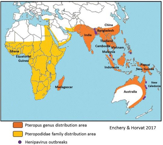
Fruit bats (‘Flying Foxes’) of the concern over the potential spread Caledonia, and Papua New Guinea
family Pteropodidae, particularly of NiV, is illustrated in Figure 1. (Sun et al., 2018). More recently,
those of the genus Pteropus, are the a six-year study of Pteropus medius
primary animal reservoir for NiV Pteropid bats of the genus Eidolon bats in Bangladesh indicates
and are asymptomatically infected range over most of the African that Nipah virus may be more
by the virus. Even experimental continent and have been found widespread than previously
infections with very high doses to be seropositive for NiV, and thought: bats throughout the
of NiV cause only sub-clinical thus are potentially an additional country, and not just those in what
infection in fruit bats and viremia reservoir (Enchery and Horvat, is referred to as the “Nipah belt”,
has not been reported, although 2017). A field survey found had similar patterns of Nipah virus
the animals do seroconvert against that 9% to 25% of fruit bats in infection throughout the year.3
the virus and virus shedding has Malaysia, Cambodia, Thailand
been observed, albeit rarely and and Bangladesh were seropositive
only in urine (Geisbert et al., 2012; for NiV (Sharma et al., 2019) and
Middleton et al., 2007). The broad NiV-seropositive bats have also
geographical distribution of these been found in China, Vietnam,
animals, one of the factors driving Indonesia, Madagascar, New
Figure 1. Geographic distribution of Pteropid fruit bats and
Henipavirus outbreaks (Enchery and Horvat, 2017)
3
Epstein et al. PNAS 2020: https://doi.org/10.1073/pnas.2000429117
5 Nipah Virus Assays and Animal Models for Vaccine Development, Landscape Analysis, January 2021A number of domesticated animals and Tan, 2014). In the 2014
– pigs, dogs, cats and horses – can NiV outbreak in the Philippines
be infected by NiV (Geisbert et al., (Malaysia strain), the majority
2012), but the virulence and rate of of cases were acquired by eating
infection is variable. Mortality in contaminated horse meat or
suckling pigs is high (40%) and 1-6 participating in slaughtering
month-old pigs show respiratory horses, and the rest were likely due
and neurological symptoms, but to human to human transmission.
mortality is less than 5%. Adult 65% of the cases presented with
pigs show less serious respiratory an acute encephalitic syndrome
signs and mortality is rare (McLean and the overall case fatality rate
and Graham, 2019). During NiV was 53% (Ching et al., 2015).
outbreaks in Malaysia many dogs
on pig farms were found to be The NiV outbreaks in Bangladesh
NiV-seropositive, but only 2 had and India beginning in 2001
active disease (Hooper et al., 2001). showed a distinct pattern of
Horses can be infected by NiV transmission and symptomology.
(Hooper et al., 2001), most likely Humans were infected by drinking
from eating fruit contaminated by raw date palm sap contaminated
bats, and were intermediate hosts with bat saliva or urine, and there
in an outbreak in the Philippines was no intermediate animal host.
in 2014. During that outbreak There was a higher incidence
investigations found disease among of respiratory illness (69% and
horses including neurological a higher fatality rate (75%; see
signs and 10 deaths. Four (4) cats Table 1). Patients infected with the
and a dog were also likely infected Bangladesh strain (NiV-B) had
by eating horse meat and died higher NiV RNA levels in the blood
(Ching et al., 2015). However, no and more virus in oral secretions
published reports of cats or dogs (Hossain et al., 2008). Finally,
serving as intermediate hosts for there was evidence of human-to-
transmission of NiV have been human transmission, primarily
found. to healthcare workers or family
caregivers, in the Indian and
The locations and human fatality Bangladeshi outbreaks (Chadha et
rates of all NiV outbreaks to date al., 2006). The mechanism(s) of
are summarized in Table 1. human to human transmission has
The first reported NiV outbreaks not been conclusively established,
occurred in Malaysia and Singapore but exposure to bodily fluids
in 1998-99. In those outbreaks (saliva, cough, vomit, blood)
the majority of human cases were elevates the risk of transmission
due to contact with infected pigs compared to physical contact
that had acquired NiV by eating alone or being near the patient
fruit contaminated by bat saliva, (Kumar et al., 2019). Experiments
urine, or feces. Humans (mostly in non-human primates have
pig farmers and slaughterhouse demonstrated infection via small
workers) acquired NiV from and medium particle size aerosols
infected pig urine or respiratory (Cong et al., 2017; Hammoud
secretions. A majority of cases et al., 2018). Comparisons in
were characterized by an acute transmission rates between the
encephalitic syndrome. The case Malaysia and Bangladesh outbreaks
fatality rate was 40% (see Table have not been found in the
1) and, at the time, there was published literature.
inconclusive evidence of human-
to-human transmission (Ahmad
6 Nipah Virus Assays and Animal Models for Vaccine Development, Landscape Analysis, January 2021Table 1. NiV Outbreaks by Year and Location*
Number of Number of Case
Month/Year Country Location
Cases Deaths Fatality (%)
Sep 1998 – Apr 1999 Malaysia Perak, Selangor, Negeri Sebilan 265 105 40
Mar 1999 Singapore Singapore 11 1 9
Jan – Feb 2001 India Siliguri 66 45 68
Apr – May 2001 Meherpur 13 9 69
Jan 2003 Naogaon 12 8 67
Jan 2004 Rajbari 31 23 74
Apr 2004 Faridpur 36 27 75
Bangladesh
Jan – Mar 2005 Tangail 12 11 92
Jan – Feb 2007 Thakurgaon 7 3 43
Mar 2007 Kushtia, Pabna, Tatore 8 5 63
Apr 2007 Naogaon 3 1 33
Apr 2007 India Nadia 5 5 100
Feb 2008 Manikgon 4 4 100
Apr 2008 Rajbari, Faridpur 7 5 71
Jan 2009 Gaibandha, Rangpur, Nilphamari 3 0 0
Jan 2009 Rajbari 1 1 100
Feb – Mar 2010 Faridpur, Rajbari, Gopalganj, Madaripur 16 14 87.5
Bangladesh Lalmohirhat, Dinajpur, Comilla, Nilphamari ,
Jan – Feb 2011 44 40 91
Rangpur
Feb 2012 Joypurhat, Rajshahi, Tatore, Rajbari, Gopalganj 12 10 83
Gaibandha, Natore, Rajshahi,Naogaon, Rajbari,
Jan – Feb 2013 24 21 87.5
Pabna, Jhenaidah, Mymensingh
Manikganj, Magura, Faridpur, Rangpur,
Feb 2014 Shaariatpur, Kushtia, Rajshahi, Tatore, 18 9 50
Dinajpur, Chapai Nawabganj, Naogaon
Mar – May 2014 Philippines Tinalon, Midtungok 17 9 53
Nilphamari, Pnchoghor, Faridpur, Magura,
Feb 2015 Bangladesh 9 6 67
Naugaon, Rajbari
May 2018 India Kozhikode, Malappuram 19 17 89
Overall Total 643 379 58.9
Malaysia/Singapore/Philippines Total 293 115 39.2
Bangladesh/India Total 350 264 75.4
*Adapted from (Thakur and Bailey, 2019) and (Sharma et al., 2019)
7 Nipah Virus Assays and Animal Models for Vaccine Development, Landscape Analysis, January 2021A comparison of key clinical and the Bangladesh outbreaks. and convulsions) were seen in
epidemiological characteristics This, coupled with the higher level the Bangladesh outbreaks at rates
of NiV outbreaks in Malaysia and of NiV-B RNA in oral secretions comparable to the Malaysia cases
Bangladesh is compiled in Table could be linked to the higher level (Ang et al., 2018; Hossain et al.,
2. Three clinical characteristics of human to human transmission, 2008). This phenomenon is largely
between the outbreaks stand out. which is likely via oral/respiratory unexplained but could reflect
The first is the shorter time from secretions or bodily fluids (Ahmad involvement of different areas of
disease onset to death (7 days vs. and Tan, 2014). Finally, in the the central nervous system (CNS)
16 days) and higher case fatality Malaysia outbreak there was a due to differences in virus tropism,
rate (74% vs. 38%; Table 1) in high incidence of segmented differences in the route of infection
the Bangladesh vs. the Malaysia myoclonus (muscle jerking), or slower disease progression
outbreaks (Ahmad and Tan, 2014; which was not reported in the allowing infection of different
Ang et al., 2018; Hossain et al., Bangladesh outbreaks, although neural tissues.
2008; Lo and Rota, 2008). The other indications of encephalitis or
second is the higher incidence neurological involvement (such as
of respiratory involvement in altered mental status, hyporeflexia
Table 2. Differences in Clinical and Epidemiological Characteristics Between NiV Malaysia
and Bangladesh Outbreaks
Characteristic Malaysia-Singapore Bangladesh-India
• Bat to human via consumption of
• Bat to pig → pig to human
Transmission contaminated date palm juice and fruits.
• Rare human to human
• Human to human
Fever 95% 100%
Headache 75% 73%
Vomiting 32% 58%
Diarrhea 18% 29%
Respiratory involvement 14-29% 62-69%
• Segmental myoclonus 32-54% • Segmental myoclonus not reported
• Hyporeflexia 60.5% • Hyporeflexia 65%
Encephalitis/neurological involvement
• Convulsion 23% • Convulsion 23%
• Altered mental status 72% • Altered mental status 100%
Disseminated small, high-signal-intensity Confluent high-signal brain lesions (limited
MRI
lesions MRIs were performed)
Relapsed and late-onset encephalitis ~5-10% 4 out of 22 patients (18%) in a follow-up study
Persistent neurological deficits ~20% ~30%
Incubation Period Mean = 10 days 6-11 days
Average (mean) time from disease onset to
16 days 7 days
death
Sources: (Ang et al., 2018; Hossain et al., 2008); (Ahmad and Tan, 2014; Chong et al., 2002; Lo and Rota, 2008)
8 Nipah Virus Assays and Animal Models for Vaccine Development, Landscape Analysis, January 2021Further clinical commonalities • Ferrets infected with NiV-B AGMs appear to reproduce the NiV
and differences in Nipah showed comparable disease strain differences in virulence and
infections across countries were progression, and histopathology pathology seen in humans more
discussed during the NIAID co- of the lungs and CNS compared faithfully than hamster and ferrets,
sponsored Nipah@20 Conference to ferrets infected with NiV-M. and the results suggest that the
in Singapore, December 20204. However, NiV-B infected ferrets distinct clinical characteristics and
The cause of the differences in shed more virus in oral secretions epidemiology seen in the Malaysia
disease between the Malaysia/ than NiV-M infected animals and Bangladesh outbreaks is at
Singapore and Bangladesh/India (Clayton et al 2012). Increased least partly genetic. However,
outbreaks remains uncertain and human shedding of NiV-B was geographic differences in virus
is complicated by the possibility of seen in the Bangladesh outbreaks transmission (including route of
differing diagnostic methods and (Hossain et al., 2008). infection and dose), population
case definitions. Experiments in health and the quality of
animal models have demonstrated • African Green Monkeys (AGM) subsequent health care may also
some differences in disease course Lethality of NiV-B was 100%, play a role. More information on
and symptomology between the compared to 50% for monkeys the major animal challenge models
Malaysia and Bangladesh strains, infected with an equivalent dose for NiV is presented in Section V.
suggesting a genetic component: of NiV-M. NiV-B also causes more
severe lung histopathology than
•H
amsters infected with NiV-M NiV-M and has a shorter window
showed accelerated virus for therapy with the monoclonal
replication, pathology and death antibody m102.4 (Mire et al.,
compared to hamsters infected 2016).
with equivalent doses of NiV-B
(DeBuysscher et al., 2013). This
finding is opposite to the human
fatality rates in the Malaysia and
Bangladesh outbreaks.
2. NiV Clinical Features and Pathogenesis
in Humans
The initial clinical presentation of rapidly to an encephalitic syndrome encephalitis survivors suffer
NiV infection (by either strain) is in approximately 60 percent long-term neurologic dysfunction
non-specific and characterized by of patients. The time course of characterized by persistent
flu-like symptoms including fever, disease progression from initial seizures, disabling fatigue and
headache, dizziness, myalgia, symptoms to the encephalitic behavioral abnormalities (Mazzola
and loose stools (Banerjee et al., syndrome has not been reported and Kelly-Cirino, 2019; Sejvar
2019). Mild or asymptomatic in detail. Neurological symptoms et al., 2007). In addition, some
infections have also been reported include meningismus (central patients with initially mild, non-
in various outbreaks, but the nervous system inflammation) and encephalitic disease develop a late-
overall incidence is relatively low seizures in approximately one- onset or recurrent neurological
and appears to be strain dependent, third of patients. A deterioration disease (Ramphul et al., 2018).
with the Malaysia strain causing in consciousness, coma and death Some patients also present with
less severe illness, a lower case typically occur within an average of severe respiratory symptoms,
fatality rate and higher prevalence 7 days of disease onset (Bangladesh and respiratory involvement
of asymptomatic infections than outbreaks) or an average of 16 has been more common in the
the Bangladesh strain (Kumar et days (Malaysia outbreak) (Ang Bangladesh outbreaks compared
al., 2019). The incubation period et al., 2018; Hossain et al., 2008; to the Malaysia outbreaks (see
ranges from 7 to 40 days and Rahman and Chakraborty, 2012). Epidemiology).
from onset the disease progresses Approximately 20-30% of NiV
4
Conference Proceedings:
https://cepi.net/wp-content/uploads/2020/06/2019-CEPI-Duke-WHO-NIAID-Nipah-Conference_FINAL.pdf.
9 Nipah Virus Assays and Animal Models for Vaccine Development, Landscape Analysis, January 2021Although the route of infection throughout the body, and the virus by severe vasculitis and syncytia
in humans has not been may then enter the blood stream formation, resulting in endothelial
conclusively determined, work with and disseminate throughout the damage due to vasculitis-induced
experimental animal challenge host, infecting the brain, spleen thrombosis and the presence of
models has shown that inhalation and kidneys (Escaffre et al., 2013). viral inclusion bodies. Necrotic
of NiV virus particles is sufficient Experiments in hamsters suggest plaques are found in both grey and
to initiate infection (Cong et al., that entry to the central nervous white matter of the CNS (Escaffre
2017; Hammoud et al., 2018). In system (CNS) may occur either et al., 2013). Lessons learned from
humans, early infection appears through olfactory neurons or via pathology and disease course in
to occur in lung epithelial cells, the choroid plexus and cerebral humans were discussed in the
and in later stages moves to lung blood vessels (Baseler et al., 2016; Transmission/Case Management
endothelial cells. Vasculitis in Munster et al., 2012). Infection of Session of the Nipah@20
small blood vessels may be present the human CNS is characterized Conference.
3. Diagnosis and Treatment
Laboratory diagnosis of NiV was not randomized and the In a lethal challenge study in
infection can be performed using treated patients may have received African Green Monkeys (AGMs),
a variety of nucleic acid-based or better overall care, thus making the monkeys could be successfully
serological assays. The currently outcome uncertain (Banerjee et al., treated up to 5 days post-infection
preferred methods for detecting 2019). Subsequent animal challenge with the NiV Malaysia strain
active NiV infection are PCR- studies in hamsters showed that (NiV-M). Although half of the
based tests such as conventional ribavirin delayed, but did not treated monkeys developed overt
reverse transcriptase (RT) PCR, prevent, death after NiV infection clinical signs (fever, respiratory
nested RT-PCR and real-time PCR (Freiberg et al., 2010; Georges- and neurological), all the animals
(qPCR). PCR-based tests usually Courbot et al., 2006). A similar fully recovered (Geisbert et al.,
target the conserved N, M or P viral result was obtained after infection 2014). The window for successful
genes. ELISA assays detecting IgM of African Green Monkeys with the treatment with m102.4 is only 3
against NiV antigens are typically closely related Hendra Virus (HeV; days post-infection when AGMs
the first-line serological tests (Rockx et al., 2010)). are challenged with the NiV
for NiV infection (Mazzola and Bangladesh strain (NiV-B) (Mire
Kelly-Cirino, 2019). Progress and The broad-spectrum antiviral et al., 2016). These results suggest
challenges in diagnostics, including drug Remdesivir (GS-5734) was that m102.4 may have utility as
a presentation on the WHO Nipah recently shown to protect African a post-exposure prophylactic
diagnostics Target Product Profile, Green Monkeys when administered or therapeutic in humans. The
were featured in Session 4 of the 24 hours post-inoculation with m102.4 antibody has also been
Nipah@20 Conference. a lethal dose of NiV (Bangladesh administered on an emergency
strain) (Lo et al., 2019). Treated basis as post-exposure prophylaxis
Treatment of NiV infection animals developed mild respiratory to a handful of humans in cases of
consists primarily of supportive symptoms, reduced appetite and high risk of exposure to NiV or HeV
care including maintaining fluids, showed local virus replication but (Broder et al., 2013). In all cases the
anticonvulsants, treatment of no viremia. All the Remdesivir- patients did not become ill, but it
secondary infection and mechanical treated animals recovered fully, is impossible to know if illness was
ventilation. (Ang et al., 2018). while all the control-treated prevented by the antibody.
No effective therapeutics for NiV animals succumbed to the
infection are currently approved infection.
for use in humans. The antiviral
drug ribavirin was administered to A human monoclonal antibody,
140 patients during the 1998-99 m102.4, targeting the Ephrin-B2/
NiV Malaysia outbreak, resulting B3 binding site on the NiV and HeV
in a 36% reduction in mortality G glycoproteins (see NiV Molecular
compared to 52 untreated control Biology and Structure), has been
patients (Chong et al., 2001). tested in NiV animal models for
However, the treatment allocation prophylactic and therapeutic use.
10 Nipah Virus Assays and Animal Models for Vaccine Development, Landscape Analysis, January 20214. NiV Molecular Biology and Structure NiV is an enveloped, negative- host cell plasma membrane. Three glycoprotein mediates target cell sense, single-stranded RNA virus non-structural proteins, W, V, attachment via the cell surface of the family Paramyxoviridae, a and C, are produced by alternative receptors Ephrin-B2 and -B3. group which also includes measles, initiation or RNA editing within the The tissue tropism of Henipavirus mumps, parainfluenza viruses P gene open reading frame (Wang infection is determined by the and Sendai Virus. NiV shares the et al., 2001). These gene products tissue distribution of these genus Henipavirus with a handful inhibit host cell antiviral responses receptors. Ephrin-B2 is expressed of other recently identified viruses, such as Type 1 interferon signaling in neurons, endothelial cells, including Hendra Virus (HeV) and and are major determinants of smooth muscle surrounding Cedar Virus (Sharma et al., 2019). A viral pathogenicity (Mathieu et arteries, placental tissue and schematic of the NiV viral structure al., 2012b; Satterfield et al., 2015; spleen. High levels of Ephrin-B2 and genome organization is shown Yoneda et al., 2010). The viral mRNA have also been detected in Figure 2 below (Sun et al., 2018). envelope is studded with two in cardiomyocytes and bronchial The 18.2 kb NiV genome encodes transmembrane glycoproteins, epithelial cells. Ephrin-B3 is six structural proteins and three the trimeric F glycoprotein and expressed in the CNS and in lymph non-structural proteins. NiV RNA the tetrameric (dimer of dimers) nodes (Xu et al., 2012). Ephrin-B2/ is associated with nucleoprotein G glycoprotein (Aguilar and Lee, B3 expression levels in target (N) and phosphoprotein (P) to form 2011). The F and G glycoproteins tissues also impact the rate of the virus ribonucleocapsid (RNP). are the major targets of NiV virus replication (Sauerhering The NiV genome encodes its own neutralizing antibody responses in et al., 2016), but have not been RNA-dependent RNA polymerase animals and humans (Satterfield et extensively characterized. This (L) which, together with N and P, al., 2016b). is an important area for future forms the catalytic subunit of the investigation to understand replicase complex that enables The Henipavirus infection and differences in NiV disease virus replication. The matrix replication cycle is depicted pathogenesis in humans and (M) protein is required for virion schematically in Figure 3 (Aguilar animal challenge models (see assembly and budding from the and Lee, 2011). The viral G Section V.) Figure 2. NiV structure and organization of the 18.2 kB ssRNA (-) genome (Sun et al., 2018). 11 Nipah Virus Assays and Animal Models for Vaccine Development, Landscape Analysis, January 2021
Following attachment, the G             which is then re-exported with G      91.8% similarity at the nucleotide
glycoprotein activates the F            glycoprotein for assembly into the    level (Rockx et al., 2012). The
glycoprotein, which then mediates       budding viral envelope. Assembly      nucleotide changes are not
fusion of the viral envelope            and budding of new viral particles    distributed uniformly; in most
with the host cell membrane.            from the plasma membrane is           cases homologies are higher in the
After cell entry the viral genome       mediated primarily by the M           coding regions than in non-coding
[vRNA(-)] serves as a template for      (matrix) protein. (Aguilar and        regions. Nucleotide homologies
transcription of mRNAs by the           Lee, 2011). The interaction of cell   range from 92.0% to 98.5% in the
viral RNA polymerase (NiV L gene        surface-displayed NiV F and G with    open reading frames. While the 5’
product) which are then translated      Ephrin B2/3 also mediates syncytia    untranslated region of the N gene
into proteins, the vRNA(-) is also      formation by infected cells (Rockx    is 100% conserved between the two
a template for cRNA(+), which is        et al., 2012).                        major strains, homologies in the 5’
then a template for production of                                             and 3’ untranslated regions of all
vRNA(-) genomes for packaging           Two genetically distinct NiV          the other viral genes range from
into new viral particles. Precursor F   strains, Malaysia (NiV-M) and         75.5% to 91.4% (Harcourt et al.,
glycoprotein (Fo) is exported to the    Bangladesh (NiV-B), have been         2005).
plasma membrane, endocytosed            identified. The two strains share
and proteolytically matured to F1/2,    92% amino acid homology and

Figure 3. The Henipavirus infection and replication cycle
(Aguilar and Lee, 2011).

12                Nipah Virus Assays and Animal Models for Vaccine Development, Landscape Analysis, January 2021
5. Vaccine Development
A number of factors suggest that        little attention. One challenge study    Due to safety issues associated
development of a safe, efficacious      conducted in pigs suggested that         with production (i.e., BSL-4
human prophylactic vaccine              cellular immune responses may            containment) and administration
against NiV is scientifically           be important for achieving full          of a live-attenuated or inactivated
feasible. Natural infection by other    protection, but the mechanism was        NiV vaccine, and the need to
paramyxoviruses, such as measles        not defined, and the conclusions         elicit neutralizing antibodies,
and mumps, results in long-term         are complicated by the fact that,        most attempts at NiV vaccine
immunity and vaccines for those         unlike with other animal hosts, NiV      development have focused on
diseases have been successfully         infects a range of porcine immune        recombinant viral vectors and
developed. A vaccine protecting         cells (Pickering et al., 2016). More     adjuvanted protein subunit
horses against the closely related      work is needed to elucidate the role     vaccines. In all cases the target
Hendra Virus (HeV; Equivac®)            of cellular immune responses in          antigen(s) have been the F and/or G
has been approved for use in            protection against NiV infection.        glycoproteins (see Tables 3 and 4).
Australia (Tan et al., 2018). Passive
immunization experiments in a NiV       The WHO developed a Target               The NiV vaccines described below
animal challenge model (hamsters)       Product Profile (TPP) for a human        are all research-stage candidates
using immune sera and monoclonal        NiV vaccine, including preferred         focused on demonstrating
antibodies have demonstrated            as well as critical or minimal           immunogenicity and protection
that neutralizing antibodies            product characteristics.1 Key            against lethal NiV challenge.
confer protection against NiV           vaccine performance attributes           No safety issues associated with
challenge (Guillaume et al., 2004;      recommended in the TPP are:              vaccination or subsequent virus
Guillaume et al., 2006). Finally, as                                             challenge (due to antibody-
discussed below, multiple modes         • Intended use: For reactive use in     dependent disease enhancement;
of active vaccination have resulted        outbreak settings                     ADE) were reported. However,
in protection from lethal NiV                                                    more in-depth safety studies
challenge in animal models.             • Efficacy: ≥ 90% efficacy in           will necessarily be performed
                                           preventing disease (preferred);       on any NiV vaccine candidates
There is broad consensus that              ≥70% (minimal); rapid onset           prior to advancing into human
neutralizing antibodies confer             of protection, less than 2 weeks      clinical testing.
protection against NiV infection           after the first dose (preferred);
and all vaccine development efforts        protection ≤ 2 weeks after the last
to date have focused on their              dose (minimal).
elicitation (Broder et al., 2012;
Prescott et al., 2012; Satterfield et   • Dose Regimen: Single-dose
al., 2016b). However, a correlate of       primary series (preferred); no
protection based on neutralizing           more than 2 doses, with some
antibody titer has not been defined.       protection after the first dose
Neutralizing antibody titers in            (minimal).
animal vaccine challenge studies
where protection was conferred          • Durability of Protection: ≥ 1
are reported in Tables 3 and               year (preferred); ≥ 6 months
4. However, since virtually all            (minimal).
animals were protected in these
studies a threshold of protection       • Product Stability and Storage:
cannot be defined. The role of             Shelf life of 5 years at 2-8oC
cell-mediated immune responses             (preferred); shelf life of at
(CMI) in either natural immunity           least 12 months at -20oC and
or vaccine-induced protection              demonstrated stability of ≥ 1
against NiV has received relatively        month at 2-8oC (minimal).

13                Nipah Virus Assays and Animal Models for Vaccine Development, Landscape Analysis, January 2021
Viral Vector Candidates A number of viral vector platforms Other recombinant viral vector expressing the NiV F or G vaccine platforms expressing NiV glycoproteins have been tested F or G have been tested, including: as vaccine candidates. The most vaccinia virus (Guillaume et widely used vector platform to date al., 2004), canarypox (ALVAC) has been the Vesicular Stomatitis (Weingartl et al., 2006), Measles Virus (VSV). Three types of VSV virus (Yoneda et al., 2013), vectors have been employed: Venezuelan Equine Encephalitis 1) replication-incompetent VSV Virus (VEEV) (Defang et al., pseudotypes expressing NiV 2010), Rabies virus (Keshwara F or G (Lo et al., 2014); 2) VSV et al., 2019), Newcastle disease virions expressing NiV F or G virus (Kong et al., 2012), Adeno that can undergo a single round Associated Virus (AAV) (Ploquin of replication (Mire et al., 2019); et al., 2013) and chimpanzee and 3) replication-competent adenovirus (ChAd; (van Doremalen recombinant viruses in which et al., 2019)). All these candidates the VSV-G protein is replaced by conferred full protection against the Ebola glycoprotein (ZEBOV) lethal challenge and/or elicited and also co-expressing NiV F high titers of neutralizing or G (DeBuysscher et al., 2014; antibodies. However, only the DeBuysscher et al., 2016; Prescott AAV and chimpanzee adenovirus et al., 2015). All three VSV-vaccine (ChAd) vectored vaccines types, whether expressing NiV F reported protection after a single or G antigens and administered vaccination. A summary of NiV viral singly, or co-administered, elicited vector vaccine candidates tested in neutralizing antibodies and fully animals is shown in Table 3. protected immunized animals from clinical disease in at least one of the 3 major NiV lethal challenge models (hamsters, ferrets or non-human primates; see Section V). Additionally, all three VSV vaccine types conferred protection after a single dose (see Table 3). Additional details on the animal challenge models and their use in vaccine studies are given in Section V. 14 Nipah Virus Assays and Animal Models for Vaccine Development, Landscape Analysis, January 2021
Table 3. NiV Viral-vector Vaccine Candidates Tested in Animals

                                                                                                                            NiV Neutralization
                                                     Animal(s)        Vaccination Route/Regimen/Challenge
     Reference         Vaccine Description                                                                                     Titers (Pre-
                                                    immunized                       (Strain3)
                                                                                                                               challenge)2

    Guillaume et al    Vaccinia virus (VV) vector                       SC/2 vaccinations 1 month apart/ challenge 3
                                                      Hamsters                                                                  ~ 1:10 – 1:25
        (2004)           expressing NiV G or F                               months after last vaccination (M)

    Weingartl et al    Canarypox vector (ALVAC)                       IM/2 vaccinations 14 days apart/ challenge on day
                                                         Pigs                                                                  1:200 – 1:1280
       (2006)            expressing NiV G or F                         28 post 2nd vaccination (NiV strain not specified)

                          Venezuelan Equine
     Defang et al                                                     Footpad inoculation/ 3 vaccinations on week 0, 5
                       Encephalitis Virus (VEEV)         Mice                                                                  ~ 1:215 – 1:217 *
       (2011)                                                                      and 18/ no challenge
                         expressing NiV G or F

                       Newcastle Disease Virus
      Kong et al
                       (NDV) vector expressing           Pigs          IM/2 vaccinations 4 weeks apart/ no challenge            ~ 1:27 – 1:212*
       (2012)
                             NiV F or G

                        Single-cycle replication
     (Mire et al.,                                                     IM/one vaccination/ challenge on day 20 post-
                       VSV-∆G vector expressing         Ferrets                                                                 ~1:40 – 1:160
       2013)                                                                         vaccination (M).
                              NiV G or F

                        Adeno-Associated Virus
    (Ploquin et al.,                                                   IM/one vaccination/ challenge at 5 weeks post-
                        (AAV) vector expressing       Hamsters                                                                  < 1:10 to 1:160
        2013)                                                                        vaccination (M).
                                 NiV G

                                                                         Hamster: IP/ 2 vaccinations 21 days apart/
                         Measles virus vaccine                                                                              Hamster: Not reported
     Yoneda et al                                     Hamsters,       challenge 7 days post 2nd Vaccination AGM. SC/2
                        vector expressing NiV G
       (2013)                                          AGM*            vaccinations 28 days apart/ challenge 2 weeks        AGM: 1:1600 – 1:3200
                             glycoprotein
                                                                       post 2nd vaccination (NiV strain not specified).

                        Replication-competent
    DeBuysscher et                                                     IP/ one vaccination/ challenge on day 28 post-
                       VSV vector expressing NiV      Hamsters                                                                  1:80 - ≥ 1:640
      al (2014)                                                                       vaccination (M).
                                 G or F

                         Replication-defective
                                                                       IM/ one vaccination/ challenge at day 32 post-
    Lo et al (2014)    VSV-∆G vector expressing       Hamsters                                                                ~ 5 x10³ – 1 x 10⁴
                                                                                     vaccination (M)
                              NiV G or F.

     (Guillaume-
                       Canarypox vector (ALVAC)
    Vasselin et al.,                                Ponies (horses)     IM/2 vaccinations 21 days apart/ no challenge              ~ 1:2128*
                         expressing HeV G or F
        2016)

     Prescott et al      Live-attenuated VSV                           IM/one vaccination/ challenge on day 29 post-
                                                        AGM1                                                                     1:80 – 1:160
        (2015)          vector expressing NiV G                                      vaccination (M).

    DeBuysscher et        Live attenuated VSV                           IP/one vaccination/ challenge one day post-
                                                      Hamsters                                                                  Not reported
       al 2016          vector expressing NiV G                              vaccination (100% survival) (M).

                        Live-attenuated Rabies                                                                                  ~1:10 to 1:600
    Keshwara et al
                          Virus vaccine vector           Mice          IM/2 vaccinations 28 days apart/ no challenge
       (2019)                                                                                                                  (no challenge)
                       (RABV) expressing NiV G.

                        Single-cycle replication
                                                                       IM/one vaccination/ challenge on day 28 post-
    Mire et al 2019    VSV-∆G vector expressing         AGM1                                                                    1:160 – 1:640
                                                                                     vaccination (B).
                              NiV G or F

         (van           Chimpanzee adenovirus                           IM/ one or two vaccinations (28 days apart)/
    Doremalen et       (ChAd) vector expressing       Hamsters         challenge 70 days post-prime or 42 days post-           ~1:40 - ~1:100
      al., 2019)                NiV G                                                 boost (M and B).

1
 =African Green Monkey 2~ Indicates titer values estimated from data presented graphically 3 Challenge strain M= Malaysia;
B=Bangladesh; *endpoint neutralization titers determined by 2-fold serial dilution and expressed as exponentials of 2.
IM = intramuscular IP = intraperitoneal, SC = sub-cutaneous

15                       Nipah Virus Assays and Animal Models for Vaccine Development, Landscape Analysis, January 2021
Subunit Vaccine Candidates
The most widely studied NiV does not efficiently generate HeV comprised of an enveloped virus-
subunit vaccine candidates have cross-neutralizing antibodies like particle (VLP) created by co-
utilized a purified, recombinant G (Ploquin et al., 2013). Various expression of the NiV M (matrix),
glycoprotein from Hendra Virus adjuvant formulations have been F and G glycoproteins and
(HeV) in which the transmembrane tested, including aluminum + CpG adjuvanted in either aluminum
domain has been removed to allow (Bossart et al., 2012; McEachern hydroxide (Alhydrogel®),
soluble G protein expression (sG). et al., 2008), CpG alone (Pallister monophosphoryl lipid A (MPLA),
The high sequence conservation et al., 2013), and Quil A/DEAE- or CpG has also been tested and
between the NiV and HeV G dextran/Montanide (Mungall et al., was 100% protective in the hamster
glycoproteins (83% amino acid 2006). All formulations were 100% challenge model (Walpita et al.,
homology; (Wang et al., 2001) efficacious, eliciting neutralizing 2017). A summary of NiV subunit
allows for the elicitation of antibodies and protecting all vaccine candidates tested in
potent NiV cross-neutralizing vaccinated animals against lethal animals is shown in Table 4.
antibodies (Sun et al., 2018), NiV challenge with no signs of
although vaccination with NiV G clinical disease. A vaccine candidate
Table 4. NiV Submit Vaccine Candidates Tested in Animals
NiV Neutralization
Animal(s) Vaccination Route/Regimen/Challenge
Reference Vaccine Description Titers (Pre-
immunized (Strain3)
challenge)2
sGNiV or sGHeV
Mungall et al adjuvanted with SC/ 3 vaccinations 2 weeks apart/ challenge 15
Cats 1:2,560 –1:20,480
(2006) Montanide/QuilA/DEAE- weeks after the first vaccination (M).
dextran
Recombinant soluble HeV
McEachern et IM/ 2 vaccinations 21 days apart/ challenge on
G glycoprotein adjuvanted Cats 1:32 – 1:512
al (2008) day 42 post 1st vaccination (M).
with CpG + AlhydrogelTM
Virus-like particles
Walpita et al SC/ 3 vaccinations on days 0, 15 and 29/ no
(VLPs) comprising NiV M, Mice 1:5 - >1:80
(2011) challenge
G and F
Recombinant soluble HeV
(Bossart et al., IM/ 2 vaccinations 21 days apart/ challenge 21
G glycoprotein adjuvanted AGM1 1:67 – 1:379
2012) days post 2nd vaccination (M).
with CpG + AlhydrogelTM
Recombinant soluble HeV
(Pallister et al., SC/ 2 vaccinations 20 days apart/ challenge 20
G glycoprotein adjuvanted Ferrets 1:16 – 1:128
2013) days or 14 months post 2nd vaccination (B)
with CpG
Recombinant soluble
Pickering et al HeV G glycoprotein in IM/ 2 vaccinations 21 days apart/ challenge 35
Pigs ~ 1:25- - 1:450
(2016) a proprietary adjuvant days post 1st vaccination (Strain not specified)
(Zoetis, Inc.)
Single dose trial: IM/ one dose/ challenge on day 3-Dose Trial: ~ 1:200
Virus-like particles 28 post vaccination (M). – 1:2500
Walpita et al
(VLPs) containing NiV M, Hamsters
(2017) 3 dose trial: 3 doses on days 0, 21 and 42/ 1-Dose Trial: ~ 1:10 –
F and G.
challenge on day 58 (M). 1:200
1
=African Green Monkey 2~ Indicates titer values estimated from data presented graphically 3 Challenge strain M= Malaysia;
B=Bangladesh; *endpoint neutralization titers determined by 2-fold serial dilution and expressed as exponentials of 2.
IM = intramuscular IP = intraperitoneal, SC = sub-cutaneous
16 Nipah Virus Assays and Animal Models for Vaccine Development, Landscape Analysis, January 2021More recently, novel antigen challenge with the Malaysian
design options have been evaluated strain of Nipah virus. Authors
using a structure-based design.5 noted immune responses were
A stabilized prefusion F (pre-F), suboptimal. It is conceivable
multimeric G constructs, and that the protection would have
chimeric proteins containing both been superior under a two-dose
pre-F and G were developed as regimen.
protein subunit candidate vaccines.
The proteins were evaluated In addition, some of the structure-
for antigenicity and structural based designs described earlier
integrity using kinetic binding for immunogen development5
assays, electron microscopy, and are being evaluated in the mRNA
other biophysical properties. platform in collaboration with
Immunogenicity of the vaccine Moderna, and clinical evaluation
antigens was evaluated in mice is planned.
using aluminum hydroxide as
adjuvant. NiV Vaccine Candidates Supported
by CEPI
mRNA Vaccine Candidates As of August 2019, CEPI has four
NiV vaccine candidates in its
The US CDC has published proof- vaccine development portfolio,
of-concept pre-clinical data on a three viral-vector platform
Hendra virus glycoprotein mRNA candidates and one candidate
vaccine in liquid nanoparticles.6 comprising an adjuvanted
A single dose of the vaccine recombinant protein antigen
protected up to 70% of hamsters (Table 5).
against a lethal, intraperitoneal
Table 5. NiV vaccine candidates supported by CEPI
Developer Vaccine Platform Development Stage
University of Tokyo Recombinant Viral Vector Pre-clinical
Profectus Biosciences/Emergent Biosolutions/
Recombinant Protein Phase 1 (USA)
PATH
Janssen Vaccines & University of Oxford Recombinant Viral Vector Pre-clinical
Replication-competent rVSV vector
Public Health Vaccine, LLC Pre-clinical
expressing NiV-G
Source: https://cepi.net/research_dev/our-portfolio/
5
https://www.frontiersin.org/articles/10.3389/fimmu.2020.00842/full
6
https://academic.oup.com/jid/article/221/Supplement_4/S493/5637464
17 Nipah Virus Assays and Animal Models for Vaccine Development, Landscape Analysis, January 2021III. STANDARDIZATION OF
ASSAYS AND ANIMAL MODELS
Assays and animal models to quantify or characterize immune responses
elicited by vaccination are, by their nature, inherently variable.
The reasons for this include the Immune Serum Reference and commercial manufacturing
molecular complexity of the Standards (https://www.who.int/biologicals/
samples (serum or other biological vaccines/en/). Recent examples
samples), the need to produce One of the most important tools include HPV 16 (Ferguson et al.,
reagents in complex biological for standardization of serological 2011), Typhoid Fever (Rijpkema et
systems such as cell culture or in assays is immune reference al., 2018), Respiratory Syncytial
vivo, variability in composition and serum. Even when similar assay Virus (McDonald et al., 2018) and
stability of these reagents, and the formats are used for detection Zika (Source: WHO/BS/2018.2345).
need to test immune responses in of antigen binding antibodies or
vivo. Nevertheless, modern vaccine virus neutralizing antibodies, Three key factors determine
development requires vaccines the resulting data can be highly the fitness of material for use
and samples from vaccinated variable between laboratories due as a biological standard. First,
humans and animals be tested to differences in assay methods the material must have similar
with the highest possible precision. and reagents. For example, a composition and in vitro behavior
The task is further complicated 10-laboratory collaborative study to the human sera test articles.
by the collaborative and global assessing the precision of assays Second, the standard should be
nature of modern vaccine for detection of serum antibodies commutable, meaning it should
development. Multiple research against Human Papillomavirus 16 work for a wide range of serological
laboratories, vaccine developers, (HPV 16) revealed inter-laboratory assays and vaccine platforms
non-governmental organizations, variations in anti-HPV titer of being tested. Finally, a blinded
and regulatory agencies are often up to 25-fold for the same test multi-laboratory collaborative
involved in the development sample (Ferguson et al., 2006). A study must demonstrate the
process and vaccine candidates similar, 15-laboratory collaborative utility of the standard for reducing
utilizing different platform study evaluating assays for serum intra-laboratory assay variability.
technologies are often evaluated for antibodies against H5N1 influenza (Source: CEPI 2nd Standards and
the same disease indication. Thus, showed inter-laboratory variations Assays Workshop; June 2019).
standardization of methods and in titer of 10 to 35-fold, depending
reagents is important to facilitate on the sample and type of assay Serum reference standards for a
development of new vaccines such (Stephenson et al., 2009). The new vaccine are often established
as for NiV. The goal is to enable purpose of establishing immune in a staged manner as the
“like versus like” comparisons reference standards is to provide development process progresses.
of data generated by different a common, external control to This mitigates the risk of producing
laboratories and derived from improve the comparability of exhaustively characterized
many assay types. Recognizing assay data between laboratories. materials which might not be
the value of phase-appropriate With the standard in place, test required if vaccine development
standardization early in the vaccine results are reported relative does not progress. For R&D and
development process, CEPI is to the activity of the reference early clinical trials a working
promoting assay, reagent, and standard. In the studies cited standard or interim standard may
animal model standardization to above, use of a common reference be established by a collaborative
accelerate development of vaccines standard significantly reduced study involving a relatively limited
for NiV and other priority diseases intra-laboratory assay variability. number of laboratories, and
in its portfolio. Reference standards have been relatively low volumes may be
developed for many vaccine sufficient in the earlier stages.
indications, both in development
18 Nipah Virus Assays and Animal Models for Vaccine Development, Landscape Analysis, January 2021A number of different sources of (Source: CEPI 2nd Standards common reagents (reference
immune sera may be considered. and Assays Workshop; June sera and antigens) provided by
For example, a collaborative 2019). A single, large lot of an regulatory agencies and using a
study for establishment of an antibody standard is preferred single, validated assay method
interim standard for antibodies to avoid potential variability to test and release new seasonal
to Ebola virus (EBOV) tested between multiple lots and the vaccine formulations. In general,
plasma samples from patients who need for subsequent bridging standards may improve inter-
recovered from Ebola infection studies. Once suitable standard laboratory test performance.
(convalescent sera), anti-EBOV sera candidates are available for However, the need, feasibility
IgG preparations from trans- evaluation, a collaborative study is and level of standardization is
chromosomal (Tc) cows immunized performed to evaluate serological typically considered on a case-
with experimental vaccines and assays performed by a number of by-case basis according to the
plasma from vaccinated volunteers participating laboratories. A broad stage of vaccine development,
participating in an EBOV vaccine panel of test samples from different the types of assays in use and the
trial (Wilkinson et al., 2017). An sources (e.g., sera from naturally potential of standards to facilitate
interim standard for NiV will infected humans, animals infected development and licensure.
probably be generated from non- in the laboratory, and vaccinated Standardized reference sera are
human primates infected with a humans or animals) is assayed and relatively easy to implement since
sub-lethal dose of NiV and which the intra-laboratory variability in they ideally should be commutable
generate high titers of neutralizing assay results is assessed. Finally, across many assay types and are
antibodies (Dhondt and Horvat, the test sample absolute values broadly recognized for improving
2013). Obtaining convalescent sera (for example, geometric mean both intra- and inter-laboratory
from NiV survivors is also being titers) are expressed relative to the assay consistency. In contrast,
considered (Source: CEPI 2nd activity of the candidate standard standardized assay formats are
Standards and Assays Workshop; and the ability of the standard more challenging to implement,
June 2019). However, given the to improve intra-laboratory especially for newer vaccines
sporadic nature of NiV outbreaks comparability of test results is and those in development, since
and relatively small number of assessed. Once the standard has vaccine antigens may differ
cases (and available survivors), been chosen a full storage stability between candidates and there is
obtaining sufficient quantities of program is conducted to ensure the less consensus on the ideal assay
convalescent sera for long-term quality of the material over time. format. With these considerations
use may be challenging. Therefore, Trending of assay performance in mind, the benefits and potential
in the case of emerging infections over time is also performed. The challenges of standardizing
such as NiV one of the alternative International Standard itself is various assay and animal model
approaches described above may not intended for routine assay use. components to accelerate
need to be employed. A working reference standard is NiV vaccine development are
established for routine use and summarized in Table 6.
Establishment of an interim a bridging study is conducted to
standard usually precedes calibrate the working standard to
establishment of an International the International Standard (Source:
Standard (IS) , often called CEPI 2nd Standards and Assays
an International Reference Workshop; June, 2019).
Preparation (IRP) under the
endorsement of the WHO Standardization of Other Biological
Expert Committee on Biological Assay Reagents and Methods
Standardization. This is a more
formal process, taking up to 36 Many aspects of biological
months and involving a larger assays for vaccine testing may
and more in-depth collaborative be standardized to improve the
study, often involving more comparability of intra-laboratory
than 25 laboratories and a wide data. Common reagents (reference
geographical distribution. This sera, antigens, virus stocks) may
is the main difference from a be produced and standardized
working or interim standard. assay methods established and
Regulatory agencies generally validated. For example, potency
expect an established International testing for release of subunit
Standard to be used in pivotal seasonal influenza vaccines
clinical trials for vaccine approval, is performed under a high
unless specifically justified degree of standardization using
19 Nipah Virus Assays and Animal Models for Vaccine Development, Landscape Analysis, January 2021Table 6. Benefits and Potential Challenges of Implementing Biological Standards for NiV
Vaccine Development
Standard Benefits Potential challenge(s)
Finding human NiV convalescent donors may
Long track record and high level of acceptance
be challenging (low numbers). Generating
for improving intra-laboratory assay
Immune Standard Reference Sera NiV convalescent animal sera by sub-lethal
comparability. Inter-laboratory performance
infection requires BSL-4 containment and
may also be improved.
can be considered an interim mitigation
Choice of genotype/strain and ensuring
Promote standardization of serum antibody reactivity to diverse serum isolates; storage
Common stocks of ELISA coating antigen detection; relatively easy to produce, test, stability; heterogeneity in post-translational
store and distribute. modifications; biochemical differences
between strains.
Current use of pseudovirus assays by
major NiV research groups is rare. Often
Common pseudovirus(es) for assaying Promote use of an assay method which can be overestimate titer compared to wild-type
neutralizing antibodies performed in low-level biocontainment NiV-based assays, so will require extensive
characterization compared to traditional NiV
neutralization assays to gain acceptance.
Production, testing, storage, stability and
Common NiV virus stocks for neutralization Promote standardization of animal challenge distribution of live NiV and requirement for
assays and animal challenge models experiments and NiV neutralization assays BSL-4; mutations during passaging of ssRNA
virus.
The large number of potential variables in
challenge model performance (ie., challenge
Standards for performance of animal Promote standardization of challenge
strain/stock, route, dose, animal species etc.)
challenge models experiments
may complicate agreement on and acceptance
of performance standards.
20 Nipah Virus Assays and Animal Models for Vaccine Development, Landscape Analysis, January 2021IV. NIV SEROLOGICAL ASSAYS Robust serological assays for quantifying and characterizing humoral immune responses in humans and animals are critical for vaccine development. A number of methods have been assays for NiV, as well as newer experiments is compiled in Table developed for NiV serology, and the assays in earlier stages of use and 8 and Table 9 to illustrate the refinement and standardization acceptance. An analysis of the pros prevalence of use, variables in of such methods will be essential and cons of different serological assay performance, how reagents for facilitating development of assays for NiV vaccine development and methods have changed over safe and effective human vaccines is presented in Table 7. The usage time and opportunities for assay against NiV. This section describes of assays in a large number of NiV standardization. the commonly used serological vaccine studies and other research 1. Detection of antigen – specific serum IgG Detection of antigen – specific The earliest ELISAs for detection the 17 ELISAs in Tables 8 and 9 serum IgG is essential to the of NiV antibodies in sera were use recombinant G or F for target vaccine development process to developed by the Centers for IgG capture. The sFNiV and sGNiV characterize the specificity and Disease Control (CDC; USA). have been produced in a variety of magnitude of the vaccine-induced Different ELISAs were developed recombinant expression systems humoral immune response. The for NiV-specific serum IgG and (E. coli, insect cells, mammalian most common assay method is IgM. These ELISAs were used cells) and are usually epitope the traditional “indirect” ELISA. for surveillance and diagnosis of tagged for ease of purification In this assay a target antigen is disease in humans and pigs, and (Eshaghi et al., 2005; Eshaghi et al., plated (adsorbed) onto a 96- well used detergent and radiation- 2004; Keshwara et al., 2019; Kurup microtiter plate. After blocking inactivated, NiV – infected Vero et al., 2015). The glycosylation and the plate to suppress non-specific cell lysates as the target antigen disulfide bonding in the NiV F and binding, dilutions of immune (Daniels et al., 2001). Several of G glycoproteins make eukaryotic or control sera are added to the NiV animal vaccination studies cells preferable for recombinant the wells. After washing away and research experiments detailed expression of these antigens. The unbound antibody, bound IgG is in Table 8 and Table 9 utilized many successful tests of vaccines usually detected by the addition inactivated crude NiV- infected targeting the G glycoprotein in NiV of a species-specific anti-IgG Vero extracts or gradient-purified animal challenge models make it secondary antibody conjugated NiV as the target antigen. However, likely that this antigen will be used to a chromogenic enzyme. While the use of NiV as an assay reagent in human vaccine candidates. serum IgG is measured to elucidate is obviously problematic since vaccine responses, as well as for the initial preparation requires surveillance and epidemiology, BSL-4 containment. ELISAs using measurement of NiV-specific NiV-infected crude extracts also serum IgM is usually performed suffered from non-specific binding for diagnosis of active infection (Daniels et al., 2001). The discovery (Mazzola and Kelly-Cirino, 2019). that NiV F and G glycoproteins are The advantages of the ELISA assay the major target of neutralizing format include its broad use and antibodies and their use in vaccine familiarity throughout biomedical formulations spurred the use science, relatively low-tech and of recombinant, soluble NiV F low-cost application and wide (sFNiV) and G (sGNiV) as the target availability of reagents. antigens in ELISA assays. Six of 21 Nipah Virus Assays and Animal Models for Vaccine Development, Landscape Analysis, January 2021
You can also read