Specific immune modulation of experimental colitis drives enteric alpha-synuclein accumulation and triggers age-related Parkinson-like brain pathology
←
→
Page content transcription
If your browser does not render page correctly, please read the page content below
Free Neuropathology 2:13 (2021)                                                                                                                                          Stefan Grathwohl et al
doi: https://doi.org/10.17879/freeneuropathology-2021-3326                                                                                                                         page 1 of 23

                                                                                                                                                                      Original Paper

Specific immune modulation of experimental colitis drives
enteric alpha-synuclein accumulation and triggers age-related
Parkinson-like brain pathology
Stefan Grathwohla, Emmanuel Quansahb, Nazia Maroofa, Jennifer A. Steinerb, Liz Spychera, Fethallah
Benmansourc, Gonzalo Duran-Pachecod, Juliane Siebourg-Polsterd, Krisztina Oroszlan-Szovika, Helga
Remya, Markus Haenggia, Marc Stawiskia, Matthias Selhausend, Pierre Maliverd, Andreas Wolferte, Thomas
Emriche, Zachary Madajb, Arel Sud, Martha L. Escobar Galvisb, Christoph Muellerf, Annika Herrmannd,
Patrik Brundinb*, and Markus Britschgia*
a
    Roche Pharma Research and Early Development, Neuroscience and Rare Diseases Discovery and Translational
    Area, Roche Innovation Center Basel, F. Hoffmann-La Roche Ltd, Grenzacherstrasse 124, Basel, Switzerland
b
    Parkinson’s Disease Center, Department of Neurodegenerative Science, Van Andel Institute, 333 Bostwick Ave.
    NE, Grand Rapids, MI, USA
c
    Roche Pharma Research and Early Development, pREDi, Roche Innovation Center Basel, F. Hoffmann-La Roche
    Ltd, Grenzacherstrasse 124, Basel, Switzerland
d
    Roche Pharma Research and Early Development, Pharmaceutical Sciences, Roche Innovation Center Basel, F.
    Hoffmann-La Roche Ltd, Grenzacherstrasse 124, Basel, Switzerland
e
    Roche Pharma Research and Early Development, Pharmaceutical Sciences, Roche Innovation Center Munich,
    Roche Diagnostics GmbH, Nonnenwald 2, Penzberg, Germany
f
    Institute of Pathology, University of Bern, Murtenstrasse 31, Bern, Switzerland
*Corresponding          authors:
Markus Britschgi · Roche Pharma Research and Early Development · Neuroscience and Rare Diseases Discovery and Translational Area
· Roche Innovation Center Basel · F. Hoffmann-La Roche Ltd · Grenzacherstrasse 124 · 4070 Basel · Switzerland
markus.britschgi@roche.com
Patrik Brundin · Van Andel Institute · 333 Bostwick Ave. NE · Grand Rapids · MI 49503 · USA
patrik.brundin@vai.org

Additional resources and electronic supplementary material: supplementary material

Submitted: 07 April 2021                     ·     Accepted: 08 May 2021                      ·      Copyedited by: Bert M. Verheijen                         ·       Published: 18. May 2020

Abstract
Background: In some people with Parkinson’s disease (PD), α-synuclein (αSyn) accumulation may begin in the
enteric nervous system (ENS) decades before development of brain pathology and disease diagnosis.
Objective: To determine how different types and severity of intestinal inflammation could trigger αSyn accumu-
lation in the ENS and the subsequent development of αSyn brain pathology.

Copyright: © 2021 The author(s). This is an open access article distributed under the terms of the Creative Commons Attribution 4.0 International License (https://creativecommons.org/licenses/by/4.0/),
which permits unrestricted use, distribution, and reproduction in any medium, provided the original author and source are credited, a link to the Creative Commons license is provided, and any changes are
indicated. The Creative Commons Public Domain Dedication waiver (https://creativecommons.org/publicdomain/zero/1.0/) applies to the data made available in this article, unless otherwise stated.
Free Neuropathology 2:13 (2021)                                                                                  Stefan Grathwohl et al
doi: https://doi.org/10.17879/freeneuropathology-2021-3326                                                                 page 2 of 23

Methods: We assessed the effects of modulating short- and long-term experimental colitis on αSyn accumulation
in the gut of αSyn transgenic and wild type mice by immunostaining and gene expression analysis. To determine
the long-term effect on the brain, we induced dextran sulfate sodium (DSS) colitis in young αSyn transgenic mice
and aged them under normal conditions up to 9 or 21 months before tissue analyses.
Results: A single strong or sustained mild DSS colitis triggered αSyn accumulation in the submucosal plexus of
wild type and αSyn transgenic mice, while short-term mild DSS colitis or inflammation induced by lipopolysac-
charide did not have such an effect. Genetic and pharmacological modulation of macrophage-associated path-
ways modulated the severity of enteric αSyn. Remarkably, experimental colitis at three months of age exacer-
bated the accumulation of aggregated phospho-Serine 129 αSyn in the midbrain (including the substantia nigra),
in 21- but not 9-month-old αSyn transgenic mice. This increase in midbrain αSyn accumulation is accompanied
by the loss of tyrosine hydroxylase-immunoreactive nigral neurons.
Conclusions: Our data suggest that specific types and severity of intestinal inflammation, mediated by mono-
cyte/macrophage signaling, could play a critical role in the initiation and progression of PD.

Keywords: Alpha-synuclein, Experimental colitis, Enteric nervous system, Parkinson’s disease, Substantia nigra

Introduction                                                           gastrointestinal dysfunction for several years during
                                                                       the prodrome [7,10,12]. Notably, αSyn-immunore-
      Parkinson’s disease (PD) is a progressively de-                  active inclusions have been found in neurons of the
bilitating neurodegenerative disease affecting 1% of                   submucosal plexus in people with PD [3,13]. Taken
the population above 60 years [1]. Typical symptoms                    together, this converging evidence suggests an early
are motor impairments including muscle rigidity,                       involvement of the enteric nervous system (ENS) in
tremor, and bradykinesia. Neuropathologically, PD                      the pathogenesis of PD. Already over a decade ago,
is hallmarked by loss of dopaminergic neurons in the                   Braak and colleagues hypothesized that αSyn-immu-
substantia nigra (SN), a concomitant reduction of                      noreactive inclusions first appear in the ENS and
striatal dopaminergic signaling [2], and the presence                  then occur in the parasympathetic (e.g., vagal out-
of intraneuronal inclusions called Lewy bodies and                     put neurons in the intestines) and sympathetic (e.g.,
neurites [3]. Lewy pathology is enriched in α-synu-                    in the celiac ganglion in the upper abdomen) nerv-
clein (αSyn), a presynaptic protein that tends to ag-                  ous system and gradually engage the brainstem, in-
gregate and become phosphorylated at serine 129                        cluding the vagal dorsal motor nucleus and midbrain
under pathological conditions [2]. Rare point muta-                    areas [3,13]. Several studies in preclinical models
tions in αSyn and gene multiplications also cause fa-                  have demonstrated that αSyn pathology in the gut is
milial forms of PD and related neurological condi-                     associated with the development of αSyn pathology
tions, and certain single nucleotide polymorphisms                     in the brain [14–21]. For a better understanding of
close to the αSyn gene (SNCA) locus are associated                     PD pathogenesis and particularly events happening
with increased risk for sporadic PD [4]. These find-                   at preclinical stages of PD, it is critical to determine
ings make αSyn a focal point of biomarker and drug                     factors that regulate αSyn accumulation in the ENS
development programs for PD.                                           and to understand whether the process underlying
                                                                       αSyn accumulation in the gut can also lead to αSyn
      Several years before the first appearance of
                                                                       pathology in the brain.
motor symptoms, many patients exhibit a variety of
non-motor symptoms including constipation, sleep                            Inflammation can potentially trigger αSyn pa-
disorder, depression, and hyposmia [5–7]. Indeed,                      thology in the ENS of the gut and in the brain. A re-
co-occurrence of some of these non-motor symp-                         cent finding in children with gastrointestinal inflam-
toms is coupled to elevated PD risk [8–11]. Constipa-                  mation suggests an immune regulatory function of
tion is an important non-motor feature of prodro-                      αSyn [22]. Immune pathways are indeed activated in
mal PD, with 28-61% of patients having exhibited                       the brain and colon of PD cases [23,24]. Also, several
Free Neuropathology 2:13 (2021)                                                                  Stefan Grathwohl et al
doi: https://doi.org/10.17879/freeneuropathology-2021-3326                                                 page 3 of 23

genes associated with an increased PD risk have an           inflammation might be a relevant upstream trigger
immune system-related function [25], and it was re-          that plays a critical role in the initiation of PD patho-
cently proposed that PD heritability is not simply           genesis and the disease progression.
due to variation in brain-specific genes, but that sev-
eral cell types in different tissues are involved [26].      Methods
Further genetic evidence supporting a role for im-
mune pathways in PD pathogenesis is provided by a            Aim, design and setting
genome-wide association study that identified com-
mon genetic pathways linking PD and autoimmune                     We aimed to combine an αSyn transgenic
disorders [27]. Most prominently, LRRK2, a major             mouse model of age-dependent development of
genetic risk factor for PD [28] also confers increased       αSyn pathology with well-established experimental
risk for developing inflammatory bowel disease               colitis paradigms in order to explore the effect of
(IBD) [29]. Certain risk alleles are shared between PD       type and severity of immune activation on the de-
and Crohn’s disease [30], and LRRK2 is known to              velopment of αSyn pathology in the colon and the
modulate the function of monocytes, macrophages              brain. The design and setting of the different studies
and other immune cells [31,32]. Intriguingly, IBD is         are illustrated in Fig. 1.
associated with an increased risk for developing PD
and specifically blocking the tumor necrosis factor          Mice
(TNF) pathway reduces this risk [33–37]. Recently, it
was reported that experimental colitis in αSyn trans-              Male C57BL/6J wild type mice (Jackson Labora-
genic mice leads to enteric accumulation of αSyn             tories, Bar Harbor, USA), hemizygous Tg(Thy1-
and the development of PD-like brain pathology and           SNCA*A30P)18Pjk ((Thy1)-h[A30P]αSyn ) [41] and
symptoms within a few months [38]. Converging                Tg(Thy1-SNCA*A30P)18Pjk          crossed        with
clinical and nonclinical data suggest that the intesti-      Cx3cr1tm1Litt ((Thy1)-h[A30P]αSyn /Cx3cr1-def; ho-
nal immune environment plays a role in triggering            mozygous for Cx3cr1-GFP knock-in allele; [42] trans-
PD or facilitating the molecular events involved in          genic mice were used for the study. (Thy1)-
the earliest phases of the disease process [39,40].          h[A30P]αSyn transgenic mice express mutant hu-
                                                             man αSyn under the neuron selective Thy1 pro-
     Here, we tested the hypothesis that specific            moter. (Thy1)-h[A30P]αSyn transgenic mice were
types and severity of intestinal inflammation are re-        crossed to Cx3cr1-def transgenic mice which express
quired to trigger the accumulation of αSyn in the            eGFP replacing fractalkine receptor gene expres-
ENS and the subsequent development of αSyn pa-               sion. All mice were maintained on a C57BL/6J back-
thology in the brain. Experimental forms of colitis in       ground for more than 10 generations and under spe-
wild type and αSyn transgenic mice demonstrated              cific pathogen-free conditions. To the extent possi-
that the type and degree of inflammation regulates           ble, littermates were used in the experiments.
the amount of αSyn accumulation in the colon. Mac-           Health status was monitored daily during experi-
rophage-related signaling limited the extent of αSyn         ments. The in vivo experiments were endorsed by a
immunoreactivity as demonstrated in a genetic and            Roche internal review board and approved by the lo-
a pharmacological immune modulation paradigm in              cal animal welfare authorities of the Canton Basel-
the experimental colitis mouse model. Most remark-           Stadt, Basel, Switzerland.
able, when αSyn transgenic mice were exposed to
experimental colitis at 3 months of age and then             Experimental colitis paradigms in mice
were allowed to age normally up to 9 or 21 months,
the accumulation of aggregated αSyn in midbrain,                   Paradigms for the induction of inflammation
including the SN, was much exacerbated in the 21-            were either 1 week (acute) or 3-4 weeks (chronic)
month-old group, but not in the 9-month-old group.           with or without an incubation phase under normal
These 21-month-old mice also exhibited loss of               conditions of 2-, 6-, or 18-months post application
nigral tyrosine hydroxylase-immunoreactive neu-              (Fig. 1). Acute systemic inflammation was induced
rons. Together, our data provide experimental evi-           by intraperitoneal (i.p.) lipopolysaccharide (LPS) ap-
dence in mice that certain specific forms of intestinal      plication [43] of 0.5 mg/kg in 100 µl injection volume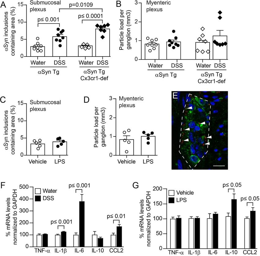
Free Neuropathology 2:13 (2021)                                                                                    Stefan Grathwohl et al
doi: https://doi.org/10.17879/freeneuropathology-2021-3326                                                                   page 4 of 23

Fig. 1 Age-dependent increase of intracellular αSyn accumulation in enteric nervous system of hemizygous (Thy1)-h[A30P]αSyn trans-
genic mice and setup of the experimental colitis paradigms.
A Confocal microscopy imaging of the inclusions of human αSyn (red, antibody clone 211; human αSyn specific) within the ganglia of the
submucosal plexus (green, peripherin; blue, DAPI/nuclei) of hemizygous (Thy1)-h[A30P]αSyn transgenic mice. Arrowhead points to one
of the typical irregularly sized and shaped αSyn inclusion bodies visualized in 2D z-stacks of rotated confocal images. Scale bar: 100 μm.
B Stereological quantification of normally occurring human αSyn inclusions in the myenteric and submucosal plexuses of 3- and 12-month-
old hemizygous (Thy1)-h[A30P]αSyn transgenic mice (n = 4 per group; mean and S.E.M. are shown; Student t-test between the two age
groups in each region). C Setup of experimental colitis paradigms employing dextran sulfate sodium (DSS, per os in drinking water) or
bacterial lipopolysaccharide (LPS, intraperitoneal injection). Except for the ‘chronic DSS paradigm, constant dose’, all paradigms were
started at the age of 3 months. The ‘chronic DSS paradigm, constant dose’ was started in mice aged 5 months and the colon were analyzed
right after. For some experiments, we used a ‘chronic DSS paradigm, increasing dose’, to mimic better the chronic nature of IBD and the
longer water intervals are generally more gentle for the mice from an animal welfare perspective as well. Under this paradigm, we induced
experimental DSS colitis intermittently as indicated over 23 days, then let the mice recover and age for two more months up to the age
of 6 months on normal drinking water, and analyzed their colon. In a separate experiment, we aged the mice further up to 9 or 21 months
and analyzed their brain pathology. Open arrows on time axis indicate that colon was analyzed and stars with closed arrows indicate that
brains were analyzed. D Hematoxylin staining of 35 μm thick colon sections of 3-month-old hemizygous (Thy1)-h[A30P]αSyn transgenic
mice. Organizational layers of the intact colon (left panel). Representative images of various severity degrees of DSS-driven colitis from
weak leukocyte infiltration (top panel of acute DSS) to more extensive leukocyte infiltration with mucosal ulceration (lowest panel of
acute DSS). Note the different appearance of enteric inflammation in acute LPS-driven peripheral inflammation compared with DSS, e.g.,
confined immune cell clustering and lymphoid hyperplasia, intact mucosal layer. Scale bar: 50 μm (intact colon), 100 μm (acute DSS), and
200 μm (LPS).
Free Neuropathology 2:13 (2021)                                                                Stefan Grathwohl et al
doi: https://doi.org/10.17879/freeneuropathology-2021-3326                                               page 5 of 23

on day 0 and 3 (Sigma-Aldrich Chemie GmbH, Stein-            anti-mIg IgG-HRP conjugate and peroxidase sub-
heim, Germany, LPS 055:B5). Acute colitis was in-            strate ABTS were used for quantitative detection of
duced by application of 36-50kDa dextran sulfate so-         mIgG-mIL10 fusion proteins.
dium (DSS) [44] (160110, MP Biomedicals, LLC, Ill-
kirch, France) at 1%, 2.5% or 5% in autoclaved drink-        Immunohistochemistry
ing water for 5 continuous days respectively, fol-                 Mice were injected with a lethal dose of pento-
lowed by 2 days of water (1 DSS application cycle).          barbital (150 mg/kg). Upon full anesthesia, mice re-
Chronic colitis was induced by two different dosing          ceived transcardial perfusion with room tempera-
protocols: i) in a constant dose of DSS (1% or 2.5%)         ture phosphate buffered saline (PBS). For biochemi-
for 5 days and changed to 2 days with normal drink-          cal and immunohistochemical analysis, one section
ing water and repeated three times (4 application            of the proximal colon was either fresh frozen and
cycles in total); ii) in an increasing dose of DSS start-    stored at -80°C or post-fixed in 4% paraformalde-
ing at 1% for 5 days followed for 4 days on normal           hyde (PFA) solution for 24 h. Following post-fixation,
drinking water, then increased to 1.5% DSS for the           organs were incubated in 30% sucrose/PBS at 4°C
next 5 days followed by 4 days of water and a final          for at least 48 h before further processing. Subse-
cycle of 2% DSS followed by aging the mice on nor-           quently, enteric tissue was cryotome-sectioned to
mal drinking water until they were sacrificed. Mice          35 µm thick longitudinal sections (approx. 1 cm
from the same littermate group were randomized               length). The brain was collected and post-fixed for
per cage into ‘exposed to inflammation inducing              24 h in 4% PFA followed by 30% sucrose in phos-
agent’ (LPS or DSS, respectively) or ‘unaffected’ (ve-       phate buffer until cryo-sectioning of floating sec-
hicle for the LPS paradigm or normal drinking water          tions at 40 μm. Histological analysis of mouse colon
for the DSS paradigms, respectively). For the long-          was performed using standard hematoxylin staining.
term experiments with the two aging cohorts ‘9               Immunohistochemical staining was accomplished
months’ and ‘21 months’, respectively, all mice in           using the Vectastain Elite ABC Kits and Peroxidase
that study were simultaneously exposed in one large          Substrate Kit SK-4100 (Vector Laboratories, Burlin-
cohort at the age of about 3 months to the increas-          game, CA, USA) or fluorescently labelled secondary
ing dose chronic DSS paradigm (Fig. 1) and DSS ex-           antibodies (Alexa 488, 555 or 647, Life Technologies,
posure was stopped for all mice on the same day af-          Zug, Switzerland). The following primary antibodies
ter the 23-day period. The mice were then kept and           have been used for overnight incubation at a dilu-
aged on normal drinking water and under normal               tion of 1:1000; monoclonal antibody to human αSyn
housing conditions in the same room until the day            (clone 211, sc-12767, Santa Cruz Biotechnology, Hei-
they were perfused and tissue was collected.                 delberg, Germany; specific to human αSyn and binds
                                                             to normal αSyn as well as abnormal αSyn inclusions
IL-10 treatment and exposure measurement                     which contain the respective epitope), monoclonal
                                                             antibody generated towards rat αSyn, cross-reactive
       Two different forms of mouse IgG bound mu-            with murine and human αSyn (Syn1/clone 42, BD
rine IL-10 (mIgG(v1)-mIL10 and mIgG(v2)-mIL10)               Transduction Laboratories, Allschwil, Switzerland;
were diluted in pre-prepared sterile formulation             used for wild type mice), polyclonal antibody to the
buffer comprised of 0.5% mouse serum supple-                 peripheral neuronal marker Peripherin (Millipore
mented with 25 mM citrate, 300 mM arginine to a              Corporation, Billerica, MA, USA), and polyclonal an-
final concentration of 0.75 mg/ml and the pH ad-             tibody to macrophage marker Iba-1 (Wako Chemical
justed to 6.7 on the day of application. Each mouse          GmbH, Neuss, Germany). To detect αSyn phosphor-
was treated once with 150 µg i.p. concurrently with          ylated at Serine 129 (pSer129-positive inclusions of
the initiation of the acute colitis paradigm with 5%         pathological/abnormal αSyn) in the free-floating
DSS. The concentrations of mIgG-mIL10 fusion pro-            brain sections, monoclonal antibody (ab51253,
teins in murine serum samples were determined by             Abcam, Cambridge, USA) was used at a dilution of
enzyme-linked immunosorbent assays (ELISA) spe-              1:10000. Prior to the pSer129 staining, the free-
cific for the Fab moiety of the administered mIgG-           floating brain sections were incubated for 10 min at
mIL10 fusion protein. Biotinylated mIgG-mIL10-spe-           room temperature in a phosphate buffered saline
cific target molecules were used for capturing, goat         solution containing 10 μg/mL proteinase K (Cat #
Free Neuropathology 2:13 (2021)                                                                 Stefan Grathwohl et al
doi: https://doi.org/10.17879/freeneuropathology-2021-3326                                                page 6 of 23

25530015; Invitrogen, California, USA). Tyrosine hy-         necting neurites. Therefore, the entire submucosa
droxylase (TH)-immunoreactive cells were detected            was set as region of interest, analyzed with the area
using a polyclonal antibody (657012, Millipore               fraction fractionator technique. Results of the sub-
Sigma) at a dilution of 1:1000. To measure the den-          mucosal plexus are displayed by percent area con-
sity of Nissl-positive cells, the TH-stained cells were      taining αSyn deposits. For the IL-10 experiment,
counter-stained with Cresyl violet. The slides were          αSyn positive inclusions from immunofluorescence
incubated in 0.1% Cresyl violet solution for 9 min           images were counted for each image. Inclusion
and then dehydrated in 95% and 100% ethanol and              body-like features were filtered based on having a
then xylene prior to cover slipping with Cytoseal 60         size between 12 and 50000 pixels and a minimal in-
mounting media (Thermo Fisher Scientific). Quanti-           tensity value greater than 300. The filtering step was
fications of the blind-coded TH/Nissl-stained slides         performed to exclude small background features
were done using Stereo Investigator (version                 and macrophages (very large spots). The counts
2017.01.1; MBF Bioscience, Williams, VT, USA) on an          were then aggregated to the animal level by sum-
Imager M2 microscope (ZEISS) coupled to a com-               ming the inclusion feature counts of all images per
puter. We analyzed 5-7 nigral sections per animal,           animal and then normalizing for (i.e., dividing by) the
and a total of 7-8 animals per treatment group. We           number of images for a given animal. Upon explora-
outlined the substantia nigra pars compacta and              tory data analysis two mice were excluded: one
counted every TH-immunoreactive and Nissl-posi-              mouse because it only had one image (technical out-
tive cell in that area (using a counting frame of 40         lier, missing data point; repeating the staining for
µm x 40 µm, grid size of 140 µm x 140 µm, a guard            this one mouse would have required re-staining the
zone of 2 µm and optical dissector height of 20 µm)          entire cohort in order to be consistent with staining
and then computed the number of cells per section,           conditions for quantification; this was unnecessary
generating the average cell count per animal. We             after statistical analysis) and another due to it being
then calculated the average count of cells per treat-        an outlier, based on its infiltration score and image
ment group and analyzed the data using unpaired              data (i.e., in contrast to the other mice that had re-
Student’s t-test after confirming normality and ho-          ceived DSS, this mouse did not show signs of inflam-
moscedasticity in Prism 7.0 (GraphPad Software).             mation or colonic tissue damage that is normally in-
                                                             duced by DSS; it could not be determined if that
Imaging and stereological quantification of αSyn
                                                             mouse was correctly dosed with DSS and thus it was
deposits in enteric nervous system                           excluded from the analysis).
      Imaging and stereological quantification were          Quantification of leukocytes infiltration
performed on a Zeiss Axio Imager Z2 fluorescence
microscope (Carl Zeiss AG, Jena, Germany). Leica                   To determine the leukocyte covered area in the
TCS SP5 confocal system using an HCX PL APO CS 40x           colon after LPS or DSS application, three adjacent
1.3 oil UV or an HCX PL APO LB 63x 1.4 oil UV objec-         hematoxylin-stained sections were quantified. Total
tive was utilized for image recording. Accumulation          area of colon sections and localizations of leukocyte
of αSyn in the ENS (i.e., punctate intracellular bod-        assemblies within the tissue architecture were iden-
ies/features) was assessed on a random set of 3 ad-          tified and outlined utilizing a stereology software
jacent 35 µm thick, αSyn-immunostained sections              (Stereo Investigator 6, MBF Bioscience, Williams, VT,
comprising the myenteric and submucosal neuronal             USA). Percentage of leukocyte covered area has
plexuses. Analysis was performed with the aid of a           been set in proportion to total area of the analyzed
stereology software (Stereo Investigator 10, MBF Bi-         colon section, e.g., to at least the length of 1 cm of
oscience, Williams, VT, USA) as described previously         proximal colon. For the IL-10 experiment, hematox-
[45]. In the myenteric plexus ganglion, volume was           ylin-stained colon slices were examined by an expert
defined by multiple outlined plexuses containing a           pathologist blinded to treatment conditions. A score
range of 5-20 neuronal cells and quantified by the           of 0-3 was assigned to each section for each of the 3
optical fraction fractionator technique. In contrast         layers lamina propria, submucosa and muscularis
to the myenteric plexus, the submucosa consists of           based on the degree of inflammatory infiltration. A
compact plexuses with 1-5 cells including intercon-          score of 0 denoted no inflammation and a score of 3
Free Neuropathology 2:13 (2021) Stefan Grathwohl et al
doi: https://doi.org/10.17879/freeneuropathology-2021-3326 page 7 of 23
indicated extensive infiltration. The mean of the val- cleus, periaqueductal gray, gray and white layer, re-
ues for all 3 layers was taken as the final measure of ticular formation, substantia nigra, ventral tegmen-
leukocyte infiltration per mouse. tal area, thalamus, hypothalamus, central amygdala,
pallidum and striatum) was determined in the water
Quantification of Iba-1/αSyn-double positive and DSS-treated animals. A NIKON Eclipse Ni-U mi-
macrophages croscope was used to acquire 20x magnification im-
ages (without condenser lens) from all the indicated
The number of Iba-1/αSyn-double positive cells
brain areas, using the same exposure time for all im-
was evaluated by quantification of 10 random re-
ages. In all cases, images were acquired on three
gions in 2 adjacent sections of the proximal colon.
sections separated by 420 μm intervals (localized
The region of interest was set to contain the myen-
between Bregma). We then processed the acquired
teric plexus/circular muscle layer and the submuco-
images using Image J64 [47], created a mask (to ex-
sal plexus.
clude background) that redirects to the original im-
Scoring of pSer129 pathology and brain heatmap age for analysis, measured the total area and the
mean grey value of the area that had inclusions. For
We evaluated pSer129 pathology on a full se- brain areas such as periaqueductal gray that do not
ries of immunostained coronal sections from 10 fill the entirety of the field to be analyzed, we drew
mice per treatment group (i.e., water vs. DSS- a contour of the area and the analysis was per-
treated groups) on blind-coded slides using a previ- formed only within that contoured area. We subse-
ously described method [46]. We visualized pathol- quently calculated the grey value of the area per
ogy from one hemisphere of all brain sections (apart square pixels for each image (i.e., A.U./px2 = mean
from the olfactory area) using a NIKON Eclipse Ni-U grey value x area stained/total area assessed). Based
microscope and assigned scores ranging from 0 to 4 on this, we calculated the average grey value per
to each brain area based on the relative abundance square pixels for each brain area for each animal (n
of proteinase K (PK)-resistant pSer129-positive in- = 6 mice/group), and then extended this calculation
clusions (i.e., cell bodies and neurites). In this case, to determine the average grey value per square pix-
0 = no aggregates, 1 = sparse, 2 = mild, 3= dense, 4 = els for each treatment group and each of the twelve
very dense. For the heatmap, we obtained the aver- brain areas of interest.
age score values of each brain area for each treat-
ment group. The average data for each treatment Blinding of experimenters for histological and im-
group (n=10 mice/group) was then represented as a munohistochemical analyses
heatmap in a sagittal mouse brain background. To For analyses of colon and brain tissue on slides,
create the brain heatmap a postscript file down- a second individual assigned unique codes to stained
loaded freely from Allen Brain Atlas (mouse, p56, slides. Therefore, the experimenter conducted the
sagittal, image 15 of 21; http://atlas.brain- analyses blinded to the identity of the mice. For ran-
map.org/atlas?atlas=2#atlas=2&structure=771&res domization of treatment groups see above.
olution=16. 75&x=%7755.7470703125&y=3899.625
&zoom=-3&plate=100883867&z=5 was converted mRNA expression
to an XML in R v 3.4.4, and the mean scores were
To assess mRNA expression levels from the
manually assigned to respective brain regions. The
proximal colon, RNA was extracted from fresh fro-
remaining brain regions were estimated via the R
zen tissue with MagnaLyser green beads (Roche Di-
package ‘Akima’, using a pointwise bivariate inter-
agnostics, Mannheim, Germany) and Qiazol Lysis
polation algorithm for irregular data on the mean X
(Reagent cat.no.79306, Hilden, Germany) purified
and Y coordinates for each brain region.
on MagnaPure LC (HP Kit no.03542394001, F. Hoff-
Densitometry of pSer129 αSyn brain pathology mann-La Roche AG, Rotkreuz, Switzerland) and am-
plified via real-time PCR (4 ng RNA/reaction; Light-
The density of pSer129 pathology in 12 major cycler 480, Roche Diagnostics Corporation, Indian-
brain areas (reticular nucleus, pontine reticular nu- apolis, USA). Amplification of mRNA was performedFree Neuropathology 2:13 (2021) Stefan Grathwohl et al
doi: https://doi.org/10.17879/freeneuropathology-2021-3326 page 8 of 23
by using TaqMan probes for human or murine spe- For the statistical analysis of the pSer129 αSyn
cific α-synuclein and for selected cytokines/chemo- brain pathology, zero-inflated negative-binomial
kines (Applied Biosystems Europe B.V., Zug, Switzer- mixed-effects models with a random intercept for
land). Target mRNA was normalized to tissue spe- each sample and variance assumed to increase line-
cific murine GAPDH levels and displayed as relative arly with the mean (verified against a quadratic in-
expression after 30 amplification cycles. crease using Akaike Information Criterion [AIC] and
Bayesian Information Criterion [BIC]) were used to
Statistics analyze the dataset via the ‘glmmTMB’ package in R
Measurements for inflammation and αSyn ac- v 3.4.4. Linear contrasts with false discovery rate
cumulation in the ENS were taken from distinct sam- (FDR) adjustments were then used to test our hy-
ples (e.g., in three to six technical replicates per potheses and account for multiple testing (for brain
mouse). Data from each mouse was used only once, area and experimental group).
thus no repeated measure of the same sample was
Results
performed. Statistical analysis of gut pathology and
inflammation was performed using GraphPad Prism Experimental colitis exacerbates αSyn load in the
6.04 or 7.0 software (GraphPad Software, Inc. La submucosal plexus of αSyn transgenic and wild
Jolla, CA, USA). The results are expressed as mean type mice
values ± standard errors of the mean (S.E.M.). Stu-
dent’s t-test (or Welch’s t-test for unequal vari- During the process of further characterizing a
ances) was used to compare two groups and ANOVA (Thy1)-h[A30P]αSyn transgenic mouse line [41], we
was used for multi-comparison of groups followed detected by immunohistochemistry human αSyn ac-
by Tukey HSD post-hoc analysis. For the statistical cumulation in all innervated organs that were ana-
analysis of the mRNA expression, data quality was lyzed (Suppl. Fig. 1). To examine the localization of
assessed by inspecting the distribution of Cp values αSyn inclusions in nervous structures in the ENS, we
of reference endogenous genes across samples, by performed an immunofluorescent co-staining for
inspecting the level of Cp variation between tech- human αSyn (clone 211) and peripherin, a specific
nical replicates and by exploring the samples multi- marker for peripheral nerves. By applying confocal
variate signal distribution as in a principal compo- microscopy, we established a process and protocol
nent analysis. Relative gene expression levels were to identify myenteric and submucosal plexuses in or-
expressed as 2-(Cpgene – CpRef). Statistical analyses to as- der to quantify the αSyn inclusions found in the
sess the effect of the experimental conditions on the nerve cells of the ganglia in the colon. The intracel-
log2 gene expression levels were done with linear lular presence of the irregularly sized and shaped
models using the limma package (Bioconductor/R, αSyn inclusion bodies was confirmed in 2D z-stacks
[48]). These analyses were implemented in R v 3.1.1. of rotated confocal images (Fig. 1A). We observed
an age-dependent (mice aged 3 months versus 12
For the statistical modelling of the effects of months) increase of baseline human αSyn inclusions
the IL-10 treatment on αSyn counts, as well as infil- in both plexuses (Fig. 1B). Given the clinical and epi-
tration scores, the levels for IgG1(v1)-IL10 and demiological link between IBD and PD, we wanted
IgG1(v2)-IL10 treatment were compared to the pos- to test whether different types and strengths of IBD-
itive (vehicle/DSS) control. Additionally, since levels related experimental inflammation in the colon ex-
of the control antibody treatment (IgG1(v1)) were acerbates this local accumulation of αSyn acutely
very similar to the positive control, the two groups (e.g., within a few days or weeks) and how the age
were pooled in further contrasts in which effects of of the αSyn transgenic mice may influence the out-
individual antibodies or control IgG was assessed. come. Administration of dextran sulfate sodium
For αSyn counts in the enteric nervous system, a lin- (DSS) in the drinking water in acute or chronic dosing
ear model on the treatment groups with one-degree paradigms are well-established mouse models of ex-
freedom contrasts was applied. For the infiltration perimental colitis mimicking aspects of IBD, i.e., by
score a Kruskal-Wallis test, with the same contrasts, exhibiting infiltration of leukocytes into the submu-
was used. All statistical tests were two-tailed with a cosa with various degrees of destruction of the co-
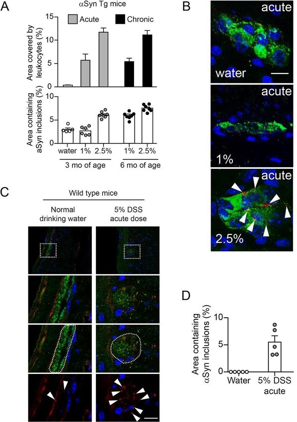
significance level of pFree Neuropathology 2:13 (2021) Stefan Grathwohl et al
doi: https://doi.org/10.17879/freeneuropathology-2021-3326 page 9 of 23
that the effects induced by the DSS paradigm can phology consistent with them being infiltrating leu-
vary substantially based on the genetic background cocytes, which was confirmed by an Iba-1 co-stain-
of the mice and due to different animal housing en- ing (Suppl. Fig. 2). This finding was relevant for the
vironments. Thus, in order to establish the DSS par- quantification of αSyn inclusions in the myenteric
adigm in our environment and with our mice, we and submucosal plexus, i.e., such features were ex-
first tested the effect of DSS administration at differ- cluded from the quantification process.
ent doses and durations in the (Thy1)-h[A30P]αSyn
transgenic mice (Fig. 1C). We observed that leuko- Wild type mice also express endogenous αSyn
cyte infiltration was appropriately modulated by the in innervated organs, but at much lower levels com-
acute dose (1% or 2.5% DSS for 5 days followed by 2 pared with the levels of human αSyn expressed by
days drinking water) and chronic constant dose (1% the hemizygous (Thy1)-h[A30P]αSyn transgenic
or 2.5% DSS alternating with normal drinking water mice (Suppl. Fig. 1). To confirm that the finding in
for 28 days, respectively) DSS paradigms (Fig. 1D and (Thy1)-h[A30P]αSyn transgenic mice was independ-
2A). In the same experiment, we wanted to test for ent of transgenic expression of human αSyn, we ap-
an age effect on a potential aggravation of αSyn ac- plied an acute 5% DSS dose paradigm (5 days DSS +
cumulation in the ENS and applied the acute dose 2 days water) in wild type mice (Fig. 1C). We ob-
paradigm on 3-month-old mice and the chronic dose served, in the submucosal plexus, small inclusion
paradigm was started at the age of 5 months leading bodies of endogenous murine αSyn (detected by ro-
to a final age of 6 months at analysis of the mice. In dent cross-reactive αSyn-specific monoclonal anti-
the acute DSS dosing paradigm with mice at the age body Syn1/clone 42, Fig. 2C, D). These features were
of 3 months, 2.5%, but not 1%, DSS triggered intra- close to undetectable in the water group that did
cellular accumulation of αSyn in peripherin positive not experience experimental colitis. A separate ex-
nerve cells of the submucosal plexus (Fig. 2A, B). In periment also confirmed that the observed effects
the 28-day chronic constant DSS dose paradigm in of elevated αSyn inclusions following acute DSS (5%
mice aged 6 months (Fig. 1C and 2A), we observed dose) could not be attributed to increased gene ex-
that the 1% constant chronic DSS dose showed a pression of murine or the transgenic human αSyn
similar degree of αSyn inclusions as the 3-month-old (Suppl. Fig. 3). Together, these results confirmed the
mice which were on an acute 2.5% DSS dose para- validity of this experimental IBD paradigm to test the
effect of inflammation on αSyn accumulation in the
digm. In addition, mice previously exposed to 2.5%
constant chronic DSS dose presented on average ENS in wild type and (Thy1)-h[A30P]αSyn transgenic
with a slightly but robust increased percent area of mice. Because the (Thy1)-h[A30P]αSyn transgenic
αSyn inclusions compared with the 6-month-old mouse model is well-established to analyze human
mice that were on 1% DSS (Fig. 2A). Together, this αSyn-related pathology, we focused for the remain-
demonstrated that different DSS paradigms can be der of the study on employing these transgenic
established in (Thy1)-h[A30P]αSyn transgenic mice mice.
and that different DSS paradigms can induce robust Colitis induced by peroral DSS but not by in-
elevation of αSyn inclusions. This first experiment traperitoneal administration of LPS aggravates
also provided us with data points to estimate a po- αSyn accumulation in colonic submucosal plexus
tential effect size for triggering αSyn inclusions in of αSyn transgenic mice
the different DSS paradigms. We observed that the
younger (Thy1)-h[A30P]αSyn transgenic mice Different inflammatory agents induce different
showed a robust increase in αSyn inclusions and bet- types of immune stimulation and thus can influence
ter signal-to-noise conditions of the autofluorescent the phenotype of experimental colitis. A well-estab-
colonic tissue than the older ones, experiments lished experimental immune trigger is the bacterial
were henceforth continued with mice of the endotoxin LPS. In order to explore the effects of dif-
younger age. In this initial experiment, it was also in- ferent approaches to induce inflammation in or
teresting to observe that (Thy1)-h[A30P]αSyn trans- nearby the gut in (Thy1)-h[A30P]αSyn transgenic
genic mice exposed to acute 2.5% DSS colitis pre- mice, we compared the outcome of acute (5 days
sented with several αSyn-positive cells with a mor- DSS + 2 days normal drinking water) 5% DSS per osFree Neuropathology 2:13 (2021) Stefan Grathwohl et al doi: https://doi.org/10.17879/freeneuropathology-2021-3326 page 10 of 23 Fig. 2 Experimental DSS colitis severity and duration-dependent aggravation of accumulation of αSyn inclusions in the colonic submu- cosal plexus of hemizygous (Thy1)-h[A30P]αSyn transgenic and wild type mice. A Administration of DSS in drinking water induced a robust increase of leukocyte infiltration in the acute (1% or 2.5% DSS for 5 days followed by 2 days of normal drinking water; one group was kept on normal drinking water) and chronic constant DSS dose (1% or 2.5% alternating with normal drinking water) paradigm in hemizygous (Thy1)-h[A30P]αSyn transgenic mice. The highest acute dose (2.5%) and the two constant chronic doses led to a very robust increase of αSyn inclusions in the submucosal plexus (stereological quantification of αSyn inclusions in the submucosal plexus of all 3- and 6-month-old hemizygous (Thy1)-h[A30P]αSyn transgenic mice; n = 5-7 per group; mean and S.E.M. are shown). B Representative 2D z-stacks of confocal images of increasing abundance of αSyn inclusions (red, human- αSyn specific monoclonal antibody clone 211) in a ganglion of the submucosal plexus (green, peripherin) with cellular nuclei in blue (DAPI) in the acute DSS paradigm. Arrow heads point to the typical irregularly sized and shaped αSyn inclusion bodies that accumulate in the highest DSS dose. Scale bar: 200 µm. C Overview of colonic region of 3-month-old wild type mice exposed to water or acute DSS (5%) with immunofluorescence analysis of murine αSyn load in the colon performed immediately after colitis. White dotted rectangles in the top row indicate the area that was zoomed-in in the lower panels. In the zoom-ins we show representative images of DAPI and αSyn (red, rodent αSyn cross-reactive monoclonal antibody syn1/clone 42) inclusions with and without the peripherin channel (green). The white dotted circled area illustrates the peripherin-positive area that was analyzed for αSyn inclusion bodies (arrow heads in bottom row). Scale bar for the lower three panels: 200 μm. D Stereological quantification of murine αSyn inclusions in the submucosal plexus of wild type mice right after acute DSS colitis (n = 5 per group). Note the regularly arranged and smoothly distributed immunoreactivity for the physi- ological αSyn with barely any inclusion bodies in the intact enteric nerves of the water group. For both panels (A) and (D), statistical analysis for αSyn accumulation was omitted as the noticeable and very robust differences between the means are self-evident (error bars indicate standard error of the mean) and an indication for an estimation for significance would be irrelevant.
Free Neuropathology 2:13 (2021)                                                                Stefan Grathwohl et al
doi: https://doi.org/10.17879/freeneuropathology-2021-3326                                              page 11 of 23

with acute 0.5 mg/kg intraperitoneal LPS admin-              petent mice (Suppl. Fig. 3A). However, a signifi-
istration (Fig. 1C and 3). To maximize the inflamma-         cantly higher level of αSyn accumulated in the sub-
tory response, we administered both DSS and LPS at           mucosal plexus in αSyn transgenic mice lacking
relatively high doses. At day 7, both agents had in-         Cx3cr1 compared to αSyn transgenic mice express-
duced variable degrees of leukocyte infiltration in          ing Cx3cr1 (p = 0.001, two-way ANOVA with Tukey
the submucosa of the colon while a marked destruc-           HSD post-hoc analysis; Fig. 3A). In the myenteric
tion of the mucosa was induced when giving only              plexus, we found no marked increase in αSyn accu-
DSS (Fig. 1D). As before, the DSS-exposed mice pre-          mulation in neither the αSyn transgenic mice with
sented with increased accumulation of αSyn in the            normal Cx3cr1 nor the αSyn transgenic mice defi-
ganglia of the submucosal plexus (Fig. 3A). In con-          cient in Cx3cr1, indicating as in the experiments
trast, we detected no change in αSyn load in the             above a possible prominent role for the localization
myenteric plexus, consistent with lack of leukocyte          of leukocyte infiltration in the process of αSyn accu-
infiltration in this part of the colonic wall (Fig. 3B).     mulation in the submucosa (Fig. 3B). Collectively,
Despite the high dose, LPS-induced inflammation              our results in Cx3cr1-deficient αSyn transgenic mice
did not increase αSyn accumulation in the colonic            provide a potential association between mono-
nervous plexuses (Fig. 3C, D). Notably, LPS and DSS          cyte/macrophage signaling and αSyn accumulation
resulted in a differential expression of cytokines,          in ENS in this experimental IBD model.
and consistent with leukocyte recruitment, CCL2
was elevated in both (Fig. 3F, G). In the LPS para-          Systemic IL-10 reduces DSS-induced colitis and
digm, mRNA for IL-10 was markedly elevated,                  associated enteric αSyn accumulation in αSyn
whereas DSS strongly increased IL-6 and also IL-1β           transgenic mice
but not IL-10. Together these results indicate that, in
our model, colonic inflammation induced by peroral                 To continue testing the hypothesis that modu-
DSS but not intraperitoneal LPS increases the accu-          lating monocytes/macrophages may affect accumu-
mulation of αSyn in the colon.                               lation of αSyn in our DSS model we moved to a phar-
                                                             macological modulation of this cellular subset. Inter-
Lack of monocyte/macrophage related Cx3cr1                   leukin-10 (IL-10) is an important regulator of mono-
signaling during DSS colitis increases αSyn load in          cytes/macrophages, and genetic ablation of IL-10
the submucosal plexus of αSyn transgenic mice                signaling or blocking IL-10 with specific antibodies
                                                             has been reported to enhance DSS colitis [53,54]. In
     Given the role of monocytes/macrophages in              the experiments with LPS we had also noted an in-
IBD and in the related DSS paradigm, we hypothe-             crease of IL-10 compared with the DSS paradigm and
sized further that modulating monocytes/macro-               LPS inflammation was in contrast to DSS colitis not
phages may affect accumulation of αSyn in our DSS            associated with increased αSyn accumulation in the
model as well. In a first set of experiments we ma-          ENS (Fig. 3). To mimic the effect of higher levels of
nipulated monocytes/macrophages genetically by               IL-10 in an acute model of DSS colitis (5% DSS for 5
crossing (Thy1)-h[A30P]αSyn transgenic mice with             days + 2 days normal drinking water, Fig. 1C), we ad-
mice that have a deletion for the fractalkine recep-         ministered intraperitoneally recombinant murine IL-
tor Cx3cr1 (Cx3cr1-GFP knock-in mice) (Fig. 3A, B).          10 (mIL10) in this paradigm. The half-life of injected
The CX3CR1-CX3CL1 axis plays an important role in            recombinant IL-10 protein in blood is very short. To
maintaining the function of the lamina propria mac-          reduce the number of injections, we extended the
rophage population of the gastrointestinal wall and          half-life of mIL-10 in circulation by engineering it
lack of this signaling pathway in experimental colitis       onto two different murine IgG variants (i.e.,
models may either aggravate or ameliorate the in-            mIgG1(v1)-mIL10 and mIgG1(v2)-mIL10, respec-
duced pathology [50–52]. In our experiment, the              tively). As described above, DSS induced a marked
area covered by infiltrating leukocytes following ex-        increase in leukocyte infiltration and αSyn accumu-
posure to DSS was near the mucosa and submucosa              lation, and we found both to be similar in the un-
and was not significantly higher in the Cx3cr1-defi-         treated and control IgG treated group (Fig. 4A, B). In
cient αSyn transgenic mice than in the Cx3cr1-com-           contrast, both mIgG1(v1)-mIL10 and mIgG1(v2)-
Free Neuropathology 2:13 (2021) Stefan Grathwohl et al doi: https://doi.org/10.17879/freeneuropathology-2021-3326 page 12 of 23 Fig. 3 Colitis induced by peroral DSS but not peritoneal LPS enhances αSyn accumulation in the colonic submucosal plexus of hemizy- gous (Thy1)-h[A30P]αSyn transgenic mice and can be increased by lack of monocyte/macrophage-related Cx3cr1 signaling. Mice received in an acute paradigm either peroral 5% DSS in their drinking water or intraperitoneally 0.5 mg/kg LPS. Effects of DSS and LPS in the colon, respectively, were compared to effects induced by vehicle (see Figure 1C for timelines). Stereological quantification of αSyn inclusions in the submucosal plexus as % area (A, C) and in the mucosal plexus as particle load per ganglion (B, D) (Two-way ANOVA with Tukey post hoc test; covariates genotype and treatment paradigm). E Representative 2D stacks of confocal images of intracellular αSyn inclusions (red, human αSyn specific monoclonal antibody clone 211; arrow heads pointing to some selected inclusions) in a ganglion of the myenteric plexus (green, peripherin) with cellular nuclei in blue (DAPI). Scale bar: 50 μm. Gene expression analysis of selected cytokines in the colon of (Thy1)-h[A30P]αSyn transgenic mice that received either acute DSS (F) or LPS (G) compared to their respective vehicle or water controls. Note the strong increase in IL-6 and the lack of elevation of IL-10 in the DSS paradigm compared to the LPS paradigm indicating a different inflammatory colonic milieu despite the abundant leukocyte infiltration in both paradigms. N = 5-8 per group; mean and S.E.M.; Student’s t-test between inflammatory agent and vehicle for individual cytokines.
Free Neuropathology 2:13 (2021)                                                                         Stefan Grathwohl et al
doi: https://doi.org/10.17879/freeneuropathology-2021-3326                                                       page 13 of 23

mIL10 significantly reduced leukocyte infiltration in
mice treated with DSS (p
Free Neuropathology 2:13 (2021)                                                                  Stefan Grathwohl et al
doi: https://doi.org/10.17879/freeneuropathology-2021-3326                                                page 14 of 23

months (Fig. 1C). At this point we wanted again to           we chose hemizygous (Thy1)-h[A30P]αSyn trans-
explore the effect of modulating monocytes/macro-            genic mice to increase the chances for a successful
phages in this chronic setting and added an experi-          outcome and potentially to aggravate the brain pa-
mental arm with (Thy1)-h[A30P]αSyn transgenic                thology from mild to strong. After exposing the mice
mice lacking Cx3cr1. As expected, after 2 months of          either to normal drinking water or a chronic increas-
recovery, the area that is usually extensively cov-          ing dose DSS paradigm, we aged them in two co-
ered by leukocytes in the submucosal plexus of the           horts on normal water and housing conditions to ei-
acute DSS paradigm had returned to normal levels             ther up to the age of 9 months or 21 months. At
following the two-month recovery period (Suppl.              these two timepoints we analyzed various brain re-
Fig. 4A). Remarkably, however, the area containing           gions for αSyn inclusions that are generally consid-
αSyn inclusions in the ganglia of the submucosal             ered pathological by being proteinase K (PK)-re-
plexus was still almost doubled when compared to             sistant and immunopositive for pSer129-αSyn.
αSyn transgenic mice that were not exposed to DSS,           When we examined the 9-month-old αSyn trans-
and this was exacerbated in αSyn transgenic mice             genic mice, we found that both experimental groups
deficient for Cx3cr1 (Suppl. Fig. 4B). The finding in        (i.e., those who were on DSS and those who stayed
the αSyn transgenic mice suggests that accumula-             on normal water throughout their entire life and
tion of αSyn is not a transient effect or response. In       thus never experienced DSS colitis) exhibited ex-
addition, modulation of monocytes/macrophages                tremely low levels of pathological αSyn aggregation
by down-regulating the CX3CR1-CX3CL1 axis con-               in the brain (Fig. 5 and Suppl. Fig. 5). Our observa-
tributes to aggravation of this accumulation.                tion of the level of pathological αSyn aggregations in
                                                             the brain of these 9-month-old hemizygous (Thy1)-
Experimental DSS colitis-induced at a young age
                                                             h[A30P]αSyn transgenic mice (Fig. 5A) is indeed con-
exacerbates αSyn brain pathology and dopamin-
                                                             sistent with earlier descriptions of the model at the
ergic neuron loss in old αSyn transgenic mice
                                                             age of 11 months [57]. The 21-month-old hemizy-
      At this point, we have established and repeat-         gous (Thy1)-h[A30P]αSyn transgenic mice that only
edly demonstrated that modulation of inflammatory            received water during their lifetime showed, in con-
mechanisms in experimental colitis induced by                trast to the 9-month-old cohort, more discernible
acute and chronic DSS administration is causatively          PK-resistant pSer129-αSyn immunoreactive fea-
linked to induction and persistence of intracellular         tures (Fig. 5B) and the abundance of these features
αSyn inclusions in the ENS of young adult mice. The          was consistent with previous observations in this
previously highlighted hypothesis by Braak and col-          transgenic line at the age of 24 months [57]. In
leagues associates αSyn brain pathology in PD with           marked contrast, the 21-month-old hemizygous
αSyn pathology in the ENS earlier in life [3,56]. To         (Thy1)-h[A30P]αSyn transgenic mice that were ex-
assess development of brain αSyn pathology and to            posed to DSS at three months of age presented with
link it to IBD risk, we exposed 3-month-old hemizy-          pSer129-positive αSyn pathology throughout vari-
gous (Thy1)-h[A30P]αSyn transgenic mice to a                 ous brain regions in a much more exacerbated fash-
chronic increasing dose DSS paradigm or normal               ion than mice that were aged up to 21 months with-
drinking water and after 23 days returned all mice to        out having experienced DSS colitis at young age (Fig.
normal drinking water until sacrifice several months         5B-E). The degree and distribution of PK-resistant
later (Fig. 1C). We chose to use the αSyn transgenic         αSyn in the brain was similar to what was previously
model rather than wild type mice for this study be-          described for homozygous (Thy1)-h[A30P]αSyn
cause of two reasons: 1) we knew that the model as           transgenic mice at the age of 8 to 9 months [57]. The
hemizygous transgenic mice exhibit some αSyn                 significant aggravation of αSyn pathology in the sub-
brain pathology that develops slowly under baseline          stantia nigra (p ≤ 0.01 in a negative-binomial mixed-
conditions. Importantly, the pathology is much less          effects model adjusting for multiple comparisons
pronounced than in homozygous (Thy1)-                        performed over all brain areas) was accompanied by
h[A30P]αSyn mice [57]; 2) at the time of the experi-         a significant loss of tyrosine hydroxylase (TH) and
ment, it was not clear whether wild type mice could          Nissl-positive cells at 21 months of age (p ≤ 0.05, Stu-
develop αSyn brain pathology upon DSS colitis. Thus,         dent’s t-test; Fig. 6). Together, we found that exper-
Free Neuropathology 2:13 (2021) Stefan Grathwohl et al doi: https://doi.org/10.17879/freeneuropathology-2021-3326 page 15 of 23 Fig. 5 A 23-days chronic DSS colitis insult at young age causes an age-dependent accumulation of proteinase K-resistant pSer129-αSyn in various brain regions of (Thy1)-h[A30P]αSyn transgenic mice. A 23-days chronic increasing dose DSS paradigm was performed with 3-month-old (Thy1)-h[A30P]αSyn transgenic mice. After recovering and further aging, various brain regions were analyzed for proteinase K (PK)-resistant pSer129-αSyn immunoreactivity in 9-month (A) and 21-month-old (B) mice, respectively. The dark brown features in (A) (barely any visible in both the water and the DSS group) and (B) (strongly visible with typical neuritic and punctated inclusion-type morphology) indicate PK-resistant inclusions with pSer129-αSyn immu- noreactivity. Densitometric quantification of pSer129-αSyn immunoreactivity in different brain regions of 21-month-old mice (C, D) (n=6 mice per group). In order to visualize better the differences between the water versus the DSS group at 21 months, brain regions with large increase were plotted on an y-axis up to 250 A.U./px2 and small increase on a y-axis up to 40 A.U./px2. The about
Free Neuropathology 2:13 (2021)                                                                                        Stefan Grathwohl et al
doi: https://doi.org/10.17879/freeneuropathology-2021-3326                                                                      page 16 of 23

imental DSS colitis at a young age caused an age-de-                        combinant αSyn fibrils to different brain regions and
pendent exacerbation of PK-resistant pSer129-αSyn                           intestines [15–17,19,20,46,66,67] together with
pathology and a loss of nigral dopaminergic neurons                         postmortem brain pathology [56,59,68], it has also
in the brains of (Thy1)-h[A30P]αSyn transgenic mice.                        been suggested that αSyn pathology propagates
                                                                            temporospatially from cell-to-cell in a prion-like
Discussion                                                                  manner [3,59,66,68,69]. However, the initial factors
      Currently, there is no therapy for PD available                       triggering αSyn aggregation in the tissue or organ of
to slow or stop disease progression and an obstacle                         origin of the pathology are yet to be established [58]
in the quest to develop one is that we do not under-                        and the involvement of peripheral stimuli in the ag-
stand how the disease develops [58]. Abnormal in-                           gregation and pathogenic spread of αSyn is only be-
traneuronal accumulation of αSyn (i.e., in Lewy bod-                        ginning to unravel.
ies and neurites) is a key neuropathological hallmark
and the distribution of Lewy pathology in postmor-                                 In this study, we provide evidence that DSS co-
tem brain is used for staging in PD [2,59]. Accumula-                       litis, i.e., an experimental IBD-like inflammation,
tion of αSyn has also been observed in the periph-                          triggers αSyn accumulation in the ENS of wild type
eral nervous system in PD, some individuals at risk                         mice and in a human αSyn transgenic mouse model
of developing the disease, and normal individuals                           of PD (Fig. 2). We found aggravation of enteric αSyn
[60–62]. Similar to this finding in humans, αSyn-im-                        accumulation in αSyn transgenic mice lacking Cx3cr1
munoreactive inclusions and signs of age-dependent                          signaling and amelioration of inflammation and as-
αSyn-related pathological changes have also been                            sociated slight reduction of enteric αSyn load by sys-
detected in the ENS of wild type rats [63,64] and sev-                      temic IL-10, demonstrating that genetic and phar-
eral transgenic mouse models prior to development                           macologic modulation of inflammation can influ-
of brain pathology [21,65]. Based on preclinical                            ence the degree of αSyn accumulation in the ENS
models employing injection of brain extracts or re-                         (Fig. 3 and 4).

Fig. 6 A 23-days chronic DSS colitis insult at young age results in loss of tyrosine hydroxylase- and Nissl-positive cells in the substantia
nigra of (Thy1)-h[A30P]αSyn transgenic mice at 21 months of age.
(Thy1)-h[A30P]αSyn transgenic mice were exposed to a 23-days chronic increasing dose DSS paradigm at the age of 3 months followed by
aging on normal drinking water up to the age to 21 months. These mice showed a significant loss of mean count of Nissl-positive cells
with tyrosine hydroxylase (TH) immunoreactivity and cellular Nissl staining in the substantia nigra compared to age-matched littermate
mice in the group that did not experience DSS colitis (water). A Representative images of two levels of the substantia nigra in one mouse
per group. B Stereological quantification of cells positive for TH or Nissl (n=7-8 mice per group). Statistical analyses of the TH dataset were
performed using Student’s t-test, while Welch’s t-test was used for the Nissl dataset to adjust for unequal variances. Scale bar: 500 μm.
You can also read