The Hippocampal Vulnerability to Herpes Simplex Virus Type I Infection: Relevance to Alzheimer's Disease and Memory Impairment
←
→
Page content transcription
If your browser does not render page correctly, please read the page content below
REVIEW
                                                                                                                                           published: 13 August 2021
                                                                                                                                     doi: 10.3389/fncel.2021.695738

                                             The Hippocampal Vulnerability to
                                             Herpes Simplex Virus Type I
                                             Infection: Relevance to Alzheimer’s
                                             Disease and Memory Impairment
                                             Shin Jie Yong 1 , Min Hooi Yong 2,3 , Seong Lin Teoh 4 , Tomoko Soga 5 , Ishwar Parhar 5 ,
                                             Jactty Chew 1* and Wei Ling Lim 1,3*
                                             1
                                               Department of Biological Sciences, School of Medical and Life Sciences, Sunway University, Petaling Jaya, Malaysia,
                                             2
                                               Department of Psychology, School of Medical and Life Sciences, Sunway University, Petaling Jaya, Malaysia, 3 Aging
                                             Health and Well-being Research Centre, School of Medical and Life Sciences, Sunway University, Petaling Jaya, Malaysia,
                                             4
                                               Department of Anatomy, Universiti Kebangsaan Malaysia Medical Centre, Kuala Lumpur, Malaysia, 5 Jeffrey Cheah School
                                             of Medicine and Health Sciences, Brain Research Institute Monash Sunway, Monash University Malaysia,
                                             Subang Jaya, Malaysia

                                             Herpes simplex virus type 1 (HSV-1) as a possible infectious etiology in Alzheimer’s
                                             disease (AD) has been proposed since the 1980s. The accumulating research thus
                                             far continues to support the association and a possible causal role of HSV-1 in the
                         Edited by:          development of AD. HSV-1 has been shown to induce neuropathological and behavioral
               Jorge Rubén Cabrera,
   Laboratory Corporation of America         changes of AD, such as amyloid-beta accumulation, tau hyperphosphorylation, as
   Holdings (LabCorp), United States         well as memory and learning impairments in experimental settings. However, a
                           Reviewed by:      neuroanatomical standpoint of HSV-1 tropism in the brain has not been emphasized
                      Gabriela O. Bodea,
                                             in detail. In this review, we propose that the hippocampal vulnerability to HSV-1 infection
     University of Queensland, Australia
                    Giovanna De Chiara,      plays a part in the development of AD and amnestic mild cognitive impairment (aMCI).
 Institute of Traslational Pharmacology,     Henceforth, this review draws on human studies to bridge HSV-1 to hippocampal-
Italian National Research Council, Italy
                                             related brain disorders, namely AD and aMCI/MCI. Next, experimental models and
                   *Correspondence:
                          Jactty Chew        clinical observations supporting the neurotropism or predilection of HSV-1 to infect
              jacttyc@sunway.edu.my          the hippocampus are examined. Following this, factors and mechanisms predisposing
                         Wei Ling Lim
                                             the hippocampus to HSV-1 infection are discussed. In brief, the hippocampus has
              weilingl@sunway.edu.my
                                             high levels of viral cellular receptors, neural stem or progenitor cells (NSCs/NPCs),
                   Specialty section:        glucocorticoid receptors (GRs) and amyloid precursor protein (APP) that support HSV-
        This article was submitted to
             Cellular Neuropathology,
                                             1 infectivity, as well as inadequate antiviral immunity against HSV-1. Currently, the
                a section of the journal     established diseases HSV-1 causes are mucocutaneous lesions and encephalitis;
    Frontiers in Cellular Neuroscience       however, this review revises that HSV-1 may also induce and/or contribute to
            Received: 15 April 2021          hippocampal-related brain disorders, especially AD and aMCI/MCI.
            Accepted: 20 July 2021
          Published: 13 August 2021          Keywords: herpes simplex virus, hippocampus, neurotropism, Alzheimer’s disease, memory impairment,
                              Citation:      infectious etiology
          Yong SJ, Yong MH, Teoh SL,
Soga T, Parhar I, Chew J and Lim WL
(2021) The Hippocampal Vulnerability
                                            INTRODUCTION
        to Herpes Simplex Virus Type I
   Infection: Relevance to Alzheimer’s
                                            Alzheimer’s disease (AD) is the leading neurodegenerative disease, accounting for about 60-80% of
    Disease and Memory Impairment.          dementia cases globally (Qiu et al., 2009; Alzheimer’s Association., 2021). AD may progress from
     Front. Cell. Neurosci. 15:695738.      a long period of subtle memory decline called amnestic mild cognitive impairment (aMCI; the
      doi: 10.3389/fncel.2021.695738        most common type of MCI) (Petersen et al., 2001). While the etiology of AD is multifaceted, the

Frontiers in Cellular Neuroscience | www.frontiersin.org                           1                                        August 2021 | Volume 15 | Article 695738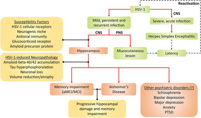
Yong et al.                                                                                                            Hippocampal Tropism of HSV-1

hypothesis for an infectious cause in AD has emerged since the                stem or progenitor cells (NSCs/NPCs) are localized (Altman
1980s. Ball (1982) and Gannicliffe et al. (1986) first suggested              and Das, 1965; Eriksson et al., 1998). The hippocampus is
that periodic reactivation of herpes simplex virus type-1 (HSV-               susceptible to various stressors, including chronic stress, aging
1) from latency in neurons may facilitate the development of                  and microbial infections. As a result, hippocampal functions
AD. In the following decades, the possible involvement of herpes              such as learning and memory would be compromised. Hence,
viruses, such as HSV-1, HSV-2, cytomegalovirus (CMV), human                   hippocampal dysfunction has been implicated in disorders that
herpesvirus types 6, 7, and 8 (HHV-6, -7, and -8), varicella-                 involve memory impairment as a symptom, such as depression,
zoster virus (VZV) and Epstein-Barr virus (EBV), in AD and MCI                schizophrenia, dementia, aMCI/MCI and AD, as reviewed in
have been investigated (Polk et al., 2002; Strandberg et al., 2003;           Small et al. (2011) and Anand and Dhikav (2012).
Carbone et al., 2014; Barnes et al., 2015; Agostini et al., 2016b; Tsai          This review first discusses the possible role of HSV-1 in
et al., 2017; Tzeng et al., 2018). The collective evidence implicates         the development of AD and aMCI/MCI in humans. Next, this
HSV-1 as the most probable infectious agent contributing to                   review describes the mechanisms of HSV-1 infection in neurons
AD and MCI, according to reviews and meta-analyses (Steel                     and theoretical model of HSV-1 infection trajectory, focusing
and Eslick, 2015; Itzhaki et al., 2016; Warren-Gash et al., 2019;             on its neurotropism or predilection to target the hippocampus
Sait et al., 2021).                                                           based on cell culture, animal and human autopsy evidence. We
    HSV-1 is an enveloped, linear double-stranded DNA virus                   suggest that the hippocampal vulnerability to HSV-1 infection
that infects more than 60% of the population worldwide (Looker                may also present a pivotal factor in initiating or facilitating the
et al., 2015; Harfouche et al., 2019; Khadr et al., 2019). Productive         development of aMCI/MCI and AD. Following this, factors and
infection of HSV-1, either from primary infection or latent                   mechanisms affecting the hippocampal susceptibility to HSV-1
reactivation, causes mucocutaneous lesion of the lips, cornea or              infection are discussed.
genitals (Darougar et al., 1985; Scott et al., 1997; Ribes et al.,
2001). HSV-1 also causes herpes simplex encephalitis (HSE), the
most common type of infection-induced encephalitis (Granerod                  BRIDGING HSV-1 TO AD AND MEMORY
et al., 2010; George et al., 2014). In pregnant mothers with                  IMPAIRMENT
genital herpes, HSV-1 can cause congenital herpes in the infant
upon vaginal delivery, resulting in mucocutaneous lesions and                 HSE, either due to primary infection or viral reactivation,
central nervous system (CNS) infection (Whitley et al., 1991;                 is known to cause long-term neurological sequelae despite
Whitley et al., 2007).                                                        immediate antiviral treatments (Riancho et al., 2013; Armangue
    At the neuronal level, HSV-1 infection has been shown                     et al., 2018). Damage to the temporal lobe and limbic system
to induce tau hyperphosphorylation, amyloid-beta 40 and 42                    (especially the hippocampus), as well as impairments in memory
(Aβ40/42) accumulation, oxidative stress, neuroinflammation                   and behavior (e.g., emotional instability and irritability), have
and apoptotic dysregulation, all of which are implicated in the               been frequently observed among HSE survivors (Kapur et al.,
pathophysiology of neurodegenerative diseases such as AD. At                  1994; Caparros-Lefebvre et al., 1996; Dagsdottir et al., 2014;
the genetic level, gene products of the HSV-1 life cycle have                 Harris et al., 2020). Degeneration of similar brain regions
been shown to interact with AD susceptibility genes, such as                  and consequent phenotypic abnormalities in HSE resemble
presenilin 1 and 2 (PSEN1 and PSEN2), apolipoprotein E allele                 that of AD. This observation has led to the hypothesis that
4 (ApoE4) and clusterin genes, to promote both viral infectivity              repeated and periodic HSV-1 reactivation may contribute to AD
and risk of AD. These molecular mechanisms of HSV-1-induced                   development, especially in the aging population with declining
neuropathology in AD have been reviewed in Harris and Harris                  immunocompetence (Ball, 1982; Esiri, 1982b; Gannicliffe et al.,
(2018) and Duarte et al. (2019). Consequently, at the behavioral              1986). Therefore, this section will describe the relationship
level, HSV-1 infection has been found to induce memory and                    between HSV-1 and AD and memory impairment, which may
learning impairments reminiscent of AD (Beers et al., 1995;                   also reflect prodromal AD, in humans.
Armien et al., 2010; De Chiara et al., 2019).
    While molecular mechanisms underpinning contributions of                  Alzheimer’s Disease
HSV-1 to AD have been reviewed extensively (Duarte et al., 2019;              HSV-1 DNA has been detected within Aβ depositions in
Marcocci et al., 2020), a neuroanatomical standpoint has not been             postmortem brain tissues of AD patients compared to non-AD
considered in detail. Deciphering the HSV-1 infection pathway                 controls (Mori et al., 2004). The same study also found HSV-1
and tropism in the brain would advance the understanding of                   antigens within cortical neurons, providing the first evidence of
the potential neurological health outcomes of HSV-1 infection.                possible HSV-1 reactivation in the AD brain (Mori et al., 2004).
This review, thus, examines which brain region is most affected               A further study reported that most HSV-1 DNA was localized
by HSV-1. Literature to date suggests that it may be the                      within Aβ plaques in the cortices of AD patients (Wozniak et al.,
hippocampus, given its cardinal role in learning and memory.                  2009b). Furthermore, transcriptome analyses of brain specimens
The hippocampus and its neuronal connections to the entorhinal                from cohorts of AD patients have revealed higher abundance
cortex, amygdala, olfactory bulb and hypothalamus comprise                    of HSV-1 latency-associated transcripts (LATs; transcribed from
the limbic system (Vilensky et al., 1982). In the mammalian                   HSV-1 DNA) than older adults without AD (Readhead et al.,
hippocampus, life-long neurogenesis has been shown to occur in                2018). These results indicate that HSV-1 can infect the brain and
the subgranular zone of the dentate gyrus (DG), where neural                  is associated with AD neuropathology.

Frontiers in Cellular Neuroscience | www.frontiersin.org                  2                                   August 2021 | Volume 15 | Article 695738
Yong et al. Hippocampal Tropism of HSV-1
When compared to age-matched healthy controls, individuals Letenneur et al., 2008; Lovheim et al., 2015b) and HSV-1
with AD and aMCI exhibited increased levels of anti-HSV-1 DNA in the brain (Jamieson et al., 1991; Hemling et al., 2003;
IgG antibodies (Costa et al., 2017; Agostini et al., 2019; Pandey Pisa et al., 2017) between individuals with and without AD.
et al., 2019), which also correlated with increased cortical volumes This could be attributed to genetic factors that predispose
(Mancuso et al., 2014a,b). Similarly, increased antibody levels and HSV-1-infected individuals to AD. For instance, the presence
avidity index against HSV-1 were found to be elevated in aMCI of HSV-1 DNA or IgG seropositivity with ApoE4 gene has
patients that did not develop AD, compared to those who did. been shown to pose a greater risk factor in AD development
In the same study, HSV-1-specific antibody titers also correlated than either one by itself (Lin et al., 1995, 1996; Itzhaki et al.,
positively with hippocampal and amygdala volumes (Agostini 1997; Steel and Eslick, 2015; Lopatko Lindman et al., 2019).
et al., 2016a). Other studies have also found that aMCI patients Another reason may be that HSV-1 IgG seropositivity and DNA
displayed higher anti-HSV-1 IgG antibody levels and avidity only indicate a history of viral exposure, as HSV-1 may remain
index compared to that of both healthy controls and AD patients latent and non-infective. Other meta-analyses have found that
(Kobayashi et al., 2013; Costa et al., 2020). Taken together, these HSV-1-specific IgM seropositivity and high IgG levels (i.e.,
findings imply that robust antibody immunity against HSV-1 indicating productive infection or reactivation) were associated
may prevent the brain atrophy progression of aMCI into AD, with dementia and MCI, but not HSV-1 IgG seropositivity and
possibly via antibody neutralization of HSV-1 that protects the DNA (Warren-Gash et al., 2019; Ou et al., 2020; Wu et al., 2020).
brain against HSV-1-induced neuropathology (Mancuso et al.,
2014a; Agostini et al., 2016a; Costa et al., 2020). Memory Impairment
When anti-HSV-1 immunity is inadequate to control HSV-1 As mounting evidence supports the link between HSV-1
infection, periodic HSV-1 reactivation and productive infection infection and AD development, HSV-1 may also be associated
may occur. One nationwide retrospective cohort study in with the prodromal stage of AD, aMCI. Patients with aMCI may
Taiwan reported that individuals diagnosed with recurrent show early signs of AD neuropathological attributes, such as
HSV-1 infection had a 2.8-fold higher risk of developing AD hippocampal shrinkage, neurofibrillary tangles (NFT; aggregates
than uninfected individuals. More importantly, antiherpetic of hyperphosphorylated tau) and Aβ40/42 accumulation,
medications reduced such risk by about 90% compared to placebo according to a systematic review (Stephan et al., 2012).
(Tzeng et al., 2018). In another nationwide retrospective cohort Hippocampal neuroimaging has also been demonstrated to
study involving participants with HSV-1 or VZV infections and predict whether MCI patients would develop AD (Jack et al.,
uninfected matched controls in Sweden, antiherpetic treatment 1999; Hu et al., 2016; Li et al., 2019). Thus, given that HSV-1
was associated with a 10% reduced risk of dementia. In infection can induce hippocampal dysfunction, HSV-1 infection
untreated patients, the risk of dementia increased by 50% may also be associated with reduced memory function.
compared to uninfected controls (Lopatko Lindman et al., 2021). For instance, HSV-1 IgG seropositivity has been associated
A four-national (i.e., Wales, Scotland, Denmark and Germany) with an 18-fold increased odds of memory deficits in middle-
retrospective cohort study found that persons with HSV infection aged adults (Dickerson et al., 2008). Associations between HSV-1
who were not given anti-herpetic medication had 18% higher IgG seropositivity and impaired cognition were also reported in
risk of dementia compared to uninfected controls, although other groups, e.g., healthy soldiers (Fruchter et al., 2015), young
this effect was present in the Germany cohort only (Schnier individuals (Tarter et al., 2014; Vanyukov et al., 2018) and older
et al., 2021). In a smaller retrospective cohort study comprising adults (Zhao et al., 2020; Murphy et al., 2021). Therefore, the
HSV-1-seropositive older adults, antiherpetic prescription was clinical biomarker of HSV-1 exposure (i.e., IgG seropositivity)
associated with 70% lower risk of AD development compared is likely to be linked to impaired memory and other cognitive
to no prescription (Hemmingsson et al., 2021). Two aging measures, while biomarkers of HSV-1 reactivation or productive
prospective cohort studies have also found that the risk of AD infection (i.e., high IgG levels or IgM seropositivity) is linked
was about twofold greater in those with IgM seropositivity for to AD (Letenneur et al., 2008; Lovheim et al., 2015a; Warren-
HSV-1 (Letenneur et al., 2008; Lovheim et al., 2015a). Taken Gash et al., 2019). This indicates that HSV-1 reactivation
together, these studies suggest that HSV-1 productive infection or productive infection may promote more severe cognitive
or reactivation may promote AD development, which may also decline than mere HSV-1 exposure. In a population study
be preventable with antiherpetic agents. comprising healthy adolescents, HSV-1 IgG seropositivity was
Interestingly, a longitudinal study reported that IgM associated with memory decline, whereas HSV-1 IgG levels
seropositivity for HSV-1 was associated with memory decline, correlated with both poor memory and executive functioning
especially amongst carriers of ApoE4 (Lövheim et al., 2019). (Jonker et al., 2014).
Likewise, in another prospective cohort study, ApoE4 carriers Population studies have also reported that IgG levels specific
had a threefold increased risk of both AD and HSV-1 reactivation for either HSV-1 or CMV independently predicted cognitive
(i.e., as indicated by IgM seropositivity or elevated IgG levels) defect in the elderly (Strandberg et al., 2003), schizophrenics
compared to ApoE4-negative individuals (Linard et al., 2020). and their non-psychotic relatives (Shirts et al., 2008; Watson
Therefore, host genetic risk factors such as ApoE4 may modulate et al., 2013), and middle-aged adults (Tarter et al., 2014).
the interactions between HSV-1 and AD risk. However, some studies found that only CMV-specific (and
However, several studies found no significant differences not HSV-1-specific) IgG seropositivity or levels correlated with
in HSV-1 IgG seropositivity (Wozniak et al., 2005; cognitive impairment in elderly populations (Aiello et al., 2006;
Frontiers in Cellular Neuroscience | www.frontiersin.org 3 August 2021 | Volume 15 | Article 695738Yong et al. Hippocampal Tropism of HSV-1
TABLE 1 | Animal infection models assessing the neuronal invasion and spread of HSV-1.
Model of Site of viral Site of latency Neurological and References
infection dissemination behavioral findings
Lip infection
Tooth pulp • Hippocampus N/A N/A Barnett et al., 1994
inoculation into • EC
BALB/C mice • TG
(young; • Amygdala
5–6 weeks) • Brainstem
• Insular cortex
• Olfactory cortex
• Cingulate cortex
• Temporal cortex
Infection by lip • Hippocampus • TG Accumulation of Li Puma et al., 2019
abrasion into Aβ40/42 peptides and
C57BL/6 mice reduced neurogenesis
(newborn; PND in the hippocampus.
0–1)
Infection by lip • Hippocampus • TG Upregulated De Chiara et al., 2019
abrasion into • Neocortex neuroinflammatory
BALB/c and • Cerebellum (astrogliosis, IL-1β and
3xTg-AD mice IL-6) and
(young; neurodegenerative
6–8 weeks) (Aβ40/42 and
hyperphosphorylated
tau) markers in
neocortex and
hippocampus. Mice
exhibited learning and
memory deficits.
Upregulated oxidative Protto et al., 2020
stress markers (HNE,
HNE-modified proteins,
protein carbonyls and
3-nitrotyrosine) in
cortex.
Intranasal infection
Intranasal • Hippocampus N/A N/A Anderson and Field, 1983
inoculation into • OB
BALB/C mice • Amygdala
(young; • Hypothalamus
3–4 weeks) • Brainstem
Intranasal • Hippocampus N/A Neuronal degeneration Tomlinson and Esiri, 1983
inoculation into • OB and acute inflammation
BALB/C mice • Trigeminal root entry in infected areas,
(young; • Brainstem especially the trigeminal
6 weeks) • Amygdala system.
• Thalamus
• Hypothalamus
• Temporal lobe
• Cingulate cortex
Intranasal • Hippocampus N/A N/A Webb et al., 1989
inoculation into • EC
BALB/C mice • OB
(young; • Trigeminal nerve
6–8 weeks) • Brainstem
Intranasal • EC • EC Acute inflammation in Stroop et al., 1990
inoculation into • TG • TG olfactory structures,
New Zealand • OB • Olfactory cortex including the EC.
White rabbits • Olfactory cortex
(adult)
(Continued)
Frontiers in Cellular Neuroscience | www.frontiersin.org 4 August 2021 | Volume 15 | Article 695738Yong et al. Hippocampal Tropism of HSV-1
TABLE 1 | Continued
Model of Site of viral Site of latency Neurological and References
infection dissemination behavioral findings
Intranasal • Hippocampus • Hippocampus Inflammatory and Beers et al., 1993
inoculation into • EC • EC haemorrhagic lesions in
Lewis rats • Amygdala • OB the TG, OB, amygdala,
(adult) • TG EC, spinal trigeminal
• OB nuclei and
hippocampus.
Viral injection • EC N/A Mice with bilateral McLean et al., 1993
into OB of • Olfactory nucleus damage to olfactory
Sprague- • Piriform cortex cortex exhibited
Dawley rats impaired learning and
(adult) memory.
Intranasal • Hippocampus • Hippocampus Impairments in spatial Beers et al., 1995
inoculation into • EC • EC memory and learning.
Lewis rats, Brain tissues remain
followed by a histologically normal.
recovery period
from HSE
(adult)
Intranasal • TG N/A Cytopathic effects Meyding-Lamade et al.,
inoculation into • Olfactory epithelium found predominantly in 1998, 1999
SJL/NBOM the hippocampus,
mice (adult) temporal and
frontobasal lobes,
thalamus, pons and
mesencephalon.
Intranasal • Hippocampus N/A HSV-1 replicated in the Boggian et al., 2000
inoculation into • EC brain without producing
Albino Swiss • OB neurological or
CD-1 mice • Amygdala behavioral anomalies.
(young; • Frontal lobe
6–10 weeks) • Temporal lobe
Intranasal • Temporal cortex N/A Aβ40/42 deposition in Wozniak et al., 2007
inoculation into the temporal cortex.
BALB/C mice
(young;
4–6 weeks)
Intranasal • Hippocampus N/A Most viral antigens Ando et al., 2008
inoculation into • OB were localized in the
Wistar • TG DG subfield of the
Hannover • Cerebral cortex Medulla hippocampus. Severe
GALAS rats neuronal loss and
(young; tissue damage in
PND 14) infected areas.
Intranasal • Hippocampus N/A Glial cells necrosis and Armien et al., 2010
inoculation into • Trigeminal nerve myelin degeneration
BALB/c mice to • OB within the hippocampus
induce • Thalamus and lateral tegmental
encephalitis • Hypothalamus nucleus. Neuronal loss
(young; • Temporal cortex in the hippocampus
8–10 weeks) • Piriform cortex (most profound), EC,
amygdala and temporal
cortex. Lymphocytic
infiltration in the
hippocampus and
temporal cortex. Mice
exhibited severe
learning deficits.
(Continued)
Frontiers in Cellular Neuroscience | www.frontiersin.org 5 August 2021 | Volume 15 | Article 695738Yong et al. Hippocampal Tropism of HSV-1
TABLE 1 | Continued
Model of Site of viral Site of latency Neurological and References
infection dissemination behavioral findings
Intranasal • Hippocampus N/A N/A Jennische et al., 2015
inoculation into • TG
Sprague– • OB
Dawley rats • Brainstem
(adult)
Corneal/Ocular infection
Corneal • TG • TG N/A Deatly et al., 1988
inoculation into • Pons (entry of trigeminal • Pons
BALB/C mice nerve) • Cerebellum
(young; • EC
4-6 weeks) • Hippocampus
Corneal • TG • TG Profound Paivarinta et al., 1994
inoculation into • Pons (entry of trigeminal • Pons neuroinflammation in
Californian nerve) the hippocampus and
rabbits (young; EC.
6 weeks)
Ocular infection • Hippocampus • Hippocampus N/A Chen et al., 2006
into BALB/c • OB • OB Brainstem
mice (young; • Brainstem Cerebellum
10 weeks) • Cerebellum Frontal lobe
• Frontal lobe
Ocular infection • Hippocampus • OB N/A Li et al., 2016
into tree shrews • OB • Brainstem
(Tupaia • Brainstem
belangeri • Thalamus
chinensis; • Cerebral cortex
young;
6-months)
Corneal • Hippocampus • TG Chronic inflammation in Menendez et al., 2016
infection into • SVZ • Hippocampus the hippocampus, SVZ
C57BL/6J mice • NPCs • SVZ and midbrain.
(young; • Midbrain • Midbrain
10 weeks) • Frontal lobe • Frontal lobe
Brain infection
Intracerebral • Hippocampus N/A N/A Anderson and Field, 1983
inoculation into • Hypothalamus
BALB/C mice • Cerebral cortex
(young;
3–4 weeks)
HSV-1 vector • Hippocampus N/A N/A Braun et al., 2006
propagation in • NPCs
an ex vivo • Ependymal cells
system of • Ventricles
brains of • Cortical areas
BALB/c mice
and SABRA
rats (newborn;
PND 1–2 and
young;
4 weeks)
Stereotactic • Cortex N/A Accumulation of Aβ42 Eimer et al., 2018
injection of peptides in the brain,
HSV-1 into the which inhibited HSV-1
hippocampus infectivity and protected
of transgenic mice from acute viral
AD mice encephalitis.
(5XFAD) (young;
5–6 weeks)
(Continued)
Frontiers in Cellular Neuroscience | www.frontiersin.org 6 August 2021 | Volume 15 | Article 695738Yong et al. Hippocampal Tropism of HSV-1
TABLE 1 | Continued
Model of Site of viral Site of latency Neurological and References
infection dissemination behavioral findings
Intracranial • Hippocampus N/A Accumulation of Aβ42 Ezzat et al., 2019
infection into • Cortex peptides.
transgenic
5xFAD mice
(young;
3-months)
Intracranial • Hippocampus N/A Neuronal loss, Toscano et al., 2020
infection into upregulated
C57BL/6 mice inflammatory markers
(age and (TNF, IL1-β, IL-6 and
weight N/A) IFNα/IFNβ) and
suppressed
anti-inflammatory
(IL-10, SOCS2 and
SOCS3) signals in the
hippocampus.
Peripheral infection
Intravenous • Brainstem N/A N/A Anderson and Field, 1983
and sciatic • Hypothalamus
nerve
inoculation into
BALB/C mice
(young;
3–4 weeks)
Intraperitoneally • Hippocampus • TG N/A Burgos et al., 2006
inoculation into • Ventricles • Hippocampus
female • Midbrain
C57BL/6 mice • Cerebellum
(young; • Cortex
14 weeks)
Aβ, amyloid-beta; EC, entorhinal cortex; HNE, 4-hydroxynonenal; IL, interleukin; NPCs, neural progenitor cells; IFN, interferon; OB, olfactory bulb; PND, postnatal day;
SOCS, suppressor of cytokine signaling; SVZ; subventricular zone; TG, trigeminal ganglion; TNF, tumor necrosis factor.
Barnes et al., 2015; Nimgaonkar et al., 2016) and bipolar disorder One key limitation of these studies is that HSV-1 seropositivity
patients (Tanaka et al., 2017). On the contrary, other studies indicates the history of prior HSV-1 exposure. Seropositivity
showed that only HSV-1-specific IgG (and not CMV-specific) alone does not inform the status of HSV-1 infection; that is,
seropositivity or levels were associated with cognitive impairment active or latent infection, or viral replication in the central
in healthy adolescents (Jonker et al., 2014) and individuals with or peripheral nervous systems or peripheral epithelial cells
or without neuropsychiatric disorders (Dickerson et al., 2003; (Dickerson et al., 2008; Murphy et al., 2021). Therefore, the
Yolken et al., 2011; Hamdani et al., 2017). possible link of causality or pathophysiological pathways between
A putative explanation for these inconsistencies could be that HSV-1 seropositivity and memory impairment remains unclear
both CMV and HSV-1 contribute to memory dysfunction. (Vanyukov et al., 2018). Besides, the majority of the global
It was suggested that CMV, which is known to induce adult population (i.e., > 60%) is seropositive for HSV-1 (Looker
immune dysregulation, may exacerbate HSV-1-induced et al., 2015; Harfouche et al., 2019; Khadr et al., 2019), whereas
neurodegeneration, leading to AD (Stowe et al., 2012; Lovheim only relatively few develop memory impairment (i.e., 10–20% of
et al., 2018). This is based upon the finding that HSV-1- adults over 50 years) (Overton et al., 2019; Pais et al., 2020; Lu
specific IgG levels increased with age only in CMV seropositive et al., 2021). This suggests that HSV-1 seropositivity may only
individuals (Stowe et al., 2012). CMV IgG seropositivity alone play a subtle role in the development of memory impairment
also did not elevate the risk of AD, but both CMV and HSV-1 in certain cases.
seropositivity did (Lovheim et al., 2018), suggesting that CMV
and HSV-1 interact to influence the risk of developing AD.
For AD with severe memory impairment, HSV-1 likely plays HSV-1 IN THE NERVOUS SYSTEM
a more predominant role in memory impairment than other
herpesviruses, as suggested by meta-analyses (Steel and Eslick, HSV-1 has four notable structures: the glycoproteins-embedded
2015; Warren-Gash et al., 2019) and reviews (Itzhaki, 2014; membrane, tegument layer, capsid and double-stranded DNA
Itzhaki and Klapper, 2014). genome (Grunewald et al., 2003). Although HSV-1 is considered
Frontiers in Cellular Neuroscience | www.frontiersin.org 7 August 2021 | Volume 15 | Article 695738Yong et al. Hippocampal Tropism of HSV-1
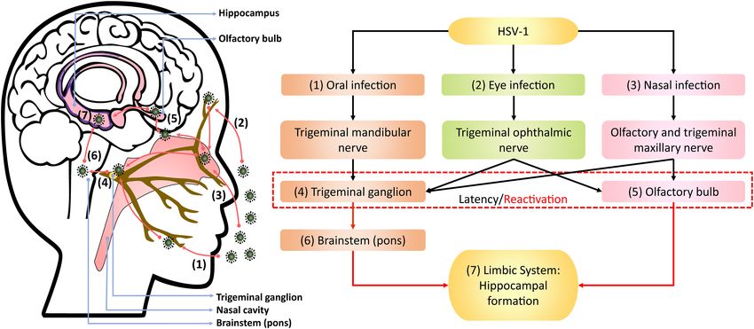
FIGURE 1 | Theoretical model of HSV-1 infection trajectory within the nervous system. Following viral replication in the (1) oral, (2) corneal, or (3) olfactory epithelial
cells, HSV-1 can exploit the neuronal retrograde machinery to reach the (4) trigeminal ganglion and (5) olfactory bulb, which are also sites for HSV-1 latency and
stress-induced reactivation. Reactivated HSV-1 relies on anterograde transport to infiltrate the brain. Therein, HSV-1 travels from the trigeminal ganglion to the (6)
pons, a part of brainstem innervated by the trigeminal nerve, and then to the (7) limbic system that inhabits the hippocampal-entorhinal circuitry. Alternatively, HSV-1
may directly infect the hippocampal-entorhinal circuity via the olfactory bulb, which is part of the limbic system.
a human pathogen, it can infect other animals in experimental are synthesized and re-assembled (Honess and Roizman, 1974).
settings, as reviewed in Karasneh and Shukla (2011). The The assembled viral particles may bud into the inner nuclear
primary target cells of HSV-1 are epithelial and neuronal cells, membrane and undergo primary envelopment for nuclear egress.
where HSV-1 can alternate between lytic and latent life cycle HSV-1 particles may subsequently undergo de-envelopment at
phases, as reviewed in Kukhanova et al. (2014). This section the outer nuclear membrane to allow for further viral assembly
will detail the mechanisms of HSV-1-neuronal interactions, in the cytoplasm, and complete the secondary envelopment at
followed by the theoretical model of HSV-1 infection trajectory Golgi vesicles before the mature virions egress out of the host
in the nervous system. cell (Hutchinson and Johnson, 1995; Granzow et al., 2001), as
reviewed in Mettenleiter (2002).
HSV-1 Infection and Latency After this lytic phase in peripheral epithelial cells, HSV-1 can
First, HSV-1 attachment to cells depends on the binding proceed to the latent phase in neurons, typically in the trigeminal
of envelop glycoprotein B (gB) to surface heparan sulfate ganglion (Takasu et al., 1992; Theil et al., 2001; Hufner et al.,
proteoglycans (HSPGs), with an optional gC to augment the 2009). The latent phase of HSV-1 starts similarly to the lytic
interaction (WuDunn and Spear, 1989; Herold et al., 1991, 1994). phase, until HSV-1 reaches the nucleopore by retrograde axonal
Alternatively, gB can bind to myelin-associated glycoprotein transport (Antinone and Smith, 2010). In the latent phase, HSV-1
(MAG) (Suenaga et al., 2009) and non-muscle myosin heavy lytic gene expression in the nucleus is silenced, and no infectious
chain IIA and IIB (NHMC-IIA and -IIB) to initiate viral entry viral progeny is produced. Instead, HSV-1 DNA undergoes
(Arii et al., 2010, 2015). The subsequent binding of gD to 3- histone modifications with nucleosomes of the chromatin in cell
O-sulfated heparan sulfate, herpesvirus entry mediator (HVEM) nucleus (Deshmane and Fraser, 1989; Kubat et al., 2004; Wang
and/or nectin-1 (Montgomery et al., 1996; Whitbeck et al., 1997; et al., 2005). During latency, HSV-1 gene expression is limited
Geraghty et al., 1998; Shukla et al., 1999) catalyzes viral fusion and to LATs and a few microRNAs, which may inhibit (i) lytic gene
entry via activation of gH/gL (Atanasiu et al., 2010). transcription to maintain latency and (ii) apoptosis to promote
Following HSV-1 entry, the DNA-containing nucleocapsid survival of infected neurons (Perng et al., 2000; Wang et al., 2005;
and tegument proteins are released into the cell cytoplasm. Umbach et al., 2008).
One of the tegument proteins may proceed to turn off HSV-1 can transition from latent to lytic phase under
protein translation in the cell (Kwong and Frenkel, 1987). The conditions of cellular stress, such as chemotherapy,
nucleocapsid moves and docks on the nuclear pore, which hyperthermia, ultraviolet radiation, fever, immunosuppression
then disassembles to release the DNA into the cell nucleus and psychological stress, as reviewed in Suzich and Cliffe (2018)
(Sodeik et al., 1997; Dohner et al., 2002). This initiates a and Yan et al. (2020). During HSV-1 reactivation, the HSV-1
cascade of HSV-1 gene expression, in the order of α-genes, β- DNA-associated chromatin relaxes and transcription of the viral
genes and γ-genes, where viral structural proteins and DNA regulatory VP16 protein begins, which consequently activates a
Frontiers in Cellular Neuroscience | www.frontiersin.org 8 August 2021 | Volume 15 | Article 695738Yong et al. Hippocampal Tropism of HSV-1
cascade of viral lytic gene expression that lead to the production Hufner et al., 2009). In animal studies, HSV-1 could also infect
of infectious viral progeny (Thompson et al., 2009; Kim et al., the eye, propagating along the corneal subbasal nerve plexus
2012; Sawtell and Thompson, 2016). Newly manufactured HSV-1 innervated by trigeminal ophthalmic nerve, to initiate latency
then travels by anterograde transport from the cell body to axon in the olfactory bulb and trigeminal ganglion (He et al., 2017;
termini to infect neighboring epithelial cells or neurons (Snyder Menendez and Carr, 2017; Figure 1). Although congenital herpes
et al., 2008; Miranda-Saksena et al., 2009). HSV-1 reactivation is usually contracted upon vaginal delivery, in utero infection can
can lead to the appearance of disease (e.g., cold sores and occur in 5% of human infant cases (Hutto et al., 1987; Marquez
HSE), asymptomatic viral replication or spread into the CNS, as et al., 2011). In such instances, using the murine model, genital
reviewed in Bearer (2012) and Marcocci et al. (2020). HSV-1 likely enters the bloodstream and crosses the placenta into
Neurons with actively replicating HSV-1 via reactivation or the fetal nervous system following the trigeminal infection route
primary infection begins to undergo various mechanisms that (Burgos et al., 2006).
lead to pathological changes, as reviewed in Harris and Harris Following reactivation from the trigeminal ganglion, HSV-1
(2018) and Duarte et al. (2019). Neuronal culture studies have may infiltrate the pons innervated by trigeminal nerves and then
demonstrated that HSV-1 induced tau hyperphosphorylation by travel along the brainstem to the limbic system, as demonstrated
upregulating several enzymes such as caspase-3, protein kinase in animal models (Tomlinson and Esiri, 1983; Webb et al., 1989;
A and glycogen synthase kinase 3β (Wozniak et al., 2009a; Barnett et al., 1994; Paivarinta et al., 1994). The olfactory bulb
Lerchundi et al., 2011; Alvarez et al., 2012). HSV-1-infected constitutes part of the limbic system and has direct projections to
neurons have also been shown to exhibit impaired autophagy the hippocampus. Thus, reactivation from the olfactory bulb may
and amyloid precursor protein (APP) processing, resulting in provide HSV-1 and other neurotropic viruses direct access to the
increased Aβ40/42 accumulation (De Chiara et al., 2010; Santana hippocampus, as proposed by Mori et al. (2005) and Duarte et al.
et al., 2012; Piacentini et al., 2015). These HSV-1-induced AD- (2019).
related neuropathology can be inhibited with antiviral treatment The theoretical model depicting neuronal pathways of HSV-
targeting HSV-1 in vitro (Wozniak et al., 2011, 2013, 2005). 1 infection in the brain (Figure 1) is also consistent with
These studies indicate that HSV-1 spread into the brain may autopsy examinations of HSV-1 antigen distribution amongst
facilitate the development of AD-related neuropathology. Animal HSE patients. Specifically, HSV-1 antigens were localized mostly
models further provided support wherein HSV-1 infection or in the hippocampus with the highest number of cases and
reactivation have been shown to induce AD neuropathology in viral abundance found, as well as in the temporal lobe,
the brain, which was also associated with learning and memory olfactory bulb and amygdala (Dinn, 1979; Twomey et al., 1979;
impairments (Martin et al., 2014; De Chiara et al., 2019). Esiri, 1982a,b). Notably, postmortem analysis of AD victims
detected HSV-1 DNA more frequently in the hippocampus
and temporal cortex compared to other brain areas (Jamieson
HSV-1 Infection Trajectory: Emphasis on et al., 1991, 1992). Damasio and Van Hoesen (1985) also
Hippocampal Tropism hypothesized that HSV-1 travels to the limbic system via
In the brain, HSV-1 invasion has been shown to target the the trigeminal nerve, wherein HSV-1 may exhibit higher
olfactory system and hippocampus, followed by the higher affinity for the hippocampus, and subsequently spread to
cortical areas in animal studies (Table 1). According to Braak’s cortices during HSE.
staging scheme in human AD samples, the hippocampus– Most animal models investigating HSV-1 neurotropism
entorhinal circuitry within the temporal lobe deteriorates the following reactivation, primary infection, or both have supported
earliest, followed by higher cortical areas (Braak and Braak, the predilection of HSV-1 to infect the hippocampus (Table 1).
1991). Increasing evidence has also suggested that dysfunction Some studies induced stress in animal models to reactivate HSV-1
of the olfactory system may indicate prodromal AD in humans, and showed that the consequent viral replication was particularly
as Murphy (2019) reviewed. With this anatomical resemblance, prominent in the hippocampus (Burgos et al., 2006; De Chiara
Fewster et al. (1991) and Ball et al. (2013) have previously et al., 2019). Further supporting evidence can be derived from
suggested that HSV-1 might induce the neuron-to-neuron animal findings that demonstrated impairment in hippocampus-
tauopathy and Aβ spread in AD as HSV-1 propagates along its dependent memory and learning tasks following HSV-1 infection
infection pathways. (McLean et al., 1993; Beers et al., 1995; Armien et al., 2010; De
Upon oral infection in animal models, HSV-1 can infect Chiara et al., 2019).
the mandibular trigeminal nerve to establish latency at the AD-associated neuropathology induced by HSV-1 can
trigeminal ganglion (Barnett et al., 1994; Lewandowski et al., be observed in the hippocampus. For instance, multiple
2002; De Chiara et al., 2019). Alternatively, the nasal cavity reactivations of HSV-1 caused memory deficits that were
can be an infection site wherein HSV-1 can travel along the correlated with increased Aβ accumulation, tau phosphorylation
olfactory and trigeminal maxillary nerves and become latent in and neuroinflammation in the neocortex and hippocampal
the olfactory bulb and trigeminal ganglion of animals (Stroop DG of mice (De Chiara et al., 2019). It was demonstrated
et al., 1990; Beers et al., 1993; Jennische et al., 2015). Autopsy that HSV-1 could form a protein corona layer that served as
studies have also detected HSV-1 DNA, including LATs, in catalytic surfaces for Aβ accumulation in the hippocampus
the trigeminal ganglion, trigeminal nerves and olfactory bulb and cortex of mice (Ezzat et al., 2019). Neurodegeneration and
of deceased humans (Liedtke et al., 1993; Theil et al., 2001; lymphocytic infiltration were also observed in the hippocampus,
Frontiers in Cellular Neuroscience | www.frontiersin.org 9 August 2021 | Volume 15 | Article 695738Yong et al. Hippocampal Tropism of HSV-1
entorhinal cortex, amygdala and temporal cortex in HSV- TABLE 2 | Susceptibility factors of the hippocampus toward HSV-1 infection.
1-infected mice (Ando et al., 2008; Armien et al., 2010;
Susceptibility factor Component Function
Toscano et al., 2020). Moreover, HSV-1 has been shown to
inhibit the proliferation and differentiation of hippocampal High expression of viral ↑ NMMHC-IIA Binds to gB for HSV-1
NSCs (Li Puma et al., 2019). In mature hippocampal neurons, receptors (MYH9) attachment Binds to gB for
↑ MAG HSV-1 fusion and entry
acute HSV-1 infection has been shown to increase Aβ42
↑ HVEM
accumulation and hyperphosphorylated tau compared to (TNFRSF14)
uninfected neurons (Powell-Doherty et al., 2020). Taken ↑ Nectin-1
together, findings from neuronal culture, animal models and (PVRL1 or
human autopsy studies implicate the hippocampus as the HveC)
nexus between HSV-1 and memory-related disorders, such as Abundance of NPCs/NSCs: ↑ HSPG Binds to gB for HSV-1
A neurogenic niche attachment
AD and aMCI/MCI.
Inadequate antiviral ↓ IL-6 Lowered resistance against
immunity ↓ Microglial HSV-1 infection
type I interferon
SUSCEPTIBILITY FACTORS TOWARD High expression of GR ↑ GR Interact with HSV-1 promoters
to enhance infectivity
HSV-1 INFECTION IN THE
High expression of APP ↑ APP Promote HSV-1 spread
HIPPOCAMPUS
Alternative names are bracketed; refer to the main text for relevant references.
APP, amyloid precursor protein; GR, glucocorticoid receptor; HSPG, heparan
Several biological factors potentially place the hippocampus sulfate proteoglycan; HveC; herpesvirus entry mediator C; HVEM, herpesvirus
at risk for HSV-1 infection compared to other brain regions, entry mediator; IL-6, Interleukin-6; MAG, myelin-associated glycoprotein; MHY9,
providing a mechanistic basis for the hippocampal tropism of myosin heavy chain 9; NMMHC-IIA, non-muscle myosin heavy chain-IIA; PVRL1,
HSV-1 (Table 2). For one, receptors for HSV-1 cellular entry poliovirus receptor-like 1; TNFRSF14, tumor necrosis factor receptor superfamily,
member 14.
are highly expressed in the hippocampus. The hippocampus
is a site of active neurogenesis throughout adulthood, which
may also favor HSV-1 infection. The impaired antiviral
disease in mice (McFarland and Hotchin, 1987). This was in
immunity in the hippocampus, especially during aging,
contrast to the pervasive viral spread and death when HSV-1 was
may further render the hippocampus vulnerable to HSV-1
inoculated into the murine hippocampus instead (McFarland and
infection. HSV-1 may also capitalize on the high levels of
Hotchin, 1987). Another study also showed that HSV-1 binds
hippocampal glucocorticoid receptors (GRs) to promote
more strongly to the murine hippocampus than the brainstem
its virulence. Additionally, the high APP levels in the
and cerebellum (McFarland et al., 1982). Based on animal models
hippocampus may facilitate HSV-1 neuronal propagation.
investigating HSV-1 spread in the brain, HSV-1 infects the
Details of these hippocampal susceptibility factors are further
hippocampus in most studies, and rarely targets the cerebellum
discussed below.
(Table 1). Therefore, the HSV-1 tropism for the hippocampus
may be attributed to the high expression of viral gB and gD
High Expression of Cellular Receptors receptors in the hippocampus.
for HSV-1
HSV-1 entry and infection in cells rely on the presence of viral
envelop gB, gD and gH/gL and cell surface receptors for gB and Abundance of NSCs/NPCs: A
gD. According to the Allen Brain Atlas transcriptome database Neurogenic Niche
of the adult human brain, the expression of receptors for the As demonstrated ex vivo, the hippocampus and periventricular
envelop glycoproteins of HSV-1, specifically gB (i.e., NHMC- areas of neonate mice were particularly susceptible to HSV-
IIA and MAG receptors) and gD (i.e., HVEM and nectin-1 1 infection (Braun et al., 2006). The viral dissemination into
receptors), were found to be highest in the hippocampus by 2– these brain regions where neuronal differentiation is active
3-fold compared to other brain regions (Lathe and Haas, 2017). suggests that dividing cells are more vulnerable to HSV-1
The same study also found similar HSV-1 receptors being highly infection (Braun et al., 2006). Using organotypic hippocampal
expressed in the murine hippocampus (Lathe and Haas, 2017). cultures, it was shown that the hippocampal DG (i.e., the chief
Immunohistochemical analyses have also revealed that nectin- neurogenic niche) was most vulnerable to HSV-1 infection
1 expression was particularly high in the hippocampus of mice compared to hippocampal glia and other neuronal types (Ando
and humans (Horvath et al., 2006; Prandovszky et al., 2008). et al., 2008). Another study also showed that HSV-1 preferentially
Similarly, nectin-1 RNA was detected in large quantities in the infects undifferentiated NSCs rather than mature hippocampal
murine hippocampus compared to other brain regions (Haarr neurons, resulting in impaired hippocampal neurogenesis (Li
et al., 2001). Aside from nectin-1, the distribution of other HSV-1 Puma et al., 2019). More recent studies have shown that HSV-
receptors in the brain has not been widely studied. 1 readily infects NSCs/NPCs and induces Aβ42 accumulation,
Furthermore, it was shown that the cerebellum lacks gD neuroinflammation and neuronal impairments, which can be
receptors (Lathe and Haas, 2017), which may explain the finding prevented with valacyclovir antiherpetic treatment (Abrahamson
that HSV-1 inoculation into the cerebellum did not induce lethal et al., 2020; Cairns et al., 2020; Zheng et al., 2020).
Frontiers in Cellular Neuroscience | www.frontiersin.org 10 August 2021 | Volume 15 | Article 695738Yong et al. Hippocampal Tropism of HSV-1 FIGURE 2 | Framework for HSV-1 pathogenicity. Green denotes established disease pathways wherein mild HSV-1 infection causes mucocutaneous lesions of the lips (cold sores), genitals (genital herpes), and cornea (keratitis). Severe HSV-1 infection causes herpes simplex encephalitis (HSE). Following productive infection (dashed arrows), HSV-1 establishes latency in the trigeminal ganglion and olfactory bulb, and periodically reactivates. Red-brown denotes current and emerging putative pathogenicity pathways, wherein HSV-1 preferentially infects the hippocampus due to several susceptibility factors. HSV-1 consequently induces neuropathological effects and, thus, compromises hippocampal functions. As a result, memory becomes impaired, which may lead to aMCI/MCI and AD. Alternatively, the progressive HSV-1-induced hippocampal damage may facilitate the progression from aMCI/MCI to AD. Given that hippocampal dysfunction may be present in other neuropsychiatric disorders such as schizophrenia, PTSD, depressive and anxiety disorders, HSV-1 may hypothetically contribute to such disorders as well. Aβ, amyloid-beta; AD, Alzheimer’s disease; aMCI, amnestic mild cognitive impairment; CNS, central nervous system; HSV-1, herpes simplex virus type 1; MCI, mild cognitive impairment; PNS, peripheral nervous system; PTSD, post-traumatic stress disorder. The vulnerability of dividing, undifferentiated NSCs in the shown to bind to HSV-1 glycoproteins, which entrapped and hippocampal DG to HSV-1 infection could be attributed to the neutralized HSV-1 to prevent encephalitis in mice, but at the high expression of surface HSPGs. HSPGs comprise a family of consequence of increased Aβ42 accumulation (Eimer et al., 2018). two glycoproteins, syndecans and glypicans, which are highly Conversely, HSV-1 infection has been shown to form a protein expressed throughout mammalian neurogenesis (Hagihara et al., corona layer that bound to amyloidogenic peptides and catalyzed 2000; Wang et al., 2012; Oikari et al., 2016; Yu et al., 2017). HSPGs Aβ42 accumulation in the hippocampus and cortex of mice regulate basic fibroblast growth factor (bFGF; NSCs mitogen) (Ezzat et al., 2019). Taken together, HSPGs-mediated interactions to initiate neurogenesis (Rapraeger et al., 1991; Yayon et al., between HSV-1 and Aβ peptides at the NSCs-rich hippocampus 1991; Vicario-Abejon et al., 1995). However, HSPGs also mediate may initiate and/or facilitate AD neurodegenerative processes. HSV-1 attachment to mammalian cell surfaces (WuDunn and Spear, 1989; Herold et al., 1991, 1994). HSV-1 infection in Inadequate Antiviral Immunity mice has also been shown to downregulate FGF-2 expression NPCs have also been found to be susceptible to HSV-1 and NSCs proliferation (Rotschafer et al., 2013). Similarly, infection and latency establishment in murine and neuronal reactivating HSV-1 in mice resulted in Aβ40/42 accumulation in 3D models (Menendez et al., 2016; Zheng et al., 2020). In the hippocampal NSCs, disrupting neurogenesis (Li Puma et al., cultured NPCs, HSV-1 infection decreased neuronal survival, 2019). Therefore, HSPGs play dual roles in promoting NSCs which was prevented in co-cultures of NPCs with microglia proliferation and HSV-1 cell attachment. (Chucair-Elliott et al., 2014). This protective effect can be In addition, surface HSPGs have been implicated in the reversed by the addition of IL-6-specific neutralizing antibodies. pathogenesis of AD (Zhang et al., 2014). HSPGs expression has Likewise, exposing NPCs to recombinant IL-6 demonstrated been detected in Aβ plaques and NFTs in cortical areas and similar protective effects against HSV-1 infection (Chucair-Elliott more frequently in the hippocampus of AD patients (Snow et al., et al., 2014). IL-6 activation has also been associated with 1992; Bignami et al., 1994; Verbeek et al., 1999). This indicates increased in vivo resistance against HSV-1 infection (Carr and that existing NFTs and Aβ plaques in the hippocampus may Campbell, 1999; LeBlanc et al., 1999a). Immunohistochemical bind to HSV-1 via HSPGs, perhaps to advance AD progression. analyses have revealed that IL-6 expression was localized in the Indeed, the heparin-binding domain of Aβ oligomers has been ventricles and lesser in other areas, including the hippocampus, Frontiers in Cellular Neuroscience | www.frontiersin.org 11 August 2021 | Volume 15 | Article 695738
Yong et al.                                                                                                           Hippocampal Tropism of HSV-1

in mice (Aniszewska et al., 2015). Hence, the low protein levels           Aβ40/42 peptides (Wozniak et al., 2007; De Chiara et al., 2010;
of IL-6 in the hippocampus may not provide sufficient antiviral            Piacentini et al., 2015). APP is ubiquitously expressed in the
immunity against HSV-1 infection.                                          brain, with higher expression found in the olfactory system,
   Microglia have been identified as the primary activator                 cerebral cortex and hippocampus (Card et al., 1988; Imaizumi
of cyclic GMP-AMP synthase-stimulator of interferon genes                  et al., 1993). These areas are also known to be targeted by
(cGAS-STING)-dependent type I interferon antiviral defense                 HSV-1 (Table 1). HSV-1 capsids have been shown to bind to
against HSV-1. Specifically, mice deficient in cGAS or STING               APP to expedite viral transport in both squid and epithelial cell
showed impaired microglial type I interferon responses and                 culture models (Satpute-Krishnan et al., 2003; Cheng et al., 2011).
elevated HSV-1 replication in the brain, leading to increased              The infected epithelial cells further displayed abnormal APP
vulnerability to HSE (Reinert et al., 2016). A genome-wide study           processing, resulting in mislocalized APP that may contribute to
analyzing the microglia immunophenotype in the adult mice                  AD (Cheng et al., 2011).
brain found that the immune vigilance (e.g., antiviral interferon             Recent studies have also demonstrated that HSV-1 infection
activities) of hippocampal microglia was more robust than other            induced Aβ42 or Aβ40/42 accumulation, indicative of
brain areas. Interestingly, the hippocampal microglia were most            pathological APP metabolism, in the hippocampus in vitro
vulnerable to age-related decline in immune function (Grabert              and in vivo (De Chiara et al., 2019; Ezzat et al., 2019; Powell-
et al., 2016). HSV-1 might become opportunistic as a result,               Doherty et al., 2020). Interestingly, Aβ40/42 accumulation was
targeting the hippocampus when microglial immunosurveillance               observed in the hippocampus in a mouse model of HSV-1
weakens. This is consistent with the findings that senescence              reactivation, but such neuropathology did not occur in mice with
or aged microglia often preceded AD-related neuropathology                 APP gene knockout (Li Puma et al., 2019). Thus, APP appears
in the brain, including the hippocampus (Kaneshwaran et al.,               imperative for the cellular propagation and spread of HSV-1,
2019; Rodriguez-Callejas et al., 2020), as also reviewed in Streit         generating Aβ40/42 peptides in the process. This might also
et al. (2009). Henceforth, specific antiviral immunity against             contribute to the hippocampal susceptibility to HSV-1 infection,
HSV-1 might be inadequate in brain regions susceptible to HSV-             given that the hippocampus has high APP expression.
1 infection. Kramer and Enquist (2013) also hypothesized that
not all cells of the nervous system are equally prone to HSV-1
infection due to variations of immune defenses involved.                   CONCLUDING REMARKS
High Expression of Glucocorticoid                                          The review discussed the interplay between HSV-1 and
                                                                           hippocampal- or memory-related brain disorders, namely AD
Receptor (GR)
                                                                           and aMCI/MCI. Next, this review outlined the theoretical
In the mammalian brain, high GR expression has been found
                                                                           pathway by which HSV-1 productive infection or reactivation
in the hippocampus throughout life (Reul et al., 1989; Wang
                                                                           infiltrates the brain, underscoring its predilection for the limbic
et al., 2013). Hence, the hippocampus is known to be highly
                                                                           system and the hippocampus therein. HSV-1 likely induces
vulnerable to glucocorticoid- or stress-related pathology, as
                                                                           neuropathological effects in the hippocampus comparable to
reviewed in McEwen et al. (2016). Activated GR is known to
                                                                           AD phenotype. Given the established role of the hippocampus
interact with viral promoters to facilitate viral replication and
                                                                           in learning and memory, aMCI/MCI likely precede AD in the
infectivity in the brain (Fouty and Solodushko, 2011). The HSV-1
                                                                           course of disease development in persistent or recurrent HSV-
genome has several GR response elements that have been shown
                                                                           1 infection.
to stimulate viral promoters (i.e., VP16 and ICP0) to initiate
                                                                              Factors and mechanisms contributing to the hippocampal
reactivation and replication (Harrison et al., 2019; Ostler et al.,
                                                                           susceptibility to HSV-1 infection are also elucidated. Several
2019). These studies further demonstrated that GR antagonists
                                                                           2D and 3D cell culture studies reported the use of antiherpetic
prevented HSV-1 shedding in neuronal cells and reactivation
                                                                           agents to prevent HSV-1-induced AD-related neuropathology,
in mice (Harrison et al., 2019; Ostler et al., 2019). Inhibiting
                                                                           including hippocampal damage (Ando et al., 2008; Wozniak
glucocorticoid synthesis with cyanoketone also inhibited HSV-
                                                                           et al., 2011, 2013; Cairns et al., 2020). This is consistent
1 reactivation in mice (Noisakran et al., 1998). In contrast,
                                                                           with three large retrospective cohort studies spanning multiple
dexamethasone (i.e., synthetic glucocorticoid) treatment has been
                                                                           countries showing that antiherpetic agents (e.g., acyclovir and
shown to induce HSV-1 reactivation and replication in vitro and
                                                                           valacyclovir) were associated with a reduced risk of dementia
in vivo (Sawiris et al., 1994; Halford et al., 1996; Hardwicke and
                                                                           (Tzeng et al., 2018; Lopatko Lindman et al., 2021; Schnier et al.,
Schaffer, 1997; Noisakran et al., 1998; Erlandsson et al., 2002; Du
                                                                           2021). However, observational cohort studies can only inform
et al., 2012; Harrison et al., 2019). Therefore, the hippocampus
                                                                           associations, not causation. To this end, an on-going 78-week
has a prominent GR expression that could promote HSV-1
                                                                           phase II randomized placebo-controlled clinical trial is assessing
virulence in the CNS.
                                                                           the efficacy of valacyclovir in attenuating symptom progression
                                                                           in patients with mild AD with HSV-1 seropositivity (Devanand
High Expression of Amyloid Precursor                                       et al., 2020). This is the first trial to investigate whether antiviral
Protein (APP)                                                              has any causal role in treating AD (Devanand et al., 2020).
HSV-1 infection has been shown to upregulate enzymes that                     It is still unknown whether the risk of AD development
cleave APP following the amylogenic pathway to generate                    or progression would remain attenuated should antiviral

Frontiers in Cellular Neuroscience | www.frontiersin.org              12                                     August 2021 | Volume 15 | Article 695738
Yong et al. Hippocampal Tropism of HSV-1
agents be discontinued as HSV-1 may reactivate thereafter. and Vigasova et al. (2021). However, there is no evidence
Current antiherpetic agents only inhibit HSV-1 replication of causation in humans yet as HSV-1 reactivation cannot be
and do not eradicate HSV-1 latency (LeBlanc et al., 1999b; measured in the living brain, as Mancuso et al. (2019) suggested.
Sawtell et al., 2001). Hence, HSV-1 may reside permanently in the Although only AD and aMCI/MCI neuropathogenesis have been
nervous system amongst those infected, with their hippocampal strongly linked to HSV-1, HSV-1 may also be involved in other
function at risk for HSV-1 infection. More research could be hippocampal-related brain disorders, such as schizophrenia,
conducted on potential treatments that may attenuate or prevent depression, anxiety and post-traumatic stress disorder (Small
HSV-1-induced neuropathology. For one, the optimal drug et al., 2011; Anand and Dhikav, 2012; Klein, 2017). This possibly
dosage, frequency and duration of antiherpetic agents in treating adds on to the list of established diseases caused by HSV-1,
AD should be determined, in light of HSV-1 latency. No vaccines namely mucocutaneous lesions and encephalitis (Figure 2).
are available for HSV-1 to date, suggesting further research on
vaccine design and development to be considered (Whitley and
Baines, 2018). Multiple phase II/III clinical trials investigating AUTHOR CONTRIBUTIONS
Aβ-based therapies (e.g., secretase inhibitors and monoclonal
antibodies) for AD have been unsuccessful, as reviewed in Oxford SY wrote the manuscript. SY, JC, and WL conceptualized and
et al. (2020). A possible reason for this could be an on-going edited the manuscript. MY, ST, TS, and IP critically revised the
HSV-1 infection or reactivation that may promote the formation manuscript. All authors read and approved the manuscript.
or prevent the clearance of Aβ40/42 in the brain, especially in
the hippocampus. Therefore, synergistic antiherpetic agent with
Aβ-based therapy may show promise in treating AD. FUNDING
All in all, persistent HSV-1 infection and reactivation may
present as risk factors, which likely interacts with and adds to This work was supported by the Long Term Research Grant
other risk factors (e.g., age, ApoE4 genotype and other microbial Scheme (LRGS) (LRGS/1/2019/SYUC/02/1/5) from the Ministry
infections) in the development of AD and other hippocampal- of Higher Education, Malaysia and Sunway University Individual
related brain disorders, as reviewed in Wainberg et al. (2021) Research Grant (GRTIN-IRG-23-2021).
REFERENCES Aniszewska, A., Chlodzinska, N., Bartkowska, K., Winnicka, M. M., Turlejski,
K., and Djavadian, R. L. (2015). The expression of interleukin-6 and
Abrahamson, E. E., Zheng, W., Muralidaran, V., Ikonomovic, M. D., Bloom, D. C., its receptor in various brain regions and their roles in exploratory
Nimgaonkar, V. L., et al. (2020). Modeling Abeta42 accumulation in response behavior and stress responses. J. Neuroimmunol. 284, 1–9. doi: 10.
to herpes simplex virus 1 infection: 2D or 3D? J. Virol. 95, e2219–e2220. 1016/j.jneuroim.2015.05.001
Agostini, S., Costa, A. S., Mancuso, R., Guerini, F. R., Nemni, R., and Clerici, Antinone, S. E., and Smith, G. A. (2010). Retrograde axon transport of herpes
M. (2019). The PILRA G78R variant correlates with higher HSV-1-Specific simplex virus and pseudorabies virus: a live-cell comparative analysis. J. Virol.
IgG titers in Alzheimer’s disease. Cell Mol. Neurobiol. 39, 1217–1221. doi: 84, 1504–1512. doi: 10.1128/jvi.02029-09
10.1007/s10571-019-00712-5 Arii, J., Goto, H., Suenaga, T., Oyama, M., Kozuka-Hata, H., Imai, T., et al. (2010).
Agostini, S., Mancuso, R., Baglio, F., Cabinio, M., Hernis, A., Costa, A. S., et al. Non-muscle myosin IIA is a functional entry receptor for herpes simplex
(2016a). High avidity HSV-1 antibodies correlate with absence of amnestic Mild virus-1. Nature 467, 859–862. doi: 10.1038/nature09420
cognitive impairment conversion to Alzheimer’s disease. Brain Behav. Immun. Arii, J., Hirohata, Y., Kato, A., and Kawaguchi, Y. (2015). Nonmuscle myosin
58, 254–260. doi: 10.1016/j.bbi.2016.07.153 heavy chain IIb mediates herpes simplex virus 1 entry. J. Virol. 89, 1879–1888.
Agostini, S., Mancuso, R., Baglio, F., Cabinio, M., Hernis, A., Guerini, F. R., et al. doi: 10.1128/jvi.03079-14
(2016b). Lack of evidence for a role of HHV-6 in the pathogenesis of Alzheimer’s Armangue, T., Spatola, M., Vlagea, A., Mattozzi, S., Cárceles-Cordon, M.,
disease. J. Alzheimers Dis. 49, 229–235. doi: 10.3233/jad-150464 Martinez-Heras, E., et al. (2018). Frequency, symptoms, risk factors, and
Aiello, A. E., Haan, M., Blythe, L., Moore, K., Gonzalez, J. M., and Jagust, W. (2006). outcomes of autoimmune encephalitis after herpes simplex encephalitis: a
The influence of latent viral infection on rate of cognitive decline over 4 years. prospective observational study and retrospective analysis. Lancet Neurol. 17,
J. Am. Geriatr. Soc. 54, 1046–1054. doi: 10.1111/j.1532-5415.2006.00796.x 760–772.
Altman, J., and Das, G. D. (1965). Autoradiographic and histological evidence of Armien, A. G., Hu, S., Little, M. R., Robinson, N., Lokensgard, J. R., Low,
postnatal hippocampal neurogenesis in rats. J. Comp. Neurol. 124, 319–335. W. C., et al. (2010). Chronic cortical and subcortical pathology with associated
doi: 10.1002/cne.901240303 neurological deficits ensuing experimental herpes encephalitis. Brain Pathol. 20,
Alvarez, G., Aldudo, J., Alonso, M., Santana, S., and Valdivieso, F. (2012). Herpes 738–750. doi: 10.1111/j.1750-3639.2009.00354.x
simplex virus type 1 induces nuclear accumulation of hyperphosphorylated tau Atanasiu, D., Saw, W. T., Cohen, G. H., and Eisenberg, R. J. (2010). Cascade of
in neuronal cells. J. Neurosci. Res. 90, 1020–1029. doi: 10.1002/jnr.23003 events governing cell-cell fusion induced by herpes simplex virus glycoproteins
Alzheimer’s Association. (2021). 2021 Alzheimer’s disease facts and figures. gD, gH/gL, and gB. J. Virol. 84, 12292–12299. doi: 10.1128/jvi.01700-10
Alzheimers Dement. 17, 327–406. doi: 10.1002/alz.12328 Ball, M. J. (1982). Limbic predilection in Alzheimer dementia: is reactivated
Anand, K. S., and Dhikav, V. (2012). Hippocampus in health and disease: an herpesvirus involved? Can. J. Neurol. Sci. 9, 303–306. doi: 10.1017/
overview. Ann. Indian Acad. Neurol. 15, 239–246. doi: 10.4103/0972-2327. s0317167100044115
104323 Ball, M. J., Lukiw, W. J., Kammerman, E. M., and Hill, J. M. (2013). Intracerebral
Anderson, J. R., and Field, H. J. (1983). The distribution of herpes simplex type 1 propagation of Alzheimer’s disease: strengthening evidence of a herpes simplex
antigen in mouse central nervous system after different routes of inoculation. virus etiology. Alzheimers Dement. 9, 169–175. doi: 10.1016/j.jalz.2012.07.005
J. Neurol. Sci. 60, 181–195. doi: 10.1016/0022-510x(83)90061-8 Barnes, L. L., Capuano, A. W., Aiello, A. E., Turner, A. D., Yolken, R. H., Torrey,
Ando, Y., Kitayama, H., Kawaguchi, Y., and Koyanagi, Y. (2008). Primary target E. F., et al. (2015). Cytomegalovirus infection and risk of Alzheimer disease in
cells of herpes simplex virus type 1 in the hippocampus. Microbes Infect. 10, older black and white individuals. J. Infect. Dis. 211, 230–237. doi: 10.1093/
1514–1523. doi: 10.1016/j.micinf.2008.09.005 infdis/jiu437
Frontiers in Cellular Neuroscience | www.frontiersin.org 13 August 2021 | Volume 15 | Article 695738You can also read