Defined neuronal populations drive fatal phenotype in a mouse model of Leigh syndrome - eLife
←
→
Page content transcription
If your browser does not render page correctly, please read the page content below
RESEARCH ARTICLE

                                    Defined neuronal populations drive fatal
                                    phenotype in a mouse model of Leigh
                                    syndrome
                                    Irene Bolea1,2†*, Alejandro Gella2,3†, Elisenda Sanz2,4,5†, Patricia Prada-Dacasa2,5,
                                    Fabien Menardy2, Angela M Bard4, Pablo Machuca-Márquez2, Abel Eraso-Pichot2,
                                    Guillem Mòdol-Caballero2,5,6, Xavier Navarro2,5,6, Franck Kalume4,7,8,
                                    Albert Quintana1,2,4,5,9‡*
                                    1
                                     Center for Developmental Therapeutics, Seattle Children’s Research Institute,
                                    Seattle, United States; 2Institut de Neurociències, Universitat Autònoma de
                                    Barcelona, Bellaterra, Spain; 3Department of Biochemistry and Molecular Biology,
                                    Universitat Autònoma de Barcelona, Bellaterra, Spain; 4Center for Integrative Brain
                                    Research, Seattle Children’s Research Institute, Seattle, United States; 5Department
                                    of Cell Biology, Physiology and Immunology, Universitat Autònoma de Barcelona,
                                    Bellaterra, Spain; 6Centro de Investigación Biomédica en Red sobre Enfermedades
                                    Neurodegenerativas (CIBERNED), Bellaterra, Spain; 7Department of Neurological
                                    Surgery, University of Washington, Seattle, United States; 8Department of
                                    Pharmacology, University of Washington, Seattle, United States; 9Department of
                                    Pediatrics, University of Washington, Seattle, United States
*For correspondence:
irene.bolea@uab.cat (IB);
albert.quintana@uab.cat (AQ)
†
                                    Abstract Mitochondrial deficits in energy production cause untreatable and fatal pathologies
 These authors contributed
                                    known as mitochondrial disease (MD). Central nervous system affectation is critical in Leigh
equally to this work
                                    Syndrome (LS), a common MD presentation, leading to motor and respiratory deficits, seizures and
Present address: ‡Institut de       premature death. However, only specific neuronal populations are affected. Furthermore, their
Neurociències, Universitat         molecular identity and their contribution to the disease remains unknown. Here, using a mouse
Autònoma de Barcelona,             model of LS lacking the mitochondrial complex I subunit Ndufs4, we dissect the critical role of
Bellaterra (Barcelona), Spain       genetically-defined neuronal populations in LS progression. Ndufs4 inactivation in Vglut2-
Competing interests: The            expressing glutamatergic neurons leads to decreased neuronal firing, brainstem inflammation,
authors declare that no             motor and respiratory deficits, and early death. In contrast, Ndufs4 deletion in GABAergic neurons
competing interests exist.          causes basal ganglia inflammation without motor or respiratory involvement, but accompanied by
Funding: See page 21                hypothermia and severe epileptic seizures preceding death. These results provide novel insight in
                                    the cell type-specific contribution to the pathology, dissecting the underlying cellular mechanisms
Received: 26 March 2019
                                    of MD.
Accepted: 11 August 2019
                                    DOI: https://doi.org/10.7554/eLife.47163.001
Published: 12 August 2019

Reviewing editor: Matt
Kaeberlein, University of
Washington, United States           Introduction
   Copyright Bolea et al. This      Leigh syndrome (LS) is the most frequent pediatric mitochondrial disorder, leading to defective mito-
article is distributed under the    chondrial energy metabolism. LS affects 1 in 40,000 births (Rahman et al., 1996), although adult
terms of the Creative Commons       onset has also been described (McKelvie et al., 2012). Mutations in more than 75 genes have been
Attribution License, which
                                    described to cause LS (Lake et al., 2016). To date, no effective treatment or cure exists. Clinically,
permits unrestricted use and
redistribution provided that the    albeit highly variable, LS symptoms usually include failure to thrive, hypotonia, rigidity, seizures,
original author and source are      ataxia, lactic acidosis, encephalopathy and premature death (Rahman et al., 1996; Lake et al.,
credited.                           2016; Sofou et al., 2014). LS is characterized by restricted anatomical and cellular

Bolea et al. eLife 2019;8:e47163. DOI: https://doi.org/10.7554/eLife.47163                                                         1 of 26
Research article                                                                                                    Neuroscience

                                    eLife digest Mitochondria are often described as the power plants of cells because they
                                    generate most of the energy that a cell needs to survive. But one in every 5,000 children is born with
                                    a mutation that leads to faulty mitochondria, which generate less energy than their healthy
                                    counterparts. This is particularly problematic for tissues with high energy demands, such as the brain
                                    and muscles. Children with such mutations are said to have mitochondrial disease, and one of the
                                    most common and severe forms is Leigh syndrome.
                                        Children with Leigh syndrome suffer from epilepsy, and have difficulties with movement and
                                    breathing. There is no treatment for Leigh syndrome, and most of those affected will die in
                                    childhood. The brains of children with Leigh syndrome show a characteristic pattern of damage and
                                    inflammation, symmetrical across both hemispheres, with two areas of the brain affected the most.
                                    First, the brainstem, which connects the brain with the spinal cord and is responsible for many vital
                                    functions such as breathing, maintaining the heart rate or swallowing. Secondly, a group of neurons
                                    deep within the brain called the basal ganglia, which has a role in voluntary movement.
                                        But although all of a patient’s neurons carry the mutation responsible for their Leigh syndrome,
                                    not every neuron is harmed by it. Knowing which neurons are affected, and why, could help develop
                                    treatments. Bolea, Gella, Sanz et al. therefore introduced the same Leigh syndrome mutation into
                                    different groups of neurons in three groups of mice. The first group had the mutation in the neurons
                                    that activate other cells, called glutamatergic or ’go’ neurons. The second group had the mutation in
                                    the neurons that inhibit other cells, known as GABAergic, or ’stop’, neurons. The third had the
                                    mutation in cholinergic neurons, which carry information from the brain to the organs.
                                        Examining the mice revealed that having faulty mitochondria in GABAergic neurons from the
                                    basal ganglia and in glutamatergic neurons of the brainstem, but not in cholinergic neurons, leads to
                                    the symptoms of Leigh syndrome. The fault in the GABAergic neurons causes the epilepsy
                                    associated with the syndrome, while faulty mitochondria in the glutamatergic neurons give rise to
                                    the observed impairments in movement and breathing. This work could help researchers identify the
                                    cellular mechanisms that make neurons more or less resistant to the effects of faulty mitochondria.
                                    This in turn will provide a stepping stone to developing new treatments, which can then be tested
                                    on the mice developed for these experiments.
                                    DOI: https://doi.org/10.7554/eLife.47163.002

                                    specificity (Arii and Tanabe, 2000), a common feature shared by mitochondrial diseases, affecting
                                    high energy-requiring tissues such as muscle and brain (Molnar and Kovacs, 2017). Pathologically,
                                    LS is characterized by the presence of bilateral symmetrical lesions predominantly in the brainstem
                                    and basal ganglia (Arii and Tanabe, 2000). Neuronal damage is responsible for most of the fatal
                                    symptoms, including respiratory failure and seizures (Barends et al., 2016). However, the identity of
                                    the affected cellular populations and the molecular determinants of neuronal vulnerability have not
                                    been adequately elucidated, representing a challenge for the development of efficient treatments.
                                       Mutations affecting the NDUFS4 subunit of mitochondrial Complex I, a key structural component
                                    for the assembly, stability and activity of the complex (Calvaruso et al., 2012), are commonly associ-
                                    ated with a severe, early-onset LS phenotype (Ortigoza-Escobar et al., 2016). Although a late-onset
                                    case of LS has been recently reported (Bris et al., 2017), prognosis is usually poor and most of the
                                    patients die in early childhood (Sofou et al., 2014).
                                       Animals with a global deletion of Ndufs4 (Ndufs4KO mice) develop a fatal encephalomyopathy,
                                    which recapitulates the classical signs of LS, including motor alterations, respiratory deficits, epilepsy
                                    and premature death (Quintana et al., 2010; Kruse et al., 2008). Behavioral and neuropathological
                                    characterization of Ndufs4KO mice revealed the pivotal role of the dorsal brainstem, particularly the
                                    vestibular nucleus (VN), in disease manifestation and progression (Quintana et al., 2012). However,
                                    the genetic identity of the neuronal populations and circuitries involved in the plethora of symptoms
                                    observed have not yet been identified.
                                       Here, we describe the contribution of genetically defined, discrete neuronal populations to the
                                    fatal phenotype of Ndufs4KO mice. To that end, we generated three mouse lines using a conditional
                                    genetic approach that selectively inactivates Ndufs4 in glutamatergic (Vglut2-expressing),

Bolea et al. eLife 2019;8:e47163. DOI: https://doi.org/10.7554/eLife.47163                                                             2 of 26
Research article                                                                                                 Neuroscience

                                    GABAergic (Gad2-expressing) or cholinergic (ChAT-expressing) neurons. The results reveal distinct,
                                    lethal phenotypes for the glutamatergic and GABAergic neuronal populations.

                                    Results
                                    Reduced lifespan and body weight in cell type-specific conditional
                                    Ndufs4KO mice
                                    To dissect the neuronal cell types contributing to the neuropathology observed in Ndufs4KO mice
                                    (Quintana et al., 2010; Kruse et al., 2008; Quintana et al., 2012), we generated three mouse lines
                                    lacking Ndufs4 selectively in glutamatergic (expressing Vglut2), GABAergic or cholinergic neurons.
                                    We did this by crossing Ndufs4 exon2-floxed mice with Cre-driver lines of mice expressing either
                                    Slc17a6-Cre, Gad2-Cre or Chat-Cre as described in Figure 1A and Figure 1—figure supplement 1;
                                    the affected mice are referred to here as: Vglut2:Ndufs4cKO, Gad2:Ndufs4cKO or ChAT:Ndufs4cKO
                                    mice and their respective controls are Vglut2:Ndufs4cCT, Gad2:Ndufs4cCT or ChAT:Ndufs4cCT.
                                       Ndusf4 gene inactivation in glutamatergic or GABAergic neurons of both male and female mice
                                    resulted in failure to thrive and premature death (Figure 1B–F); however, there was no effect on sur-
                                    vival, body weight, or motor function when Ndufs4 expression was abolished in cholinergic neurons
                                    (Figure 1—figure supplement 1). Selective deletion of Ndufs4 in glutamatergic or GABAergic neu-
                                    rons was confirmed by western blot analysis of NDUFS4 levels in brain areas where Vglut2 or Gad2
                                    are preferentially expressed (Lein et al., 2007) (Figure 1—figure supplement 2). Vglut2:Ndufs4cKO
                                    mice had a median lifespan of 67 days with a mortality rate of 90% at postnatal day (P) 128. Similarly,
                                    Gad2:Ndufs4cKO mice had a median lifespan of 60 days (90% mortality at P70) (Figure 1B). This
                                    premature death was preceded by a reduction in body weight gain in male and female mice of both
                                    genotypes (Figure 1C–F). At 7 weeks of age for females and 9 weeks of age for males, Vglut2:
                                    Ndufs4cKO mice stopped gaining weight, which resulted in an overall reduction in body weight
                                    when compared to age-matched controls (Figure 1C,D). Similarly, male and female Gad2:
                                    Ndufs4cKO mice body weight reached a plateau 2–3 weeks before manifesting a sudden unex-
                                    pected death (Figure 1E,F). Both Vglut2:Ndufs4cKO and Gad2:Ndufs4cKO mice were also signifi-
                                    cantly smaller than their littermate controls (Figure 1G–H). This lack of weight gain and reduced size
                                    appeared to be due to decreased food intake in both genotypes (Figure 1I,J); however, this was
                                    not significantly different when food consumption was normalized to body weight (Figure 1—figure
                                    supplement 2).

                                    Vglut2:Ndufs4cKO and Gad2:Ndufs4cKO mice manifest clinically
                                    distinct phenotypes
                                    The reduced lifespan and decreased body weight observed in both Vglut2:Ndufs4cKO and Gad2:
                                    Ndufs4cKO mice were the result of two prominently different clinical presentations. Gad2:
                                    Ndufs4cKO mice were, for the most part, phenotypically indistinguishable from controls, without any
                                    overt clinical alteration beyond a reduced growth rate for a few weeks prior to a sudden premature
                                    death. On the other hand, Vglut2:Ndufs4cKO mice manifested progressive motor and respiratory
                                    deterioration with most of the clinical signs visibly apparent (Table 1).
                                       Vglut2:Ndufs4cKO mice presented body tremor and a decline in balance as early as at 5 weeks
                                    (early stage of the disease), which dramatically worsened as the disease progressed. In a mid-stage
                                    of the disease, mice increased body tremor and showed a prominent decline in balance and motor
                                    coordination (Table 1). Subsequently, animals started exhibiting ataxia and a progressive loss of the
                                    righting and hindlimb extension reflexes (clasping) (Figure 1—figure supplement 3). At a late stage
                                    of the disease, these mice showed increased tremor, were completely docile and hypotonic
                                    (Table 1). Animals also had difficulty maintaining a regular breathing pattern at an early stage of the
                                    disease. These breathing abnormalities worsened as the disease progressed with mice presenting
                                    noticeably shorter and deeper respirations at advanced stages of the disease (Video 1). Further-
                                    more, at a late stage of the disease (over P60), about 40% of Vglut2:Ndufs4cKO developed hindlimb
                                    dragging and eventually hindlimb paralysis. We observed increased glial reactivity but no neuronal
                                    loss in the spinal cord of these mice when compared to Vglut2:Ndufs4cCT mice, which was deter-
                                    mined by immunoblot analysis of spinal cord lysates from Vglut2:Ndufs4cCT and Vglut2:Ndufs4cKO
                                    mice using antibodies against GFAP (glial fibrillary acidic protein), Iba-1 (ionized calcium-binding

Bolea et al. eLife 2019;8:e47163. DOI: https://doi.org/10.7554/eLife.47163                                                          3 of 26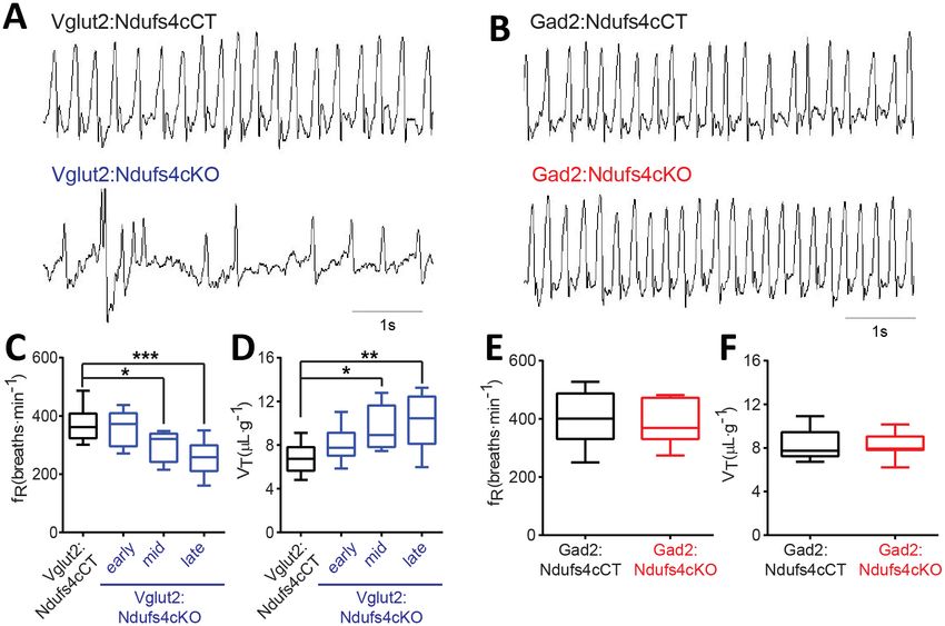
Research article                                                                                                          Neuroscience

Figure 1. Generation of two mouse lines lacking Ndufs4 selectively in Vglut2-expressing glutamatergic neurons or Gad2-expressing GABAergic
neurons. (A) Breeding strategy to obtain mice with conditional Ndufs4 deletion in glutamatergic neurons (Vglut2:Ndufs4cKO) or GABAergic neurons
(Gad2:Ndufs4cKO) and their respective controls (Vglut2:Ndufs4cCT, Gad2:Ndufs4cCT). (B) Kaplan-Meier survival curve for Vglut2:Ndufs4cKO (n = 96;
blue), Gad2:Ndufs4cKO (n = 40; red) and cCT mice (n = 50; black). (C–D) Body weight curves for Vglut2:Ndufs4cKO (n = 57 males, n = 48 females; blue)
and Vglut2:Ndufs4cCT (n = 52 males, n = 50 females; black). (E–F) Body weight curves for Gad2:Ndufs4cKO (n = 30 males, n = 32 females; red) and
Gad2:Ndufs4cCT (n = 41 males, n = 34 females; black). Data are presented as the mean ± SEM. Statistical analysis was performed using a two-way
ANOVA (***p
Research article                                                                                                 Neuroscience

                                    Neuronal identity defines distinct neuroinflammatory patterns after
                                    Ndufs4 deletion
                                    LS is characterized by symmetrical brain lesions and neuroinflammation in select nuclei, predomi-
                                    nantly brainstem and/or basal ganglia (Arii and Tanabe, 2000). Accordingly, late-stage Ndufs4-defi-
                                    cient mice present overt lesions in brainstem (namely vestibular nucleus (VN), cerebellar fastigial
                                    nucleus (FN) and inferior olive (IO)), olfactory bulb and basal ganglia (Quintana et al., 2010;
                                    Quintana et al., 2012; Chen et al., 2017a). To define the contribution of Ndufs4 deficiency in either
                                    excitatory or inhibitory neurons to the overall neuroinflammatory phenotype and identify the specific
                                    brain areas with exacerbated astroglial and microglial reactivity, we performed immunofluorescence
                                    analysis using anti-GFAP and anti-Iba1 antibodies on brain sections of Vglut2:Ndufs4cKO and Gad2:
                                    Ndufs4cKO mice, and their respective controls (Figure 2A–D). Analysis of these sections showed
                                    marked glial reactivity in VN, IO and FN (Figure 2A, C; Figure 2—figure supplement 1), accompa-
                                    nied by increased caspase eight activation in affected areas (such as the VN) in Vglut2:Ndufs4cKO
                                    mice (Figure 2E,F), recapitulating most of the neuroinflammatory profile described for the global
                                    Ndufs4KO mice (Quintana et al., 2010). In contrast, Gad2:Ndufs4cKO mice presented a more
                                    restricted glial reactivity pattern, including marked microglial and astroglial reactivity in primarily
                                    GABAergic nuclei such as the external globus pallidus (GPe) in the basal ganglia and the substantia
                                    nigra pars reticulata (SNr) (Figure 2B, D), without affecting neighboring non-GABAergic areas like
                                    the dopaminergic substantia nigra pars compacta (SNc) (Figure 2—figure supplement 2). Other
                                    prominently GABAergic areas such as the olfactory bulb also showed increased immunoreactivity for
                                    GFAP and Iba-1 in Gad2:Ndufs4cKO mice when compared to control mice (Figure 2B,D; Figure 2—
                                    figure supplement 1). In a percentage of Gad2:Ndufs4cKO mice, prominent microglial reactivity
                                    with Purkinje neuron loss (as assessed by calbindin staining) was also observed in the cerebellar ver-
                                    mis and flocculus (Figure 2—figure supplement 2). Normal Slc17a6 (Vglut2) or Gad2 transcript
                                    abundance was observed in the VN and OB of late-stage Vglut2:Ndufs4cKO or Gad2:Ndufs4cKO
                                    mice, respectively, suggesting preservation of Ndufs4-deficient neuronal populations (Figure 2G,H).
                                        The extensive neuroinflammation present in the brainstem of Vglut2:Ndufs4cKO mice, and its
                                    similarity to the phenotype of the global Ndufs4KO, allowed us to further define this inflammatory
                                    phenotype using whole-tissue transcriptional profiling. Gene expression analysis in the brainstem of
                                    late-stage (over P68) Vglut2:Ndufs4cKO mice using Illumina Beadchips (MouseRef-8 V2; Illumina)
                                    showed that differentially expressed (DE) mRNAs were, for the most part, upregulated in Vglut2:
                                    Ndufs4cKO mice (p 2) (Figure 2—figure supplement 2). Selected genes
                                    included transcripts directly involved in the regulation of the immune system such as chemokines
                                    and their receptors (Ccl3, Ccl4 and Cxcr3), toll-like receptors (Tlr2, Tlr7), complement proteins
                                    (C1qa, C1qb, C3 and C4b), surface antigens (Cd84, Cd86 and Ly9), and markers of the myeloid cell
                                    lineage (infiltrating macrophages and microglia) including Lyz, Lyz2, Lyzs and Slc11a1, among
                                    others.

                                    Table 1. Neurological signs observed in Vglut2:Ndufs4cKO mice according to the stage of disease.
                                                                                     Early                Mid                      Late
                                    Body tremor                                      +                    ++                       +++
                                    Motor alterations
                                     Decline in balance                              +                    ++                       +++
                                     Ataxia                                          -                    +/++                     +++
                                     Loss of motor coordination                      -                    +/++                     +++
                                     Inability of righting                           -                    +                        ++/+++
                                     Hindlimb clasping                               -                    +                        ++
                                    Respiratory abnormalities
                                     Breathing irregularities                        +                    ++                       +++

                                    -, absent; +, mild; ++, moderate; +++, severe.
                                    DOI: https://doi.org/10.7554/eLife.47163.007

Bolea et al. eLife 2019;8:e47163. DOI: https://doi.org/10.7554/eLife.47163                                                          5 of 26
Research article Neuroscience
Functional enrichment analysis of differentially
expressed mRNAs (1.4-fold or higher) using over-
representation analysis (ORA) (Wang et al.,
2017) showed that the top 10 most overrepre-
sented Gene Ontology (GO) categories (biologi-
cal process, non-redundant) were all related to
defense and immune responses, and also uncov-
ered components of the adaptive immune
response (GO:0002250; p=2.0177e-12,
FDR = 7.2033e-10) including ‘leucocyte mediated
immunity’ (GO:0002443; p=1.7533e-12,
Video 1. Breathing irregularities in a late-stage Vglut2:
FDR = 7.2033e-10) and ‘myeloid leukocyte activa-
Ndufs4cKO mice.
tion’ (GO:0002274; p=8.7458e-10,
DOI: https://doi.org/10.7554/eLife.47163.008
FDR = 9.0537e-8) (Figure 2—figure supplement
2). Therefore, to further characterize the immune
cell composition in these whole tissue gene
expression profiles, we applied recently developed deconvolution tools that use leucocyte gene
expression signature matrices to computationally infer the relative proportions of each immune cell
type in gene expression mixtures (Newman et al., 2015; Chen et al., 2017b) (Figure 2—figure sup-
plement 2). This analysis revealed a gene expression profile consistent with an increased proportion
of proinflammatory CD4 cells (Follicular cells, Th1, Th17 and Treg), dendritic cells, mast cells and
macrophages, and an underrepresentation of CD4 Th2 cells, CD8 cells and NK cells in the brainstem
of Vglut2:Ndufs4cKO mice compared to controls, showing that Ndufs4 deficiency in glutamatergic
neurons promotes a neuroinflammatory environment that involves a commitment to distinct proin-
flammatory TH cell lineages and a defined profile of tissue defense cells.
Vglut2:Ndufs4cKO but not Gad2:Ndufs4cKO mice present motor
alterations and respiratory deficits
Motor dysfunction is a prominent feature in LS and Ndufs4KO mice pathology (Sofou et al., 2014;
Quintana et al., 2010). To genetically define the neuronal cell types mediating this functional disor-
der, we assessed motor coordination in Vglut2:Ndufs4cKO mice, Gad2:Ndufs4cKO mice, and their
respective controls (Figure 3). Starting at P40, and concurring with the onset of clinical signs,
Vglut2:Ndufs4cKO mice showed impaired rotarod performance when compared to control litter-
mates (Figure 3A). While control mice maintained rotarod performance, Vglut2:Ndufs4cKO mice
failed to properly execute the task, presenting a progressive decline in motor coordination, in line
with the clinical phenotype. Conversely, and in agreement with the lack of apparent clinical signs, no
differences in rotarod performance were observed in Gad2:Ndufs4cKO mice compared to control lit-
termates (Figure 3B). Exposure to a novel environment revealed a hypoactive phenotype in Vglut2:
Ndufs4cKO mice as assessed by a reduction in the total distance traveled and the speed of explor-
atory movement in the open-field test (Figure 3C–E). The severity of this phenotype in Vglut2:
Ndufs4cKO mice positively correlated with age and disease stage (Figure 3—figure supplement 1).
In contrast, no significant differences were observed in either distance traveled or speed in the
open-field test between Gad2:Ndufs4cKO mice and their respective controls (Figure 3F–H). No dif-
ferences in the innervation of neuromuscular junctions were observed in the gastrocnemius muscle
of either Vglut2:Ndufs4cKO or Gad2:Ndufs4cKO mice (Figure 3I–K), suggesting a central origin of
the motor phenotype.
Respiratory abnormalities are frequently associated with disease mortality in LS patients and
global Ndufs4KO mice (Arii and Tanabe, 2000; Quintana et al., 2012; Gerards et al., 2016). To
provide insight into the genetic identity of the neurons responsible for this respiratory phenotype,
we assessed respiratory function by unrestrained whole-body plethysmography in awake Vglut2:
Ndufs4cKO and Gad2:Ndufs4cKO mice. Vglut2:Ndufs4cKO mice exhibited erratic plethysmographic
recordings (Figure 4A). In these mice, the frequency of respiration (fR) was markedly reduced at a
mid-stage (P40-P60) of the disease and worsened as disease progressed (Figure 4C). In addition,
significant differences were also seen in the volume of air inspired by the animal during one breath
(tidal volume, VT). VT was increased in Vglut2:Ndufs4cKO mice at a mid-stage of the disease and
became significantly larger than in Vglut2:Ndufs4cCT at late disease stages (Figure 4D). In contrast,
Bolea et al. eLife 2019;8:e47163. DOI: https://doi.org/10.7554/eLife.47163 6 of 26Research article Neuroscience Figure 2. Distinct histopathological pattern in Vglut2:Ndufs4cKO and Gad2:Ndufs4cKO mice. (A) Whole-mount composite sagittal sections showing GFAP (red) and Iba-1 (green) staining in late-stage Vglut2:Ndufs4cKO mice and their respective controls (Vglut2:Ndufs4cCT). Affected areas are indicated: Vestibular nucleus (VN), inferior olive (IO) and fastigial nucleus (FN). (B) Whole-mount composite sagittal sections showing GFAP (red) and Iba-1 (green) staining in Gad2:Ndufs4cKO mice and their respective controls (Gad2:Ndufs4cCT). Affected areas are indicated: External globus pallidus Figure 2 continued on next page Bolea et al. eLife 2019;8:e47163. DOI: https://doi.org/10.7554/eLife.47163 7 of 26
Research article                                                                                                             Neuroscience

Figure 2 continued
(GPe), the sustantia nigra pars reticulata (SNr) and the olfactory bulb (OB). (C) Close-up micrographs showing GFAP and Iba1 staining in the VN, IO,
and FN of late-stage Vglut2:Ndufs4cKO and Vglut2:Ndufs4cCT mice. (D) Close-up micrographs showing GFAP and Iba-1 staining in the GPe, SNr and
OB in Gad2:Ndufs4cKO and Gad2:Ndufs4cCT mice. Scale bar A-B:1000 mm. C-D: 50 mm. (E–F) Western blot analysis (E) and quantification (F) for active
caspase eight and b-Actin (loading control) levels in the vestibular nucleus of late-stage Vglut2:Ndufs4cKO mice (n = 3) and Vglut2:Ndufs4cCT mice
(n = 3). (G) Slc17a6 (Vglut2) transcript levels in the vestibular nucleus of late-stage Vglut2:Ndufs4cKO mice (n = 3) and Vglut2:Ndufs4cCT mice (n = 3).
(H) Gad2 transcript levels in the olfactory bulb of Gad2:Ndufs4cKO mice (n = 3) and Gad2:Ndufs4cCT mice (n = 3). Data are presented as the
mean ± SEM. Statistical analysis was performed using an unpaired t-Test (*p
Research article                                                                                                           Neuroscience

                                    Figure 3. Motor impairment in Vglut2:Ndufs4cKO mice. Latency to fall (seconds) in the Rotarod test for (A) Vglut2:
                                    Ndufs4cKO (n = 8) and Vglut2:Ndufs4cCT mice (n = 10), and (B) Gad2:Ndufs4cKO (n = 4) and Gad2:Ndufs4cCT
                                    mice (n = 10). Statistical analysis was performed using two-way ANOVA followed by Bonferroni post-test (*p
Research article Neuroscience
Figure 3 continued
open-field for (C–E) Vglut2:Ndufs4cKO (n = 36) and Vglut2:Ndufs4cCT mice (n = 70), and (F–H) Gad2:Ndufs4cKO
(n = 11) and Gad2:Ndufs4cCT mice (n = 16). Statistical analysis was performed using an unpaired t-Test
(***pResearch article Neuroscience Figure 5. Vglut2-expressing glutamatergic cells in the vestibular nucleus show reduced in vivo electrophysiological activity in Vglut2:Ndufs4cKO mice. (A) Representative identification of two different glutamatergic cells with raster plots (upper part) and peri-stimulus histograms (PSTH, lower part) using optogenetic stimulation. 0 represents the onset of optogenetic stimulation. Red line represents mean firing rate from the 500 ms before the stimulus plus three time the SD. Blue shading indicates period of laser stimulation. (B) Firing rate of Vglut2-expressing glutamatergic cells (n = 17 from Vglut2: Ndufs4cCT mice and n = 12 from Vglut2:Ndufs4cKO mice) during a 10 min session in an open-field. Reduced electrophysiological activity was observed in freely-moving late-stage Vglut2:Ndufs4cKO mice when compared to Vglut2:Ndufs4cCT mice regardless behavioral state (resting or active). Data are presented as the mean ± SEM. Statistical analysis was performed using two-way repeated measures ANOVA with Bonferroni post-test (***p
Research article                                                                                                       Neuroscience

Figure 6. Decreased body temperature in Vglut2:Ndufs4cKO and Gad2:Ndufs4cKO mice. (A) Telemetric body temperature measurements in Vglut2:
Ndufs4cKO (n = 4–8) and Vglut2:Ndufs4cCT mice (n = 7–10) at different stages of the disease. (B) Telemetric body temperature measurements in Gad2:
Ndufs4cKO (n = 7–11) and Gad2:Ndufs4cCT mice (n = 5–7) at different ages. Data are presented as the mean ± SEM. Statistical analysis was performed
using two-way ANOVA followed by Bonferroni post-test (*p
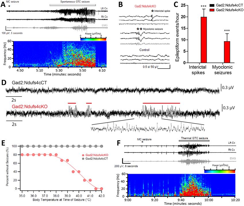
Research article                                                                                                         Neuroscience

Figure 7. Gad2:Ndufs4cKO mice present epileptic seizures. (A) EEG-EMG recordings and spectrogram analysis showing the frequency and power
density of a typical spontaneous seizure in a Gad2:Ndufs4cKO mouse. A preceding myoclonic seizure (MC) is marked with an asterisk. (B)
Representative EEG-EMG traces and (C) number of epileptiform events (interictal spikes and myoclonic seizures) in Gad2:Ndufs4cKO (n = 9) and control
(Gad2:Ndufs4cCT; n = 5) mice. Data are presented as the mean ± SEM. Statistical analysis was performed using two-way ANOVA (***p
Research article                                                                                                               Neuroscience

                                    Table 2. Clinical signs inNdufs4-LS patients, Ndufs4KO mice and conditional Vglut2:Ndufs4cKO and
                                    Gad2:Ndufs4cKO mice.
                                                               Ndufs4-LS           Ndufs4KO          Vglut2:Ndufs4cKO            Gad2:Ndufs4cKO
                                                               patients            mouse             mouse*                      mouse*
                                    Decreased lifespan         22/22               Yes               Yes                         Yes
                                    Feeding impairment         8/22                n.d               Yes                         Yes
                                    Reduced body weight        n.a.                Yes               Yes                         Yes
                                    Brainstem lesions          14/22               Yes               Yes                         No
                                    Basal ganglia lesions      9/22                Yes               No                          Yes
                                    Ataxia, motor alterations 22/22                Yes               Yes                         No
                                    Growth retardation         11/22               Yes               Yes                         Yes
                                    Hypotonia                  22/22               Yes               Yes                         No
                                    Respiratory                22/22               Yes               Yes                         No
                                    abnormalities
                                    Increased sensitivity to   n.a.                Yes               Yes†                        No†
                                    VAs
                                    Seizures                   4/22                Yes               No                          Yes
                                    Hypothermia                n.a.                Yes               Yes‡                        Yes

                                    n.d, not determined; n.a, not available. *Reported in this study. †Reported in Zimin et al. (2016). ‡Only in late-stage
                                    mice. VAs: volatile anesthetics. Adapted from Quintana et al. (2012).
                                    DOI: https://doi.org/10.7554/eLife.47163.021

                                    equivalent to controls, indicating no overt contribution of this cell type to the pathology observed in
                                    Ndufs4KO mice. In contrast, Vglut2:Ndufs4cKO and Gad2:Ndufs4cKO mice had reduced lifespan
                                    and body weight, which was accompanied by a decrease in food intake, which are common clinical
                                    signs that appear in Leigh Syndrome patients (Rahman et al., 1996; Smeitink, 2003). Recent reports
                                    have shown that neuronal cell-specific NDUFS4 knock down in Drosophila also leads to severe feed-
                                    ing abnormalities and premature death (Foriel et al., 2018). Our results indicate a conserved role
                                    for neurons in the onset and progression of the pathological condition of global Ndufs4 deficiency
                                    and reveal that both glutamatergic and GABAergic systems contribute to the growth and lethality
                                    phenotype. Noteworthy, the slight reduction in phenotype severity of conditional neuronal Ndufs4
                                    deficiency, with relative neuronal preservation, compared to the global Ndufs4KO mice suggests a
                                    concerted contribution of different cell types. In this regard, our previous work has shown the impor-
                                    tance of neuron-astrocyte crosstalk in the development of neurodegeneration in the context of mito-
                                    chondrial disease (Liu et al., 2015). However, astrocytic Ndufs4 deficiency is not sufficient to
                                    recapitulate the phenotype of global Ndufs4KO mice (Ramadasan-Nair et al., 2019), underscoring
                                    the central role of neurons in the disease. Even so, recent studies have shown enhanced neuronal
                                    survival in global Ndufs4KO after disruption of hepatic S6K1 (Ito et al., 2017). Hence, the role of cell
                                    and tissue non-autonomous effects on disease progression have to be taken into account to fully
                                    understand the phenotype of neuronal-specific conditional Ndufs4KO mice.
                                       Apart from the premature death and feeding deficits, Vglut2:Ndufs4cKO and Gad2:Ndufs4cKO
                                    mice present two markedly distinct clinical entities (summarized in Table 2). With Vglut2:Ndufs4cKO
                                    mice, the lethality was associated with severe motor and respiratory alterations, whereas with Gad2:
                                    Ndufs4cKO mice, sudden unexpected death was associated with epilepsy (SUDEP) (Abdel-
                                    Mannan et al., 2019; Manolis et al., 2019; DeGiorgio et al., 2019) with no overt clinical alteration
                                    beyond the weight loss.
                                       Histologically, Vglut2:Ndufs4cKO mice present with prominent neuroinflammation and lesions in
                                    areas of the brainstem such as the VN, IO and the cerebellar FN, reminiscent of the pathology found
                                    in Ndufs4KO mice (Quintana et al., 2010). We have identified a critical role of brainstem lesions in
                                    the development of fatal breathing alterations observed in the Ndufs4KO mice (Quintana et al.,
                                    2012), in agreement with human LS patients (Arii and Tanabe, 2000). Glutamatergic signaling in the
                                    brainstem has been shown to regulate breathing (Whitney et al., 2000). In addition, VN glutamater-
                                    gic neurons have been suggested to modulate respiratory responses (Xu et al., 2002). Furthermore,

Bolea et al. eLife 2019;8:e47163. DOI: https://doi.org/10.7554/eLife.47163                                                                        14 of 26
Research article Neuroscience
the Pre-Bötzinger (PreBotC) complex, a key respiratory center, is composed of glutamatergic
neurons (Stornetta et al., 2003) and receives extensive glutamatergic inputs (Bochorishvili et al.,
2012). We have shown that Ndufs4KO mice present intrinsic PreBotC alterations and that vestibular
projections to the PreBotC are necessary for maintaining respiration (Quintana et al., 2012). Our
electrophysiological recordings in Vglut2-expressing VN neurons from Vglut2:Ndufs4cKO mice show
relatively normal basal firing rate but fail to increase spiking in response to locomotor activity. Neu-
ronal firing is a highly energy-requiring process, mostly dependent in mitochondrial function
(Harris et al., 2012), especially in glutamatergic synapses (Juge et al., 2010; Zimin et al., 2016).
Hence, our results suggest that glutamatergic VN neurons are unable to achieve the energy require-
ments of increased firing rates. Thus, it is likely that failure of glutamatergic VN projections to the
PreBotC allowing appropriate responses to physiological needs underlies the breathing deficits
observed in Vglut2:Ndufs4cKO mice.
Development of brainstem lesions correlate with motor deficits in animals constitutively lacking
Ndufs4 (Quintana et al., 2010). Accordingly, strategies that improve motor symptoms in Ndufs4KO
mice, such as AAV-based gene therapy (Quintana et al., 2012; Di Meo et al., 2017), rapamycin
administration (Johnson et al., 2013), or hypoxia (Jain et al., 2016; Ferrari et al., 2017) cause a
marked reduction in brainstem lesions. Here, we have identified a critical role for Vglut2-expressing
glutamatergic neurons, in the brainstem and cerebellum, in the development of the motor deficits
observed after Ndufs4 deficiency. In keeping with this, conditional deletion of Ndufs4 in dopaminer-
gic neurons does not cause cell loss (Kim et al., 2015) or motor deficits (Choi et al., 2017). How-
ever, other areas, such as the striatum, may participate in the delayed, mild, progressive motor
dysfunction observed in LS patients (Chen et al., 2017a; Di Meo et al., 2017).
Our gene expression analysis in brainstem of Vglut2:Ndufs4cKO mice has enabled the generation
of an in-depth profile of the transcriptomic landscape in an affected brain area after Ndufs4 defi-
ciency. This analysis revealed a prominent increase in inflammatory mediators in the affected tissue.
However, anti-inflammatory or immunotherapeutic approaches have been mostly ineffective as treat-
ments for LS (Johnson et al., 2013; Finsterer and Zarrouk-Mahjoub, 2017) with only a few success-
ful cases reported (Chuquilin et al., 2016). Our deconvolution data show a marked infiltration of
distinct leucocyte populations, underscoring the complex cellular milieu elicited by mitochondrial
dysfunction that may underlie the failure of global anti-inflammatory approaches. Delineation of the
immune cells recruited to the brain lesions may lead to novel therapeutic approaches tailored for LS.
The Gad2-expressing GABAergic neurons do not participate in the appearance of respiratory or
motor deficits in Ndufs4 deficiency. However, they are critical for body temperature control and the
onset and development of the fatal epileptic seizures, features that are observed in both global
Ndufs4KO mice (Quintana et al., 2010) and LS patients (Finsterer, 2008; Koenig, 2008;
Finsterer and Zarrouk Mahjoub, 2012). Lack of Ndufs4 in Gad2-expressing neurons leads to the
appearance of neuroinflammation in the basal ganglia nuclei such as the GPe and SNr, in agreement
with the increased vulnerability of basal ganglia neurons to mitochondrial dysfunction
(Gubellini et al., 2010). Furthermore, we show that electrophysiological alterations in the GPe neu-
rons predate cortical epileptic events, suggesting a primary role of the basal ganglia circuitry in the
development of epileptic seizures in Gad2:Ndufs4cKO mice. Basal ganglia are involved in
epilepsy (Rektor et al., 2012; Badawy et al., 2013; Vuong and Devergnas, 2018), likely by acting
as an inhibitory input to cortical seizure spread via feedback mechanisms (Rektor et al., 2012).
Hence, we hypothesize that basal ganglia inhibitory network is affected in Gad2:Ndufs4cKO mice,
being unable to control the activity of cortical excitatory neurons, thus leading to epilepsy. Gad2:
Ndufs4cKO mice are resistant to different antiepileptic approaches, such as the widely-used antiepi-
leptic drugs carbamazepine, perampanel, and levetiracetam. Although earlier administration of
these drugs may have led to a better antiepileptic outcome, epilepsy-induced death in mitochondrial
disease patients is usually linked to refractory epileptic seizures (Finsterer and Zarrouk Mahjoub,
2012). Hence, Gad2:Ndufs4cKO mice may represent an excellent model to study epileptic mecha-
nisms in LS, a much needed area of research, especially considering that most commonly used anti-
epileptic drugs may promote mitochondrial toxicity (Finsterer, 2017).
As described, both LS patients (Finsterer, 2008) and NDUFS4-LS patients (Ortigoza-
Escobar et al., 2016) present predominant basal ganglia and brainstem affectation. Accordingly,
basal ganglia and brainstem lesions are prominent features in global Ndufs4KO (References:
Quintana et al., 2010; Quintana et al., 2012; Table 2), and GABAergic and glutamatergic
Bolea et al. eLife 2019;8:e47163. DOI: https://doi.org/10.7554/eLife.47163 15 of 26Research article Neuroscience
conditional Ndufs4KO mice, respectively. However, alterations in other areas such as thalamus, cere-
bellum and spinal cord are also frequently observed, contributing to the clinical complexity of the
pathology (Arii and Tanabe, 2000; Lake et al., 2015). In line with our previous studies
(Quintana et al., 2010; Quintana et al., 2012), here we show the glutamatergic origin of cerebellar
and spinal cord alterations, probably contributing motor deficits observed in LS and Ndufs4KO. Clin-
ically, LS patients commonly present hypothermia and failure to thrive (Finsterer, 2008), which are
recapitulated in global Ndufs4KO mice (Quintana et al., 2010). Our work shows reduced body
weight and hypophagia in both Vglut2:Ndufs4cKO and Gad2:Ndufs4cKO, while hypothermia is
mainly restricted to the latter. Central control of food intake and thermoregulation heavily rely on
glutamatergic and GABAergic hypothalamic neuronal populations (Tan and Knight, 2018;
Sternson and Eiselt, 2017; Zhao et al., 2017). Hence, our results indicate, that even in the absence
of overt neuroinflammation, neuronal Ndusf4 deficiency may lead to hypothalamic impairment, as
observed in LS patients (Zinka et al., 2010).
One remaining question is the characterization of the underlying molecular mechanisms leading
to the specific vulnerability of defined neuronal populations to Ndufs4 deficiency. In this regard,
increased oxidative stress is one of the hallmarks of the phenotype (Quintana et al., 2010). Accord-
ingly, antioxidant treatments have proven moderate effectivity in the global Ndufs4KO mice
(Liu et al., 2015; de Haas et al., 2017). Initiation of extrinsic apoptotic cascades has also been
found in Ndufs4KO mice (Finsterer and Zarrouk-Mahjoub, 2017, and this work), even though EM
imaging demonstrated mostly necrotic death in affected brain regions of Ndufs4KO mice
(El Sabbagh et al., 2010). In this regard, the remarkable immune cell infiltration described in this
work may contribute to the initiation of these cascades. Finally, different studies have pointed at
metabolic dysregulation as a potential contributor to the pathology. In this regard, mTOR inhibition
or hypoxic conditions modify glycolytic levels, leading to clinical sign amelioration and extended life-
span in Ndufs4KO mice (Johnson et al., 2013; Jain et al., 2016).
In conclusion, we provide new insights on the genetic identity of affected neuronal populations in
LS by dissecting the associated cell type-specific molecular, biochemical, clinical and behavioral fea-
tures in a model of LS. Our work highlights the importance of addressing mitochondrial disease at
the cell type-specific level. The advent of new tools to assess transcriptomic and biochemical
changes at this level of resolution (Sanz et al., 2009; Bayraktar et al., 2019) bodes well for more
progress. Hence, our work broadens current understanding of the etiology of LS and paves the way
for future studies at the cell type-specific level to unravel the molecular determinants of neuronal
pathology in LS.
Materials and methods
Key resources table
Reagent type (species) Additional
or resource Designation Source or reference Identifiers information
Genetic reagent Slc17a6Cre (BAC-Vglut2::Cre) Borgius L, et al. MGI:4881727 Tg(Slc17a6-icre)1Oki
(M. Musculus) Mol Cell Neurosci. 2010;
45(3):245–57
Genetic reagent Gad2Cre/+ Taniguchi H, et al. MGI:4418713 B6J.Cg-Gad2tm2
(M. Musculus) (Gad2-IRES-Cre) Neuron. 2011; (cre)Zjh/MwarJ
71(6):995–1013. JAXs Stock No: 028867
Genetic reagent ChatCre/+ Rossi J, et al. MGI:6121363 B6.129S-Chattm1(cre)
(M. Musculus) (Chat-IRES-Cre) Cell Metab. 2011; Lowl/MwarJ
13(2):195–204 JAXs Stock No: 031661
Genetic reagent Ndufs4D/+ Kruse SE, et al. MGI:5614215 B6.129S4-Ndufs4tm1.
(M. Musculus) Cell Metab. 2008; 1Rpa/J
7 (4):312–20 JAXs Stock No: 027058
Genetic reagent Ndufs4lox/lox Kruse SE, et al. MGI:5613135 B6.129S4-Ndufs4tm1
(M. Musculus) Cell Metab. 2008; Rpa/J
7 (4):312–20 JAXs Stock No: 026963
Bolea et al. eLife 2019;8:e47163. DOI: https://doi.org/10.7554/eLife.47163 16 of 26Research article Neuroscience
Study approval
All experiments were conducted following the recommendations in the Guide for the Care and Use
of Laboratory Animals and were approved by the Animal Care and Use Committee of the Seattle
Children´s Research Institute and the Universitat Autònoma de Barcelona.
Animal husbandry
Mice were maintained with Teklad Global rodent diet No. 2014S (HSD Teklad Inc, Madison, Wis.)
and water available ad libitum in a vivarium with a 12 hr light/dark cycle at 22˚C.
Mouse genetics
The following mouse lines were used in this study: Slc17a6Cre (BAC-Vglut2::Cre) (Borgius et al.,
2010) mice were generated by Ole Kiehn. Gad2Cre/+ (Gad2-IRES-Cre) (Taniguchi et al., 2011) and
ChatCre/+ (Chat-IRES-Cre) (Rossi et al., 2011) mice were obtained from The Jackson Laboratory
(Stock No: 028867 and 031661, respectively) (Bar Harbor, ME). Ndufs4lox/lox and Ndufs4D/+ were pre-
viously generated by our group (Quintana et al., 2010; Kruse et al., 2008). Male and female mice
of different ages were used in this study. Sex and age of the animals are described in the figure
legends. All mice were on a C57BL/6J background after backcrossing for at least 10 generations.
Mice with conditional deletion of Ndufs4 in Vglut2-expressing glutamatergic neurons (Slc17a6Cre,
Ndufs4D/lox or Vglut2:Ndufs4cKO) were generated by crossing mice with one Ndufs4 allele deleted
and expressing a codon-improved Cre recombinase (iCre) under the Slc17a6 promoter (Slc17a6Cre,
Ndufs4D/+) to mice with two floxed Ndufs4 alleles (Ndufs4lox/lox). Mice with conditional deletion of
Ndusf4 in Gad2-expressing GABAergic neurons (Gad2Cre/+, Ndufs4lox/lox or Gad2:Ndufs4cKO) were
obtained by crossing mice with one floxed Ndufs4 allele and expressing Cre recombinase under the
control of the Gad2 promoter (Gad2Cre/+, Ndufs4lox/+) to mice carrying two floxed Ndufs4 alleles
(Ndufs4lox/lox). Similarly, mice with conditional Ndufs4 deletion in ChAT-expressing cholinergic neu-
rons (ChatCre/+, Ndufs4lox/lox or ChAT:Ndufs4cKO) were obtained by crossing mice carrying one
floxed Ndufs4 allele and expressing Cre recombinase driven by the ChAT promoter (ChatCre/+,
Ndufs4lox/+ mice) to Ndufs4lox/lox mice. Littermate controls were Slc17a6Cre, Ndufs4lox/+ (Vglut2:
Ndufs4cCT); Gad2Cre/+, Ndufs4lox/+ (Gad2:Ndufs4cCT) and ChatCre/+Ndufs4lox/+ (ChAT:Ndufs4cCT)
mice. In all cases, genotype of the offspring and absence of ectopic recombination (i.e. presence of
recombination bands in tail DNA samples) was determined by PCR analysis. Primer sequences have
been described (Kruse et al., 2008).
Clinical evaluation
Vglut2:Ndufs4cKO and Gad2:Ndufs4cKO mice were examined every other day for clinical signs
resulting from cell type-specific Ndufs4 inactivation. Physiological (body weight) and behavioral
(locomotor activity, motor coordination, gait/postural alterations) parameters were evaluated in
more than 50 animals for each mouse line and were grouped into the following categories based on
visual observation: ‘+++” severe manifestation of the clinical sign, ‘++” moderate manifestation of
the clinical sign, ‘+” mild clinical sign, “- “absence of clinical sign. Mice were humanely euthanized
after losing 20% of their peak body weight. Only Vglut2:Ndufs4cKO mice presented the overt and
progressive clinical signs. Albeit the presence of individual variability in the development of the dis-
ease, early stage was defined in the range P20-P40, mid-stage between P40-P60, and late stage at
ages over P60 in Vglut2:Ndufs4cKO.
Food intake analysis
Food consumption was recorded from 7 to 11 weeks of age using a Physiocage system (Panlab,
Spain). Data at 8 weeks of age (right before the median survival value) are presented, including
enough individuals to ensure sufficient statistical power.
Behavioral assays
Rotarod test
A standard rotarod device (Rotarod, San Diego Instruments, USA) was used to assess motor coordi-
nation and global physical condition of animals. Mice were placed on the spindle, which linearly
accelerated from 4 to 40 r.p.m and increased 2 r.p.m. every 10 s. Each mouse received five trials per
Bolea et al. eLife 2019;8:e47163. DOI: https://doi.org/10.7554/eLife.47163 17 of 26Research article Neuroscience
day over 3 days with a 5 min rest period between trials. The trial ended when the mouse fell off the
spindle or after 3 min (cut-off time).
Open-field
Mice were placed in the open-field arena (560 [W]365 [D]400 [H] mm) and allowed to move freely
for 10 min. Locomotor activity of mice was next monitored and total distance traveled (m) and veloc-
ity (cm/s) measured using the EthoVision tracking software (Noldus).
Whole-body plethysmography
Ventilatory function was assessed by whole–body plethysmography (EMMS, England, UK) under
unrestrained conditions. The system was calibrated to 1 ml volume. An acclimation period of 45 min
was allowed for mice adaptation to the chamber, followed by a 15 min experimental period. For
Vglut2:Ndufs4cKO mice, plethysmography recordings were performed at different stages of the dis-
ease according to the clinical examination, and compared to littermate controls. Studies were also
conducted on Gad2:Ndufs4cKO mice between 50–60 days of age and compared to corresponding
controls. Respiratory frequency (FR; breathsminute 1) and tidal volume normalized per body weight
(VT; mLg 1) were measured.
Tissue preparation
For immunofluorescence, mouse brains were collected and fixed overnight in 4% paraformaldehyde
(PFA) in PBS (pH 7.4). Subsequently, brains were cryoprotected in a PBS solution containing 30%
sucrose and frozen in dry ice. Frozen brains were embedded in OCT, sectioned at 30 mm in a cryo-
stat and rinsed in PBS prior to staining. For western blot analysis, brain areas (olfactory bulb, thala-
mus, spinal cord and globus pallidus) were dissected according to the Paxinos mouse brain
atlas (Paxinos and Frank, 2013) and flash-frozen in liquid nitrogen before homogenization.
Immunofluorescence
Tissue sections were rinsed in PBS-0.2% Triton X-100 (PBST) solution. Non-specific binding was
blocked with 10% normal donkey serum (NDS) in a PBST solution for 60 min at room temperature,
followed by overnight incubation at 4˚C with primary antibodies diluted in 1% NDS-PBST (1:2000 for
mouse anti-GFAP, Sigma; 1:1000 for chicken anti-GFAP, Abcam; 1:1000 for anti-Iba-1, Wako; 1:1000
for anti-TH, Millipore). Sections were then washed in PBST and incubated for 1 hr at room tempera-
ture with the corresponding Cy- (1:200, Jackson Immunoresearch) or Alexa Fluor-conjugated sec-
ondary antibodies (1:500, Thermo Fisher Scientific) in 1% NDS-PBST. Sections were finally washed in
PBS and rinsed in water before mounting onto slides with Fluoromount G (Electron Microscopy Sci-
ences) for microscopic analysis.
Immunofluorescence labeling of neuromuscular junctions
Gastrocnemius muscles were sectioned in 60 mm longitudinal sections, collected in 24-well plates in
sequential series of 4 slices per well in antifreezing solution. Sections were then blocked with PBS-
0.3%Triton-5%Normal Donkey serum and incubated 48 hr at 4˚C with primary antibodies anti-synap-
tophysin (1:500; AB130436, Abcam, UK) and anti-neurofilament 200 (NF200, 1:1000; AB5539, Milli-
pore, USA). After washes, sections were incubated overnight with Alexa 594-conjugated secondary
antibody (1:200; A11042, Invitrogen, USA) and Alexa 488 conjugated alfa-bungarotoxin (1:200;
B13422, Life Technologies, USA). Slides with the sections were then mounted in Fluoromount-G
(Southern Biotech, USA). Confocal images were captured with a scanning confocal microscope (LSM
700 Axio Observer, Carl Zeiss 40x/1.3 Oil DIC M27, Germany). Maximum projections images shown
in this study were created from 1.5 mm z projections. For neuromuscular junctions analysis, the pro-
portion of fully occupied endplates was determined by classifying each endplate as fully innervated
(when presynaptic terminals overly the endplate), partially innervated (when presynaptic terminals
were not clearly within the endplate) or vacant (no presynaptic label in contact with the endplate).
At least 3–4 fields with more than 80 endplates were analyzed per each muscle.
Bolea et al. eLife 2019;8:e47163. DOI: https://doi.org/10.7554/eLife.47163 18 of 26Research article Neuroscience
Western blotting
Brain tissue samples were homogenized in iced-cold RIPA buffer (Santa Cruz Biotechnology) and
protein concentration determined by the BCA assay (Thermo Fisher Scientific). Thereafter, 20 mg of
protein lysates were heat-denatured in Laemmli sample buffer (Bio-Rad Laboratories, Inc), subjected
to 4–20% gradient SDS-PAGE and transferred to nitrocellulose membranes (Bio-Rad Laboratories,
Inc). Membranes were then blocked for 1 hr with 5% (w/v) dried skimmed milk in Tris-buffered saline
containing 0.1% Tween-20 (TBS-T) and incubated overnight at 4˚C with primary antibodies against
NDUFS4 (Abcam, mouse, 1:500), NSE (Dako, mouse, 1:1,000), GFAP (Sigma, mouse, 1:50,000), Iba1
(Wako, rabbit, 1:10,000), Active (cleaved) caspase 8 (Cell Signaling Technologies, 1:1000), b-actin
(Sigma, mouse, 1:20,000) or GAPDH (GeneTex, mouse, 1:40,000). After incubation with the corre-
sponding HRP-conjugated secondary antibodies (1:10,000; Jackson ImmunoResearch), membranes
were washed in TBS-T and developed using an enhanced chemiluminescence (ECL) detection system
(Pierce). Bands were quantified using Image J software (National Institutes of Health, USA).
Whole-genome gene expression (WGGEX) analysis
For WGGEX analysis, 150 ng of total RNA extracted from the brainstem of late-stage (over P68)
Vglut2:Ndufs4cKO (n = 4) and Vglut2:Ndufs4cCT mice (n = 4) was amplified and biotin-labeled using
the Illumina TotalPrep RNA Amplification kit (Ambion). 750 ng of the labeled cRNA was hybridized
to MouseRef-8 v2 expression beadchips (Illumina) for 16 hr before washing and analyzing according
to the manufacturer’s directions. Signal was detected using a BeadArray Reader (Illumina), and data
were analyzed for differential expression using the GenomeStudio data analysis software (Illumina).
Average normalization, the Illumina custom error model, and multiple testing corrections using the
Benjamini and Hochberg false discovery rate were applied to the analysis. Only transcripts with a dif-
ferential score of >13 (pResearch article Neuroscience
described (Oakley et al., 2009), body temperature was increased by 0.5˚C every 2 min until seizure
occurred or a 42˚C temperature was reached. Mice were immediately cooled using a small fan.
Electrophysiological studies
Surgery
Vglut2:Ndufs4cKO and Gad2:Ndufs4cKO mice and their respective controls (30 to 40 days old) were
anesthetized with 1.5% isoflurane and implanted with a homemade implant in either the vestibular
nuclei (AP: 6.0; ML: 1.0; DV: 4.00) or the lateral globus pallidus (AP: 0.46; ML: 1.95; DV:
4.00), according to Paxinos (Paxinos and Frank, 2013). Briefly, a small craniotomy window was
made above the desired recording site and a 4-tetrode bundle was lowered into the brain until its
destination at 0.1 mm/sec (Robot Stereotaxic, Neurostar, Germany). As ground, a stainless steel
wire (0.075 mm diameter, Advent Research Material Ltd., England) was placed between skull and
dura over the cerebellum or visual cortex. Then the entire implant was attached and secured to the
animal head with dental cement. After surgery, animal welfare and body weight were documented
daily and mice allowed to recover for 10–14 days. In Vglut2:Ndufs4cKO mice and their respective
controls, a viral vector expressing the light-sensitive cation channel Channelrodopsin-2 in a Cre-
dependent manner (AAV-DIO-ChR2) was delivered into the VN prior to implantation of the tetrode
to identify glutamatergic Vglut2-expressing cells after blue light (473 nm) stimulation.
Electrophysiological data acquisition
To explore presence of seizures, the electrophysiological activity was recorded from Gad2:
Ndufs4cCT and Gad2:Ndufs4cKO freely-moving mice. After the recovery period, animals were
placed on a daily basis in an open-field arena and both local field potentials (LFPs) and extracellular
single-unit activity were recorded until the death of the animal. In Vglut2:Ndufs4cCT and Vglut2:
Ndufs4cKO mice, a tetrode bundle was attached along a fiber optic (Ø200 mm Core, 0.22 NA,
FG200UCC, Thorlabs, USA) to deliver light and record neuronal cells at the same time. Again, all ani-
mals were daily recorded in an open-field until death.
For all experiments, local field potential activity was amplified, A-D converted and sampled at 1
KHz and bandpass filtered at 0.1 to 250 Hz (DigitalLynx SX and Cheetah Data Acquisition System,
Neuralynx, USA). Continuous spike signals were also recorded, amplified, band-pass filtered (300 Hz
to 8 kHz) and sampled at 32 kHz. Optogenetic stimulations (ranging from 50 to 200 ms blue light
stimuli, 473 nm, delivered frequency from 1 to 40 Hz) were delivered through a blue-emitting diode
laser (473 nm DPSS Laser System, Laserglow Technologies, CA).
Electrophysiological data analysis
For the presence of seizures, data were analyzed in Spike2 (Cambridge Electronic Design Limited,
UK) for visual inspection and report presence of epileptic events. Offline single-unit spike sorting
was performed with Offline Sorter software (Offline Sorter, Plexon Inc, USA). Briefly, for each chan-
nel, a specific manual threshold was defined and all events bypassing this threshold were assumed
to be an action potential. After an overall waveform shape and three principal component analysis
(PCA) inspection, all spikes were sorted with an automatic K-mean algorithm to separate clusters of
cells. After sorting, a final inspection of ISI histogram and sorting statistics (MANOVA F statistics, J3
and the Davies-Bouldin validity index) was performed to ensure the best single-unit clustering. In
Vglut2:Ndufs4cCT and Vglut2:Ndufs4cKO mice, to identify glutamatergic neurons from all sorted
cells, response to optogenetic stimulations were plotted. For all stimulation trains, during a baseline
activity corresponding to 500 ms before stimulation, the mean number of spikes per bins was calcu-
lated. Then, a single cell was identified as glutamatergic only if, during optogenetic stimulation,
there were bins expressing a number of spikes superior to the baseline mean plus three times the
standard deviation of the baseline mean. Finally, after analysis of spiking activity (Matlab, Math-
Works, USA), cells with a coefficient of variation of interspike interval (CV.ISI) over three were
exclude from the data set.
Body core temperature measurement
The baseline temperature was acquired as follows: As for thermal seizure induction (Oakley et al.,
2009), a T-type implantable rectal probe was placed and taped to the tail with a lab tape. The probe
Bolea et al. eLife 2019;8:e47163. DOI: https://doi.org/10.7554/eLife.47163 20 of 26Research article Neuroscience
was connected to a temperature monitoring device (T-pod) and the resting body core temperature
was monitored in unrestrained mice using a PowerLab 8/35 data acquisition unit and Labchart 7.3.3
software (AD Instruments, Colorado Spring, Co) for 10 min. The telemetric data are used for long-
term monitoring of temperature and its oscillation during circadian rhythm and sleep. Note that
baseline data obtained via the two methods are consistent.
Drug treatments
Levetiracetam (Keppra parental formulation), perampanel (Clinisciences) and carbamazepine (Sigma-
Aldrich) antiepileptic drugs were injected intraperitoneally in Gad2:Ndufs4cKO mice daily, starting
at P40. Levetiracetam and perampanel were dissolved in saline solution and injected at a dose of 60
mg/kg and 0.75 mg/kg, respectively. Carbamazepine was slowly diluted in PEG300, dissolved in
saline, and injected at 40 mg/kg. A control group for each drug was obtained by injecting each
respective vehicle to Gad2:Ndufs4cKO mice.
qRT-PCR analysis
qRT-PCR assays were performed as described (Sanz et al., 2015). Briefly, equal amounts of RNA
were assayed using the Power SYBR green RNA-to-Ct 1-Step Master Mix (Applied Biosystems) or
the TaqMan RNA-to-Ct 1-Step Master Mix (Applied Biosystems), depending on the system used
(SYBR or Taqman). Relative expression values were obtained using the standard curve method and
normalized to Actb levels. Amplification efficiencies were calculated using the AriaMx software (Agi-
lent) and were within accepted parameters (80–120%). Gad2 mRNA was determined using a specific
Taqman assay (Mm00484623_m1; Applied Biosystems), and sequences for the different primer sets
used in SYBR assays (Slc17a6 and Actb) were obtained from Primerbank (Spandidos et al., 2010).
Statistics
Data are shown as the mean ± SEM. GraphPad Prism v5.0 software was used for statistical analyses.
Appropriate tests were selected depending on the experimental design as stated in the figure
legends. Statistical significance, when reached (pYou can also read