The Bidirectional Relationship Between Sleep and Inflammation Links Traumatic Brain Injury and Alzheimer's Disease - Frontiers
←
→
Page content transcription
If your browser does not render page correctly, please read the page content below
REVIEW
                                                                                                                                            published: 25 August 2020
                                                                                                                                        doi: 10.3389/fnins.2020.00894

                                            The Bidirectional Relationship
                                            Between Sleep and Inflammation
                                            Links Traumatic Brain Injury and
                                            Alzheimer’s Disease
                                            Tabitha R. F. Green 1,2 , J. Bryce Ortiz 1,2 , Sue Wonnacott 3 , Robert J. Williams 3 and
                                            Rachel K. Rowe 1,2,4*
                                            1
                                             BARROW Neurological Institute at Phoenix Children’s Hospital, Phoenix, AZ, United States, 2 Department of Child Health,
                                            University of Arizona College of Medicine – Phoenix, Phoenix, AZ, United States, 3 Department of Biology and Biochemistry,
                                            University of Bath, Bath, United Kingdom, 4 Phoenix Veteran Affairs Health Care System, Phoenix, AZ, United States

                                            Traumatic brain injury (TBI) and Alzheimer’s disease (AD) are diseases during which the
                                            fine-tuned autoregulation of the brain is lost. Despite the stark contrast in their causal
                                            mechanisms, both TBI and AD are conditions which elicit a neuroinflammatory response
                                            that is coupled with physical, cognitive, and affective symptoms. One commonly
                            Edited by:      reported symptom in both TBI and AD patients is disturbed sleep. Sleep is regulated by
                    Marilyn J. Duncan,      circadian and homeostatic processes such that pathological inflammation may disrupt
 University of Kentucky, United States
                                            the chemical signaling required to maintain a healthy sleep profile. In this way, immune
                        Reviewed by:
                                            system activation can influence sleep physiology. Conversely, sleep disturbances can
                         Mark R. Opp,
       University of Colorado Boulder,      exacerbate symptoms or increase the risk of inflammatory/neurodegenerative diseases.
                         United States      Both TBI and AD are worsened by a chronic pro-inflammatory microenvironment
                     Radhika Basheer,
             Harvard Medical School,        which exacerbates symptoms and worsens clinical outcome. Herein, a positive
                         United States      feedback loop of chronic inflammation and sleep disturbances is initiated. In this
                 *Correspondence:           review, the bidirectional relationship between sleep disturbances and inflammation is
                    Rachel K. Rowe
                                            discussed, where chronic inflammation associated with TBI and AD can lead to sleep
           rkro222@email.arizona.edu
                                            disturbances and exacerbated neuropathology. The role of microglia and cytokines in
                   Specialty section:       sleep disturbances associated with these diseases is highlighted. The proposed sleep
         This article was submitted to
       Sleep and Circadian Rhythms,
                                            and inflammation-mediated link between TBI and AD presents an opportunity for a
               a section of the journal     multifaceted approach to clinical intervention.
            Frontiers in Neuroscience
                                            Keywords: sleep, inflammation, traumatic brain injury, concussion, Alzheimer’s disease, cytokines, microglia,
           Received: 06 April 2020          neurodegeneration
           Accepted: 31 July 2020
         Published: 25 August 2020
                             Citation:      INTRODUCTION
                   Green TRF, Ortiz JB,
        Wonnacott S, Williams RJ and        In diseased states affecting the central nervous system (CNS), the fine-tuned autoregulation of
    Rowe RK (2020) The Bidirectional
                                            the brain is lost, which contributes to the affective and cognitive symptoms seen in psychiatric,
           Relationship Between Sleep
    and Inflammation Links Traumatic
                                            neurological, and mental health disorders (Charrier et al., 2017). Alzheimer’s disease (AD) is a
 Brain Injury and Alzheimer’s Disease.      neurodegenerative disorder that impacts approximately 1 in 10 people over the age of 65 and 1 in 3
              Front. Neurosci. 14:894.      people over the age of 85 (Newcombe et al., 2018). AD has a long prodromal phase and progresses
       doi: 10.3389/fnins.2020.00894        slowly, across a span of typically around 10 years. In contrast, traumatic brain injury (TBI) is a rapid

Frontiers in Neuroscience | www.frontiersin.org                                    1                                             August 2020 | Volume 14 | Article 894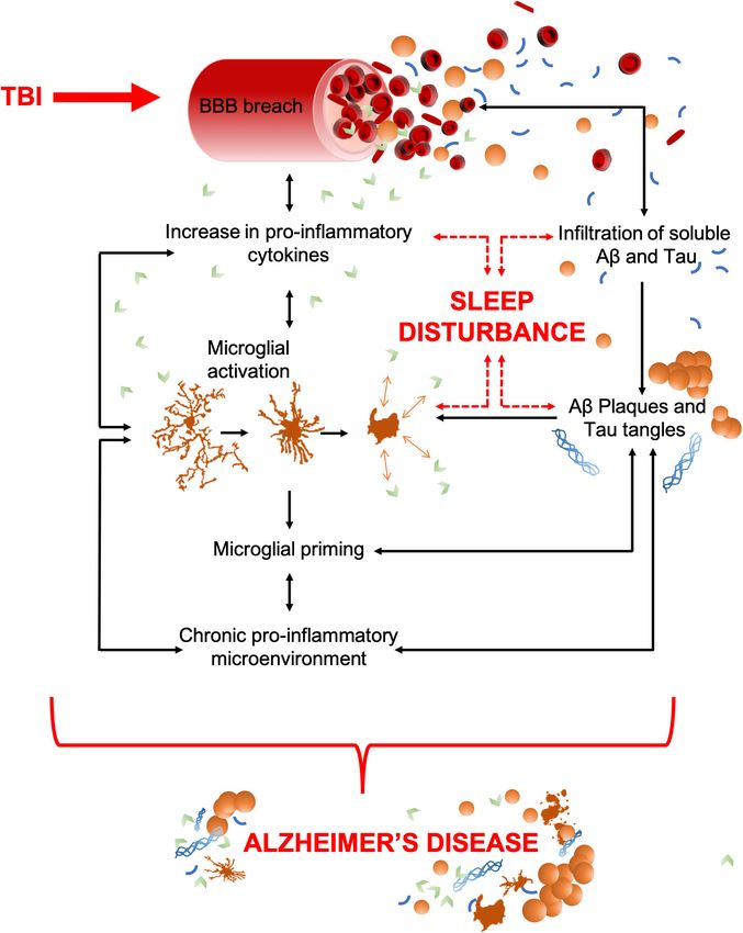
Green et al.                                                                                                       Sleep, Inflammation, Neurodegeneration

onset condition which can occur at any age due to impact to
the head. It has been suggested that TBI heightens the risk of
subsequent development of AD (Mendez, 2017). Despite the
stark contrast in their causal mechanisms, both AD and TBI
are conditions which elicit a neuroinflammatory response that
is coupled with physical, cognitive, and affective symptoms.
A commonly reported symptom in both AD and TBI is sleep
disturbance (Vitiello and Borson, 2001; Castriotta et al., 2007;
Viola-Saltzman and Watson, 2012; Sandsmark et al., 2017).
Sleep is regulated by circadian and homeostatic processes,
such that pathological inflammation may disrupt the chemical
signaling required to maintain a healthy sleep profile. In this
way, immune system activation can influence sleep physiology.
Conversely, sleep disturbances can exacerbate symptoms or
increase the risk of inflammatory/neurodegenerative diseases.
The coupling between sleep and inflammation may indicate
clinical disease status or outcome of disease processes (Wiseman-
Hakes et al., 2013; Rao et al., 2014). The self-perpetuating cycle
of sleep disturbance and neuroinflammation seen in TBI and AD
encourage a comparison of these seemingly disparate conditions
to gain greater insight into the shared mechanisms of progressive
symptomology. This review will discuss the possibility of a sleep-
and inflammation-fueled progression from TBI to AD.
    TBI is characterized by a primary insult, initiated by
mechanical forces applied to the head or brain. Immediately,
sequential pathophysiological processes are initiated, which often
result in a chronic inflammatory microenvironment. Microglia
                                                                           FIGURE 1 | TBI to AD, an inter-disease trajectory. TBI disrupts the blood brain
and resident mononuclear phagocytes throughout the CNS are
                                                                           barrier (BBB) upon insult which results in an infiltration of peripheral
activated as part of this secondary injury process in an attempt           pro-inflammatory cytokines and any soluble pools of amyloid-β (Aβ) and Tau.
to preserve homeostasis and repair injured tissue. TBI-induced             Together, these can precipitate AD. As some pro-inflammatory cytokines have
inflammation is communicated by pro- and anti-inflammatory                 dual (opposing) roles as sleep regulatory substances, their increase can also
cytokines, which are below detectable levels in healthy tissue but         lead to sleep disturbances, a characteristic that commonly precedes the
                                                                           cognitive decline in AD. Pro-inflammatory cytokines upregulate the activation
rapidly increase upon impact (Wang and Shuaib, 2002; Lucas
                                                                           of microglia, which act as a positive feedback mechanism, resulting in
et al., 2006). Cytokines are powerful chemical communicators,              increased pro-inflammatory cytokine production and an increased breach of
essential for maintaining homeostasis throughout the body.                 the BBB. Unregulated cytokine release also sustains microglial activation and
However, unregulated release of pro-inflammatory cytokines                 priming which results in a chronic pro-inflammatory microenvironment. This
following an injury can cause pathological functions that lead             includes astrocytosis, hypoxia, reactive oxygen species (ROS), elevated
                                                                           cytokine levels, and microglial activation. The movement of amyloid-β (Aβ) and
to detrimental inflammation and progressive tissue damage                  Tau through the breach in the BBB could potentially seed protein
(Kumar and Loane, 2012; Morganti-Kossmann et al., 2018).                   oligomerization and aggregation, thereby acting as possible drivers of central
Cytokines released by microglia act as sleep regulatory substances         plaque and tangle pathology. Such aggregates in the brain further contribute
(SRSs) and help to maintain healthy sleep (Krueger and Majde,              to microglial activation, the pro-inflammatory microenvironment, and neuronal
1995). Therefore, TBI-induced elevation of cytokines can lead              apoptosis. Together, these contribute to cognitive dysfunction and brain
                                                                           atrophy, the key pathological features of AD. Both brain atrophy and neuronal
to sleep disturbances. Hereon, cytokine-mediated inflammatory              death help to sustain the pro-inflammatory microenvironment creating a
cascades and sleep enter a self-perpetuating positive-feedback             self-perpetuating feedback loop.
loop, containing many of the components seen in the molecular
circuitry of sleep disturbances in AD (as shown in Figure 1).
    AD, much like TBI, is associated with a range of cognitive and
non-cognitive symptoms that include memory loss, disturbed                AD (Faden and Loane, 2015). These include TBI, a common
sleep, speech difficulties, depression, and loss of executive             injury which increases the frequency and onset of AD and
function (Burns and Iliffe, 2009). Preceding cognitive decline, the       other related dementias (McKee et al., 2009; Gavett et al., 2010;
aggregation of amyloid-β plaques and tau neurofibrillary tangles          Gavett et al., 2011; Baugh et al., 2012). Similar to TBI, AD
occurs in the brain, both of which are required for a retrospective       has a neuroinflammatory component that is orchestrated by
AD diagnosis after death (Murphy and LeVine, 2010). While a               pro-inflammatory cytokines and activated microglia. Although
minority of cases are genetically linked, most do not have an             inflammation has a beneficial role in clearing cellular debris
identified cause which makes AD hard to predict, characterize,            and apoptotic cells associated with AD, chronic inflammation
diagnose, and treat (Newcombe et al., 2018). Preexisting diseases         can be damaging due to pro-inflammatory intermediates that
and lifestyle factors can also contribute to an increased risk of         compromise future clearance mechanisms, synaptic pruning,

Frontiers in Neuroscience | www.frontiersin.org                       2                                              August 2020 | Volume 14 | Article 894
Green et al. Sleep, Inflammation, Neurodegeneration
and neuronal survival (Solito and Sastre, 2012; Sarlus and the damage caused by the initial impact if left unresolved.
Heneka, 2017). Moreover, the cytokine storm associated with Although the primary brain injury from TBI is irreversible, the
chronic inflammation can disrupt sleep-wake cycles, a symptom subsequent injury processes occur in a delayed fashion and may
frequently observed in AD patients. Sleep disturbances cause be responsive to treatment.
further cytokine release and microglial activation, hence a Ongoing cellular events post-TBI often cause further damage
positive feedback loop of chronic inflammation and sleep and lead to physiological consequences (Werner and Engelhard,
disturbance is initiated (as shown in Figure 1). 2007; Prins et al., 2013). Among these, sleep disturbances after
In this review, we discuss the bidirectional relationship TBI are commonly reported in the acute timeframe post-injury by
between sleep disturbance and inflammation, such that chronic up to 70% of TBI survivors (Cohen et al., 1992; Orff et al., 2009).
inflammation associated with TBI and AD can lead to sleep Sleep disturbances may persist chronically (Castriotta et al., 2007;
disturbances that exacerbate neuropathology. We also highlight Verma et al., 2007; Kempf et al., 2010), and are experienced
the role of microglia and cytokines in sleep disturbances across the spectrum of TBI patients, including children and
associated with these diseases. adolescents (Tham et al., 2012). These sleep disturbances in TBI
survivors ultimately impact their quality of life. Identification
and treatment of sleep-wake disturbances following TBI can
THE ROLE OF SLEEP ON HOMEOSTASIS improve outcomes in vigilance, working memory, and capacity
of language processing (Wiseman-Hakes et al., 2013). Excessive
While sleep is an evolutionarily conserved phenomenon that daytime sleepiness is the most common sleep-wake disturbance
is essential for survival (Banks and Dinges, 2007), its function reported among TBI patients (Castriotta et al., 2007; Kempf
is not fully understood. The roles of sleep in the elimination et al., 2010; Baumann, 2012) and is characterized primarily by an
of waste, restoration of depleted energy sources (Oswald, 1980; increase in sleep propensity. Other commonly reported disorders
Dworak et al., 2010), and energy conservation (Walker and include post-traumatic hypersomnia, narcolepsy, delayed sleep
Berger, 1980; Xie et al., 2013) have long been characterized. phase, insomnia, and fatigue (Ouellet and Morin, 2006; Verma
Sleep plays a physiological role in the cardiovascular, metabolic, et al., 2007; Kempf et al., 2010; Baumann, 2012; Billiard and
thermoregulatory, respiratory, and sexual systems, as well as Podesta, 2013). These sleep disturbances negatively impact
having a major role in regulating brain health and function. More rehabilitation of TBI patients and can exacerbate symptoms
recent hypotheses suggest a cellular need for sleep in regulating such as pain and cognitive deficits (Mathias and Alvaro, 2012;
brain plasticity (Puentes-Mestril and Aton, 2017), learning, and Bhalerao et al., 2013).
memory (Tononi and Cirelli, 2003; Huber et al., 2004; Tononi Extensive pre-clinical research has focused on the detrimental
and Cirelli, 2014; Miyamoto et al., 2017; Raven et al., 2018). effects of inflammation on the injured brain. In rodent models
Indeed, in the absence of sleep, there is significant detriment to of TBI, microglia respond immediately to brain injury-induced
cognitive function (Krueger et al., 1999). tissue damage and release inflammatory mediators such as
The sleep-wake cycle is essential in regulating many of the inflammatory cytokines and chemokines that have been shown
body’s fine-tuned homeostatic processes, even on a molecular to alter sleep (Morganti-Kossmann et al., 2001; Davalos et al.,
level (Cirelli and Tononi, 2008). A study, using two-photon 2005; Nimmerjahn et al., 2005; Frugier et al., 2010; Semple
imaging of live mice after infusion of fluorescent tracers, showed et al., 2010; Ziebell and Morganti-Kossmann, 2010; Bachstetter
that during sleep there was a 60% increase in the interstitial et al., 2013). Two-photon microscopy images of fluorescently
space, allowing an increase in exchange between cerebrospinal labeled microglia following a laser-induced injury demonstrated
fluid (CSF) and interstitial fluid. This has important implications rapid proliferation and migration of microglia to the site of
for the clearance of interstitial disease-associated proteins (Xie injury, where their processes fused (Davalos et al., 2005). It
et al., 2013) and highlights a role for sleep in maintaining was hypothesized that this fusion event was to create a barrier
brain homeostasis. between healthy and injured tissue (Davalos et al., 2005). These
findings suggest that microglia may be the first line of defense
following TBI (Kumar and Loane, 2012). However, microglia can
TBI – INFLAMMATION AND SLEEP become over-activated and induce detrimental neurotoxic effects
PATHOLOGY through the overproduction of cytotoxins, including cytokines
(Block and Hong, 2005). Uncontrolled cytokine production
Mechanical forces applied to the head or brain initiate TBI, by activated microglia can significantly influence activation of
wherein sequential pathophysiological processes permanently astrocytes, which can increase neuronal cell death and worsen
change neurological function (Masel and DeWitt, 2010; Corrigan outcomes after TBI (Myer et al., 2006). Together, these results
and Hammond, 2013). TBI survivors suffer irreversible cognitive, support the view that the inflammatory response to TBI possesses
sensory, sleep, mental health, and emotional morbidities both beneficial and detrimental effects that likely differ in the
as a consequence of injury-induced pathological processes acute and delayed phase after injury.
(McAllister, 1992; Kempf et al., 2010). TBI can cause neuronal cell In the mouse, diffuse brain injury has been shown to increase
death, ischemia, hemorrhage, and the disruption of the blood- the parameters of sleep for 6 h post-injury, regardless of
brain barrier (BBB) which elicit neuroinflammatory cascades sex, injury severity, or time of day the injury occurs (Rowe
(Pleines et al., 2001). Such secondary injury can exacerbate et al., 2014b; Saber et al., 2019). This period of post-traumatic
Frontiers in Neuroscience | www.frontiersin.org 3 August 2020 | Volume 14 | Article 894Green et al. Sleep, Inflammation, Neurodegeneration
sleep correlates with elevated central and peripheral cytokine TBI in the mouse decreased wake and increased non-rapid eye
levels, particularly SRSs. Subsequent studies have shown sleep movement (NREM) sleep during the dark period and reduced
impairment at light-dark transitions, suggesting possible TBI- orexin cells as a function of injury severity up to 2 weeks post-
induced disruption of circadian rhythms (Lim et al., 2013; injury (Thomasy et al., 2017). Orexin (hypocretin) knockout
Rowe et al., 2014a). Whether post-traumatic sleep is beneficial mice subjected to TBI did not exhibit altered sleep, whereas
or detrimental to neurological outcome from TBI remains control wild type mice had increased NREM sleep and decreased
unresolved. To advance the understanding of post-traumatic wake up to 1 month post-TBI (Thomasy and Opp, 2019). A
sleep, a gentle handling approach was used to keep both prospective clinical study found that TBI survivors who reported
uninjured and brain-injured mice awake over the 6 h of post- excessive daytime sleepiness had reduced CSF orexin levels up to
traumatic sleep. In this study, a single period of sleep deprivation 6 months post-injury (Baumann et al., 2007). Together, these data
demonstrated no adverse, long-standing neurological effects support that the orexinergic system may be involved in long-term
(Rowe et al., 2014a), although, the course of recovery from sleep-wake alterations that persist after the acute inflammatory
injury may have been altered. Three days of transient sleep response to TBI has subsided. Therefore, orexins should be
disruption following experimental TBI in the mouse worsened considered when discussing the sleep-mediated links between
inflammation and altered stress-immune pathways (Tapp et al., TBI and AD (Liguori, 2017). However, the complexity of this
2020). Total sleep deprivation for 24 h following diffuse TBI relationship must be noted as not only has increased orexinergic
in rats reduced morphological damage and improved functional signaling been associated with the progression of AD, orexins
outcome in the acute recovery period (Martinez-Vargas et al., have been shown to increase amyloid-β in the interstitial fluid
2012). The effect of sleep deprivation prior to TBI has also (Kang et al., 2009) and there is a documented correlation between
been investigated and 48 h of sleep deprivation or chronic sleep tau proteins and orexin CSF levels and sleep-wake dysregulation
restriction (10 days, 6-h sleep/day) prior to mild or moderate in AD patients (Liguori et al., 2020b). Furthermore, damage to
TBI in rats did not exacerbate brain injury-induced neuronal the hypothalamus and pituitary can affect the endocrine system in
damage (Caron and Stephenson, 2015). Sleep after TBI has also general (Sundaram et al., 2013), with potential for further impact
been inhibited pharmacologically. The therapeutic treatment of on sleep architecture that is currently poorly understood.
mice with an anti-inflammatory lipid mediator, and treatment
with a novel TNF-α inhibitor, attenuated TBI-induced sleep and
subsequently improved functional outcome (Harrison et al., 2015; DYSREGULATED IMMUNE FUNCTION
Rowe et al., 2018b). When a second TBI was introduced during AND SLEEP
post-traumatic sleep (3 h apart), more severe functional and
histopathological outcomes were observed, when compared with The immune system is the body’s defense system which
mice that received a second TBI after sleep from the initial impact monitors, detects, and attempts to eliminate threats to health and
subsided (Rowe et al., 2018a). Taken together, these studies homeostasis. Immune cells communicate via chemical mediators
suggest that more evidence is needed to determine if acute sleep such as cytokines and chemokines allowing them to respond
after TBI is beneficial or detrimental, however, the underlying to pathological changes within tissues with a high degree of
pathological response to brain injury that results in increased specificity. While the peripheral immune system plays a role
sleep may be amenable to therapies. Furthermore, post-traumatic in the disease progression of TBI and AD, the inflammatory
sleep may be a physiological response to brain injury that could reactions observed in these conditions are largely driven by
serve as a personalized biomarker to pathological conditions. In microglia, the immune cells exclusive to the CNS. There are
moderate to severe clinical cases of TBI, sleep-wake cycles can be bidirectional links between sleep and the immune system, such
altered due to a loss of consciousness. Clinical data indicate when that sleep loss impairs immune function and physiological
the brain has not recovered consciousness from a moderate to sleep is modified in response to an immune challenge (Ingiosi
severe TBI, it is not able to consolidate sleep or generate a 24-h and Opp, 2016). Sleep/circadian disturbances, particularly states
sleep-wake cycle (Duclos et al., 2017). These clinical data parallel of sleep disruption and deprivation, can lead to serious
experimental data and suggest in the acute phase of TBI, injury- consequences; for example, dysregulation of the inflammatory
induced pathophysiology contributes to sleep disturbances. response (Castanon-Cervantes et al., 2010; Besedovsky et al.,
Although inflammation has emerged as a major player in sleep 2019), leading to a state of systemic inflammation with increased
disturbances post-TBI, there are other influences that should be pro-inflammatory cytokines in the brain (Marshall and Born,
considered. TBI can cause widespread mechanical damage to 2002; Opp and Toth, 2003; Maurovich-Horvat et al., 2008;
regions of the brain, many of which exert a role in sleep and Aldabal and Bahammam, 2011).
inflammation. One region of particular interest to understanding
sleep disturbances post-TBI is the hypothalamus (Sundaram Cytokines and Sleep
et al., 2013). Damage or loss of hypothalamic neurons can disrupt Cytokines have an essential role in regulating the immune
orexin (a.k.a. hypocretin) secretion which has a well-established system at multiple stages throughout the body and have
role in arousal and wakefulness. Extensive loss of orexin neurons long been characterized as a contributing factor to sleep and
and consequent excessive daytime sleepiness have been observed sleep disturbances. Under normal conditions, cytokines have
both pre-clinically and clinically after TBI (Baumann et al., 2009; been shown to play a role in NREM sleep (Shoham et al.,
Thomasy et al., 2017; Thomasy and Opp, 2019). Experimental 1987; Morrow and Opp, 2005; Krueger, 2008), as well as
Frontiers in Neuroscience | www.frontiersin.org 4 August 2020 | Volume 14 | Article 894Green et al. Sleep, Inflammation, Neurodegeneration
altering neuronal firing patterns in both the hypothalamus and injury (Gyoneva and Ransohoff, 2015; Plesnila, 2016). However,
brainstem, with consequent effects on sleep regulation (Opp, chronically elevated cytokine levels can further perpetuate the
2005). Interleukin (IL)-6, tumor necrosis factor (TNF)-α, and initial damage and lead to robust secondary damage (Morganti-
IL-1β, are well characterized for their essential role in both Kossman et al., 1997; Zeiler et al., 2017). Similarly, in AD, a
sleep and inflammation and mediate the crosstalk between the dysregulation of cytokines may potentiate the pathogenesis of the
peripheral immune system and the brain (Krueger and Majde, disease leading to irreversible damage (Su et al., 2016; Figure 1).
1995; Krueger, 2008; Besedovsky et al., 2019). Mechanistically, Indeed, plasma cytokines are higher in the AD population when
this property of IL-6, TNF-α, and IL-1β, could be linked to their compared with healthy controls (Swardfager et al., 2010). In the
neuromodulatory effect in contributing to the fine-tuned control context of this review, we assert that sleep disturbances, brought
of synaptic transmission and plasticity (Jewett and Krueger, about by either TBI or AD, lead to changes in cytokine production
2012; Vezzani and Viviani, 2015), which contributes to the and may further exacerbate disease-related damage to the brain.
function of memory formation during intermittent rapid eye
movement (REM) sleep and NREM sleep (Walker, 2017). As
such, increased production of these pro-inflammatory cytokines ALZHEIMER’S DISEASE –
mediates increases in sleep under inflammatory conditions INFLAMMATION AND SLEEP
such as TBI or AD. PATHOLOGY
There is also a relationship between sleep disturbances and
cytokine production [Please see (Irwin, 2019) for an excellent Sleep disturbances have long been a symptom associated with AD
review on sleep and cytokines]. The sleep disturbances that but more recently have been recognized as an early feature of the
occur in neurological disease and injury (Musiek and Holtzman, disease, thought to precede cognitive decline and contribute to
2016; Leng et al., 2019; Logan and McClung, 2019) are likely disease progression (Musiek et al., 2015; Musiek and Holtzman,
due to common pathological pathways that activate the immune 2016). Literature reports that 25–70% of all suspected AD patients
response; specifically, inflammation and cytokine production. report disturbances in sleep which are linked to poorer disease
Prolonged wakefulness increased cytokines in the CSF (Borbely prognosis (Moran et al., 2005; Craig et al., 2006; Beaulieu-
and Tobler, 1989; Krueger et al., 2001), which can lead Bonneau and Hudon, 2009; Lim et al., 2014; Wennberg et al.,
to subsequent sleep disturbances. In one study, REM sleep 2017). A recent study found that 20% of people between 25 and
deprivation for 72 h in rats led to significant increases in plasma 45 were sleeping 90 min less than the recommended amount for
IL-6, IL-1β, and TNF-α expression, but the expression of IL-6 their age group (Léger et al., 2011). This raises the question as
and IL-1β returned to levels of controls 1-week after the period to whether irregular sleep in early life contributes to developing
of sleep deprivation ended, while TNF-α remained elevated AD later in life.
(Yehuda et al., 2009). Moreover, sleep deprivation increased IL- Sleep is important for clearing pathological and inflammatory-
1β and TNF-α mRNA expression in cortical and subcortical associated proteins from the brain. The recently discovered
brain regions in rats (Zielinski et al., 2014). Similarly, in humans glymphatic system clears unwanted or pathological proteins from
with experimental chronic circadian sleep disruption, there was the interstitial space in the brain by exchange between the
a significant increase in plasma TNF-α expression (Wright CSF the interstitial fluid (ISF) (Xie et al., 2013). Aquaporin-4
et al., 2015). A meta-analysis also showed that in humans, water channels, located on the end feet on astrocytes, mediate
sleep disturbances increased plasma IL-6, although no significant the process by allowing ISF flow from the interstitial space
changes were found in TNF-α expression, likely owing to the into the paravenous region, which in turn drains through the
low statistical power in the analysis (Irwin et al., 2016). These lymphatic system (Verheggen et al., 2018). This is the process
studies support a bidirectional relationship between these pro- by which solutes are removed from the brain. Further, it has
inflammatory cytokines and sleep. Increases in these cytokines been shown that the level of amyloid-β in the brain fluctuates
lead to increased sleep, and conversely, sleep deprivation and rapidly with the daily sleep-wake cycle. During sleep, there is
disruption can increase these cytokines. However, a recent study a 60% increase in the flow of ISF to CSF. Therefore, it is
showed that TNF-α knockout mice display similar patterns of postulated that the disturbed sleep of AD patients decreases
sleep and no deficiency in sleep regulation compared with wild the exchange of ISF and CSF which may lead to reduced
type mice (Szentirmai and Kapás, 2019). In fruit flies, knockdown clearance of soluble amyloid-β, contributing to the formation
of the TNF-α homologue in astrocytes, but not neurons, reduced of plaques (Xie et al., 2013; Rasmussen et al., 2018). In
sleep duration and disrupted sleep-rebound following sleep turn, increased amyloid burden can lead to an upregulated
deprivation (Vanderheyden et al., 2018). As such, although IL- level of inflammation, perpetuating the cycle. Clinically, this
6, IL-1β, and TNF-α promote sleep, these cytokines may have a effect has been observed using accelerated neuroimaging;
larger role in regulating sleep following inflammatory insults such blood oxygen level, functional magnetic resonance imaging,
as after TBI or during the chronic inflammatory events of AD. electroencephalogram, and CSF flow. Using these techniques,
Moreover, cytokines have functional implications in both TBI waves of CSF flow were observed during slow wave sleep,
and AD. Following a TBI, there is a profound increase in the which appeared to be coupled with the hemodynamic rhythm
production and release of cytokines (Figure 1). Acutely, the during sleep (Fultz et al., 2019). Further, a recent clinical
increased release of cytokines is believed to have therapeutic study that examined the sleep architecture and CSF biomarkers
implications that can help improve primary and secondary (tau and amyloid-β42 ) in patients with AD (which progressed
Frontiers in Neuroscience | www.frontiersin.org 5 August 2020 | Volume 14 | Article 894Green et al. Sleep, Inflammation, Neurodegeneration
from having subjective cognitive impairment to mild cognitive
impairment to AD), found that both REM and NREM sleep
were disturbed. They also found that AD patients displayed
a decrease of CSF amyloid-β42 (Liguori et al., 2020a). In the
clinic, amyloid-β deposition has been shown to accumulate
prior to cognitive and memory decline (Liguori et al.,
2020a) and this effect was significantly higher in those who
also had poor sleep efficiency (Molano et al., 2017). Other
experimental studies on human volunteers have also suggested
that dysregulated sleep patterns increase amyloid-β deposition
in the human brain (Shokri-Kojori et al., 2018). Together,
these data demonstrate how the self-perpetuating cycle of sleep
disturbance and inflammation are associated with a decline in
cognitive function.
Pre-clinically, transgenic mice overexpressing disease-
associated APPswe/PS1δE9 mutations which drive amyloid-β FIGURE 2 | Depiction of a coronal brain slice showing the global expanse of
pathology, exhibit disturbed sleep patterns prior to developing microglial phenotypes in TBI and AD. The left illustrates localized effects of TBI
amyloid-β plaques (Roh et al., 2012). This disturbance further which include increased microglial activation near the injury site and
emphasizes the potential bidirectional relationship between decreased activation in distal regions. In comparison, the right represents the
AD brain with widespread changes in microglia morphology, gross structural
amyloid-β deposition and sleep regulation. However, whether changes to the cortex, and an enlargement of ventricles (gray). Both TBI and
the detrimental cycle is between sleep and amyloid-β directly, AD lead to increased inflammation and activated microglia. Despite this
or rather associated inflammatory factors (or a combination similarity, distinct microglial morphologies are observed in these conditions. (1)
of both) is unknown. The extent to which amyloid-β acts Microglia are activated, migrate to the injury site, and display an amoeboid or
phagocytic morphology. (2) Rod-cell morphology is often observed in the
as a driver of AD progression remains controversial, but it
cortex after TBI and have also been documented in AD tissue. However, the
does appear very likely that inflammation increases amyloid-β function of this cell phenotype is currently unknown, and the morphology is
deposition (Irwin and Vitiello, 2019). Clinically, the AD brain not restricted to TBI/AD or the region in which they are shown. (3) Distal to the
presents with activated microglial phenotypes, most notably injury site, microglia are ramified and occur at a lower density. (4) Microglia
aggregated in the penumbra of amyloid-β plaques (Navarro that surround amyloid-β plaques show activated, amoeboid, and dystrophic
morphologies. Cells of each phenotype pictured are not restricted to the brain
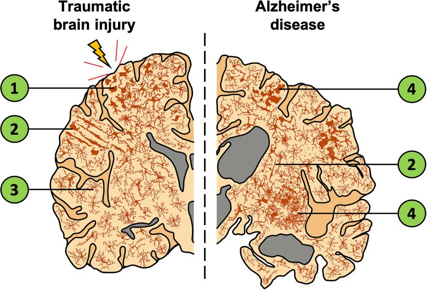
et al., 2018) (see Figure 2; further discussed in section “TBI as
regions shown.
a Contributor to AD”), where their likely/presumed function
is to reduce neuronal exposure to soluble amyloid β-mediated
toxicity (Wang et al., 2016). In the post-mortem AD brain,
other morphological changes in microglia are seen, such as of acetylcholine from basal forebrain neurons in AD. Although
rod and dystrophic phenotypes, as well as an overall increase the acetylcholinesterase inhibitors donepezil, galantamine, and
in microglial density (Figure 2; Bachstetter et al., 2015). This rivastigmine (drugs currently licensed to treat the symptoms
suggests that the role of microglia in AD is probably more of AD) would be anticipated to boost acetylcholine levels,
complex than simply clearing amyloid-β and associated proteins they have a poorly defined effect on sleep architecture (Cooke
(as represented in Figure 2). Despite the elusiveness of their et al., 2006). One study found an 81.8% improvement in sleep
function, these altered microglial phenotypes are associated with quality with galantamine treatment, 75% with rivastigmine,
an increased release of pro-inflammatory cytokines known to and 50% from donepezil (Naharci et al., 2015). Despite these
cause sleep disturbances and neuronal toxicity (Lull and Block, reported improvements, the treatment of sleep disturbances
2010), thus, through this mechanism microglia can contribute in AD patients remains a clinical challenge. Glutamatergic
to the progression of AD. In summary, both pre-clinical signaling, which plays an integrated role in the regulation of
and clinical research suggests a complex interaction between sleep stages and arousal (Shi and Yu, 2013), also deteriorates
sleep and the microglia-mediated inflammatory response in AD patients. Memantine, a moderate affinity N-methyl-D-
in the AD brain. aspartate receptor antagonist, has been shown to increase total
Both sleep and inflammation are tightly coupled to the time spent sleeping in AD patients treated with 20 mg/day for
cholinergic system, which promotes wakefulness and REM 4 weeks (Ishikawa et al., 2016). However, due to the nature
sleep (Platt and Riedel, 2011). Irreversible damage to the of receptor desensitization and transient efficacy of this drug,
basal forebrain cholinergic system correlates with the memory it is not a good long-term pharmacological candidate to break
and cognitive symptoms seen in AD (Ferreira-Vieira et al., the altered sleep architecture and inflammation cycle discussed
2016). Moreover, the immune response is in part regulated here. In the search for a more effective intervention, multiple
by the cholinergic anti-inflammatory pathway by control of neuroinflammatory molecules have been targeted as potential
macrophages and microglia through α7 nicotinic receptors. Such treatments for AD but have had limited success in measurable
modulation attenuates the release of pro-inflammatory cytokines, outcomes. Currently, there is renewed interest in microglia
namely TNF-α, as shown in vitro and in vivo (Shytle et al., as a therapeutic target to stop the pathological inflammatory
2004; Lehner et al., 2019) and might be compromised by the loss cascades seen in AD which could then potentially treat sleep
Frontiers in Neuroscience | www.frontiersin.org 6 August 2020 | Volume 14 | Article 894Green et al. Sleep, Inflammation, Neurodegeneration
disturbances, as discussed in section “TBI to AD – A Microglial causes further loss of blood vessel integrity leading to brain
Mediated Progression?”. hemorrhage (Smith and Greenberg, 2009). As both mechanical
damage and infiltration of pro-inflammatory molecules that
TBI as a Contributor to AD travel in the blood increase the inflammatory microenvironment
Meta-analyses have found that a history of TBI is associated after TBI, this may increase the likelihood of developing AD, and
with development of AD (Mortimer et al., 1991; Fleminger et al., its consequent sleep disturbances, that are known to be tightly
2003). Despite different mechanisms of onset, these conditions associated with inflammation.
are mechanistically interlinked by a complex web of pathologies, Matrix metalloproteases (MMPs) are key enzymes that cause
as demonstrated by Figure 1. Furthermore, increased amyloid- breakdown of BBB integrity, and therefore the infiltration of
β levels are seen after TBI (Sivanandam and Thakur, 1995; products into the brain (Nissinen and Kähäri, 2014). MMP9
Rasmusson et al., 1995; Nemetz et al., 1999; Jellinger et al., 2001) knockout mice have shown the deleterious role of MMPs in
and post-mortem examination of TBI brains found that amyloid- maintaining a chronic ischemic environment after a transient,
β plaques, a hallmark of AD, had developed acutely in 30% of focal ischemic injury (Asahi et al., 2001), as the absence of
TBI brains (Roberts et al., 1991; Roberts et al., 1994; Johnson MMP9 reduced proteolytic breakdown of the BBB. Further,
et al., 2010). However, as amyloid-β plaques are also seen in MMPs increase the bioavailability of both TNF-α (Haro et al.,
the healthy brain (Rodrigue et al., 2009), the meaning of these 2000; Vandenbroucke et al., 2013) and IL-1β (Schönbeck et al.,
findings are unclear. While there is an increased risk of AD after 1998; Nissinen and Kähäri, 2014), two major inflammatory
TBI, most individuals with AD do not have a history with TBI, cytokines that can cause breach of the BBB independent of injury
thus, other factors may interact with TBI-induced damage and (Siegel et al., 2009). Not only does this lead to a chronic pro-
lead to exacerbated cognitive decline and dementia (Plassman inflammatory environment of the brain, but TNF-α and IL-1β
and Grafman, 2015). As discussed above, sleep disturbances post- act as SRSs, the increase of which can cause sleep disturbances.
TBI correlate with microglial activation and a concomitant rise in The disruption of sleep patterns feeds into the upregulation of
pro-inflammatory cytokine levels (Rowe et al., 2014b). As sleep inflammation and is a key feature of AD which can precede
disturbances have been identified as an early symptom of AD, it cognitive decline. When considered together, this body of
is plausible that TBI-induced sleep disturbances are a risk factor literature suggests that MMPs are mediators of the perpetual
for AD. Until this is understood, the opportunity for therapeutic pathological feedback loop initiated by TBI and postulated to
intervention in this inter-disease period is stunted. increase the risk of developing AD.
Breach of the Blood Brain Barrier – Amyloid-β and Tau as Mediators of
Unregulated Infiltration of Plasma Inflammation and Sleep Disturbance
Proteins It is well established that TBI can cause disruption to
TBI, despite the varying causal mechanisms, initiates breach of the BBB, henceforth, crosstalk between the CNS and
the BBB. Whether it be a transient opening due to the shearing peripheral organ systems is initiated. Not only can this allow
of blood vessels, or a chronic inflammation-mediated opening, infiltration of peripheral cytokines (which further exacerbate
cross-talk between the CNS and the blood can occur. This is due neuroinflammation), but soluble forms of amyloid-β and tau
to disruption of the tight junctions that prevent uncontrolled can move across the BBB. The following section will discuss
infiltration of substances from the blood, as summarized by the role of these proteins in the inflammatory mediated sleep
Figure 1. A scanning electron microscopy (SEM) study of the disturbances seen during the progression of both TBI and AD.
human brain after death from TBI showed altered vascular There is little known about the brain-wide concentration of
features (Rodríguez-Baeza et al., 2003). Microvascular casts oligomeric and unaggregated forms of amyloid-β, a protein that
examined by SEM showed a sunken vascular surface, longitudinal is found in high abundance peripherally with respect to the
creases, and flattened luminal morphology (Rodríguez-Baeza brain. As TBI creates an opportunity for amyloid-β proteins
et al., 2003). A similar breach in neurovascular membranes has to cross the BBB, the clinical and biochemical impact of this
been observed in many pre-clinical studies (Tanno et al., 1992; is under much dispute. One study examined patients with a
Baldwin et al., 1996; Sangiorgi et al., 2013). Not only does the severe TBI and found that amyloid-β peptides (amyloid-β 1–42
initial brain injury cause BBB breach from the sheering caused and 1–40) were increased in the CSF, peaking 1 week post-
by the mechanical force exerted on the brain, but secondary injury (Raby et al., 1998). Increased levels of tau and amyloid-
processes such as inflammation and hypoxia contribute to β were also observed in blood exosomes (Gill et al., 2018).
prolonged breach of the BBB creating a self-perpetuating, Such increases in these proteins may be reflected in increased
unregulated cycle of cross-talk between the CNS and the blood. levels in CSF, however, evidence has proven inconsistent. For
Alongside TBI, BBB breach is a well-known consequence of many example, two studies reported that amyloid-β in the CSF was
other inflammation-inducing conditions such as viral or bacterial significantly lower than in controls during the week following
infection. Amyloid-β has also been shown to cause cerebral- TBI (Franz et al., 2003; Kay et al., 2003). Despite the disparity
amyloid angiopathy (the presence of amyloid-β aggregates in the of these findings, it could be interpreted that CSF amyloid-β
vasculature of the brain) which adds to the perpetual, unregulated decreased post-injury due to possible deposition in the brain.
infiltration of blood components into the brain as this also However, as sleep disturbance post-TBI peaks in the acute
Frontiers in Neuroscience | www.frontiersin.org 7 August 2020 | Volume 14 | Article 894Green et al. Sleep, Inflammation, Neurodegeneration
time phase, and sleep disturbance increases soluble amyloid- Johnson et al., 2012). NFTs impair axonal transport, notably
β in the CSF (Lucey et al., 2018), the findings of these of mitochondria (which are necessary for supporting nerve
studies are conflicting. Moreover, researchers have shown that terminal function), contributing to the neuronal death associated
the anti-inflammatory antibiotic doxycycline can attenuate the with TBI (Kondo et al., 2015). Tau acts intracellularly as a
acute behavioral deficits associated with intracerebral delivery microtubule stabilizer; however, its accumulation can occur
of oligomeric amyloid-β (Forloni and Balducci, 2018), which both intracellularly and extracellularly (Swanson et al., 2017).
acts as proof of principle; unaggregated forms of amyloid-β and Intracellularly tau abnormalities may cause neuronal death
inflammation are tightly coupled. through disruption of intracellular cargo transport, whereas
Not only does infiltration of amyloid-β and tau directly affect extracellular oligomers of tau are released from degenerating
the formation of plaques and tangles and increase oligomeric neurons. This was shown by in-vitro studies, revealing addition
toxic species, but infiltration of associated proteins can also have of un-aggregated tau protein increased the amount of cell
an effect. For example, S100A9 (a pro-inflammatory protein) has death in tau-treated cells compared to controls (Gómez-Ramos
been shown to contribute to amyloid plaques that accumulate et al., 2006). This suggests that extracellular tau is able to
rapidly after TBI. These plaques were found to be positive for transmit the pathology to neighboring neurons, which causes
oligomeric forms of amyloid-β and not fibrillar forms, suggesting spread of neuronal death from tauopathy (Swanson et al.,
that these could be a precursor to the plaques seen in later 2017). Further, tau induces synaptic loss through microglial
stage AD (Wang et al., 2014). Further, it has been well accepted phagocytosis of synaptic compartments containing tau, leading
that the levels of iron, copper and zinc are altered in the to synaptic loss and cognitive decline (Jadhav et al., 2015).
brain of AD patients and that amyloid-β plaques are sites of Alongside mediating the spatiotemporal spread of pathology, tau
metal aggregation. Experimental models of TBI have also shown can prolong microglial activation, exacerbating a chronic pro-
that there is increased infiltration of metal ions (iron, copper inflammatory environment in the brain, as shown in Figure 2.
and zinc) post-TBI. Under normal physiological conditions, A recent study using positron emission tomography has
such ions are found in the blood and are prevented from identified an increased level of tau deposition in patients that
freely moving across the BBB. Amyloid-β has been shown to had suffered a single, moderate-severe TBI compared to controls
associate with zinc, copper and iron ions (Lovell et al., 1998), (Gorgoraptis et al., 2019). Pre-clinical rodent models have also
which are all present in the blood, therefore, the increased found an increase in oligomeric tau in the brain acutely (4 h) after
level of blood in the brain after TBI is likely to exacerbate parasagittal fluid percussion injury, which remained elevated for
this association. This increase in ion concentration is therefore 2 weeks post-injury compared to uninjured shams (Hawkins
likely to augment plaque formation, increasing plaque-associated et al., 2013). These studies support an increase in tau protein in
inflammation. Another well characterized contributor to AD is the brain after TBI and provides another link for an inter-disease
oxidative stress caused by the rapid production of free radicals. As progression to AD triggered by TBI-induced BBB disruption.
sleep fosters antioxidant activity, sleep disruption may exacerbate Not only has tau been shown to have inflammatory
ROS activity and associated inflammation post-TBI, which can consequences, but a recent human study, using regression
lead to neuronal cell death and further aggregation of amyloid-β analysis on positron emission tomography measures of amyloid-
(Molina-Holgado et al., 2007). β and tau, and EEG sleep recording, showed that the extent
TBI can also augment the formation of amyloid-β plaques of slow wave sleep disturbance predicted a greater presence of
and tau neurofibrillary tangles (NFTs) through inflammation- tau in the medial temporal lobe (Winer et al., 2019). Numerous
dependent gene expression and transcription factor activation. studies using animal models also associate aggregates of tau with
Experimental TBI in the 3xTg mouse model of AD, showed disturbance to sleep or circadian regulation (Musalek et al., 1989;
that TBI activated transcription factors (CCAAT/Enhancer Witton et al., 2016; Holth et al., 2017; Arnes et al., 2019). Taken
Binding Protein Beta), and increased the expression of delta- together the data presented in this section suggest a plausible
secretase (Wu et al., 2019). Delta-secretase is an enzyme that inter-condition link between TBI, AD through the inflammatory
mediates pathology by cleaving amyloid-β and tau which allows and sleep mediated pathological cycle.
the formation of amyloid-β plaques and hyperphosphorylated
tau, and consequently induces the neuroinflammatory cascade
(Zhang et al., 2020). Support for the hypothesis that TBI TBI TO AD – A MICROGLIAL MEDIATED
augments the formation of amyloid-β plaques and NFTs PROGRESSION?
was strengthened by the fact that the authors reversed the
effect using viral expression of tau in 3xTg mice, that was Microglia, the immune cells exclusive to the CNS (Loane and
resistant to cleavage by delta secretase (Wu et al., 2019). Byrnes, 2010), play a role in perpetuating homeostasis in the
These data demonstrate how amyloid- β and tau contribute CNS (Inoue, 2002), where they act as a biological surveillance
to the inflammatory microenvironment (and consequent sleep system continuously monitoring the microenvironment. This
disturbances) associated with both TBI and AD. allows them to be the first responders upon detection of
NFTs are another hallmark of AD. In both mice and humans, pathological stimuli (Glenn et al., 1992). Microglial activation
NFTs have been found post-TBI (Kondo et al., 2015) and play displays a morphological (and genetic) continuum, with changes
a critical role in post-injury neuronal damage (Smith et al., in characteristics enabling the cells to rapidly respond to stimuli.
1999; Duda et al., 2000; Uryu et al., 2007; Yao et al., 2008; Under normal conditions, microglia have a small cell soma
Frontiers in Neuroscience | www.frontiersin.org 8 August 2020 | Volume 14 | Article 894Green et al. Sleep, Inflammation, Neurodegeneration
and highly branched processes. After detection of pathological other cells of the glial network also show a more reactive
stimuli, or sleep disturbance, the cell soma enlarges, and the phenotype in the aging brain. Activation of astrocytes, with
processes retract. This activated phenotype permits phagocytic their physical role in maintaining the integrity of the BBB,
activity (Liu and Quan, 2018). Upon activation, microglia release may allow greater influx of soluble β-amyloid proteins into
cytokines to trigger pro-inflammatory pathways. Despite some the brain (see section “Amyloid-β and Tau as Mediators of
immediate benefit seen from microglial activation, such as Inflammation and Sleep Disturbance”), thus advancing the
clearing of cellular debris, their reactivity has been associated pathologies of the disease.
with potentially damaging intermediates. These include reactive Much like in AD, a primed microglial phenotype has been
oxygen species and pro-inflammatory cytokines (Norden and observed after TBI. One study in rodents showed that after TBI,
Godbout, 2013; Iizumi et al., 2016), which can cause damage microglia remained sensitized and quickly became activated after
to nearby neurons, prolong the inflammatory response, hinder a peripheral immune challenge provided by lipopolysaccharide
CNS repair, and exacerbate neurological symptoms, such as (LPS). Mice that had received a TBI and LPS appeared to have
inflammation-induced sleep (Dheen et al., 2007; Ziebell and a poorer cognitive outcome, suggesting that microglial priming
Morganti-Kossmann, 2010; Bilbo and Stevens, 2017). Further, worsens cognition (Muccigrosso et al., 2016). Another study
chronic microglial activation increases astrocyte activation, found that microglia displayed a primed morphology up to a
which can worsen the outcome after an inflammatory challenge, year after controlled cortical impact which appeared to augment
increasing neuronal cell death (Myer et al., 2006). pathologies such as lesion volume, degeneration of hippocampal
In response to pathological stimuli, microglia upregulate neurons and myelin loss, which suggests microglial priming
their release of certain cytokines, including TNF-α, IL-1β, and may be, at least in part, a contributor to neurodegeneration
IL-6 which act as SRSs (see section “Cytokines and Sleep”). post-TBI (Loane et al., 2014). However, this relationship is
A further consideration is that higher levels of microglial hard to pinpoint as not only do microglia express a primed
activation are observed during waking hours; microglia return morphology with age and TBI, but also with other inflammatory-
to their non-activated status during sleep (Fonken et al., 2015). associated conditions such as HIV (Hains et al., 2010), stroke
As microglia upregulate their release of IL-1β and TNF-α upon (Espinosa-Garcia et al., 2017), and even exposure to air pollution
activation, it is likely that these cytokines fluctuate in response (Mumaw et al., 2016).
to endogenous circadian regulation, though this is beyond the After TBI, activated microglia have an increase in the
scope of current experimental literature. If microglia return to number of lysosomes (Tanaka et al., 2013). This has been
non-activated states during sleep, one would predict sleep is suggested to provide a link to the development of amyloid-
neuroprotective following pathological stimuli, however, these β plaques, because of the acidic lysosomal pH in comparison
data are inconclusive and require further research (see section with the extracellular microenvironment (Spangenberg et al.,
“Alzheimer’s Disease – Inflammation and Sleep Pathology”). 2019). This work was continued using histological staining
The inflammatory cascade present in both AD and TBI in human tissue and found an association between microglia
presents as an attractive target for therapeutic intervention. containing amyloid-β-aggregates which are hypothesized to lead
Historically, cyclooxygenase, a key enzyme in prostaglandin to progressive amyloid-β pathology (Spangenberg et al., 2019). To
production, was identified as a target for reducing the further demonstrate the role of microglia in plaque formation,
inflammatory response seen in AD, however, its inhibition with elimination of microglia in a transgenic mouse model of AD
nonsteroidal anti-inflammatory drugs (Aisen, 2002) had limited eliminated plaque formation, but plaques returned with the
efficacy in stalling the progression of AD. Due to observed recovery of microglia, which supports them having a role in
clustering of microglia around amyloid-β plaques, microglia have plaque formation.
emerged as a therapeutic target. Despite the growing volume of Eliminating and repopulating microglia have shown
multidisciplinary research on the activation of microglia and pro- therapeutic efficacy in both AD and TBI. Eliminating microglia
inflammatory cytokines as SRSs, the nuances of their involvement using a bioavailable colony stimulating factor 1 receptor (CSF1R)
in sleep are not yet fully understood. As dysregulation of inhibitor, a receptor necessary for microglia survival, in a
sleep has been associated with neurological deficits, a complex, 5xFAD mouse model of AD showed amyloid-β plaques failed
multifaceted feedback loop is created. This poses a difficult target to form in the absence of microglia but when the CSF1R
for therapeutic intervention, until the role of microglia in sleep inhibitor was withdrawn, microglia repopulated and amyloid-β
is fully unveiled. plaques developed similar to those in controls (Spangenberg
Multiple areas of neuroscientific research have presented et al., 2019). Similarly, the elimination of microglia with a
cogent evidence suggesting that microglia can become “primed” low dose of a bioavailable CSF1R inhibitor in 3xTg-AD mice
and display an activated phenotype chronically (Fenn et al., prevented microglia association with plaques and improved
2014; Witcher et al., 2015; Hoeijmakers et al., 2016; Ziebell cognitive function (Dagher et al., 2015). Another study showed
et al., 2017). Microglia in the aged brain express “primed” chronic microglia elimination did not alter amyloid-β loads
morphology, even under healthy conditions, a facet not observed but prevented neuronal loss, rescued dendritic spine loss,
in the younger adult brain (Norden and Godbout, 2013; Loane and improved cognitive behavior (Spangenberg et al., 2016).
et al., 2014). Hypotheses suggest priming allows microglia to Microglia elimination has been used in experimental TBI
respond more rapidly to injury and infection, however, the studies where microglia elimination with a CSF1R inhibitor
link between chronic microglial priming and neurodegeneration prevented acute and chronic inflammatory responses in the
is under-explored (Norden and Godbout, 2013). Furthermore, mouse and attenuated astrogliosis (Witcher et al., 2018). As a
Frontiers in Neuroscience | www.frontiersin.org 9 August 2020 | Volume 14 | Article 894Green et al. Sleep, Inflammation, Neurodegeneration
therapeutic intervention, chronically activated microglia were in a highly dynamic inter-disease trajectory. To this end, the
eliminated using a CSF1R inhibitor 1 month following a focal proposed sleep and inflammation-mediated link between TBI
TBI, and the inhibitor was withdrawn 1 week later to allow and AD presents an opportunity for a multifaceted approach to
microglia to repopulate. These studies indicated that removal clinical intervention.
of microglia after TBI reduced chronic neuroinflammation and
associated neurodegeneration and mice had improved functional
outcome (Henry et al., 2020). Taken together, these studies AUTHOR CONTRIBUTIONS
support that microglia play a role the chronic inflammatory
response following TBI and contribute to AD pathology. If TG and RR are responsible for the conceptualization and
eliminating microglia changes sleep profiles in the context of TBI organization of this review. TG wrote the first draft of the
or AD has yet to be explored, however, these experiments may manuscript. JO, SW, RW, and RR contributed to content and
offer further therapeutic approaches to mitigate inflammation- edited the manuscript. All authors contributed to the article and
mediated sleep disturbances. approved the submitted version.
CONCLUSION
FUNDING
This review documents the complexity of the multidirectional
relationship between sleep, microglia, and inflammation in the TG, RR, and JO are funded by Phoenix Children’s Hospital and
clinical and pre-clinical contexts of TBI and AD. It is posited the University of Arizona College of Medicine—Phoenix Mission
that the inflammatory cascades triggered by the disturbed sleep Support Funds. RW acknowledges the support of Alzheimer’s
observed in TBI and AD creates the perfect storm, resulting Society (Project Grant 288).
REFERENCES Beaulieu-Bonneau, S., and Hudon, S. (2009). Sleep disturbances in older adults
with mild cognitive impairment. Int. Psychogeriatr. 21, 654–666. doi: 10.1017/
Aisen, P. S. (2002). Evaluation of selective COX-2 inhibitors for the treatment of s1041610209009120
Alzheimer’s disease. J. Pain Symptom Manage. 23 4(Suppl.), S35–S40. Besedovsky, L., Lange, T., and Haack, M. (2019). The sleep-immune crosstalk in
Aldabal, L., and Bahammam, A. S. (2011). Metabolic, endocrine, and immune health and disease. Physiol. Rev. 99, 1325–1380. doi: 10.1152/physrev.00010.
consequences of sleep deprivation. Open Respir. Med. J. 5, 31–43. doi: 10.2174/ 2018
1874306401105010031 Bhalerao, S. U., Geurtjens, C., Thomas, G. R., Kitamura, C. R., Zhou, C., and
Arnes, M., Alaniz, M. E., Karam, C. S., Cho, J. D., Lopez, G., Javitch, J. A., et al. Marlborough, M. (2013). Understanding the neuropsychiatric consequences
(2019). Role of tau protein in remodeling of circadian neuronal circuits and associated with significant traumatic brain injury. Brain Inj. 27, 767–774. doi:
sleep. Front. Aging Neurosci. 11:320. doi: 10.3389/fnagi.2019.00320 10.3109/02699052.2013.793396
Asahi, M., Wang, X., Mori, T., Sumii, T., Jung, J. C., Moskowitz, M. A., et al. Bilbo, S., and Stevens, B. (2017). Microglia: the brain’s first responders. Cerebrum
(2001). Effects of matrix metalloproteinase-9 gene knock-out on the proteolysis 2017:cer–14–17.
of blood-brain barrier and white matter components after cerebral ischemia. Billiard, M., and Podesta, C. (2013). Recurrent hypersomnia following
J. Neurosci. 21, 7724–7732. doi: 10.1523/jneurosci.21-19-07724.2001 traumatic brain injury. Sleep Med. 14, 462–465. doi: 10.1016/j.sleep.2013.
Bachstetter, A. D., Rowe, R. K., Kaneko, M., Goulding, D., Lifshitz, J., and Van 01.009
Eldik, L. J. (2013). The p38alpha MAPK regulates microglial responsiveness Block, M. L., and Hong, J. S. (2005). Microglia and inflammation-mediated
to diffuse traumatic brain injury. J. Neurosci. 33, 6143–6153. doi: 10.1523/ neurodegeneration: multiple triggers with a common mechanism. Prog.
jneurosci.5399-12.2013 Neurobiol. 76, 77–98. doi: 10.1016/j.pneurobio.2005.06.004
Bachstetter, A. D., Van Eldik, L. J., Schmitt, F. A., Neltner, J. H., Ighodaro, Borbely, A. A., and Tobler, I. (1989). Endogenous sleep-promoting substances and
E. T., Webster, S. J., et al. (2015). Disease-related microglia heterogeneity sleep regulation. Physiol. Rev. 69, 605–670. doi: 10.1152/physrev.1989.69.2.605
in the hippocampus of Alzheimer’s disease, dementia with Lewy bodies, and Burns, A., and Iliffe, S. (2009). Alzheimer’s disease. BMJ 338:b158.
hippocampal sclerosis of aging. Acta Neuropathol. Commun. 3:32. Caron, A. M., and Stephenson, R. (2015). Sleep deprivation does not affect neuronal
Baldwin, S. A., Fugaccia, I., Brown, D. R., Brown, L. V., and Scheff, S. W. (1996). susceptibility to mild traumatic brain injury in the rat. Nat. Sci. Sleep 7, 63–72.
Blood-brain barrier breach following cortical contusion in the rat. J. Neurosurg. Castanon-Cervantes, O., Wu, M., Ehlen, J. C., Paul, K., Gamble, K. L., Johnson,
85, 476–481. doi: 10.3171/jns.1996.85.3.0476 R. L., et al. (2010). Dysregulation of inflammatory responses by chronic
Banks, S., and Dinges, D. F. (2007). Behavioral and physiological consequences of circadian disruption. J. Immunol. 185, 5796–5805. doi: 10.4049/jimmunol.
sleep restriction. J. Clin. Sleep Med. 3, 519–528. doi: 10.5664/jcsm.26918 1001026
Baugh, C. M., Stamm, J. M., Riley, D. O., Gavett, B. E., Shenton, M. E., Lin, A., Castriotta, R. J., Wilde, M. C., Lai, J. M., Atanasov, S., Masel, B. E., and Kuna,
et al. (2012). Chronic traumatic encephalopathy: neurodegeneration following S. T. (2007). Prevalence and consequences of sleep disorders in traumatic brain
repetitive concussive and subconcussive brain trauma. Brain Imaging Behav. 6, injury. J. Clin. Sleep Med. 3, 349–356. doi: 10.5664/jcsm.26855
244–254. doi: 10.1007/s11682-012-9164-5 Charrier, A., Olliac, B., Roubertoux, P., and Tordjman, S. (2017). Clock genes
Baumann, C. R. (2012). Traumatic brain injury and disturbed sleep and and altered sleep-wake rhythms: their role in the development of psychiatric
wakefulness. Neuromol. Med. 14, 205–212. doi: 10.1007/s12017-012-8178-x disorders. Int. J. Mol. Sci. 18:938. doi: 10.3390/ijms18050938
Baumann, C. R., Bassetti, C. L., Valko, P. O., Haybaeck, J., Keller, M., Clark, E., et al. Cirelli, C., and Tononi, G. (2008). Is sleep essential? PLoS Biol. 6:e216. doi: 10.1371/
(2009). Loss of hypocretin (orexin) neurons with traumatic brain injury. Ann. journal.pbio.0060216
Neurol. 66, 555–559. doi: 10.1002/ana.21836 Cohen, M., Oksenberg, A., Snir, D., Stern, M. J., and Groswasser, Z. (1992).
Baumann, C. R., Werth, E., Stocker, R., Ludwig, S., and Bassetti, C. L. (2007). Sleep- Temporally related changes of sleep complaints in traumatic brain injured
wake disturbances 6 months after traumatic brain injury: a prospective study. patients. J. Neurol. Neurosurg. Psychiatry 55, 313–315. doi: 10.1136/jnnp.55.
Brain 130(Pt 7), 1873–1883. doi: 10.1093/brain/awm109 4.313
Frontiers in Neuroscience | www.frontiersin.org 10 August 2020 | Volume 14 | Article 894You can also read