Ferroptosis in Liver Diseases: An Overview - MDPI
←
→
Page content transcription
If your browser does not render page correctly, please read the page content below
International Journal of
            Molecular Sciences

Review
Ferroptosis in Liver Diseases: An Overview
Martina Maria Capelletti 1,2 , Hana Manceau 1,3 , Hervé Puy 1,4 and Katell Peoc’h 1,3, *
 1    Centre de Recherche sur l’Inflammation, INSERM U1149, Université de Paris,
      Laboratory of Excellence GR-Ex, 75018 Paris, France; m.capelletti@campus.unimib.it (M.M.C.);
      hana.manceau@aphp.fr (H.M.); herve.puy@aphp.fr (H.P.)
 2    Department of Biotechnology and Biosciences, University of Milano-Bicocca, I-20126 Milan, Italy
 3    Laboratoire de Biochimie, Hôpital Beaujon, APHP, 92110 Clichy, France
 4    Centre Français des Porphyries, Hôpital Louis Mourier, APHP, 92701 Colombes, France
 *    Correspondence: katell.peoch@aphp.fr

 Received: 11 June 2020; Accepted: 10 July 2020; Published: 11 July 2020

 Abstract: Ferroptosis is an iron-dependent form of cell death characterized by intracellular lipid
 peroxide accumulation and redox imbalance. Ferroptosis shows specific biological and morphological
 features when compared to the other cell death patterns. The loss of lipid peroxide repair
 activity by glutathione peroxidase 4 (GPX4), the presence of redox-active iron and the oxidation of
 polyunsaturated fatty acid (PUFA)-containing phospholipids are considered as distinct fingerprints
 of ferroptosis. Several pathways, including amino acid and iron metabolism, ferritinophagy, cell
 adhesion, p53, Keap1/Nrf2 and phospholipid biosynthesis, can modify susceptibility to ferroptosis.
 Through the decades, various diseases, including acute kidney injury; cancer; ischemia–reperfusion
 injury; and cardiovascular, neurodegenerative and hepatic disorders, have been associated with
 ferroptosis. In this review, we provide a comprehensive analysis of the main biological and
 biochemical mechanisms of ferroptosis and an overview of chemicals used as inducers and inhibitors.
 Then, we report the contribution of ferroptosis to the spectrum of liver diseases, acute or chronic.
 Finally, we discuss the use of ferroptosis as a therapeutic approach against hepatocellular carcinoma,
 the most common form of primary liver cancer.

 Keywords: ferroptosis; cell death; liver; iron metabolism

1. Introduction
      Cell death is the concluding event of all essential cellular processes in living cells [1]. Under certain
conditions, this lethal process occurs suddenly, making cells incapable of preventing the spreading of
cell death; therefore, this event is referred as “accidental/explosive” or necrotic cell death [1]. In contrast,
organisms have developed highly controlled cell death modalities (RCD, regulated cell death). The first
example of RCD was apoptosis, and then necroptosis, netosis, entosis and autophagy [1]. Ferroptosis
(derived from the Greek word “ptosis”, meaning “a fall”, and Ferrum or iron) was first described in
2012 [1,2]. Iron chelators, such as deferoxamine (DFO), prevent the formation of reactive radical species
such as the hydroxyl radical, protecting from cell death. Ferroptosis is defined as an iron-dependent,
non-apoptotic type of cell death characterized by the increase of reactive oxygen species (ROS) [3].
      Moreover, ferroptosis is morphologically distinguishable from other types of cell death. Ferroptosis
plays a critical regulatory role in many diseases (e.g., tumorigenesis, ischemia–reperfusion injury, renal
failure, nervous system diseases and hematological diseases). Because of the increasing relevance of
ferroptosis, several projects have been undertaken focusing on ferroptosis, which rapidly became the
new hotspot of research on the treatment and improvement of prognosis of related disorders [3]. Given
the trend of an increasing number of publications on ferroptosis, we firmly believe, as authors, in the
need of an extensive and detailed review of this topic. Therefore, in the first part of the article, we give

Int. J. Mol. Sci. 2020, 21, 4908; doi:10.3390/ijms21144908                            www.mdpi.com/journal/ijms
Int. J. Mol. Sci. 2020, 21, 4908                                                                                      2 of 23

a comprehensive description of the different processes involved, while, in the second part, the focus is
on the role of ferroptosis in hepatic diseases.

2. Discovery of Ferroptosis
      In 2003, a study was conducted to identify new molecules with lethal effects on Ras-mutated
cells. Among the 23,550, different chemical compounds screened, NSC146109, renamed erastin, had a
selectively lethal effect on Ras-expressing cancer cells [4]. In 2008, two other molecules, Ras-selective
poisonous small molecules (RSL3-5), were identified, which kill selectively human foreskin fibroblasts
(BJeLR) in a non-apoptotic manner [5]. Inhibitors specific for RCDs cannot undo RSL-induced cell
death. In contrast, antioxidants (vitamin E) and iron chelators can block and reverse RSL-induced
cell death [1]. Therefore, the term “ferroptosis” refers to an iron-dependent, non-apoptotic cell death
characterized by lipid peroxidation [3].

3. An Overview of Ferroptosis
      In vitro ferroptosis inducer (e.g., erastin) treatment leads to a round shape of tumor cells and their
detachment from the flask surface. Ferroptosis is characterized by the presence of small mitochondria
with condensed mitochondrial densities, reduction or vanishing of mitochondrial cristae and outer
mitochondrial membrane (OMM) rupture [1]. In contrast to apoptosis, ferroptotic cells do not show
cell shrinkage, chromatin condensation and formation of apoptotic bodies and disintegration of the
cytoskeleton [1]. Moreover, no autophagic vacuoles have been found after treatment with ferroptosis
inducers. The main morphological and biochemical features of the different types of cell death are
summarized in Table 1. The ferroptotic cell appears morphologically and biochemically distinguishable
from the other four types of cells Table 1).

      Table 1. The main morphological and biochemical features of regulated cell deaths (adapted from
      Xie et al., 2016 [5] and Lie et al., 2020 [3]). MAPKs, mitogen-activated protein kinases; GSH, glutathione;
      DAMPs, damage-associated molecular patterns; PS, phosphatidylserine; PARP1, Poly [(ADP-ribose)]
      polymerase 1; LC3-I/II, Microtubule-associated protein light chain 3.

     Cell Death                          Morphological Features                          Biochemical Features
                                                                                         Iron and ROS overload
                             No rupture of the plasma membrane [3]
                                                                                          Activation of MAPKs
                                    Rounding up of the cell [3]
                                                                                      Inhibition of system Xc − and
     Ferroptosis         Small mitochondria, outer mitochondrial rupture,
                                                                                        decreased cystine uptake
                                    reduction of the cristae [3]
                                                                                             GSH depletion
                       Normal nuclear size and no chromatin condensation [3]
                                                                                Release of arachidonic acid mediators [3]
                                   Plasma membrane blebbing [3]
                                     Rounding up of the cell [3]
                                Pseudopod retraction and reduction                     Activation of caspases
     Apoptosis                   of cellular and nuclear volume [3]             Oligonucleosomal DNA fragmentation
                         Nuclear fragmentation, chromatin condensation                     PS exposure [3]
                                  Formation of apoptotic bodies [3]
                        No significant changes in mitochondrial structure [3]
                                   Rupture of the plasma membrane [3]
                                        Cytoplasmic swelling [3]                         Decrease in ATP level
    Necroptosis                    Moderate chromatin condensation [5]                     Release DAMPs
                                     Spillage of cellular constituents                 PARP1 hyperactivation [3]
                                       into microenvironment [3]
                             Lack of change in the plasma membrane [3]
                              Accumulation of autophagic vacuoles [3]
                                 Lack of chromatin condensation [5]                  LC3-I to LC3-II conversion [3]
     Autophagy
                          Formation of double-membraned autolysosomes,                 Substrate degradation [5]
                          including macroautophagy, microautophagy and
                                 chaperone-mediated autophagy [3]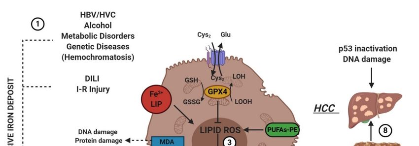
Accumulation of autophagic vacuoles [3]
                                                                                                    LC3-I to LC3-II
                                    Lack of chromatin condensation [5]
 Autophagy                                                                                           conversion [3]
                          Formation of double-membraned autolysosomes,
                                                                                                Substrate degradation [5]
                          including macroautophagy, microautophagy and
   Int. J. Mol. Sci. 2020, 21, 4908 chaperone-mediated autophagy [3]                                                   3 of 23

4. Mechanisms of Ferroptosis
     4. Mechanisms of Ferroptosis
     Ferroptosis is a sum of many biological pathways, acting simultaneously. In Figure 1, ferroptosis
         Ferroptosis is a sum of many biological pathways, acting simultaneously. In Figure 1, ferroptosis
regulatory pathways
    regulatory pathways areare
                            reported.
                               reported.Three
                                         Threemain biologicalaxes
                                              main biological  axesare
                                                                     are roughly:
                                                                       roughly:
1.   1.glutathione/glutathione
           glutathione/glutathione   peroxidase
                                       peroxidase44 (GSH/GPX4)          pathway,
                                                       (GSH/GPX4) pathway,          inhibition
                                                                                  inhibition      of system
                                                                                             of system          Xc , sulfur
                                                                                                         Xc − , sulfur
                                                                                                                         -

      transfer  pathway,
           transfer pathway, and  p53
                                and  p53regulatory
                                          regulatory axis;
                                                      axis;
2.    iron
     2.    iron metabolism with the regulation ofautophagy
            metabolism     with   the  regulation    of                protein5 5and
                                                         autophagy protein         and  7 (ATG5-ATG7)
                                                                                      7 (ATG5-ATG7)    and and     nuclear
                                                                                                             nuclear
      receptor    coactivator
           receptor  coactivator44 (NCOA4)
                                    (NCOA4)pathway pathwayand and    iron-responsive
                                                              iron-responsive             element-binding
                                                                                element-binding                  protein 2
                                                                                                   protein 2 (IREB2)
      (IREB2)   related
           related       to ferritin
                   to ferritin        metabolism,
                               metabolism,    and theand   the p62-Kelch-like
                                                       p62-Kelch-like             ECH-associated
                                                                        ECH-associated               protein 1 (Keap1)-
                                                                                          protein 1 (Keap1)-nuclear
           factor
      nuclear     erythroid
                factor        2-related
                       erythroid         factor (Nrf2)
                                    2-related    factorregulatory   pathwayspathways
                                                        (Nrf2) regulatory     [3]; and [3]; and
3.   3.    lipid metabolism    pathways    as p53, arachidonate
      lipid metabolism pathways as p53, arachidonate lipoxygenaselipoxygenase  15 (ALOX15), 15 acyl-CoA
                                                                                                 (ALOX15),synthetase
                                                                                                                 acyl-CoA
           long-chain
      synthetase       family member
                    long-chain     family4 (ACSL4),
                                             member lysophosphatidylcholine        acyltransferase 3 (LPCAT3)
                                                        4 (ACSL4), lysophosphatidylcholine                        [3].
                                                                                                      acyltransferase     3
     (LPCAT3) [3].

          Figure 1. This figure summarizes the regulatory core of ferroptosis, approximately divided into
          three
     Figure      axes.figure
              1. This   The first axis is represented
                              summarizes                in the core
                                             the regulatory    middle.    It includes the
                                                                     of ferroptosis,        GSH/GPX4, sulfur
                                                                                        approximately     dividedtransfer
                                                                                                                    into three
          and  p53  pathways.     The second   axis (right  part of theF)  consists  of  the iron  metabolism
     axes. The first axis is represented in the middle. It includes the GSH/GPX4, sulfur transfer and p53       pathway,
          including IREB2 related to ferritin metabolism, the regulation of ATG5-ATG7-NCOA4 pathway and the
     pathways.    The second axis (right part of theF) consists of the iron metabolism pathway, including
          p62-Keap1-Nrf2 regulatory pathway. These elements can influence the concentration of intracellular iron,
     IREB2 related to ferritin metabolism, the regulation of ATG5-ATG7-NCOA4 pathway and the p62-
          mandatory for the development of ferroptosis. On the left, the third axis implies that lipid metabolism
     Keap1-Nrf2     regulatory pathway. These elements can influence the concentration of intracellular iron,
          p53-spermidine/spermine N1-acetyltransferase 1 (SAT1)-ALOX15, ACSL4, LPCAT3, etc. impact on
     mandatory
          fatty acids the
                   for     development
                       regulation           of ferroptosis.
                                   and ferroptosis           On themitochondria
                                                      [3]. Finally,   left, the thirdare
                                                                                       axis  implies
                                                                                          also        thatsince
                                                                                                involved,  lipid VDACs
                                                                                                                 metabolism
     p53-spermidine/spermine
          (voltage-dependent anion   N1-acetyltransferase
                                        channels) are inhibited1 (SAT1)-ALOX15,         ACSL4,
                                                                  by erastin. In parallel,         LPCAT3, etc.
                                                                                              the independent      impact on
                                                                                                                pathway
     fattyferroptosis
            acids regulation
                       suppressorand  ferroptosis
                                   protein           [3]. Q10
                                            1-coenzyme     Finally,  mitochondria
                                                               (FSP-1-CoQ10)           are GSH/GPX4
                                                                                 acts with   also involved,    sincelipid
                                                                                                        to contrast   VDACs
          peroxidation [3]. anion channels) are inhibited by erastin. In parallel, the independent pathway
     (voltage-dependent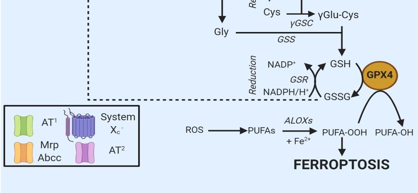
Int. J. Mol. Sci. 2020, 21, 4908                                                                     4 of 23

4.1. Radical Oxygen Species (ROS)
      Oxygen (O2 ) is an essential element for aerobic living beings. Cellular membranes are permeable
to gas; thus, oxygen permeates freely into the cells, where it undergoes several processes. Oxygen
is used in the mitochondria to provide energy through oxidative phosphorylation (OXPHOS) [6].
During cellular respiration and the degradation of organic compounds via graded oxidative reactions,
radical oxygen species (ROS) are inevitably produced [7]. Therefore, while essential for life, oxygen
participates also in the demise of the organism [8]. Before discussing ROS, it is necessary to define
the term “radical”. In chemistry, a radical is an atom, molecule or ion with unpaired highly reactive
valence electron. A radical undergoes dimerization reactions spontaneously.
      ROS are tightly monitored by cells since they play a pivotal role in tissue homeostasis [9].
In excessive amount, ROS cause cellular toxicity and their concentrations need to be tightly controlled;
at the same time, low concentrations of ROS are used as a defensive weapon, since their release in
the extracellular environment protects cells from bacterial invasion as well as intracellular signaling
molecules. Cellular oxidative stress is defined as a disequilibrium between the oxidative and antioxidant
systems, and it appears to be essential in the occurrence of ferroptosis.
      Several types of ROS are generated during the ferroptotic process, which can be divided into four
main groups: (i) superoxide (O2 − ), which is produced because of electron reaction with O2 at complex
I/III of the mitochondrial electron transport chain and which is rapidly dismutated to hydrogen
peroxide (H2 O2 ) [9]; (ii) hydrogen peroxide (H2 O2 ), which is moderately reactive and responsible for
protein oxidation [9]; (iii) peroxyl radical (OH• ), which is formed from H2 O2 via the Fenton reaction
and is the most reactive ROS [9]; and (iv) lipid peroxides, which are obtained from the oxidation of
polyunsaturated fatty acids (PUFAs) [9].
      During ferroptosis, the Fenton reaction is the primary source of ROS. The Fenton reaction
describes the formation of hydroxide (OH− ) and hydroxyl (OH• ) radicals by a reaction between Fe2+
and hydrogen peroxide (H2 O2 ) (Equation (1)).

                                    Fe2+ + H2 O2 → Fe3+ + OH− + OH                                      (1)

     When Henry J.H. Fenton discovered the Fenton reaction, he never mentioned the existence of
the hydroxyl radical intermediate (OH• ). In 1934, Haber and Weiss proposed a modified version of
the Fenton reaction. They hypothesized the existence of OH• via reaction of hydrogen peroxide and
superoxide with iron as a catalyst. The Haber–Weiss (Equations (2) and (3)) cycle is a two-step reaction,
where Fe3+ is reduced to Fe2+ via reaction with superoxide (O2 − ), which in turn reacts with H2 O2 to
form OH− and OH• , regenerating ferric ion.

                                        O2 − + Fe3+ → Fe2+ + O2                                         (2)

                                    Fe2+ + H2 O2 → Fe3+ + OH− + OH                                      (3)

4.2. Regulation of Ferroptosis via Cysteine-Glutathione Redox Axis
     Oxidative stress triggers ferroptosis. Redox glutathione (GSH) plays a crucial role in ferroptosis,
since it represents the ideal substrate for glutathione peroxidase 4 (GPX4) and is indispensable for
preventing ferroptosis. In vitro ferroptosis is typically induced after cysteine starvation, GSH depletion
and inhibition of GPX4. Intracellular cysteine concentration is usually maintained low partly due to its
cytotoxic effects; regardless, cysteine is directly required for the synthesis of the glutathione tripeptide
(Cys-Gly-Glu). Additionally, cysteine is a versatile molecule; cells carry out conversions of the amino
acid into a variety of reactive compounds such as taurine, cysteamine, coenzyme A and Fe-S clusters.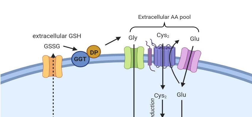
Int. J. Mol. Sci. 2020, 21, 4908                                                                     5 of 23

4.2.1. Biosynthesis of Glutathione
      Glutathione is a conserved antioxidant in plants, mammals, bacteria and archaea.
Moreover, the conjugation of toxic molecules with GSH helps in the detoxification of the cells
from dangerous substances ([7–10]). GSH, the most abundant and available form of thiol in the
cell is found in both the cytosol and the organelles, with concentrations ranging between 0.5 and
10 mM [7]. Glutathione exists in reduced (GSH) and oxidized states (GSSG), and the ratio between
the two indicates the cellular oxidative stress degree. Typically, cells contain more than 90% of GSH,
while the rest is in the disulfide form [7]. In biological reactions, GSH is a cofactor, that donates its
hydrogen while generating GSSG. Then, the enzyme glutathione reductase (GSR) converts GSSG to
GSH using NADPH/H+ as a cofactor:

                                   NADPH + GSSG + H2 O → 2 GSH + NADP+ + OH−                            (4)

      The biosynthesis of GSH involves two ATP-dependent steps: The first reactions is the synthesis of
γ-glutamylcysteine from the Glu and Cys. This reaction is under the control of γ-Glu-Cys ligase (GLC),
and it represents the rate-limiting step of GSH synthesis. Besides, it underlines GSH’s importance for
life since GLC knockout is lethal [11]. The second reaction is the addition of Gly to the C-terminal
γ-glutamylcysteine, and this passage is catalyzed by glutathione synthetase (GSS).

4.2.2. Uptake of Cysteine
     A balanced diet plan provides the correct amount of Cys required for GSH biosynthesis [11]. In the
extracellular environment, Cys and cystine (cysteine dipeptide, Cys2) are present as GSH and GSSG,
extruded from the cells via the multidrug resistance-associated proteins (MRP/ABCC), expressed under
the control of nuclear factor erythroid 2-related factor (Nrf2) [11]. Once outside the cell, both Cys and
GSH are oxidized to GSSG and Cys2.
     In most cells, ubiquitously expressed and rather nonspecific neutral amino acid transporters
internalize Cys [12]. Besides, cells express two specific Cys transporters: system b0+ and system Xc − .
The first one is exclusively expressed in kidneys [7]. In contrast, system Xc − is constitutively expressed
in various organs (e.g., brain and immune system) or induced in others in the presence of oxidants
or electrophiles [12]. Cystine is internalized by the cells after the reduction of Cys with unknown
modalities. At the same time, GSH or GSSG are hydrolyzed by γ-glutamyl transpeptidase (GGT),
an enzyme facing the outer layer of the membrane (Figure 2) [11]. The hydrolysis produces di- or
tetrapeptides containing Gly and Cys or two Gly and Cys2, allowing the recycling of cysteine.
     Finally, some other amino acids or peptide transporters exist, which complicate the general scheme.
Of note, in 2015, Newstead proposed the existence of proton-coupled oligopeptide transporter family
members (PEPT1 and PEPT2) at the extracellular membrane in mammalian cells. These transporters
internalize the Cys-Gly dipeptide generated after the dissociation of GSSG by GGT [13].
Int. J. Mol. Sci. 2020, 21, 4908
Int. J. Mol. Sci. 2020, 21, 4908
66of 23
of 23
Figure 2. Oxidized
Oxidized GSHGSH (GSSG)
(GSSG) isis exported
exported from from the
the cell
cell via
via the MRP/ABCC transporter and
hydrolyzed by byGGT
GGTand anddipeptidase
dipeptidase to to
Gly,Gly,
GluGluand and
Cys2Cys 2, thus
, thus contributing
contributing to theto the extracellular
extracellular amino
amino γ-glutamylcysteine
γ-glutamylcysteine acidSpecific
acid pool. pool. Specific amino
amino acid acid transporters
transporters (AT1 and (AT1
AT2)and AT2) internalize
internalize Gly
Gly and Glu,
and Glu,
while Cys2while
is taken 2 is
Cysup bytaken
systemupXby − [11].
system X
Once c - [11]. Once in the cell, Cys2 is reduced to Cys and
in the cell, Cys is reduced to Cys and combined with
c 2
combined with Glu
Glu to generate to generate
γGlu-Cys γGlu-Cys
[11]. The addition[11].
of GlyThetoaddition of Glycatalyzes
the dipeptide to the dipeptide catalyzes
the formation the
of GSH,
formation of GPX4.
a cofactor of GSH, aThecofactor
box onof the
GPX4. The left
bottom boxindicates
on the bottom left indicates
the proteins the proteins involved.
involved.
4.2.3. System
4.2.3. Xcc- −
System X
The impact
The impact of of GSH
GSH levels
levels on on ferroptosis emerged from
ferroptosis emerged from thethe use
use of
of erastin,
erastin, aa compound
compound that that
decreases the intracellular concentration of GSH by targeting the cystine/glutamate
decreases the intracellular concentration of GSH by targeting the cystine/glutamate antiporter system antiporter system
X − [2]. In system X − , sodium-independent antiporter imports cystine and exports glutamate with a
Xcc- [2]. In system Xc-c, sodium-independent antiporter imports cystine and exports glutamate with a
− determines a decrease in GSH
ratio 1:1 in
ratio 1:1 in anan ATP-dependent
ATP-dependent manner manner [2].
[2]. Inhibition
Inhibition of of system
system XXcc- determines a decrease in GSH
levels and
levels and the
the beginning
beginning of of ferroptosis.
ferroptosis. The The system
system was was initially
initially characterized
characterized in in human
human fetalfetal lung
lung
fibroblasts in 1980 [14]. Then, it became clear that the induction of the antiporter
fibroblasts in 1980 [14]. Then, it became clear that the induction of the antiporter was tissue-specific, was tissue-specific,
and that
and that itit occurred
occurred in in several organs such
several organs such asas lung,
lung, kidney and liver,
kidney and liver, under
under conditions
conditions of of oxidative
oxidative
stress [12].
stress [12].
Once internalized,
Once internalized, cystine
cystine is is rapidly
rapidly reduced
reduced to to cysteine,
cysteine, the
the limiting
limiting precursor
precursor forfor glutathione
glutathione
GSH synthesis [11]. To assure the
GSH synthesis [11]. To assure the import of Cys import of Cys , intracellular level of glutamate is maintained
2 2, intracellular level of glutamate is maintained high so
high
thatthat
so it can be easily
it can be easilyexported.
exported.Increasing the concentration
Increasing the concentration of extracellular glutamate
of extracellular mightmight
glutamate be a solid
be a
competitive inhibition strategy to prevent cystine uptake and induce
solid competitive inhibition strategy to prevent cystine uptake and induce ferroptosis. ferroptosis.
The system − structure consists of two subunits, the 4F2 heavy chain (4F2hc/CD98/SLC3A2)
The system X Xcc- structure consists of two subunits, the 4F2 heavy chain (4F2hc/CD98/SLC3A2)
and a light chain (xCT/SCL7A11).
and a light chain (xCT/SCL7A11). The The latter
latter subunit
subunit exhibits
exhibits homology
homology with with the
the light
light chains
chains ofof
heterodimeric amino acid transporters (HATs), a family of amino acid transporters
heterodimeric amino acid transporters (HATs), a family of amino acid transporters formed by a light formed by a light
and aa heavy
and heavy chain
chain linked
linked by by aa disulfide
disulfide bridge
bridge [15].
[15].Int. J. Mol. Sci. 2020, 21, 4908 7 of 23
4.2.4. Cysteine Synthesis via the Transsulfuration Pathway
The transsulfuration pathway, in conjunction with methionine metabolism, metabolically produces
Cys. Methionine can be converted to Cys to fulfill the requirement in some organs under normal
physiological conditions [7]. Methionine is first converted to homocysteine, which is transformed into
cystathionine and finally to Cys. In this peculiar context, the carbon backbone of Cys derives from
serine, while cystathionine serves as a sulfur donor [7].
This pathway helps to maintain the supply of Cys in some organs, notably in the liver,
thus preventing ferroptosis from arising after inhibition of system Xc − . Primary hepatocytes can
survive for several days without Cys or cystine in culture media, thanks to the protective role of this
pathway [16]. When cysteinyl-tRNA synthetase is knocked down, the transsulfuration pathway is
activated, and this leads to the inhibition of Cys-deprivation ferroptosis [17]. This result underlines a
connection between protein synthesis and Cys metabolism. It is also necessary to mention that Cys can
derive from intra-lysosomal protein degradation. After proteolysis, Cys2 is released into the cytosol via
cystinosin, H+ -driven lysosomal transporter (SLC66A4) and it becomes available for GSH synthesis.
In humans, a defect of cystinosin leads to rare hereditary disease cystosinosis, caused by crystallization
of Cys2 in the lysosomes [7–10].
4.3. The Enzyme Glutathione Peroxidase 4 and Ferroptosis
The O2 -dependent oxidation of carbon fuels in the mitochondria allows for the generation
of abundant ATP, but gives rise to partially reduced oxygen species [18]. ROS are generated
by mitochondria as well as by enzymes involved in lipid metabolisms such as lipoxygenases,
cyclooxygenase, cytochrome P450 and NADPH oxidases [8–10]. Oxidation of cholesterol and
phospholipids containing PUFAs by lipoxygenases can lead to lipid peroxidation, membrane damage
and ultimately cell death [18]. Lipid hydroperoxides (R-OOH) can modify membrane structure and
function. Moreover, these lipid hydroperoxides are unstable, and, in the presence of redox-active
iron, their structures can change generating small-molecule reactive electrophiles, which lead to new
free radical reactions [8–10]. The rupture of fatty acids backbone can also lead to the formation of
reactive aldehydes such as 4-hydroxy-nonenal (4-HNE) and malondialdehyde (MDA), which can
attack proteins or DNA, amplifying the cellular damage [8].
To ensure membrane integrity and minimize ROS-incurred damage, lipid hydroperoxide
glutathione peroxidase 4 (GPX4) converts lipid hydroperoxides (R-OOH) to lipid alcohols (R-OH)
using reduced GSH as a cofactor [8]. This process prevents the iron (Fe2+ )-dependent formation and
accumulation of toxic lipid ROS, more toxic than cytosolic ROS in ferroptosis [18]. However, lipids
such as eicosanoids, derived from enzymatically oxygenated PUFAs, play a significant signaling role,
especially in mediating inflammation. Thus, GPX4 has a double function in cellular homeostasis: as a
guardian against oxidative damage as well as the physiological regulator.
For this reason, GPX4 seems to be vital for development. Indeed, GPX4−/− mouse embryos die
right after implantation at Day 7.5. GPX4 is defined as the master regulator of ferroptosis, and its direct
or indirect inhibition with small compounds leads to ferroptosis [8–19].
From a structural point of view, GPX4 is a monomeric enzyme with a molecular size smaller
than the subunits of the other GPXs, and this structure affords a broader substrate specificity than
other glutathione peroxidases [20]; this allows GPX4 to react directly with LOOHs in membranes with
high efficiency.
The pharmacological inhibition of GPX4 triggers the ferroptotic process by the accumulation of
lipid peroxides. On the one hand, the induction of ferroptosis may help get rid of therapy-resistant
cancer cells, thus enhancing the effects of chemotherapy or radiotherapy and opening up new
therapeutic fields [8]. On the other hand, GPX4 could be neuroprotective, and increasing GPX4 function
could be a therapeutic strategy for neurodegenerative diseases [8–10].Int. J. Mol. Sci. 2020, 21, 4908 8 of 23
4.4. Lipid Metabolism and Ferroptosis
Oxidative damage of membrane lipids is central to the execution of ferroptosis. Peroxidation
can occur on both free PUFAs and PUFA-containing membrane phospholipids (PUFA-PLs)
(e.g., phosphatidylethanolamine, phosphatidylcholine and cardiolipin). Treatments with different
ferroptosis inducers produce a different type of oxidized PL species, and only some of these oxidized
species contribute directly to cell death [21]. PUFA-containing phosphatidylethanolamines (PEs) are
more susceptible to peroxidation than PUFAs with phosphatidylcholine (PC), because of PEs’ molecular
geometry. PEs assume a more conical shape due to the smaller size of the PE head-group. Peroxidation
can develop through enzymatic reactions or non-enzymatic processes. In particular, lipoxygenases are
the enzymes most involved in the generation of lipid peroxides. The second model affirms that the
majority of lipid peroxidation arises due to the spontaneous, non-catalytic process of autoxidation
with Fenton chemistry [10–22].
4.4.1. Role of Lipoxygenases
The formation of lipid peroxides of arachidonic acid (AA) and other ω-6 fatty acids containing
PE requires three enzymes: arachidonate lipoxygenases (ALOXs), acyl-CoA synthetase long-chain
family 4 (ACSL4) and lysophosphatidylcholine acyltransferase 3 (LPCAT3) [23]. ACSL4 catalyzes the
formation of AA-CoA, then LPCAT3 controls the esterification of AA-CoA into AA-PE, and the final
oxidation of AA-PE to AA-OOH-PE is run by ALOXs.
The ACSL proteins are mainly localized at the endoplasmic reticulum (ER) and at the outer
mitochondrial membrane (OMM). ACSLs are responsible for the formation of fatty acyl-CoA esters
from free long-chain fatty acids. There are five isoforms of ACSLs (ACSL1, -3, -4, -5 and -6), but only
ACSL4 correlates with ferroptosis, and it is considered the marker of ferroptosis sensitivity. GPX4−/−
leads to ferroptotic cell death, while GPX4−/− and ACSL4−/− double knockout cells survive and usually
grow [24]. ALOXs are crucial regulators of enzymatic lipid peroxidation. ALOX enzymes are non-heme
iron-containing dioxygenases that oxidize PUFAs in a regio-, stereo-, and enantio-specific manner [22].
They catalyze the insertion of oxygen in a molecule of arachidonic acid to generate lipid peroxides.
ALOX enzymes prefer free PUFAs as substrates for oxidation. Because of this, phospholipases are
likely associated with LOX in cleaving the PUFA acyl chain from PLs. The mechanisms by which LOXs
drive ferroptotic cell death and the isoforms that drive this process remain elusive [25].
4.4.2. Lipid Peroxidation
Non-enzymatic lipid autoxidation accounts for three main steps. In membranes, initiation is
operated by electron transfer from Fe2+ to LOOH through the Fenton reaction, producing the alkoxy
radical (LO• ). In particular, Fenton chemistry refers to a series of reactions between LOOHs and Fe2+
to produce oxygen radicals (Section 4.1). In physiological conditions, cellular iron is complexed with
ferritin or with heme proteins, and only a small amount of iron remains free, soluble and chelatable.
The latter is called the labile iron pool (LIP), and it represents the source of iron for the Fenton reaction.
LOOH + Fe2+ → LO• + Fe3+ + H2 O (5)
At this point, LO• interacts with a neighboring lipid collecting an H (L’H), thus forming a
carbon-centered radical (L’ • ) and then L’ • reversibly adds oxygen to form a lipid hydroperoxyl radical
(L’OO• ). The creation of new lipid radicals represents the second step (propagation).
LO• + L’H → LOH + L’ (6)
L’• + O2 ← → L’OO (7)Int. J. Mol. Sci. 2020, 21, 4908 9 of 23
From LOO• , LOOH is generated by H transfer, which stabilizes the reversible oxygen addition.
When H is donated by a CH2 carbon of an unsaturated fatty acid esterified in a complex lipid, the newly
formed L’ • adds oxygen, thereby propagating lipid peroxidation.
LOO• + L’H → LOOH + L’ (8)
The final step is the termination of the process. It happens either in the presence of antioxidants
or by another radical PUFA. LOO• can be stabilized by H donors such as tocopherols or coenzymes
Q, which act as antioxidants (Aox-OH). This reaction is called “chain-breaking” because Aox-O• is
not able to propagate the peroxidative chain reaction. In the presence of Fe2+ , LOOH reacts with the
reduced transition metal, and the peroxidation-initiating species LO• is produced (Reaction 1).
LOO• + Aox-OH → LOOH + Aox-O (9)
In the presence of a large amount of LOO• , radicals react together to form stable products and
arrest the reaction. A hydroxyl and a keto derivative of the fatty acid chain (LOH and L = O) are
generated, while oxygen is released in an electronically excited state that decays while emitting light.
2 LOO• → LOH + L = O + O2 (10)
4.4.3. Where Does Lipid Peroxidation Take Place?
Lipid peroxidation affects all cellular membranes. It affects the lipid bilayer and the subcellular
membranes of mitochondria, endoplasmic reticulum (ER) and lysosomes. Mitochondria might promote
ferroptosis under specific conditions. Mitochondrial respiration may contribute to the overall pool of
ROS in the cell. However, these organelles are not essential for ferroptosis, because cells deprived of
mitochondria are exposed to ferroptosis [25]. However, mitochondrial membranes may also be oxidized
during ferroptosis; although this oxidation does not appear to be required for death, the accumulation
of LOOHs at the mitochondrial membranes could increase permeability, thus explaining mitochondrial
swelling [2] and outer membrane rupture [26]. The ER is the first intracellular site of de novo lipid
synthesis; thus, lipid peroxidation might happen here. A possible association between ER and
ferroptosis could be evaluated, but the onset of lipid peroxidation in the ER and its relationship to
ferroptosis is still unclear [25].
Finally, lysosomes can be engaged in ferroptosis. Indeed, ferrostatins, which are ferroptosis
inhibitors, localize at the lysosomes, and structural modifications reduce their trapping by the lysosomes,
thus increasing their efficacy in suppressing ferroptosis [27]. Moreover, the discovery of ironomycin,
an iron-dependent lethal compound localized at the lysosomes, suggests that additional studies
concerning the role of lysosomes in ferroptosis are needed [25].
The role of lipid peroxidation is undisputed, but it remains unclear how lipid peroxidation exerts
a toxic effect on the cell and leads to cell death. At least three hypotheses have been proposed to
answer these questions [22]: (i) LOOH accumulation could modify membrane integrity and alter its
biophysical proprieties; (ii) LOOHs may change the localization or function of membrane-associated
proteins; and (iii) degradation of LOOHs into highly reactive products may help in the permeabilization
of membranes, and this could be directly toxic.
Lipid peroxidation disrupts ion gradients, decreases membrane fluidity, slows lateral diffusion
and increases membrane permeability [25]. Moreover, the formation of protein-based pores during
ferroptosis has been hypothesized, leading to loss of ionic homeostasis [28].
Lipid peroxidation of PUFAs generates different oxidation products, some of them highly cytotoxic
and pathogenic. The best known are MDA and 4-HNE. MDA is obtained from the decomposition of
AAs and larger PUFAs via enzymatic and non-enzymatic pathways [25]. MDA reacts with proteins and
DNA to form crosslinked adducts. Overload of MDA is associated with many human diseases such as
Alzheimer’s and Parkinson’s diseases, cancer, cardiovascular diseases and diabetes [22]. The followingInt. J. Mol. Sci. 2020, 21, 4908 10 of 23
product generated is 4-HNE, an electrophile compound, widely studied as a signaling molecule, known
for its ability to stimulate the cell cycle and cell proliferation and as a cytotoxic molecule. In particular,
4-HNE can inhibit gene expression and promote the onset of diseases.
4.5. Iron Metabolism and Ferroptosis
Iron homeostasis is strictly controlled at the cellular and systemic levels to provide the right
amounts of iron needed for the vital biological functions of cells and tissues, avoiding iron overload
and iron-related toxicity [29]. Iron trafficking is an example of a circular economy [30]; the human
body’s daily iron requirement is around 20–25 mg, which is mainly used in erythropoiesis. A balanced
diet provides about 14 mg of iron as inorganic or organic iron [29]. In an adult at a steady-state, 1–2 mg
iron is absorbed daily in the gut and compensates for an equal loss (bleeding and desquamation).
Since an insufficient amount of iron is absorbed, most of the iron is recycled from the phagocytosis of
senescent red blood cells by macrophages.
In our body, iron is found in both ferrous (Fe2+ ) and ferric (Fe3+ ) forms; it is used for the synthesis
of metalloproteins, to form organic (e.g., heme) or inorganic cofactors (e.g., Fe/S clusters) [31].
Heme and non-heme iron from dietary sources exhibit different absorption, once they arrive in
the gut. Heme carrier protein-1 (HCP-1) internalizes iron in the heme form, but not the non-heme
form. Heme is rapidly catabolized by heme oxygenase 1 (HO-1), and iron is released [31].
Inorganic iron is found in the gut in its ferric form. Duodenal cytochrome B reductase (DCYTB)
reduces the ferric into the ferrous form, and then the apical divalent metal transporter 1 (DMT1)
imports Fe2+ . At the basolateral membrane of enterocytes, ferroportin (FPN), the principal iron
exporter, transports Fe2+ . At the basolateral side, Fe2+ is oxidized to Fe3+ by membrane ferroperoxidase
(hephaestin or ceruloplasmin) before being captured by transferrin (Tf) in the plasma. Hepatocytes
produce Tf, which distributes iron to all organs, including the sites of use (bone marrow) and storage
(liver) [31]. Binding to its ubiquitous transferrin receptor protein-1 (TRF1), transferrin delivers iron to
cells through the endosomal cycle: the Fe-Tf/TRF1 complex is internalized by endocytosis, and iron is
released from Tf in the acidic endosomes. Here, the six-transmembrane epithelial antigen of prostate 3
metalloreductase (STEAP3) reduces Fe3+ , thus enabling the endosomal DMT1 to export iron. At this
point, the acquired iron can be stored in ferritin or transported by FPN to maintain the labile iron pool
at a low level to avoid cell toxicity. Excess cellular iron promotes lipid peroxidation through the Fenton
reaction and production of ROS, which then triggers ferroptosis. Several studies have demonstrated
the connection between iron metabolism genes and increased sensitivity to ferroptosis. Silencing of
TRFC, the gene encoding TRF1, can inhibit erastin-induced ferroptosis, preventing the accumulation of
LIP, while HO-1 accelerates the ferroptotic process by supplementing iron [32].
Furthermore, shRNA-mediated silencing of iron-responsive element-binding protein 2 (IREB2)
alters the expression of many iron genes such as TRFC, FTH1 and FTL, thereby modifying iron uptake,
metabolism and storage [2]. In 2015, Sun et al., described the potential involvement of heat shock
proteins (HSPs) in ferroptosis. They demonstrated that heat shock factor-binding protein 1 (HSPB1)
was highly inducible after treatment with erastin in several types of cancer cells. In HeLa cells,
phosphorylation of protein kinase C (PKC) activates HSBP1. Once activated, HSBP1 reduces iron levels
by inhibiting TRF1 expression [33]. In summary, phosphorylated HSPB1 acts as a negative regulator of
ferroptosis, reducing the uptake of iron and lipid peroxidation, while inhibition of HSBP1 increases
erastin-mediated ferroptosis both in vivo and in vitro [33].
The role of iron in ferroptosis is now established, but the connection between the inorganic metal
and the biological process remains unclear. The development of new fluorescent probes to detect LIP
levels in various cells or organs in live animals could help improve understanding of the role of iron in
ferroptosis in multiple conditions. In 2019, Hirayama et al., managed to establish an imaging method
to monitor organelle-specific labile Fe2+ , in mitochondria, lysosomes and endoplasmic reticulum at the
same time during erastin-mediated ferroptosis [34].Int. J. Mol. Sci. 2020, 21, 4908 11 of 23
4.6. p53-Mediated Ferroptosis
p53 is a tumor suppressor defined as the “guardian of the genome” because of its role in preserving
the stability of the DNA by preventing mutations. p53 works as an antitumor protein with precise
functions in apoptosis, necrosis and autophagy. p53 antitumoral activity is due to its DNA-binding
transcriptional factor role. [23]. In 2012, Li et al., showed that posttranslational modifications of p53 have
a substantial impact on its transcriptional and oncosuppressive functions. The acetylation-defective
mutant referred to as p533KR fails to induce apoptosis and cell cycle arrest; however, the tumor
suppression capacity is retained by p533KR as a wild-type p53, suggesting an additional pathway
that mediates tumor development. One target p533KR is SLC7A11, a gene encoding for the subunit
xCT/SCL7A11 of system Xc − . The binding downregulates the expression of SLC7A11, thereby affecting
the activity of GPX4. The transcriptional repression of SCL7A11 results in the reduction of antioxidant
capacity, ROS accumulation and ferroptosis instauration [3]. p534KR98 , a genetic variant with four
mutations of acetylated lysine residues, loses tumor suppression activity [35]. Therefore, acetylation of
the residue Lysine 98 is essential for p53-mediated ferroptosis [23]. Genetic inactivation of SCL7A11 and
inhibition of system Xc − impair cystine uptake and GSH synthesis, causing lipid peroxide accumulation
and ferroptosis.
p53 is also involved in polyamine metabolism, which impacts cellular growth, development,
and programmed cell death [35]. p53 can also negatively regulate ferroptosis: this anti-ferroptosis
regulator activity depends on p53 protein stabilization and trans-activation of CDKN1A/p21
(cyclin-dependent kinase inhibitor 1A), responsible for cycle cell arrest. Cell treatment with nutlin-3
(inhibitor of MDM2/p53 interaction) increases p53 expression and stability and, at the same time,
inhibits erastin-induced ferroptosis in p53+/+ cells, but not in p53−/− cells [10]. Cells treated before with
nutlin-3 and then with erastin show a delay in the onset of ferroptosis. Moreover, nutlin-3 upregulates
CDKN1A mRNA and p21 protein in p53+/+ cells, but not in p53−/− cells [10–35]. The p53/p21 axis
delays the execution of ferroptosis by reducing the consumption of GSH, but it is not clear how p21
promotes GSH biosynthesis. In colorectal cancer (CRC), the translocation of dipeptidyl-peptidase 4
(DPP4) by p53 to the nucleus prevents the onset of ferroptosis. Moreover, p53 promotes the expression
of SCL7A11 in CRC cells, but, at the same time, it inhibits the expression of the same in other tumor
cells (U2OS and MCF7 cells). It is possible to conclude that p53 plays dual regulatory functions in
ferroptosis regarding its target genes (SCL7A11, SAT1 and CDKN1A/p21), and its role could depend on
tumor cell type [23].
4.7. Ferroptosis and the Keap1–Nrf2 Pathway
The nuclear factor erythroid 2-related factor (Nrf2) is a crucial regulator of the antioxidant
response [36] and the inducible cell defense system [37]. It is a basic leucine zipper (bZIP) transcription
factor, and it heterodimerizes with its partners, small Maf proteins [37]. Under physiological conditions,
Kelch-like ECH-associated protein 1 (Keap1) helps maintain low levels of Nrf2, stimulating its
ubiquitination and proteasomal degradation. Conversely, during oxidative stress, Nrf2 protein is
derepressed and stabilized, and it initiates a multistep pathway activation [36]. This process is strictly
under the control of p62, an autophagy receptor, a multifunctional protein, which inhibits Keap1
directly and promotes Nrf2 activation simultaneously. In 2016, Sun et al., demonstrated that the
p62-Keap1-Nrf2 pathway inhibits ferroptosis in hepatocellular carcinoma (HCC) [36].
Nrf2 translocates to the nucleus, it binds to antioxidant response elements (AREs) located
in the regulatory regions and activates transcription of cytoprotective genes while undergoing
heterodimerization. Nrf2 activates multiple cell defense mechanisms, thereby potentiating the
detoxification activity of cells [37]. Nrf2 regulates a plethora of targets such as genes involved in the
regulation of synthesis and conjugation of GSH (e.g., GCL, GSR, etc.) or genes encoding antioxidant
proteins (e.g., thioredoxin (TXN) and thioredoxin reductase 1 (TXNRD1)). Heme oxygenase 1 (-HO-1),
ferrochelatase (FECH) and both ferritin heavy and light chains are strictly under the control of Nrf2,
as well.Int. J. Mol. Sci. 2020, 21, 4908 12 of 23
Nrf2−/− mice are more susceptible to drug-induced toxicity and oxidative stress [38].
Therefore, the enhancement of Nrf2 expression through pharmacological or genetic approaches
could reduce the level of ROS, avoiding the intensification of stress-induced diseases (e.g., acute lung
injury, diabetic nephropathy, heart failure and cancer) [37]. However, the activation of Nrf2 shows
considerable side effects, since an elevated activity of Nrf2 decreases the therapeutic response of
cancer cells to radiotherapy and chemotherapeutic treatments. Thanks to this, cancer cells increase
their capacity for detoxification of dangerous secondary products (e.g., ROS) and can generate
continuously [39]. Several lines of evidence of Nrf20 s double role are progressively accumulating in
the literature. For instance, in 2020, Liu et al., demonstrated that prolonged administration of erastin
induced ferroptosis resistance in ovarian cancer cells [40]. They observed a sustained upregulation of
cystathionine-β-synthase (CBS), which converts homocysteine into cystathionine in the transsulfuration
pathway (see Section 4.2.4). In addition, Nrf2 was found to be constitutively activated and positively
correlated with CBS induction. In conclusion, the activation of Nrf2/CBS accounted for ferroptosis
resistance in ovarian cancer cells [40]. However, Nrf2 guards cells against stress-induced diseases,
such as ischemia–reperfusion (I-R) injury. The protective role of Nrf2 was evaluated in renal proximal
tubular epithelial cells (RPTECs) of the native hibernator, the Syrian hamster [41]. The reoxygenation
enhanced the expression of Nrf2, leading to the upregulation of antioxidant enzymes (e.g., superoxide
dismutase 3 (SOD3) and GSR) and of anti-ferroptotic proteins (FTH1/FTL) in hamster RPTECs, rescuing
cells from reoxygenation-induced cell death [41].
4.8. Other Related Signaling Pathways
Other cellular pathways may trigger ferroptosis. Ferroptosis suppressor protein 1 (FSP1) is
a potent resistance factor and acts as a potent ferroptosis suppressor in vitro and in vivo. FSP1
contains a short N-terminal hydrophobic sequence and a FAD-dependent oxidoreductase domain [42].
The N-terminal consensus sequence is subjected to myristoylation, a fatty acid modification that
mediates FSP1 recruitment to the plasma membrane. Here, FSP1 acts as an oxidoreductase reducing
coenzyme Q10 (CoQ10) and generating a lipophilic radical-trapping antioxidant (RTA) that halts the
propagation of lipid peroxides [42]. FSP1 could provide a new target for the development of drugs
targeting the inhibition of ferroptosis [43].
Currently, the role of mitochondria in ferroptosis remains controversial. During ferroptosis,
mitochondria undergo morphological changes, including outer membrane fragmentation and cristae
enlargement. Furthermore, mitochondrial voltage-dependent anion channels (VDACs) are targets of
erastin, and their inhibition induces ferroptosis in human tumor cells. Additionally, mitochondria
have a central role in oxidative metabolism, which is indispensable for ferroptosis. In 2019,
Gao et al., demonstrated that the mitochondrion is a crucial and proactive player in cysteine
deprivation-induced ferroptosis, but not in GPX4 inhibition-induced ferroptosis [44]. They also
showed that the TCA cycle and electron transport chain promote ferroptosis, acting as a significant
source of lipid ROS. In a recent study, the role of CDGSH iron sulfur domain 1 (CISD1, also
termed mitoNEET) was investigated. CSD1 is an iron-containing outer mitochondrial membrane
protein. Functionally, it regulates the mitochondrial iron uptake and respiratory capacity. The genetic
inactivation of CSD1 increases iron-mediated intramitochondrial lipid peroxidation, which contributes
to erastin-induced ferroptosis [45].
5. Crosstalk between Autophagy and Ferroptosis
Autophagy is an evolutionarily conserved catabolic process that allows lysosomes to degrade
cytoplasmic components (e.g., unused proteins, damaged organelles and pathogens). Various stresses
can stimulate autophagy, and excessive degradation of cytosolic components can lead to the triggering
of ferroptosis. In particular, some selective autophagy, such as ferritinophagy, contribute to iron
accumulation and free radical damage during ferroptosis. Ferritin, a globular protein, is the major iron
storage protein (~4500 Fe atoms) [46].Int. J. Mol. Sci. 2020, 21, 4908 13 of 23
Ferritin is composed of two distinct ferritin subunits, the heavy (FTH1) and ferritin light chain
(FTL). The enzymatic activity of the FTH1 subunit rapidly oxidizes Fe2+ to Fe3+ , and it incorporates
iron into the shell [46].
Ferritin depletion results in an iron release, which increases the labile iron pool and subsequent
cellular toxicity. Consequently, ferritinophagy plays an essential role in maintaining iron homeostasis.
Ferritin can be degraded by two mechanisms: lysosomes and proteasomes [47]. The degradation of
ferritin via lysosome was discovered in 2014 by Mancias’s team. They identified nuclear receptor
coactivator 4 (NCOA4), which is a specific cargo receptor for the autophagic degradation of ferritin [48].
Under iron-depleted conditions, NCOA4 binds the iron-rich ferritin in the autophagosome and delivers
it to the lysosome for iron release [46]. The binding between NCOA4 and ferritin is the FTH1-specific.
In the presence of a high concentration of iron, NCOA4 is ubiquitinated by ubiquitin ligase, HERC2,
and degraded, affecting protein stability [47]. NCOA4-deficient cells fail to induce ferritinophagy,
and they are associated with decreased bioavailability of iron. The initiation of ferroptosis activates
ferritinophagy to increase the labile iron pool (LIP), which promotes ROS accumulation, which drives
the ferroptotic process. Therefore, inhibition of NCOA4 represses ferritin degradation and suppresses
ferroptosis, while its overexpression has the opposite effects [47].
6. Pharmacological Modulation of Ferroptosis
The ferroptosis pathway can be either triggered using small exogenic compounds or by modulating
physiological conditions (e.g., high/low concentration of extracellular glutamate) [49]. Ferroptosis
chemical inducers were discovered before the biological process.
6.1. Ferroptosis Inducers
A variety of different substances can lead to the induction of ferroptosis with diverse cellular
targets [3] (Table 2). The first category includes erastin, sulfasalazine and sorafenib (Figure 3).
These three small molecules bind the same transporter system Xc − .
Erastin is the prototype of the inhibitor, which directly targets system Xc − . Structural analysis of the
molecule demonstrates that the quinazolinone scaffold (Part 1) ensures drug lethality while increasing
the flexibility of the piperazine linker (Part 2) would decrease drug activity [49]. Moreover, modifications
of Parts 3 and 4 would reduce or even eliminate the inhibitory ability of erastin (Figure 3) [49]. Only
the chlorine atom of erastin is designated as a binding site with the cellular environment. Additional
groups (e.g., bromo, phenyl or furanyl groups) on Part 5 can improve the effectiveness of erastin at
its target [49]. VDAC is directly inhibited by erastin, and this can lead to mitochondrial dysfunction.
VDAC 2/3 are outer mitochondrial membrane channels, and their inhibition leads to the rupture of
the OMM. Moreover, there are pieces of scientific evidence suggesting that the cellular knockout of
VDAC2 and VDAC3, but not VDAC1, leads to erastin resistance [5].
Table 2. Ferroptosis inducers (adapted from [25]).
Class Compound Suitable for
Class Impact on Ferroptosis
Characteristics Examples In Vivo Use
Erastin,
Inhibition of Prevention of cystine import, GSH Sorafenib,
Class 1 sulfasalazine,
system Xc − depletion, loss of GPX4 activity sulfasalazine
sorafenib
Direct inhibition of Covalent interaction with GPX4
Class 2 RSL3, RSL5 No
GPX4 and inhibition of the enzyme
Depletion of GPX4
Class 3 Depletion of GPX4 and CoQ10 FIN56 Unknown
protein and CoQ10
Oxidation of iron drives lipid
Induction of lipid
Class 4 peroxidation and indirect FINO2 Unknown
peroxidation
inactivation of GPX4potently.
Sorafenib is a clinically approved multikinase inhibitor for the treatment of advanced carcinoma
(e.g., renal cell carcinoma, hepatocellular carcinoma and thyroid carcinoma) [49]. In some cancer cell
lines, drug resistance has been described after treatment with sorafenib: it has been demonstrated
that upregulation of Nrf2 and pRB can inhibit sorafenib-induced ferroptosis in hepatocellular
Int. J. Mol. Sci. 2020, 21, 4908 14 of 23
carcinoma cell lines [50].
Erastin Sulfasalazine Sorafenib
Figure 3.
Figure 3. Exogenous
Exogenous ferroptosis
ferroptosis inducers [49].
inducers [49].
After the discovery
Sulfasalazine (SAS)ofiserastin,
broadlyother
usedinducers were found
as a first-line thanks to high-throughput
anti-inflammatory drug for the screening.
treatment
Ras-selective
of rheumatoidlethal small[49].
arthritis molecules (RSL3 ferroptosis
SAS induces and RSL5) are included in
by inhibiting Xc − similar
this second
system category. These
to erastin,
molecules require
but less potently. the presence of iron, ROS and MEK in an activated state to trigger ferroptosis in
Sorafenib is a clinically approved multikinase inhibitor for the treatment of advanced carcinoma
(e.g., renal cell carcinoma, hepatocellular carcinoma and thyroid carcinoma) [49]. In some cancer cell
lines, drug resistance has been described after treatment with sorafenib: it has been demonstrated that
upregulation of Nrf2 and pRB can inhibit sorafenib-induced ferroptosis in hepatocellular carcinoma
cell lines [50].
After the discovery of erastin, other inducers were found thanks to high-throughput screening.
Ras-selective lethal small molecules (RSL3 and RSL5) are included in this second category.
These molecules require the presence of iron, ROS and MEK in an activated state to trigger ferroptosis
in Ras-mutated tumor cells. [49]. RSL3 can directly bind and inactivate GPX4, increasing the production
of lipid ROS. RSL3 binds enzymes with a nucleophilic site (e.g., cysteine, serine, selenocysteine, etc.).
RLS3 inactivates GPX4-mediated alkylation of the selenocysteine present at the reactive site [49].
The third category of ferroptosis inducers includes FIN56. Two different pathways contribute to
the induction of ferroptosis by FIN56. First, FIN56 promotes the degradation of GPX4, which requires
the catalysis of the enzyme acetyl-CoA carboxylase (ACC). Second, FIN56 binds to and supports
the activation of the enzyme squalene synthase [49]. This stimulation leads to the depletion of the
endogenous antioxidant coenzyme Q10, which is essential for preventing the accumulation of lipid
ROS, and it enhances the sensitivity of the cell to ferroptosis.
The last category includes FINO2, 1,2-dioxolane, which causes cell death in two ways: (i) the direct
oxidation of LIP; and (ii) the inactivation of GPX4 [49].
6.2. Ferroptosis Inhibitors
With the increasing relevance of the ferroptosis pathway as a pharmacological target, strategies
aiming at the inhibition of lipid peroxidation have been developed over the years. Since lipid
peroxidation can occur via enzymatic and non-enzymatic pathways, ferroptosis inhibitors can be
divided into two major groups: lipid autoxidation inhibitors (e.g., radical trapping antioxidant) and
lipoxygenase inhibitors.
The lipoxygenase inhibitors group consists of several different compounds, including iron
chelators, which can inactivate the enzyme by removal of the active site iron [51]. RTAs are compounds
that react with chain-carrying radicals, interrupting the autoxidation chain reaction. This category
includes α-tocopherol, the most biologically active form of vitamin E, ferrostatins and liproxstatins.
High-throughput screening of small molecule libraries brought to the identification of two
potent ferroptosis inhibitors: ferrostatin-1 (Fer-1) and liproxstatin-1 (Lip-1) [5]. In this screening
assay, knockout for the gene GPX4 or pharmacological inhibition of system Xc − reproduced the ideal
condition for the stimulation of ferroptosis cell death [5].
Ferrostatins inhibit lipid peroxidation associated with erastin- and RSL3-induced ferroptosis.
The activity of the first generation of ferrostatin, termed ferrostatin 1, depends on the primary aromaticInt. J. Mol. Sci. 2020, 21, 4908 15 of 23
amine (Figure 4), which specifically reduces the accumulation of lipid ROS. Pharmacokinetics analysis
on the second generation (SRS 11-92) and the third generation (SRS 16-86) of ferrostatins showed an
increased plasma and metabolic stability of these two compounds, associated with higher protection
against tissue injury in vivo [5].
Lip-1 suppresses ferroptosis in the low nanomolar range and demonstrates good pharmacological
proprieties, including
Int. J. Mol. Sci. 2020, 21, 4908a half-life of 4.6 h in plasma in a mouse model [51]. Lip-1 decreases VDAC1 15 of 23
protein synthesis, and oligomerization is observed, but the same result has not been found for VDAC
been
2/3 found
[51]. VDAC1 for isVDAC
highly2/3 [51]. VDAC1
permeable to Ca2+is highly
, and permeable
in cardiac to Ca2+, and ininjury
ischemia–reperfusion cardiac ischemia–
(IRI), VDAC1
reperfusion injury (IRI), VDAC1 promotes damage, while VDAC2 may
promotes damage, while VDAC2 may have a protective role [52]. VDAC1 knockdown decreases have a protective role [52].
VDAC1
cell deathknockdown
by reducing decreases cell death
the apoptotic Ca2+by reducing
signal the apoptotic
conveyed from theCa 2+ signal conveyed from the
mitochondrial VDAC1 to the
mitochondrial VDAC1 to the endoplasmic reticulum. Therefore, Lip-1 could have
endoplasmic reticulum. Therefore, Lip-1 could have a protective role in cardiac IRI by targeting a protective role in
cardiac IRI
VDAC1. by targeting
Moreover, Lip-1VDAC1. Moreover,
plays a role downstreamLip-1ofplays
GPX4,a role
whichdownstream of GPX4,
converts reduced GSHwhich converts
into oxidized
reduced
GSSH: GSHameliorates
Lip-1 into oxidized GSSH:
acute renalLip-1 ameliorates
failure in a geneticacute
GPX4renal
KOfailure
mouseinmodel,
a genetic GPX4suggesting
strongly KO mouse
model,
an in vivostrongly suggestingactivity
anti-ferroptosis an in vivo
[51].anti-ferroptosis activity [51].
Ferrostatin-1 Liproxstatin-1 α-Tocopherol
Figure 4.
Figure 4. Ferroptosis
Ferroptosis inhibitors
inhibitors [52].
[52].
7.
7. Ferroptosis
Ferroptosis in
in Liver
Liver Diseases
Diseases
Hepatocytes
Hepatocytes play play aa crucial
crucial role
role in
in humans
humans by by helping
helping maintain
maintain stablestable glucose
glucose and and lipoprotein
lipoprotein
concentrations
concentrations in in the
the plasma. Usually, hepatocytes
plasma. Usually, hepatocytes are are quiescent,
quiescent, but but aa radical
radical change
change in in liver
liver
physiology
physiology can can occur
occur when
when liver
liver tissue
tissue is
is exposed
exposed to to viruses,
viruses, toxic
toxic agents
agents or or metabolites
metabolites in in excess.
excess.
Moreover,
Moreover,hepatocytes
hepatocytesare arethetheprimary
primarysite siteofofthe
thestorage
storage ofof
iron
ironininthethe
body.
body. Clinicians
Clinicians setset
13–15 mgmg
13–15 of
iron/g of liver
of iron/g tissue
of liver as aascritical
tissue threshold,
a critical which
threshold, is associated
which is associatedwithwith
an increased
an increased risk of cirrhosis.
risk [29].
of cirrhosis.
The type of liver damage depends on the nature and the severity of the
[29]. The type of liver damage depends on the nature and the severity of the lesion. Different kindslesion. Different kinds of RCDs
may
of RCDscoexist
may in coexist
the progression of metabolic
in the progression of liver diseases
metabolic livertodiseases
inflammation, fibrosis and,
to inflammation, ultimately,
fibrosis and,
cirrhosis
ultimately,[53]. Cirrhosis,
cirrhosis a slow
[53]. process
Cirrhosis, spread
a slow over decades,
process spread overis thedecades,
most advanced stageadvanced
is the most of liver fibrosis
stage
and is associated
of liver fibrosis withand aishigher risk ofwith
associated malignant
a higher liver risk
transformation
of malignant intoliver
hepatocellular
transformation carcinoma
into
(HCC). Accumulating
hepatocellular carcinoma evidence
(HCC). suggests that lyticevidence
Accumulating cell deathsuggests
modalities that(e.g., necroptosis,
lytic cell death pyroptosis
modalities
and
(e.g.,ferroptosis)
necroptosis, elicit strong inflammatory
pyroptosis and ferroptosis) responses due to cell
elicit strong membrane responses
inflammatory permeabilization
due to and cell
release
membrane permeabilization and release of cellular components, contributing to the recruitment of
of cellular components, contributing to the recruitment of immune cells and activation of
hepatic
immunestellate
cells andcells [53].
activation of hepatic stellate cells [53].
The
The association
association between
between liver liver damage
damage and and bothboth inherited
inherited and and acquired
acquired iron iron overload
overload is is
indisputable. Ferroptosis could determine iron overload because
indisputable. Ferroptosis could determine iron overload because the induction of ferritinophagy the induction of ferritinophagy
induces
induces thethe active
activemobilization
mobilizationofofcellularcellular iron.
iron. In any
In any case,case, uncontrolled
uncontrolled free exerts
free iron iron exerts
a toxica effect
toxic
effect on the liver, stimulating the advancement of hepatic diseases and
on the liver, stimulating the advancement of hepatic diseases and leading to severe collateral effects leading to severe collateral
effects
[54]. [54].
7.1.
7.1. Ferroptosis
Ferroptosis and
and Drug-Induced
Drug-Induced Liver
Liver Injury
Injury
Drug-induced
Drug-inducedliverliverinjury (DILI)
injury (DILI)is the predominant
is the predominantcausecause
of acute liver diseases
of acute (ALD) in
liver diseases Europe
(ALD) in
and the USA, with acetaminophen as the paradigmatic example [55]. The exposure
Europe and the USA, with acetaminophen as the paradigmatic example [55]. The exposure of hepatic of hepatic tissue
to acetaminophen
tissue leads to
to acetaminophen the hepatocyte
leads cell death
to the hepatocyte [10–56].
cell death Its transformation
[10–56]. by cytochrome
Its transformation p450
by cytochrome
provokes liver-toxicity through its reactive metabolite NAPQI (N-acetyl-p-benzoquinone
p450 provokes liver-toxicity through its reactive metabolite NAPQI (N-acetyl-p-benzoquinone imine) imine) [56].
NAPQI bindsbinds
[56]. NAPQI to GSH and and
to GSH leads to severe
leads depletion
to severe of of
depletion GSHGSH inin
hepatocytes
hepatocytes[56].
[56]. Yamada’s
Yamada’s team
team
has recently found that ferroptosis driven by ω-6 PUFAs is associated with acetaminophen-induced
ALD [57]. Besides, ferrostatin-1, DFO and vitamin E could exert a protective effect on hepatocytes by
suppressing lipid peroxidation and GSH depletion [57]. Moreover, it was recently shown using
CRISPR-Cas9 that cytochrome P450 oxidoreductase, which is directly implied in the detoxification of
xenobiotics by hemoprotein, was necessary for ferroptotic cell death by upregulating the PUFAsYou can also read