Pronounced cancer resistance in a subterranean rodent, the blind mole-rat, Spalax: in vivo and in vitro evidence - Manov et al.
←
→
Page content transcription
If your browser does not render page correctly, please read the page content below
Pronounced cancer resistance in a subterranean
rodent, the blind mole-rat, Spalax: in vivo and
in vitro evidence
Manov et al.
                                           Manov et al. BMC Biology 2013, 11:91
                                 http://www.biomedcentral.com/1741-7007/11/91
Manov et al. BMC Biology 2013, 11:91
http://www.biomedcentral.com/1741-7007/11/91

 RESEARCH ARTICLE                                                                                                                                Open Access

Pronounced cancer resistance in a subterranean
rodent, the blind mole-rat, Spalax: in vivo and
in vitro evidence
Irena Manov1, Mark Hirsh2, Theodore C Iancu3, Assaf Malik1, Nick Sotnichenko4, Mark Band5, Aaron Avivi1*†
and Imad Shams1*†

  Abstract
  Background: Subterranean blind mole rats (Spalax) are hypoxia tolerant (down to 3% O2), long lived (>20 years)
  rodents showing no clear signs of aging or aging related disorders. In 50 years of Spalax research, spontaneous
  tumors have never been recorded among thousands of individuals. Here we addressed the questions of (1)
  whether Spalax is resistant to chemically-induced tumorigenesis, and (2) whether normal fibroblasts isolated from
  Spalax possess tumor-suppressive activity.
  Results: Treating animals with 3-Methylcholantrene (3MCA) and 7,12-Dimethylbenz(a) anthracene/
  12-O-tetradecanoylphorbol-13-acetate (DMBA/TPA), two potent carcinogens, confirmed Spalax high resistance to
  chemically induced cancers. While all mice and rats developed the expected tumors following treatment with both
  carcinogens, among Spalax no tumors were observed after DMBA/TPA treatment, while 3MCA induced benign
  fibroblastic proliferation in 2 Spalax individuals out of12, and only a single animal from the advanced age group
  developed malignancy 18 months post-treatment. The remaining animals are still healthy 30 months
  post-treatment. In vitro experiments showed an extraordinary ability of normal Spalax cultured fibroblasts to restrict
  malignant behavior in a broad spectrum of human-derived and in newly isolated Spalax 3MCA-induced cancer cell
  lines. Growth of cancer cells was inhibited by either direct interaction with Spalax fibroblasts or with soluble factors
  released into culture media and soft agar. This was accompanied by decreased cancer cell viability, reduced colony
  formation in soft agar, disturbed cell cycle progression, chromatin condensation and mitochondrial fragmentation.
  Cells from another cancer resistant subterranean mammal, the naked mole rat, were also tested for direct effect on
  cancer cells and, similar to Spalax, demonstrated anti-cancer activity. No effect on cancer cells was observed using
  fibroblasts from mouse, rat or Acomys. Spalax fibroblast conditioned media had no effect on proliferation of
  noncancerous cells.
  Conclusions: This report provides pioneering evidence that Spalax is not only resistant to spontaneous cancer but
  also to experimentally induced cancer, and shows the unique ability of Spalax normal fibroblasts to inhibit growth
  and kill cancer cells, but not normal cells, either through direct fibroblast-cancer cell interaction or via soluble
  factors. Obviously, along with adaptation to hypoxia, Spalax has evolved efficient anti-cancer mechanisms yet to be
  elucidated. Exploring the molecular mechanisms allowing Spalax to survive in extreme environments and to escape
  cancer as well as to kill homologous and heterologous cancer cells may hold the key for understanding the
  molecular nature of host resistance to cancer and identify new anti-cancer strategies for treating humans.

* Correspondence: aaron@research.haifa.ac.il; Imad.Shams@univ.haifa.ac.il
†
 Equal contributors
1
 Institute of Evolution, University of Haifa, Haifa 31095, Israel
Full list of author information is available at the end of the article

                                         © 2013 Manov et al.; licensee BioMed Central Ltd. This is an Open Access article distributed under the terms of the Creative
                                         Commons Attribution License (http://creativecommons.org/licenses/by/2.0), which permits unrestricted use, distribution, and
                                         reproduction in any medium, provided the original work is properly cited.
Manov et al. BMC Biology 2013, 11:91                                                                              Page 2 of 17
http://www.biomedcentral.com/1741-7007/11/91

Background                                                     and through reassembling of the ECM [14]. The majority
Throughout the last 50 years, several thousand Spalax          of published studies report the cancer-enhancing effects
individuals have been housed and studied in the Animal         of fibroblasts in their activated form [15,16]. However,
Facility at the Institute of Evolution of Haifa University.    early studies from co-culture experiments indicate that
Despite this small rodent’s (approximately 100 to 200 gr.)     normal fibroblasts may have a tumor suppressor function
long lifespan (>20 years), none of the animals have ever       [16]. Unfortunately, little attention has been given to the
developed spontaneous tumors, nor do they show any             protective role of normal fibroblasts.
aging-related phenotypic changes. The mole rat, Spalax           Based on our earlier observations that Spalax is resistant
ehrenbergi, is a wild, solitary rodent of the Eastern          to spontaneous cancer, and assuming that normal fibro-
Mediterranean region. Spalax inhabits a system of poorly       blasts apparently play a role in this phenomenon, we took
ventilated, dark, sealed underground tunnels protected         two experimental approaches in the present study: (1)
from climatic extremes, pathogens and predation. During        to directly confirm the hypothesis that Spalax is highly
the Mediterranean rainy season animals are engaged in in-      resistant to induced tumorigenesis, we used a two-step 7,12-
tensive digging to collect food, mate, and repair and extend   Dimethylbenz (a) anthracene/12-O-tetradecanoylphorbol-
their territory under extreme hypoxic conditions. Spalax       13-acetate (DMBA/TPA) skin carcinogenesis protocol [17],
has evolved a unique adaptive complex mechanism for            and 3-Methylcholantrene (3MCA) protocol for local fibro-
surviving underground, including a special ability to          sarcoma induction [18] in mice, rats and Spalax; and (2)
cope with extreme hypoxia and hypercapnia [1]. Spalax can      co-culture experiments were conducted to study the in-
conduct intensive aerobic work under low O2 pressures          teractions between normal primary fibroblasts isolated
(down to 3% O2) due to increased muscular mass, and high       from different rodent species (Spalax, mouse, rat, naked
density of blood vessels and mitochondria, resulting in        mole rat Heterocephalus glaber and spiny mice Acomys
reduced oxygen diffusion distance and efficient oxygen         cahirinus), with human hepatocellular carcinoma (Hep3B
delivery even at low capillary PO2 [1,2].                      and HepG2) and breast cancer cells (MDA-MB-231
   Hypoxia can result in a failure to maintain essential       and MCF7), as well as 3MCA-induced, Spalax-derived
cellular functions and contributes to cardio- and cerebro-     fibrosarcoma cells (SpFS2240).
vascular failure, pulmonary diseases and cancer, which to-       We provide evidence that (1) Spalax is extremely resist-
gether are the primary sources of morbidity in the Western     ant to experimentally induced cancer, and (2) Spalax’s nor-
world. A long and growing list of genes exhibits hypoxia-      mal fibroblasts, originated from adult or newborn animals,
related adaptations in structure and function in Spalax        target tumor cells and restrict malignant behavior either
[3-6]. Noteworthy are VEGF, constitutively highly expressed    through direct fibroblast-cancer cell interaction or via sol-
as compared to rats [7]; p53 that harbors substitutions in     uble factors produced by a monolayer of Spalax fibroblasts.
the DNA-binding site, identical to the most common p53
mutations in tumors; however, in Spalax it renders a           Results
bias against apoptosis but favors cell cycle arrest/DNA        Spalax is resistant to chemically-induced cancer
repair both in vitro and in vivo [8]; and a unique Spalax      To assess experimentally if Spalax is resistant to chemically-
heparanase splice variant that was shown to decrease           induced carcinogenesis, we treated animals from different
tumor size in mice by a factor of 7 and reduce metastatic      rodent species according to the following protocols:
activity compared to native mice heparanase [9]. Further-
more, assessment of Spalax transcriptome assembly and          DMBA/TPA treatment
expression data has revealed enrichment of genes that          Spalax and C57BL/6 mice were treated with DMBA/TPA
overlap cancer resistance, apoptosis, angiogenesis pathways    to induce skin cancer [19]. Spalax animals developed skin
and hypoxia-tolerance [10,11]. This suggests that Spalax is    lesions within 10 days (Figure 1A, upper middle panel).
potentially resistant to malignant transformation. Elucidat-   Histological examination of hematoxylin and eosin-stained
ing the mechanisms evolved in this wild, non-inbred, natur-    tissue sections demonstrated skin necrosis involving the
ally cancer resistant rodent should have great importance      deep parts of the dermis, massive infiltration of the affected
as preventative measures and may present an efficient way      areas with neutrophil leukocytes, and ulcerated epidermis
of dealing with increasing cancer incidence.                   focally covered with fibrino-purulent exudates (Figure 1A,
   Tumors contain malignant cells and tumor stroma             lower middle panel). The subcutaneous skeletal muscle and
consisting of fibroblasts, extracellular matrix (ECM)          bone tissues were not affected, and no tumor was identified.
and vasculature with endothelial cells [12,13]. Cancer         The wounds completely healed within seven to nine weeks,
progression requires a permissive stromal environment in       resulting in epidermal thickening (Figure 1A, right panels),
which mutant cells can survive, proliferate and invade.        and no further progression to skin tumors was observed,
Fibroblasts are ubiquitous stromal cells interlinked with      even though TPA treatments were extended to six months
tumors via regulation of growth factors and cytokines,         (November 2010 to April 2011). In the control group,
Manov et al. BMC Biology 2013, 11:91                                                                                                           Page 3 of 17
http://www.biomedcentral.com/1741-7007/11/91

 Figure 1 Effect of DMBA/TPA carcinogenic applications on Spalax and mice skin. Macroscopic and microscopic skin changes in Spalax
 (A) and mice (B). (A) Normal tissues (left images). Necrosis of skin and subcutaneous adipose tissue (middle images). Completely healed skin lesion
 showing epidermal thickening with hyperkeratosis and dermal fibrosis (right images). Hematoxylin and eosin staining, ×40 (left and middle images)
 and ×100 (right image). (B) Normal tissues (left images). Intra-epidermal blisters, partially ruptured with erosion formation and crusting, congestion and
 inflammatory cell infiltrate within the dermis indicate ongoing inflammation (middle images). Skin papillary outgrowths with thickened, dysplastic
 epidermis, numerous mitoses and foci are suggestive of squamous cell carcinoma (right image). Hematoxylin and eosin staining, ×40
 (left and middle images) and ×100 (right image). DMBA/TPA, 7,12-Dimethylbenz(a) anthracene/12-O-tetradecanoylphorbol-13-acetate.

Spalax animals treated with acetone only did not show                           tumors found in mice and rats (Figure 2B). Histological
any changes in their skin macro- and microstructure,                            examination revealed benign spindle cell proliferation
similar to non-treated animals (Figure 1A, left panels).                        most probably reflecting fibrosis at the site of an incom-
Following 7 to 10 days of DMBA/TPA treatment, mice                              pletely resolved inflammatory reaction.
demonstrated small intra-epidermal blisters; some of them
ruptured, forming superficial erosions with extensive                           A case of fibrosarcoma development in Spalax
crusting (Figure 1B, middle panels), which subsequently                         A single, old Spalax individual developed a 3-MCA-induced
underwent transformation into multiple skin tumors within                       tumor 18 months after initial treatment (Figure 3). A biopsy
two to three months (Figure 1B, upper right panel). Histo-                      was performed, and the histological examination revealed a
logical examination revealed papillary and flat epidermal                       partially necrotic and heavily inflamed, spindle and epitheli-
outgrowths with dysplastic features, focally similar to                         oid cell tumor with infiltrative borders and myxoid stroma.
squamous cell carcinoma (Figure 1B, right panels).                              Cells demonstrated dyscohesion, polymorphism in size and
                                                                                shape (bizarre and giant cells present) and prominent
3-MCA treatment                                                                 nuclear atypia (Figure 3A). This hypercellular tumor
The ability of a single subcutaneous 3-MCA injection to                         demonstrated high mitotic activity (above 30 mitoses
induce fibrosarcoma is well documented [20]. The expected                       per 10 high power fields) with abundant atypical mi-
tumors appeared within two to three months in mice, and                         totic figures. Transmission electron microscopy revealed
in four to six months in rats. Hypercellular spindle cell                       fibrosarcoma-like findings [21]: deformed nuclei, some
tumors with highly pleiomorphic, extensively proliferating                      with monstrous appearance; long branching and dilated
cells (30 and more mitotic figures per 10 high power fields)                    rough endoplasmic reticulum and abundance of extra-
arranged into intersected bundles or wide sheets were                           cellular collagen fibers (Figure 3B,C). Myofibroblastic
identified. Scant, partially myxoid stroma and areas of                         differentiation features were not observed. An immor-
hemorrhagic necrosis were typical findings (Figure 2A).                         tal cell line was established from the tumor sample.
All examined tumors developed in 3-MCA-treated mice                             The cultured adherent cells show a typical fibroblast
and rats were histologically identified as fibrosarcomas.                       phenotype (Figure 3D), which has remained unchanged
Importantly, Spalax did not show any pathological process                       throughout a long culture time (40 passages, 8 months
for over a year. However, by 14 to 16 months following the                      after isolation).
3-MCA treatment, 2 of the Spalax animals (out of 6 old                            The remaining treated Spalax individuals showed no
individuals and a total of 12 animals) developed a tissue                       phenotypic or behavioral changes, and were still under
overgrowth at the site of the injection. These lesions                          observation in the Animal House over two years following
were well circumscribed in shape, unlike the ill-defined                        treatment (October 2010 to July 2013).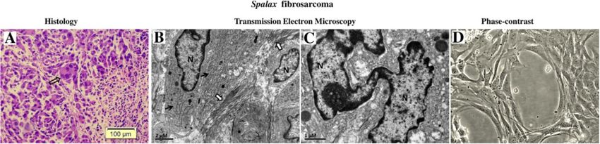
Manov et al. BMC Biology 2013, 11:91                                                                                                     Page 4 of 17
http://www.biomedcentral.com/1741-7007/11/91

 Figure 2 Effect of 3-Methylcholantren treatment on soft tissue tumor induction in Spalax and mice. Animals treated with a single
 injection of 3MCA as described in the Materials and methods section. Representative images show macroscopic and microscopic observations.
 Mice (A): An ill-defined, soft mass, with foci of necrosis and hemorrhage; diagnosed as high-grade fibrosarcoma by histology. Spalax (B): a
 well-circumscribed, firm, whitish nodule composed of benign spindle cells organized into long regular bundles - benign reactive fibrosis.
 Hematoxylin and eosin staining, ×100. 3MCA, 3-Methylcholantrene.

Spalax fibroblasts suppress growth of human cancer cells                     co-culture (Figure 4). Prolonged co-cultivation up to 11
in vitro                                                                     days resulted in further destruction of cancer cell colonies
To compare the effects of normal fibroblasts isolated                        by the presence of Spalax fibroblasts and the spaces previ-
from different rodents on the growth of human cancer                         ously occupied by Hep3B cells were invaded by fibroblasts
cells, we used a co-culture approach, where fibroblasts                      (Figure 4). In contrast, the number of cancer cells co-
were cultured together with cancer cells on a shared                         cultured with mouse fibroblasts increased gradually, and
surface (Figures 4 and 5). In these experiments, hepatoma-                   on Day 6, Hep3B cells surrounded by mouse fibroblasts
derived Hep3B cells as well as breast cancer MCF7 cells                      reached approximately 80% confluence, similar to control
were tested. Obvious inhibition of cancer cell growth                        (Hep3B only). Overgrown Hep3B colonies were found
was found when Hep3B cells were co-cultured with                             after 11-day co-culture with mouse fibroblasts. An obvi-
Spalax normal lung and skin fibroblasts: the foci of                         ous inhibitory effect was demonstrated when Spalax nor-
destroyed cancer cells were visible after six days of                        mal skin fibroblasts were co-cultured with breast cancer

 Figure 3 3MCA-induced tumor in Spalax. (A) Light microscopic examination. Note spindle, epithelioid and giant multinuclear cells
 (empty arrow); nuclei are variable in shape, size and chromatin distribution; nucleoli vary in frequency. Hematoxylin and eosin staining, ×100.
 (B) Transmission electron microscopy (TEM): dilated, elongated rough endoplasmic reticulum (black arrows) and abundant collagen fibers
 (white arrows) (C) TEM: a giant, monstrous nucleus (N). (D) Cell line established from Spalax tumor, phase contrast image after six months of
 continuous cultivation (×200). 3MCA, 3-Methylcholantrene.
Manov et al. BMC Biology 2013, 11:91                                                                                                    Page 5 of 17
http://www.biomedcentral.com/1741-7007/11/91

 Figure 4 Effects of Spalax and mouse fibroblasts on growth of co-cultured human hepatoma cells. Tumor cells (TC) were cultured either
 alone or in the presence of Spalax or mouse fibroblasts in the ratio of 1:10 (5 × 10⁴ fibroblasts and 5 × 10³ cancer cells in six-well plates) in
 RPMI/DMEM-F12 media (1:1) containing 10% FBS. White arrows point to the foci of destroyed cancer cells, and black arrows show the
 fibroblast-tumor cell colony boundaries. Cells in mono- and co-cultures were observed and photographed daily. Representative images for each
 sample at different time intervals are shown (×200).

MCF7 cells as well (Figure 5). After 10 days of co-culture                  experiments using Hep3B cancer cells with skin fibro-
with Spalax fibroblasts, massive rounding and detach-                       blasts isolated from two different wild, natural rodents:
ment of cancer cells were observed. On the other hand,                      Acomys, a short-lived, wild, above-ground rodent; and
mouse fibroblasts stimulated proliferation of MCF7                          naked mole rat (Heterocephalus glaber), a long-lived
cells, and by Day 10 densely populated colonies of cancer                   cancer-resistant wild subterranean rodent [22]. As shown
cells developed.                                                            (Figure 6), no growth inhibitory effect was found when
                                                                            Acomys fibroblasts were co-cultured with Hep3B cells.
In vitro anticancer activity by other wild, natural rodents’                On the contrary, Acomys fibroblasts promoted cancer
fibroblasts                                                                 cell invasion similar to the effect of rat fibroblasts.
Since we compare a wild mammal with laboratory ani-                         Heterocephalus cells, similar to Spalax, evidently destroyed
mals that are sensitive to cancer, we conducted co-culture                  cancer cell growth (Figure 6).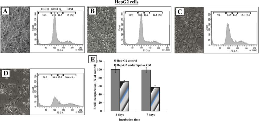
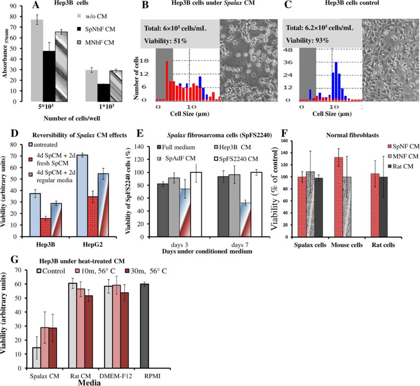
Manov et al. BMC Biology 2013, 11:91                                                                                                    Page 6 of 17
http://www.biomedcentral.com/1741-7007/11/91

 Figure 5 Morphologic alterations in human breast cancer MCF7 cells triggered by co-culture with Spalax fibroblasts. MCF7 cells were
 co-cultured with skin fibroblasts of Spalax or mouse in the ratio of 1:15 (5 × 10⁴ fibroblasts and 2.5 × 10³ cancer cells in six-well plates) in
 DMEM/DMEM-F12 media (1:1) containing 5% FBS. Representative phase contrast images after 10 days of co-culture are presented (×200). Note
 rounding and detachment of MCF7 cells co-cultured with Spalax fibroblasts. Black arrows point to rounding cells. White arrows show shrunken
 “floating” cells.

Conditioned medium generated by Spalax fibroblasts                          normal fibroblasts, but was not affected by normal, full
induces cancer cell death, but does not affect normal                       medium and CM derived from Hep3B cells or CM derived
primary fibroblasts                                                         from the SpFS2240 cells themselves (Figure 7E). Note-
To determine whether the anti-cancer activity of Spalax                     worthy, no inhibitory effects were detected on mouse,
fibroblasts was mediated by fibroblast-secreted soluble                     rat and Spalax normal fibroblasts following exposure
factors, conditioned media (CM) obtained from Spalax,                       to homologous or heterologous CM (Figure 7F). To get
mouse and rat monolayers were tested. Cancer cells of                       a preliminary idea of the nature of the secreted factors
different origins were incubated under CM of normal                         responsible for cancer cell growth inhibition, CM from
fibroblasts, which had never been exposed to cancer cells                   Spalax and rat fibroblasts, and the regular medium
or other stimuli. Effects of CM generated by cancer cells                   of fibroblasts (DMEM-F12) were heated to 56°C for 10
were also tested (Figure 7). As demonstrated in Figure 7A,                  minutes, and 30 minutes. The different heat-treated
exposure of Hep3B cells to CM from cultured newborn                         media was mixed 1:1 with RPMI (the optimal growth
Spalax fibroblasts decreased cancer cell viability as mea-                  medium for the hepatoma cell lines used in this study)
sured by mitochondrial respiratory function. Exposure to                    and was added to Hep3B cancer cells. After seven days,
mouse CM hardly had an effect on cancer cell viability.                     the viability of the cancer cells was measured. The
Similarly, nine-day exposure of Hep3B cells to CM gener-                    heat-treated CM generated from Spalax fibroblasts reduced
ated by adult (>5.5 years old) Spalax fibroblasts obviously                 its anticancer activity, expressed as a partial increase in
reduced cancer cell viability as was determined by a trypan                 Hep3B cells viability (Figure 7G).
blue extrusion assay (Figure 7B,C): cancer cells exposed to
Spalax fibroblast-conditioned CM reached 49% death,                         Soluble factors generated by Spalax fibroblasts cause cell
whereas unexposed cells remained completely adherent                        cycle arrest, nuclear fragmentation, and impair
and viable (Figure 7C).                                                     mitochondrial dynamics in cancer cells
   We next evaluated the reversibility of the inhibition of                 To investigate the mechanisms by which Spalax fibroblasts
cancer cells initiated by Spalax CM. HepG2 and Hep3B                        induce cancer cell death, we examined nuclear and
were grown with Spalax CM for four days, then the                           mitochondrial shape dynamics, as well as cell cycle dis-
medium was changed by either fresh unused regular                           tributions in Hep3B and HepG2 cells. No changes in the
media or with fresh Spalax CM. Cancer cell viability                        morphology of cells, nuclei and mitochondria as well as
was measured after another two days. Recovery of the                        in cell cycle distribution were found when Hep3B cells
cancer cells was demonstrated when the CM was changed                       were incubated with rat CM (Figure 8, middle row)
with fresh unused regular media (Figure 7D). Importantly,                   compared to Hep3B grown with their own medium
growth of Spalax-derived fibrosarcoma cells (SpFS2240)                      (Figure 8, upper row; control). In contrast, following ex-
was gradually suppressed by CM generated by Spalax                          posure to Spalax CM, Hep3B cells undergo phenotypic
Manov et al. BMC Biology 2013, 11:91                                                                                          Page 7 of 17
http://www.biomedcentral.com/1741-7007/11/91

                                                                             fibroblast CM could induce mitochondrial dynamic
                                                                             changes in cancer cells, Hep3B cells were stained with
                                                                             MitoTracker-Red® probe after eight days of incubation.
                                                                             Compared with control and rat CM, the mitochondrial
                                                                             network of cells after eight-day growth with Spalax CM
                                                                             demonstrated the presence of damaged fragmented mito-
                                                                             chondria (Figure 8, lower row, MitoTracker® + DAPI).
                                                                             Similar to Hep3B cells, HepG2 cells under Spalax CM also
                                                                             showed morphological changes and accumulation of cells
                                                                             in sub-G0/G1 whereas mouse and rat CM did not affect
                                                                             cellular morphology and cell cycle distribution (Figure 9).
                                                                             BrdU incorporation into DNA, a marker for cell prolifera-
                                                                             tion, confirmed a time-dependent anti proliferative effect of
                                                                             Spalax CM on HepG2 cancer cells (Figure 9E).

                                                                             Spalax normal fibroblasts inhibit colony formation in soft
                                                                             agar of the breast carcinoma cell lines MDA-MB-231 and
                                                                             MCF7 as well as Spalax-derived fibrosarcoma
                                                                             To study whether soluble factors generated by Spalax
                                                                             fibroblasts may influence colony formation in soft agar,
                                                                             breast cancer cells were cultivated for three weeks in
                                                                             the absence or presence of Spalax fibroblasts (Figure 10).
                                                                             Spalax fibroblasts strongly reduced the formation of MDA-
                                                                             MB-231 colonies (Figure 10A,B). The ability of MDA-MB-
                                                                             231 to form large colonies was completely inhibited by
                                                                             Spalax fibroblasts (Figure 10C), while rat fibroblasts had no
 Figure 6 Comparison of the effects of Spalax, Acomys,                       effect on colony formation (Figure 10A,B). Cells from
 Heterocephalus and rat skin fibroblasts on growth of Hep3B                  another human breast cancer cell line, MCF-7, were in-
 cells. Hep3B tumor cells were cultured either alone or in presence          cubated with monolayers of Spalax and mouse fibroblasts
 of Spalax, Acomys, Heterocephalus or rat fibroblasts in the ratio of        (Figure 10D). Remarkably, after 11 days, and compared
 1:10 (5 × 10⁴ fibroblasts and 5 × 10³ cancer cells in six-well plates) in
                                                                             to the control, more colonies were formed when human
 RPMI/DMEM-F12 media (1:1) containing 10% FBS. After seven days
 incubation cells were photographed. Representative images for each          MCF7 cells were co-cultured with mouse fibroblasts,
 sample are shown (×200). White arrows point to the foci of                  whereas a monolayer of Spalax fibroblasts significantly
 damaged cancer cells. TC, tumor cells.                                      reduced MCF7 colony-formation.
                                                                               Importantly, Spalax normal fibroblasts suppressed
                                                                             growth and colony formation of the homologous tumor,
changes observed under phase contrast microscopy: cellu-                     Spalax-derived fibrosarcoma (SpFS2240) (Figure 11). In
lar shrinkage, irregularities in the plasma membrane and                     contrast, both rat and mouse normal fibroblasts stimulated
blebs formation (Figure 8, lower row, phase-contrast).                       growth of Spalax tumor cells in soft agar (Figure 11A).
Cell cycle analysis revealed a noticeable accumulation of                    Integrating the number of colonies and their total occupied
dead cells in sub-G1 (36.7% versus 16.4% in control), a                      area, calculated from five independent fields, revealed
reduction in the number of cells in G0/G1 (28.9% versus                      a 36% reduction when SpFS2240 were grown above a
49.6% in control), and a modest arrest of proliferation in                   Spalax fibroblast monolayer compared to blank plates
G2/M (21.7% versus 17.1% in control) (Figure 8, lower                        (Figure 11B, 2240 alone). In contrast, mouse and rat fibro-
row, cell cycle). Nuclear staining with DAPI of Hep3B                        blasts enhanced colony formation by factors of 1.7 and 2.1,
cells that were grown with Spalax CM for eight days,                         respectively, compared to the blank plates (Figure 11B).
revealed heterogeneous chromatin appearance within
irregularly shaped nuclei, and in many cells extensive                       Discussion
chromatin condensation and nuclear fragmentation were                        Notwithstanding the importance of laboratory mice
conspicuous (Figure 8, lower row, DAPI staining). On the                     in comprehension of carcinogenesis mechanisms, this
other hand, homogeneous patterns with regular-shaped                         cancer-prone model organism failed to provide satisfactory
nuclei were mainly represented in the cells incubated with                   knowledge of cancer preventive mechanisms and treatment
rat CM as well as in the control cells (Figure 8, upper and                  strategies in humans. (http://www.safermedicines.org/
middle row, DAPI staining). To examine whether Spalax                        quotes/cancer.shtml). Therefore, elucidating mechanisms
Manov et al. BMC Biology 2013, 11:91 Page 8 of 17 http://www.biomedcentral.com/1741-7007/11/91 Figure 7 Effects of conditioned media (CM) on viability of cancerous and non-cancerous cells. (A) Hep3B cells were seeded in a 96-well plate at a density of 5 × 10³ and 1 × 10³ cells/well in RPMI-DMEM/F12 medium conditioned by Spalax or mouse skin newborn fibroblasts (SpNbF and MNbF, respectively). Hep3B cells were incubated for four days; viability was estimated by PrestoBlue® Reagent. (B,C) Hep3B cells (1 × 10⁴ cell/well) were cultured in six-well plates under conditioned medium of Spalax adult skin fibroblasts (B) or grown in medium generated by Hep3B cells (C). After nine days, the cells’ survival rates were assessed by a Countess® cell counter (Life Technologies); red: dead cells, blue: viable cells. (D) Hep3B and HepG2 cells were incubated under Spalax CM for four days, followed by changing the media either to fresh media or new Spalax CM. After two days, viability was estimated by PrestoBlue® Reagent. (E) Spalax fibrosarcoma cells (SpFS2240) were incubated for three or seven days in full medium or under CM of Spalax adult skin normal fibroblasts (SpAdF CM), Hep3B (Hep3B CM), Spalax fibrosarcoma (SpFS2240 CM). Cell viability was evaluated by using PrestoBlue® reagent. Results are presented as percentage of control (SpFS2240 CM); mean ± S.D. (F) Effects of CM generated by Spalax or mouse normal fibroblasts (SpNbF CM and MNbF CM, respectively) on the growth of non-cancerous cells. The viability was estimated after four days by PrestoBlue® reagent; mean ± S.D. (G) Heat treatment of conditioned media. Seven-day CM, generated by Spalax or rat fibroblasts, was heat-treated at 56°C for 10 minutes and 30 minutes prior to addition to Hep3B cancer cells (2,000 cell/well) in 96-well plates. Cells were incubated for seven days followed by PrestoBlue® test. All results were obtained from three independent experiments performed in three to six technical repeats. employed by a wild, non-inbred mammal that is naturally to environmental hypoxia (for example, [1,23]). During the cancer-resistant raises promising opportunities. last decade a growing number of genes involved in hyp- Spalax has been thoroughly investigated at the Institute oxic response have been studied and exhibited Spalax- of Evolution of Haifa University as a model for adaptation specific features [4,11]. Directly related to this study on
Manov et al. BMC Biology 2013, 11:91                                                                                               Page 9 of 17
http://www.biomedcentral.com/1741-7007/11/91

 Figure 8 Spalax fibroblast-conditioned medium compromises cell cycle, causes nuclear and mitochondrial fragmentation in Hep3B
 cells. Hep3B cells were grown on cover slips under medium conditioned by Spalax or rat fibroblasts for seven days. Representative
 phase-contrast images demonstrating morphological changes (×200) are depicted. Cells were harvested and stained with PI, and cell cycles were
 analyzed by flow cytometry. Representative flow cytometry histograms of three independent experiments performed in duplicate are presented.
 Hep3B cells were stained with MitoTracker®Red, fixed with formaldehyde and counterstained with DAPI. Representative fluorescence microscopy
 images demonstrating nuclear and mitochondrial changes are present. White arrows point fragmented nuclei; empty arrows point chromatin
 condensation. Scale bars represent 10 μm. PI, Propidium iodide.

cancer-resistance of Spalax are the tumor suppressor                      In contrast, treatment of Spalax led to necrotic wounds,
p53 favoring cell cycle arrest over apoptosis [24] and a                  which completely healed with no signs of malignancy.
unique Spalax heparanase splice variant that was shown                    The carcinogen 3-MCA is known to produce fibrosarco-
to significantly decrease tumor size and metastatic activity              mas through persistent inflammation and reactive metab-
compared to native heparanase [9]. Furthermore, despite                   olites causing severe oxidative damage [27]. In our study,
the extremely long lifespan of Spalax (>20 years), even                   100% of 3-MCA-injected mice and rats developed tumors
after studying thousands of individuals for decades, we                   at the injection site within two to three and four to six
have never observed animals that developed spontaneous                    months, respectively. One year after 3MCA treatment no
tumors, nor show any aging-related phenotypic changes.                    Spalax animals showed any pathological process. How-
                                                                          ever, 2 out of 6 old individuals (from a total of 12 animals)
In vivo studies of carcinogen-induced tumor                               developed benign fibrotic overgrowths after 14 and 16
We report here that Spalax is resistant to two-stage                      months, respectively, and only one case of malignant
DMBA/TPA, and 3-MCA carcinogen treatments. DMBA/                          transformation in a >10 year-old Spalax animal was
TPA is commonly used to study malignant transformation,                   recognized, 18 months after 3-MCA injection.
resembling formation of human squamous cell carcinoma                       It is well established that oxidative stress drives tumor
[25]. A single dose of DMBA induced substantial oxidative                 progression and metastasis [28]. Thus, the mechanisms
stress [26], and when followed by repetitive application                  that Spalax evolved to survive hypoxia might be related
of TPA led to persistent inflammation supporting tumori-                  to resistance to induced or spontaneous cancers. Spalax
genesis [17]. In the present study, mice treated with                     have recently been shown to have higher levels of reactive
DMBA/TPA initially developed benign papillomas, which                     oxygen species (ROS) processing enzymes compared to
subsequently transformed to squamous cell carcinomas.                     hypoxia-intolerant rodents [4]. Nrf2, a transcription factor
Manov et al. BMC Biology 2013, 11:91 Page 10 of 17 http://www.biomedcentral.com/1741-7007/11/91 Figure 9 Effects of Spalax, mouse and rat conditioned media on morphology and cell cycle progression in HepG2 cells. HepG2 cells were incubated under conditioned media for eight days; thereafter, cell morphology was documented using phase contrast microscopy, harvested, stained with PI and analyzed by flow cytometry. Representative images (×200) and flow cytometry histograms are presented: (A) control media; (B) rat CM; (C) mouse CM; (D) Spalax CM; (E) BrdU incorporation assay: HepG2 were grown in 96-well plates (2000 cells/well) for four and seven days under Spalax-generated CM. BrdU Cell Proliferation ELISA (Exalpha) was used. Time-dependent decrease in cell proliferation under Spalax-generated CM is depicted. CM, Conditioned media; PI, Propidium iodide. critical for defense against oxidative stress, has a unique synthesis of ferritin, an iron storage protein, thus preventing structure in Spalax. Whereas it is highly conserved among oxidative damage caused by free heme and ROS [32]. most mammals [4], Spalax Nrf2 carries 27 specific amino Indeed, most Spalax individuals, showing no external acid replacements, 6 within the Neh6-domain, which is lesions following exposure to carcinogens, have probably critical for stabilizing the protein under ambient oxida- resolved the initial inflammatory insult without excessive tive stress and for its transcriptional activity [29]. Studies fibroplasias which can be attributed to more efficient anti- performed on Nrf2−/− mice have shown the essential oxidation mechanisms. The benign fibroblastic prolifera- role of Nrf2 for detoxification of DMBA metabolites tions observed in two 3-MCA-treated Spalax animals after and protection against DMBA-induced carcinogenesis 14 and 16 months suggest that Spalax is able to effect- [30]. Unraveling the molecular mechanisms resulting ively arrest cancerous transformation. Nonetheless, whether in the healing of Spalax skin and inhibition of progression Spalax tissues are able to prevent conversion of the to tumor formation is the goal of our ongoing research. 3-MCA pro-carcinogen into an active carcinogen, over- Hence, we have just initiated a comprehensive repetition of come its effect, or to inhibit previously transformed cells, DMBA/TPA treatment where we will have a representative remains to be clarified in future studies. sample of animals from different stages following the application of this carcinogen in order to answer this In vitro studies of Spalax cancer resistance question through quantification of apoptosis and senes- Tumor growth and invasion are dependent on growth cence of Spalax skin and muscle tissue at the area of the factors and cytokines produced by stromal cells [33]. carcinogen application. Furthermore, considering the Normal stroma contains a relatively small number of fi- high tolerance of Spalax to oxidative stress and the fact broblasts associated with ECM. However, during wound that DMBA is metabolized among others into ROS that healing, fibrosis or malignant transformations, stromal cause oxidative DNA damage in the skin [31], the above fibroblasts proliferate, intensively produce growth factors experiment will allow us to compare the ROS levels upon and cytokines, express α-smooth muscle actin and become DMBA application in Spalax and mice. cancer-associated fibroblasts (CAFs) [12,34,35]. CAFs are Another antioxidant enzyme, heme oxygenase-1 (HO-1), removed from the granulation tissue rapidly after healing, was shown to be elevated in Spalax tissues, and further though in cancer stroma they persist, contributing to increased under hypoxia [4,11]. HO-1 is involved in epithelial mesenchymal transition of cancer cells. The the degradation and catabolism of heme and supports later phenomenon is important for cancer progression
Manov et al. BMC Biology 2013, 11:91                                                                                                           Page 11 of 17
http://www.biomedcentral.com/1741-7007/11/91

 Figure 10 Spalax fibroblasts suppress colony formation of human breast cancer cells MDA-MB-231 and MCF7 in soft agar. (A) MDA-MB-231
 cells (5 × 10³ cells) cells were suspended in 0.35% agar and added as the cancer cell top layer to base layer either empty (blank) or containing the Spalax
 or rat fibroblast monolayer. At Day 21, colonies larger than 50 μm were counted under an inverted microscope and photographed (×40). Representative
 microscopic images out of 15 fields are shown. (B) Average number of colonies counted in soft agar (n = 15). The experiment was performed in duplicate
 plates at least three times; mean ± S.D. (C) A representative colony in soft agar was formed by MDA-MB-231 only, or by co-culturing with a Spalax
 fibroblast monolayer. The size bar shows equivalent magnification in both images (× 200). (D) MCF7 cells (5 × 10³ cells) were grown in soft agar on top of
 a monolayer of mouse newborn (MNbF), or Spalax newborn fibroblasts (SpNbF) in 35-mm culture dishes. After 5 and 11 days of incubation colonies
 containing >20 cells were counted by using an inverted microscope (× 200), mean ± S.D.

and is mediated, at least in part, by metalloproteinases                         added to culture medium, are conceived as cancerous
secretion and ROS generation [36,37]. Although the                               transformation-like stimuli, driving Spalax fibroblast
reports published to date have been mainly addressed                             necrotic death, triggered through release of interferon-β
to a cancer-promoting role of stromal fibroblasts, evidence                      (IFN-β). Nonetheless, in the same study, higher and
suggests that normal stroma and normal fibroblasts could                         earlier death rates were also shown in serum-reduced
impede tumorigenesis [14-16,38,39]. Early studies [15]                           or serum-free media. Furthermore, the possibility that
demonstrated that normal dermal fibroblasts suppressed                           CM from “dying” cells may lack beneficial nutrients, or
development of malignant phenotypes of RAS-transformed                           contain toxic metabolites, or other factors beyond IFN-β,
keratinocytes when grafted into animals. Similarly, normal                       was not addressed. Additionally, measurements of IFN-β in
fibroblasts were able to retard melanomagenesis in its early                     Spalax CM were performed indirectly using human cell
stages [38]. Inhibition of growth and induction of differenti-                   lines [40]. The first, VSV (Vesicular Stomatitis Virus)-GFP
ation were found in breast cancer pre-neoplastic MCF10-                          (encoding a Green Fluorescent Protein) gene assay, mea-
AT1-EIII8 cells when co-cultivated with normal fibroblasts,                      sures IFN-β expression levels by VSV-GFP reporter assay.
even in the presence of estrogen [39]. It is still unclear what                  In this assay, HT1080 cell line (human fibrosarcoma cells)
events in the stroma, along with its interaction with precan-                    had been incubated with Spalax CM, and then infected
cerous cells, lead to a transition of the stromal function                       with a GFP-encoding VSV. The level of IFN-β in the media
from cancer-protective to cancer-promoting, or, as in the                        corresponds to the reduction in the number of GFP
present case of Spalax, what are the molecular mechanisms                        positive human HT1080 cells. In the second assay,
that Spalax evolved to escape cancerous transformation                           IFN-β release by “dying” Spalax cells is determined by
and to develop anti-cancer ability.                                              HEK (Human Embryo Kidney cells)-Blue cells assay. In
   In a recent study [40], cancer resistance in Spalax                           this assay the induction of β-gal reporter in human EK
was discussed. It was suggested that pro-growth signals                          cells under IFN-β-inducible promoter is measured. Both
originating from the fetal bovine serum, routinely                               assays use human cells for indirectly measuring Spalax
Manov et al. BMC Biology 2013, 11:91                                                                                      Page 12 of 17
http://www.biomedcentral.com/1741-7007/11/91

                                                                        compare the fate of the cells when grown with CM of the
                                                                        other species tested in the study, namely, mice or human.
                                                                        Additionally, the method used in this study for declaring
                                                                        necrotic death is based on the Annexin V/propidium
                                                                        iodide assay [40]. Briefly, floating and adherent cells were
                                                                        harvested, stained with Annexin-V and propidium iodide,
                                                                        and analyzed by flow cytometery. The known disadvan-
                                                                        tage of this method is that it cannot conclusively prove
                                                                        that cell death is solely the result of necrosis, nor eliminate
                                                                        the possibility of apoptotic mechanisms. Also, the authors
                                                                        have not provided evidence for interrelations between their
                                                                        three declared observations (transformation-like stimuli,
                                                                        necrotic death and release of IFN-β). Overall, it is our im-
                                                                        pression that the above mentioned study [40] does not pro-
                                                                        vide direct evidence to Spalax cancer resistance, certainly
                                                                        not its anti-tumor properties. Alternatively, we show here
                                                                        that viable, proliferating Spalax fibroblasts, from adult and
                                                                        newborn animals, inhibit growth of cancer cells derived
                                                                        from different tissues and species, most importantly hu-
                                                                        man, but do not affect non-cancerous cells, including
                                                                        those of Spalax (Figure 7F), thereby highlighting a strategy
                                                                        used by Spalax to identify and target malignancies. This
                                                                        unique interaction is further strengthened by the observa-
                                                                        tion that the growth of cancer cells is regained once the
                                                                        immediate interaction with Spalax cells is terminated
                                                                        (Figure 7D). Importantly, no inhibitory effect on cancer
                                                                        cell growth was found when fibroblasts from above-ground
                                                                        species (rat, mice and Acomys) were tested.
                                                                          Recently, several studies investigated the unique cancer-
                                                                        resistance properties of the naked mole rat (Heterocephalus
                                                                        glaber), another subterranean, long-lived, rodent species.
                                                                        The most recent study suggested a connection between
                                                                        a high viscosity of media conditioned by Heterocephalus
                                                                        fibroblast cells due to exceptional secretion of high-
                                                                        molecular mass hyaluronan (HMM-HA) [41], which was
                                                                        suggested to mediate what was named by the authors “early
                                                                        contact inhibition”, previously described by the same group
                                                                        as an anticancer mechanism in Heterocephalus cells, and
                                                                        was initially ascribed to p16(Ink4a) and p27(Kip1) activity
 Figure 11 Effect of Spalax, rat and mouse fibroblasts on               [42]. In the same paper [41], it is reported that HMM-HA
 Spalax-derived fibrosarcoma cells colony formation.                    was detected also in Spalax fibroblasts even in higher levels
 (A) SpFS2240 Cancer cells were grown in soft agar on top of
                                                                        compared to Heterocephalus fibroblasts, though no experi-
 monolayers of mouse, rat and Spalax fibroblasts. After three weeks,
 colonies were counted. At least 10 fields were recorded for each       ments were carried out to clarify its role in Spalax fibro-
 observation. Two representative images demonstrating effects of        blasts. Nevertheless, this may explain the prevalent high
 different fibroblasts on colony-formation are shown (×40).             viscosity of the medium of cultured Spalax fibroblasts we
 (B) Colony numbers and cumulative total colony area (μm2) from five    noticed, though we find that it does not prevent Spalax
 fields were calculated to demonstrate the effects of the fibroblasts
                                                                        cells from reaching confluence or influences their anti-
 monolayer on the cancer cell colony formation and growth.
                                                                        cancer properties. Furthermore, CM from Spalax with
                                                                        apparent normal viscosity was also able to inhibit cancer
IFN-β, which is inconsistent with the authors’ declaration              cells proliferation (ongoing study). In light of the fact that
that human cells are nonresponsive to Spalax CM stimuli                 hyaluronan-cancer cell interactions were shown to pro-
possibly due to species divergence of IFN-β [40]. Likewise,             mote, and not inhibit, cancer invasion [43], the correlation
no proof was given that the ability to kill “dying” fibro-              between HMM-HA, the potential of cells to reach conflu-
blasts is unique to Spalax’s CM, for example, by trying to              ence and the resistance to oncogenic transformation or
Manov et al. BMC Biology 2013, 11:91 Page 13 of 17 http://www.biomedcentral.com/1741-7007/11/91 anti-cancer activity, requires further direct experimental restrict growth and effectively kill cancer cells, while support, especially in the case of our model organism, the Acomys cells behave similarly to rat and mice, that is, Spalax. Another study endorsed Heterocephalus cells’ have no anti-cancer activity (Figure 6). We may assume cancer-resistance to rapid cell crisis following oncogenic that this anti-cancer ability might be shared by species transformation, which is characterized by abnormal living under extreme conditions and adapted to stress, such chromatin material and nuclei, leading to a failure to as hypoxia, which is directly related to cancer initiation and successfully complete cell division, hence the inability progression. It would be interesting to investigate this of the cells to progress into malignancy [44]. These phenomenon in other hypoxia-tolerant species, such as observations are somewhat similar to our findings of other subterranean, high altitude and diving mammals. fragmented and deformed nuclei and chromatin con- Previous studies showed that key hypoxia-regulatory densation (Figure 8), disturbed cell division and prolif- genes in stromal fibroblasts, such as HIF1-α and VEGF, eration (Figure 9E) of human cancer cells, as well as the negatively influence tumorigenesis [46]. HIF1-α is a known 3MCA-induced Spalax and mice fibrosarcoma cell line, tumor-promoting transcription factor in most malignancies upon their interaction with Spalax fibroblasts. In view [47]; however, its expression in tumor stromal fibroblasts of the similar ability of Heterocephalus fibroblasts to kill could suppress cancer cell growth [46]. We have previously cancer cells (Figure 6), and as the efficiency of experimental shown that HIF1-α [23] as well as ROS-scavenging enzymes oncogenic transduction of cells is never 100%, it is possible [4] are constitutively highly expressed in Spalax. Similar to that the Heterocephalus cells that escaped malignant trans- our explanations of the failure to induce cancer in vivo formation killed the oncogenic-transduced ones. in live Spalax animals, in vitro studies, using fibroblast Our findings demonstrated that Spalax fibroblasts or cells, demonstrated a significant role in adaptive response their CM target human cancer cells growth machinery, to oxidative stress, at least in part, via expression of HO-1 triggering programmed cancer cell death (Figures 4, 5, [48]. High levels of mitochondrial ROS produced by cancer 8 and 9). Following co-culture with Spalax fibroblasts cells were shown to drive tumor development via remodel- or their CM, cancer cells (Hep3B, HepG2 and MCF7) ing of the stromal environment and enhancing invasion. undergo morphological changes typical of apoptosis [45]: Recently, the roles of ROS produced by fibroblasts in their swelling, rounding, detachment, shrinkage and floating. trans-differentiation to myofibroblasts and in cancer Moreover, nuclear condensation and abnormal mitochon- cell invasiveness were reported [37]. ROS-generating CM of drial fission as well as accumulation of cells in sub-G1 mutated fibroblasts promoted metastasis of A375 melanoma (Figures 8 and 9) also suggest apoptotic modes of cancer through the increasing of ROS and HIF1-α stabilization in cell death. BrdU incorporation, reflecting cell proliferation, melanoma cells. However, when N-acetyl cysteine, a ROS confirmed that Spalax CM contains anti-proliferative fac- scavenger, was added to the system, HIF1-α accumulation tors, inhibiting cell division in a time-dependent pattern. and melanoma cell invasion were inhibited [37]. We further showed that the effect of Spalax CM on cancer Adaptive tolerance to hypoxia stress in Spalax, both cells is transient and reversible. That is, replacing the CM in vivo and in vitro, may grant the unique resistance to with regular fresh medium leads to recovery of those cancer cancer through strong antioxidant mechanisms, among cells that had not been affected by the CM. Last but not others (for example, as mentioned here, the unique activity least, Spalax fibroblasts presumably impair the aggressive of its p53 [8] and heparanse [9]), that quench ROS before behavior of tumor cells: the invasive phenotype of highly they spread and damage DNA and other macromolecules, metastatic MDA-MB-231 breast carcinoma cells was thus providing cellular homeostasis and cancer protection. markedly reduced (Figure 10). Noteworthy, the ability As such, they are a milestone in our efforts in understand- to form colonies in soft agar by 3MCA-induced, Spalax- ing the mechanisms by which the long-lived, hypoxia- derived fibrosarcoma was significantly suppressed by tolerant Spalax hinders cancer initiation and progression. homologous fibroblasts, whereas heterologous fibroblasts Collectively, we have shown here an outstanding cancer (rat and mouse) increased tumor formation (Figure 11). resistance of the whole, live Spalax, and not just in cultured Spalax fibroblasts also inhibited colony formation in cells, and anticancer activity of Spalax cells on human soft agar by 3MCA-induced, mouse-derived fibrosarcoma. cancer cells, and not just resisting transformation of its In order to strengthen our findings of Spalax cells’ own healthy cells. This phenomenon extensively described anti-cancer activity, compared to cells from laboratory, here using different methodologies on cells from different in-bred, aboveground mice and rats, we decided to follow ages of Spalax, together with our initial observation of a the cancer activity pattern of two other, wild, out-bred, similar ability of cells originated from another subterra- species. Hence, fibroblast cells were propagated from the nean, long-lived, hypoxia- and cancer-resistant animal, the aboveground, wild, short-lived rodent Acomys, and the sub- Heterocephalus, highlight the importance to adopt such terranean, wild, long-lived Heterocephalus. We have shown animal models with exceptional genetic-embedded tolerance here that, similar to Spalax cells, Heterocephalus fibroblasts to environmental stress, in cancer research.
Manov et al. BMC Biology 2013, 11:91                                                                             Page 14 of 17
http://www.biomedcentral.com/1741-7007/11/91

   Our ongoing research is focused on identifying the              “The classical mice model for cancer research has little
factors secreted by Spalax cells, and their selective            predictive value and a negligible relation to that of human.
interaction with cancer cells to suppress tumorigenesis. Our     Far more than anything else, the lack of good animal
first step to exploring the nature of the secreted factors was   models has become the rate-limiting step in human cancer
the heat-inactivation preliminary experiment presented here      research” (Prof. Robert Weinberg, MIT; Newsweek,
(Figure 7G). The heat treatment of Spalax CM caused only         September 6th, 2008). Therefore, it would be extremely
partial loss of the anti-cancer activity of Spalax-generated     useful to study naturally cancer-resistant animals, as models
CM. Although not conclusive, this may indicate the in-           to find ways to prevent cancer before it occurs. Our results
volvement of protein factors in the observed phenomenon.         may lead to a breakthrough in the conservative paradigm
We are also studying the signaling mechanisms and death          of cancer research, completely dependent on laboratory, in-
receptors whose activation triggers cancer cell death. These     bred rodents, and place Spalax as the ‘missing’, appropriate
studies will hopefully contribute to the identification of new   candidate model for such studies. We anticipate that the
anti-cancer mechanisms and future tumor preventive or            long lived, hypoxia- and cancer-tolerant Spalax will turn
therapeutic strategies. To our knowledge, the present study      out to be a significant biological resource to biomedical
demonstrates, for the first time, Spalax tolerance to chem-      research as a model organism for understanding cancer
ically induced carcinogenesis along with direct anticancer       and its prevention.
effect of Spalax fibroblasts on human cancer cells.
                                                                 Methods
Conclusions                                                      Animals
During 50 years of studies, with thousands of animals            All animal protocols were approved by the Institutional
that crossed our Animal House, we have never observed            Ethics Committee.
spontaneous tumors in Spalax. Similar phenomenon                   Blind mole-rat (Spalax), rats (Rattus norvegicus) and
was observed also in another subterranean long-lived             C57BL/6 mice were subjected to DMBA/TPA or 3MCA
rodent, Heterocephalus. Based on this observation, a few         treatments. For DMBA/TPA treatment, eight Spalax and
studies [41,42,44] have tried to explain cancer resistance in    six mice were used. For 3MCA treatment 12 Spalax, 6 mice
Heterocephalus through testing known molecular mecha-            and 6 rats were used. Spalax and Acomys were captured in
nisms of malignant transformation on healthy fibroblast          the field and housed under ambient conditions in individual
cells. Though different molecular mechanisms were                cages in the Animal Facility of the Institute of Evolution,
suggested, none of these studies dealt with anti-cancer          University of Haifa. Noteworthy, Spalax do not undergo
properties of the whole, live animal in vivo, or on the          uniform acclimatization upon transfer from their natural
direct interaction of its normal cells with cancer cells.        habitat to a standardized laboratory environment but rather
Hence, our presentation is a pioneering, genuine, break-         behave differentially according to their eco-geographic
through study. We have tried to induce cancer in Spalax          origins [49]. The C57BL/6 mice were purchased from
with chemical carcinogens that induced cancer in 100% of         Harlan Laboratories (Jerusalem, Israel). Rats were supplied
mice and rats, and the results allow us to state that Spalax     by the Animal House of the Psychology Department of
is extremely resistant not only to spontaneous cancer but        Haifa University. Heterocephalus was a gift from Tisch
also to induced cancer. Furthermore, fibroblast cells from       Family Zoological Gardens in Jerusalem. All animals were
Spalax inhibit growth and kill cancer cells from vari-           kept with free access to food and water at 21 to 23°C in a
ous species and cell lines. This is exhibited in both the        12:12 light-dark cycle. All animals used for experiments
co-culture system or by exposure to conditioned media            were healthy. Animals were sacrificed with an inhalation
harvested from Spalax fibroblasts. Cancer cell death was         anesthesia agent (isoflurane) overdose.
accompanied by decreased cancer cell viability and prolifer-
ation, reduced colony formation in soft agar, disturbed cell     DMBA/TPA treatment
cycle progression, chromatin condensation, nuclei deform-        Four Spalax individuals approximately 2 years old and
ation and mitochondrial fragmentation.                           four individuals over 10 years old; and six individuals of
   This phenomenon is prominent in Spalax fibroblasts,           C57BL/6 mice, 3 to 4 months old, were used in the 7,12-
regardless of the animal age, from newborns a few days old       Dimethylbenz(a)anthracene/12-O-tetradecanoylphorbol-
to animals over 10 years old, as proved by the reversibility     13-acetate (DMBA/TPA) experiments. A single application
of cancer cells death, once their immediate interaction with     of 200 μg of DMBA dissolved in 100 μl of acetone for mice,
Spalax fibroblasts is terminated. The anti-cancer activity of    and 500 μg in 250 μl for Spalax was used. The solution was
Spalax is specific to cancer cells and not to normal cells. It   applied onto the shaved back skin of the animal. Three days
may be shared by other stress-adapted mammals as we ini-         after the initial DMBA dose, mice were treated with 30 μg
tially showed here by co-culturing Heterocephalus fibro-         of TPA (Sigma Aldrich, Inc.) dissolved in 100 μl of acetone,
blasts with cancer cells that also leads to cancer cell death.   and Spalax with 60 μg of TPA dissolved in 250 μl of
Manov et al. BMC Biology 2013, 11:91 Page 15 of 17
http://www.biomedcentral.com/1741-7007/11/91
acetone. TPA was applied three times per week for two to RPMI or DMEM/DMEM-F12 (1:1) supplemented with
three months, until all mice developed advanced cancer 15% FBS. Fibroblasts were plated first (5 × 10⁴), and
and were subsequently sacrificed. Spalax continued to be cancer cells were added within 1 h (5 × 10³), with a
treated for an additional three months twice a week. 10:1 fibroblast-to-cancer cell ratio. In parallel, control
cultures of cancer cells and fibroblasts were plated at
3-MCA carcinogen treatment the same number of cells. The medium was changed
3-methylcholanthrene (3-MCA) has been commonly used every three days. Fibroblast-cancer cell co-interactions
for induction of tumors in rodents [50]. In this experimental were observed and photographed by using an inverted
system, mice and rats develop local fibrosarcomas in microscope (Optika XDS2, Italy).
two to three and four to six months, respectively [20].
The recommended dose of 3MCA (Sigma Aldrich, Inc.) Generation of conditioned media
for treatment of mice is 200 μg dissolved in 200 μl of olive Normal fibroblasts or cancer cells (1 × 10⁶ cells) were
oil [20,51]. We calculated the amount applied to rats and plated in 10-cm tissue culture dishes and cultured in
Spalax according to their average weight. Hence, animals full medium containing 10% FBS for four days; there-
were treated with a single injection of 3MCA as follows: after, media were collected and cells were removed by
200 μg/200 μl for mice; 1 mg/500 μl for Spalax; and 1.5 centrifugation. The cell-free supernatants were then diluted
mg/500 μl for rats. Animals used in this experiment were: with the appropriate fresh culture medium (1:1) and used
six approximately 2-year-old Spalax individuals; six 10-year for further experiments. Trypan-blue standard treatment
old or older Spalax individuals; six 3- to 4- month-old mice; followed by cell count using a Countess® automatic cell
and six 3-month-old white rats. Animals were observed counter (Life Technologies), and PrestoBlue® dye reagent
once a week until tumors could be palpated, and then two (Invitrogen) (as described in [53]) were used to investigate
to three times a week. Animals were sacrificed; tissues were the viability of cancer cells exposed to CM of normal fibro-
removed for cell isolation or fixation. For histological exam- blasts from different species. For BrdU incorporation assay,
ination, the samples were fixed in 4% paraformaldehyde Cell Proliferation ELISA (Exalpha) was used following the
dissolved in PBS, dehydrated in increasing concentrations of manufacturer’s instruction.
ethanol, and embedded in paraffin. Five-micrometer sec-
tions were cut from paraffin blocks and routinely stained
with hematoxylin and eosin for microscopic examination. Soft agar colony formation assay
Colony formation assay was performed as described [54].
Cell culture In brief, 2 × 10⁵ fibroblasts were seeded in 35-mm culture
Primary Spalax, mice, rat, Acomys and Heterocephalus dishes and cultured for two to three days to reach con-
fibroblasts were isolated from under arm skin and lungs fluence. After washing with PBS, 1 ml of 0.5% agar in
as described [52], and grown in DMEM-F12 medium DMEM-F12 containing 2% FBS was added on top of the fi-
(Biological Industries, Beit Haemeq, Israel), supplemented broblasts to form a base layer. After the agar was solidified,
with 15% fetal bovine serum (FBS). Human cancer cell lines 5 × 10⁵ cancer cells were suspended in 1 ml of 0.35% agar
Hep3B and HepG2 (hepatoma-derived), MCF7 and MDA- in DMEM containing 5% FBS and then added into the dish
MB-231 (breast cancer cells) were obtained from ATCC, to form a cancer cell layer. Visible colonies were studied
and were grown in RPMI (Hep3B, HepG2) and DMEM and photographed under phase-contrast microscope.
(MCF7, MDA-MB-231) supplemented with 10% FBS, Finally, colonies larger than 50 μm were counted. At least
L-glutamine, penicillin and streptomycin (Biological 10 fields on each plate duplicate were used for counting at
Industries). Cells were incubated in a humidified atmos- a magnification of × 40. For total colony area, five fields
phere of 5% CO2 and 95% ambient air at 37°C. Spalax- were analyzed using ImageJ software [55].
derived fibrosarcoma cells were isolated from a tumor
developed after 3MCA injection. Tumor specimens Cell cycle analysis
were minced and treated with 1 mg/ml of collagenase The cell cycle distribution was assessed by flow cytometry
(Sigma-Aldrich) under aseptic conditions to obtain a of propidium iodide (PI)-stained nuclei as described
single-cell suspension, which was plated in culture dishes in previously [56]. In brief, following incubation, cells were
DMEM-F12 medium supplemented with 15% FBS and harvested by trypsin, combined with medium containing
penicillin-streptomycin-amphotericin B solution. Cells were floating cells, washed with PBS and stained with hypotonic
serially cultured more than 40 times. PI solution (PI 50 μg/ml in 0.1% sodium citrate and 0.1%
Triton X-100). The PI fluorescence of individual nuclei was
Co-cultures of cancer cells and fibroblasts recorded by FACSaria (Becton Dickinson, NJ, USA). A total
Normal fibroblasts and human-derived cancer cells were of 10,000 events were acquired and corrected for debris
co-plated in six-well plates in 2 ml of culture medium and aggregate population.You can also read


