Skeletal Aging and Osteoporosis: Cellular Senescence and Be- yond
←
→
Page content transcription
If your browser does not render page correctly, please read the page content below

Preprints (www.preprints.org) | NOT PEER-REVIEWED | Posted: 3 February 2021                              doi:10.20944/preprints202102.0127.v1

     Type of Paper: Review

     Skeletal Aging and Osteoporosis: Cellular Senescence and Be-
     yond
     Abhishek Chandra1,2,3*, Jyotika Rajawat4

                                          1    Department of Physiology and Biomedical Engineering,
                                          2    Division of Internal Medicine, Geriatric Medicine and Gerontology,
                                          3    Robert and Arlene Kogod Aging Center, Mayo Clinic, Rochester, Minnesota, USA
                                          4    Department of Zoology, University of Lucknow, Lucknow, UP, India; jrajawat@gmail.com

                                          * Correspondence: chandra.abhishek1@mayo.edu; Tel.: (+1-507-266-1847)

                                          Abstract: Bone is a dynamic organ maintained by tightly regulated mechanisms. With old age, bone
                                          homeostasis which is maintained by an intricate balance between bone formation and bone resorp-
                                          tion, undergoes deregulation. Oxidative stress-induced DNA damage, cellular apoptosis and cellu-
                                          lar senescence are all responsible for this tissue dysfunction and the imbalance in the bone homeo-
                                          stasis. These cellular mechanisms have become a target for therapeutics to treat age-related osteo-
                                          porosis. Pharmacological and genetic mouse models have shown the importance of senescent cell
                                          clearance in alleviating age-related osteoporosis. Senescent cells have an altered secretome, which
                                          may have autocrine, paracrine, or endocrine function. The current review discusses the current and
                                          potential pathways which lead to a senescence profile in an aged skeleton. The review was written
                                          following an extensive literature survey of published studies, mostly excluding articles published
                                          on pre-print servers. The review discusses potential therapeutics targeting cellular senescence and
                                          the senescent secretome as an underlying pathogenesis of an aging bone.

                                          Keywords: Osteoporosis, Senescence, SASP, Aging, Radiation, Senotherapeutic

                                          Introduction

                                          Bone as a tissue has its own complexities with one of the largest pools of diverse cell
                                          types. These complexities are stressed to its maximum limits during old age leading to
                                          osteoporosis. Advent of loss of bone with aging are also early signs of increased fracture
                                          risk, morbidity, and mortality. Osteoporotic fractures exceed incidences of cardiovascu-
                                          lar disease or cancer by ~3-4 fold[1] and is a substantial strain on the economy. Only 31-
                                          36% people above the age of 70 have normal bones, with the remainder suffering from
                                          some form of osteopenia or osteoporosis. Moreover, it is well understood that loss of
                                          estrogen is a key driver of bone loss in women and to some extent in men, which is only
                                          exacerbated by aging [2, 3]. However estrogen is not the only cause of bone loss during
                                          aging[4], and it has been recognized and discussed in detail that aging stands as a sepa-
                                          rate entity with distinct mechanisms[5]. This review will expand on known and poten-
                                          tial mechanisms underlying the pathogenesis of skeletal aging, mainly DNA damage
                                          and cellular senescence, with special emphasis on certain proteins such as poly(ADP-
                                          ribose) polymerase 1 (PARP1) due to their role in DNA repair, telomere maintenance,
                                          senescence and in the production of pro-inflammatory cytokines (Fig.1).

     Osteology 2021, 1, Firstpage–Lastpage. https://doi.org/10.3390/xxxxx                                         www.mdpi.com/journal/osteology
             © 2021 by the author(s). Distributed under a Creative Commons CC BY license.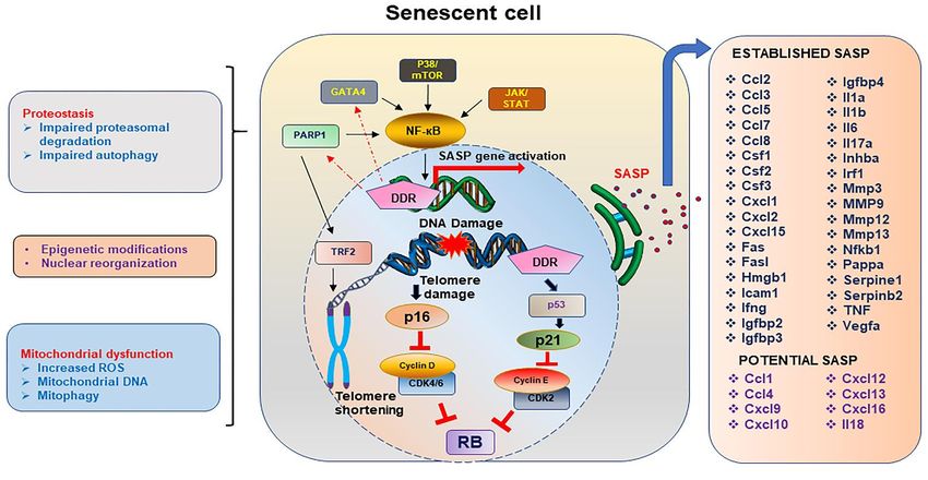
Preprints (www.preprints.org) | NOT PEER-REVIEWED | Posted: 3 February 2021              doi:10.20944/preprints202102.0127.v1

                                Biology of skeletal aging
                                       Bone is a dynamic organ incorporating several cell types which generally work syn-
                                 chronously and maintain the bone homeostasis resulting in the deposition of a mineral-
                                 ized bone matrix. The two processes which maintain this homeostasis are bone formation
                                 and bone resorption which are under an equilibrium in a normal physiological condition.
                                 Cells of the mesenchymal origin regulate the bone formation process including bone mar-
                                 row stem cells (BMSCs) which are the progenitors which give rise to osteoblasts, cells re-
                                 sponsible to deposit mineral and form the collagen enriched bone matrix supported by
                                 the multifaceted osteocytes, the most abundant cell type with an extensive canalicular
                                 network.
                                       Osteoclasts, a large multinucleate cell, having a hematopoietic origin, is responsible
                                 to resorb the bone matrix and is regulated by extracellular signals secreted by the osteo-
                                 blasts and osteocytes while being supported by progenitors such as bone marrow mono-
                                 cytes or macrophage precursors [6-8]. Differentiation of osteoclasts require binding of re-
                                 ceptor activator of nuclear factor kappa-Β (RANK) ligand (RANKL) to the RANK receptor
                                 on the osteoclast surface [9, 10] together with the secretion of macrophage colony-stimu-
                                 lating factor (M-CSF) by osteoblasts and bone marrow stromal cells [11] which then acti-
                                 vates the proliferation of the osteoclast precursors by binding to the colony stimulating
                                 factor-1 receptor (CSF-1R) also known as c-FMS. Parathyroid hormone (PTH), 1,25-vita-
                                 min D3, IL-1, IL-6, IL-11 and tumor necrosis factor (TNF) are some of the factors that di-
                                 rectly or indirectly influence osteoclast differentiation[12]. Osteoprotegerin (OPG), a hu-
                                 moral tumor necrosis factor (TNF) receptor family protein, secreted by several cell types
                                 including osteoblasts, acts as a decoy receptor to RANK blocking the binding of RANKL
                                 to RANK (Fig.2). Reduction of osteoprogenitors and osteoblasts with age reduces the OPG
                                 levels which in turn allows the activation of osteoclast-based resorption, thus tilting the
                                 balance of bone homeostasis and causing osteopenia and osteoporosis. Resorption of bone
                                 matrix that include type I collagen, the predominant component of the matrix [13] allows
                                 the release of matrix associated proteins such as transforming growth factor β1 (TGF-β1)
                                 and Insulin-like growth factor type I (IGF-1), which then promote mesenchymal cell dif-
                                 ferentiation to form mature osteoblasts [14-17]. Many such factors, some of which are se-
                                 creted by the osteoclasts as well, are known as coupling factors. Osteocytes, the terminally
                                 differentiated osteoblasts, were a major source of RANKL thus promoting osteoclastogen-
                                 esis and an osteocyte specific deletion of RANKL resulted in osteopetrosis [18].
                                        Osteocytes are one of the most abundant cell types in the bone tissue and contribute
                                 to sensing mechanical load [19], through their extensive network of lacuna-canalicular
                                 area amounting to 215m2 [20]. Osteocytes may positively or negatively regulate bone re-
                                 modeling [21], a dynamic which tilts to a negative regulation with aging during which the
                                 lacunar density declines[22].
                                       Age related functional decline in osteoblasts due to increased apoptosis[23], de-
                                 creased proliferation, impaired osteoblast differentiation[24], increased osteoblast senes-
                                 cence[25] and dysfunctional osteoprogenitors[26], leading to more marrow adipogenesis
                                 as the favored pathway[27]. The decline in bone mass with old age is inversely propor-
                                 tional to the bone marrow adipose tissue (BMAT) accumulation. BMAT is also reported
                                 in post-menopausal women, due to immobilization as seen in spinal cord injury and with
                                 steroid treatments, with age being a synergistic confounder.

                                 Senescence
                                       For many years it was believed that cells in vitro could grow uncontrollably. It was
                                 first shown by Hayflick and Moorehead that cells in culture do undergo replicative senes-
                                 cence[28]. Replicative senescence became the surrogate term for cellular aging for many
                                 years suggesting that cells that are required for tissue homeostasis, regeneration and dur-
                                 ing development, may be depleted over a period. The term senescence was loosely used
                                 for many years to describe organ aging among various pathologies[29-33]. Apart from
                                 replicative senescence, which is linked to telomere shortening during cell cycle, cytotoxic
                                 stress-induced premature senescence (SIPS) can be triggered by oncogene activation[34],
Preprints (www.preprints.org) | NOT PEER-REVIEWED | Posted: 3 February 2021 doi:10.20944/preprints202102.0127.v1
accumulation of free radicals, reactive oxygen species, DNA damage in general and of the
telomeres, proteostasis, mitochondrial dysfunction, activation of pro-survival pathways,
etc.[35, 36]. These changes eventually lead to a cellular morphology which looks enlarged
and flattened, with ruffled cellular surfaces[37], increased cellular debris, and often ac-
companied with chromatin modification, also known as senescence-associated hetero-
chromatin foci (SAHF)[38]. While the triggers for senescence may occur during normal
physiological aging, they are aggravated during pathological conditions such as disease
or their treatments, such as corticosteroids, chemotherapy, and radiotherapy, etc. Senes-
cence has now been well established as one of the major mechanisms of aging, but the
cellular pathways that play a role in the regulation of cellular senescence are only partially
discovered. Increased life and health span has been achieved by clearance of senescent
cells using pharmacological and genetic mouse models[39, 40]. In this review we have
discussed the role of DNA damage repair (DDR) pathways in cellular senescence and
skeletal aging.
Two signaling pathways, ataxia telangiectasia mutated (ATM)/p53/p21 Cip1 and
p16INK4a/RB, regulate the senescence spectrum. DDR initiates the stabilization of tumor
suppressor, p53 which in turn induces cyclin dependent kinase (CDK) inhibitor (CDKi),
p21Cip1, which initiates cell cycle arrest [41, 42]. p16INK4a was shown to directly bind and
inhibit the catalytic activity of CDK4[43] and CDK6[44]. The ultimate result of activation
of p21Cip1 or p16Ink4a and the blockade of D type cyclin and CDK4/6, thereby activating the
tumor suppressor retinoblastoma protein (RB). Accumulation of p16Ink4a has been shown
to promote tumor progression and age dependent co-morbidities, and clearance of p16-
positive cells starting from mid-life suppressed tumor progression with aging, and other
age-related tissue dysfunction[45].
Cellular senescence has been shown to play a role in development, tissue reprogram-
ming and wound healing. While these are some beneficial roles of senescent cells, it is
better understood now that senescent cells may belong to a reversible and an irreversible
phase. Moreover, during normal physiology, senescent cells are cleared by the immune
system, and thus a short presence of these senescent cells in a tissue environment may
promote programming and regeneration. However, with normal physiological aging the
senescent cells accumulate, and the immune system is often incapable to clear all these
senescent cells. Often the senescent cells are in tissues where clearance by the immune
system may be a physical challenge. One such example is the bone cell osteocyte, which
is embedded in the matrix and a senescent osteocyte is hard to clear by the immune sys-
tem. Immune cells have also been shown to undergo senescence implicating a much larger
systemic profile of senescence[46, 47].
Inducers of Cellular Senescence and osteoporosis - Since the early descriptions of cellular
senescence, several inducers of senescence have been defined over the decades including
telomere dysfunction, DNA damage, chromatin aberrations, reactive oxygen species (ROS),
and oncogenes, among others.
DNA Damage and Genomic instability- Accumulation of mutations and DNA damage
throughout life is a major factor for aging. DNA is continuously exposed to exogenous as
well as endogenous threats leading to genetic lesions. Chemical exposure, physical dam-
age and biological agents are exogenous agents causing DNA damage. Endogenous
threats include error prone DNA replication, generation of reactive oxygen species (ROS)
and hydrolytic reactions [48]. Genetic lesions caused by these agents lead to compromised
genetic integrity and gradually aging. DNA damage triggers a DDR response which is
regulated by several pathways. A damaged DNA may undergo: (i) simple reversal in an
error-free manner capable of fixing simple alkylated bases, (ii) a base excision repair (BER)
attends to oxidative, deamination, alkylation and abasic single base damage, (iii) nucleo-
tide excision repair (NER) addressing bulky base repairs, (iv) mismatch repair (MMR)
which maintains the replicative fidelity, (v) inter cross-link repair (ICL) fixes covalent link-
ages of adjacent DNA strands, and (vi) the DNA break repair which include single strand
break (SSB) repair and double strand break (DSB) repair.Preprints (www.preprints.org) | NOT PEER-REVIEWED | Posted: 3 February 2021 doi:10.20944/preprints202102.0127.v1
Figure 1. Spectrum of changes in a senescent cell.
DNA damage response (DDR) is one of the key inducers of cellular senescence, and if the DNA damage is in the telomere
sites, this drives the cell towards a senescent state which has several characteristics, also acting as sustainers or inducers
of the senescent state of the cell. Telomere shortening or damage driven DDR initiates the p16Ink4a or p21 driven pathways
which block the cyclin D, CDK2/4/6, and cyclin E to thereby stabilizing the Rb protein, allowing the cell to enter the arrest
phase. Activation of NF-кB through indirect activation of PARP1, GATA4, p38/MTOR or JAK/STAT pathways activate
the transcription of SASP genes. Proteostasis, either by impairment in the ubiquitin proteasome system or the autophagy
pathway, allows aggregation of unwanted proteins, contributing to senescent profile of the cell. Mitochondrial dysfunc-
tion, including changes in the mitochondrial DNA, increased ROS and altered autophagy of the mitochondrial compart-
ments, contributing to the overall stressed environment leading to senescence. Chemokines, interleukins, and matrix mod-
ifying enzymes form the bulk of the proinflammatory SASP genes which may work in an autocrine, paracrine, or endo-
crine manner. The list of SASP proteins is incomplete and several potential proteins actively expressed in senescent cells
may be characterized as SASP proteins based on their pro-inflammatory profile.
Key proteins of these pathways are also used to often define DDR pathways such as, ATM
kinase, ataxia telangiectasia and Rad3 related (ATR) kinase, PARP1] and three DSB repair
pathways [classical nonhomologous end joining (c-NHEJ), alternative (alt)-NHEJ, and ho-
mology-directed repair (HDR)].
One of the earliest reports showing accelerated aging caused due to a mutation in Xeroderma
pigmentosum (XP)-type D (XPD), a gene encoding a DNA helicase that functions in both
repair and transcription. Mutation in this gene resulted in a human disorder trichothi-
odystrophy (TTD). TTD mice were found to exhibit many symptoms of premature aging,
including osteoporosis[49]. Reduced bone mass was observed in ATM kinase deficient mice
with defects on osteoblast differentiation and increase in osteoclastogenesis [50]. ATM-/-
mice also reported reduced osterix protein levels in the calvarial osteoblasts. A similar reduc-
tion in bone mass was observed in an inducible deletion of ATR kinase, together with other
premature aging phenotypes[51]. Excision repair cross complementary group 1–xeroderma
pigmentosum group F (ERCC1-XPF) is an endonuclease that plays a role in several DNA
repair pathways. Genetic mutations in the ERCC1-XPF gene in humans have been shown to
have progeria like state with osteoporosis as one of the phenotypic pathologies. Ercc1-null
and hypomorphic mice both displayed severe osteoporosis, with bone resorption outpacingPreprints (www.preprints.org) | NOT PEER-REVIEWED | Posted: 3 February 2021 doi:10.20944/preprints202102.0127.v1
bone formation[52]. These mice also displayed increased cellular senescence and SASP,
which was reduced by downregulating the nuclear factor kappa B (NF-κB)[52].
Exogenous DNA damage caused by ionizing radiation (IR) has also been shown to be partly
responsible in reduction of bone forming cells in mice. Anabolic agents such as PTH 1-34 and
neutralizing antibody against sclerostin (Scl-Ab) and anti-resorptive drug zoledronate have
been shown to counter DNA damage seen in radiated bones or BMSCs[53-55]. Stabilization
of DNA repair proteins Ku70 and DNA-PKC were also shown to protect osteoporosis in ra-
diated bones[56].
Telomere dysfunction- Telomeres are repeated DNA sequences of TTAGGG and may comprise
up to a thousand repeats located at the end of the chromosome forming a cap of proteins.
Telomeres serve as a molecular clock and maintain the replicative potential of a cell. Telo-
mere length is generally maintained by an enzyme complex called telomerase, comprising of
an RNA subunit and a catalytic protein subunit called telomerase reverse transcriptase
(TERT). Exhaustion of telomeres is a major factor of normal aging and with each cell divi-
sion there is shortening of the telomere length [57, 58]. Apart from replicative senescence,
telomere damage due to oxidative stress can also lead to cellular senescence. Telomere dys-
function accelerates aging as was evident from experimental evidence in mice [59]. The dam-
aged telomere is identified as a double strand break (DSB) and initiates a DDR [60]. Recruit-
ment of DDR pathway proteins follows the initial triggers and colocalization of DDR pro-
teins to the telomere have been successfully used to identify dysfunctional telomeres, defined
by different acronyms such as Telomere dysfunction-induced foci (TIF)[25], [61] or Telomere
associated foci (TAF). These events trigger the activation of p53/p21Cip1 and p16Ink4a senescent
pathways which ceases the growth of the cell[62].
Telomere sequence is generally protected by the shelterin complex, which in humans consists
of six distinct proteins, TRF1, TRF2, Rap1, TIN2, TPP1 and POT1[63]. In absence of shelterin,
the telomere is identified as a DNA damage site and has been shown to be targeted by six
different DDR pathways, including ATM and ATR kinases, PARP1, c-NHEJ, (alt)-NHEJ and
HDR [63, 64]. Different shelterin subunits have been shown to suppress different DDR path-
ways.
Two human genetic diseases namely Werner’s syndrome (WS) and dyskeratosis congenita
with premature aging symptoms such as osteoporosis, which was confirmed in an acceler-
ated model of aging in mice where WS helicase and telomerase were genetically re-
moved[65]. It was later reported that single mutation in the telomerase gene (Terc) and dou-
ble mutants of WS helicase and telomerase (Wern-/-Terc-/-) showed accelerated age-associ-
ated osteoporosis[66].
Epigenetic alterations- DNA methylation, histone modification and transcriptional changes are
associated with aging. The Sirtuin family of proteins have been extensively studied as poten-
tial anti-aging targets. Overexpression of Sirtuin members increased the longevity, sirt6 ex-
pression in mice increased the lifespan [67]. Altered heterochromatin, also known as senes-
cence-associated heterochromatin foci (SAHF) has been linked to senescent cells [68, 69].
Epigenetic alterations are shown to be associated with age-related osteoporosis, with several
such markers are also used as predictors of bone loss with aging. Osteoporosis and osteoar-
thritis were correlated with methylation levels at CpG loci in aged women [70]. Trimethyla-
tion of the histone H3 at lysine 27 (H3K27) by histone methyltransferase EZH2 has been
shown to regulate osteogenesis [71-74]. A genome-wide methylation analysis among osteo-
porotic and osteoarthritic populations identified unique methylation sites, suggesting a role
of epigenetic regulation in the two bone pathologies [75]. However, detecting DNA methyla-
tion in blood was not found to be a good sample type as a predictor for osteoporosis in aged
patients [76].
Loss of proteostasis- Aggregated or misfolded proteins are known to induce age related disor-
ders like, Parkinson’s and Alzheimer’s diseases. Accumulation of proteins occurs due to aPreprints (www.preprints.org) | NOT PEER-REVIEWED | Posted: 3 February 2021 doi:10.20944/preprints202102.0127.v1
dysfunction of the cellular machinery which breaks down proteins, shared between autoph-
agy and the 26S-proteasome system. Timely removal of unfolded/misfolded- and short-lived-
proteins is performed by the proteasome, while most long-lived proteins, protein aggregates
or degenerated micro-organelles, are degraded by the autophagy-lysosomal pathway. Repli-
cative or hyperoxia-induced senescent cells were shown to have reduced protein turnover
correlating well with reduced 26S proteasome activity, resulting in accumulation of oxidized
or cross-linked proteins [77-79]. A reduction in autophagy causes loss of proteostasis leading
to cellular senescence[80]. A genetic deletion of the autophagy related 7 (ATG7), a key com-
ponent of the autophagy machinery, showed deterioration in bone mass in mass[81]. In an-
other study autophagy inhibitor 3-methyladenine made BMSCs senescent reducing their
osteogenic ability, while autophagy induced rapamycin could restore bone mass in aged
mice [82].
In some other cell types, proteasome inhibition induces senescence[83-85]. Proteasome inhib-
itors are successful therapeutics for treatment of multiple myeloma and negatively affect
cancer cell growth. While proteasome function is important during aging and any reduction
in function leads to senescence, this story is not without caveats. Based on our work and oth-
ers, proteasome inhibition improves osteoblast function and improves bone formation, while
suppressing osteoclast-based resorption and suppressing proteasome function at least by
certain inhibitors, has anabolic effects on bone formation[56, 86, 87]. The question then be-
comes, why some cells undergo senescence on proteasome inhibition, while bone cells seem
to be unaffected. The only explanation which can rationalize this is that there is a different
threshold for different cell type which is also dependent on how metabolically active a cell is.
Bone cells are mostly quiescent, and hence the proteasome is not very active, while prolifer-
ating cells with a high protein turnover and a hyperactive proteasome. It was shown that
higher the metabolic activity of a cell, the more susceptible the cell is to undergo senescence
due to proteasome inhibition, while quiescent cells are shown to be resistant to proteasome
inhibitor induced toxicity[88]. Endogenous proteasome suppression during aging does result
in senescence, and in bone loss. This can be attributed to the cumulative cellular events such
as impaired autophagy, mitochondrial dysfunction, and impaired endoplasmic reticulum.
Mitochondria and ROS- Being the cellular powerhouse, mitochondria utilize the maximum
intracellular oxygen, while producing energy and generating ROS in the process. ROS pro-
duced by mitochondria in turn causes DSB in the DNA and activates the DDR. Oxidative
stress has been a known inducer of senescence shown in cells grown in high oxygen concen-
tration[89]. It was recently reported that in the absence of mitochondria, senescent cells had
reduced ROS, reduced cytoplasmic chromatin fragmentation and a reduced pro-inflamma-
tory SASP profile[90]. Low levels of ROS can maintain bone homeostasis and a balance be-
tween osteoblasts and osteoclasts [91]. Abnormal levels of ROS have been shown to cause
cell death in osteoblasts and osteocytes and reduction in bone architecture [92]. Increase in
ROS and a reciprocal decrease in antioxidant levels accounts for an elevated osteoclast activ-
ity and reduced osteogenic potential of osteoblasts causes bone deterioration as seen in hu-
man studies [91, 93, 94]. Osteoclasts are multinucleated cells, and thus have a high energy
requirement provided by the mitochondria, which helps in the acid production during bone
resorption.
Mitochondrial DNA is another focus of research in aging and its associated comorbidities.
mtDNA polymerase gamma (Polg), a lone DNA polymerase found in mitochondria, when
mutated, showed accelerated age-related osteoporosis with reduced osteogenic potential and
increased osteoclasts activity[95].
Cellular Senescence and skeletal aging
The earliest studies that defined the role of cellular senescence in bone deterioration came in
the senescence accelerated mice (SAM-R/3 and SAM-P/6)[96]. Phenotypically these mice
showed all the characteristics of aging and over the years there were several strains emerged
that incorporated more of age-associated co-morbidities[97]. By 2001 cellular senescence was
not considered as a mechanism for osteoporosis[98].Preprints (www.preprints.org) | NOT PEER-REVIEWED | Posted: 3 February 2021 doi:10.20944/preprints202102.0127.v1
Figure 2. Mechanisms underlying an aging skeleton and potential therapeutic options.
Bone formation which entails recruitment of BMSCs to the bone surface, differentiation into osteoblasts and mineraliza-
tion by the osteoblasts is followed by further differentiation of osteoblasts into osteocytes, which embed in the matrix,
thereby communicating with other osteocytes, or cells in the bone environment through canalicular networks. HSCs and
precursors to the osteoclasts are activated by the binding of the RANKL to the RANK receptor, promoting osteoclasto-
genesis and bone resorption. OPG a decoy receptor to RANKL, secreted by the osteoblasts, blocks the binding of
RANKL to RANK and blocks osteoclastogenesis. With aging, osteoblasts and osteocytes undergo apoptosis or cellular
senescence, and in the process, this internal regulation by OPG is disturbed, leading to more resorption. Production of
pro-inflammatory SASP exacerbates the suppression of osteoblast function while triggering an activation of osteoclast
precursors towards osteoclastogenesis. Moreover, reduction in BMSCs due to an altered fate to adipogenesis, also con-
tributes to the suppression of osteoblast function. Reduction in osteoclast numbers, but increased activity, also disturbs
the recruitment of more BMSCs to the bone surface, thus causing uncoupling of the bone homeostasis. Bone anabolics
such as PTH 1-34 and neutralizing antibody against sclerostin (Sost), and anti resorptives as shown in the figure have
been effective treatments for post-menopausal osteoporosis, but their efficacy in an aging population is not determined.
Genetic removal of senescent cells was shown to restore bone homeostasis in aged mice hence pharmacological targeting
senescent cells became a lucrative therapeutic option. Drugs that can remove the senescent cell (Senolytic drugs) or sup-
press the production of SASP (SASP modulators), collectively called senotherapeutics, may remove the triggers for un-
coupling and restore bone homeostasis. Several of these senotherapeutics are listed in the figure and the ones which
have been tested in some form of skeletal aging are underlined.
In fact, it was not until recently that senescent osteoblasts, osteocytes and myeloid popula-
tions were identified during physiological aging [25] and in a pathological model of acceler-
ated aging using focal radiation[61]. Markers of senescence p21, p16Ink4a and p53 were identi-
fied not only in mice but in aged bones from human biopsies [25]. Targeted removal of senes-
cent cells, either pharmacologically, using senolytic drugs or genetic clearance of p16-posi-
tive cells in INK-ATTAC mice, or by the targeted inhibition of Janus kinase pathway, which
in turn blocked SASP production, alleviated age-related osteoporosis in mice [99]. p16-3MR
mice, is another genetic model for the clearance of p16-positive cells. Clearance of p16-ex-
pressing cells failed to show any recovery in the age-related bone loss [100]. However, thePreprints (www.preprints.org) | NOT PEER-REVIEWED | Posted: 3 February 2021 doi:10.20944/preprints202102.0127.v1
p16-3MR mice was not a good model for clearance of senescent osteocytes [100] as seen in the
INK-ATTAC mice [99]. Indeed, the p16-3MR mice showed that the clearance of senescent
osteoclast progenitors did not have any effect on the bone architecture of aged mice. These
data suggest a direct role of senescent osteocytes in the pathophysiology of age-related osteo-
porosis. A genetically targeted clearance of senescent osteocytes may answer this question in
future. In a model of high oxidative stress induced senescence, it was shown that countering
senescent cells with senolytic drugs could alleviate radiation-induced skeletal aging like phe-
notypes[61].
Senescence Associated Secretory Phenotype (SASP)
Senescent cells have a unique secretome, known as senescence-associated secretory pheno-
type (SASP), pro-inflammatory in nature becoming one of the hallmarks of senescent cells.
SASP proteins may have diverse functions, but the primary function is to recruit immune
cells for the clearance of senescent cells[101]. When the senescent cells overwhelm the body
as seen during aging, the impaired immune function fails to remove the senescent cells, re-
sulting in a sustained SASP production causing systemic morbidity.
As discussed earlier, SASP production is dependent on ROS production and can distinguish
quiescent cells from senescent cells. SASP proteins are produced in response to a DDR[102]
and may comprise of proteins such as cytokines, chemokines, and interleukins. ROS induces
a DSB, which triggers a DDR finally resulting in activation of NF-кB stimulating the SASP
secretion. It was shown that activation of DDR induces the transcriptional upregulation of
GATA4, which then activates NF-кB and elevated SASP gene activation[103]. The idea that a
senescent cell is always associated with a SASP was questioned by the findings when studies
showing senescent cells with p16Ink4a expression, were reported without a significant
SASP[104]. Hence production of SASP in a senescent cell relied on the presence of a DDR.
Mechanistic target of rapamycin (mTOR) pathway has also been shown to play an important
role in cellular senescence and aging[105, 106], and the activation of p38/mTOR pathway is
required for a sustained SASP production[107]. Glucocorticoids, such as corticosterone and
cortisol were shown to suppress the SASP production without the reversal of the senescent
state of the cell[108].
The list of proteins that can be called SASP factors is ever growing (Fig. 1)[109]. Production
of SASP during physiological skeletal aging shares some common features with pathological
skeletal aging[25] such as that seen with radiation[61]. Suppression of SASP using Janus ki-
nase inhibitors (JAKi) alleviated age-related bone loss[99]. A better understanding of hetero-
geneity of SASP production was seen in an enriched population among different bone cells
in mice, with varied expression levels, with larger fold changes seen in myeloid cells of aged
bones, as compared to aged osteocytes (Fig.1)[25]. These results were largely replicated in
human bone biopsies[25] and radiated mouse bones[61]. In another instance increasing doses
of radiation induced proportional levels of senescence and gene expression for SASP markers
in rat BMSCs[110]. Lipopolysaccharides have also been shown to induce senescence in alveo-
lar bone together with the SASP factors such as Icam1, Il6, Mmp13 and TNF-alpha[111].
However, with a better understanding of senescence as a driver of age-related osteoporosis,
but not post-menopausal osteoporosis [4], the correlation between senescence and bone loss
in general is not a linear relationship. The SASP profile in the bones of mice which have un-
dergone either orchidectomy or ovariectomy in young mice did not have resemblance with
aged bones and remained mostly non-significant. Similar results were obtained with INK-
ATTAC mice, in which ovariectomy induced bone loss was not recovered post-clearance of
p16-positive senescent cells, and clearance of senescent cells did not have any effect on senes-
cence markers. However, a short-term estrogen treatment could suppress age-related senes-
cence and SASP markers[4], suggesting that estrogen may regulate senescence-pathways
during old age. Since DDR is a key factor in SASP production, there may be several kinds of
pathological osteoporosis where SASP is different from age-related osteoporosis. It was re-
cently shown that ATM, other DDR proteins and NF-кB pathways were greatly elevated in
Ercc1 deficient mice, in which the NER pathway of DDR was affected. These mice had a
higher senescence and SASP profile which was reduced following the suppression of ATM
kinase[112]. These studies suggested that targeting ATM pathway could slow the progres-
sion of aging, however there are contradictory studies as well where ATM activation allevi-
ates senescence[113]. Moreover, histone variant macroH2A1, an epigenetic modified form ofPreprints (www.preprints.org) | NOT PEER-REVIEWED | Posted: 3 February 2021 doi:10.20944/preprints202102.0127.v1
the canonical H2A histone and a marker for SAHF, is one of the recent additions to the pro-
teins that in response to oncogene activation, may regulate SASP production and a persistent
DDR, controlled by both positive and negative feedback loops[114]. Variants of macroH2A1,
macroH2A1.1 and macroH2A1.2 increase with old age[115]. While a lot has not been re-
ported on the role of macroH2A in bone homeostasis, macroH2A1.2 has been shown to nega-
tively regulate breast cancer-induced osteoclastogenesis, by cooperating with Ezh2 [116].
Interactions between macroH2A1.1 and PARP1 regulate mitochondrial activity and a stress
response, which can then regulate the SASP production, an area open for further exploration.
PARP1: Role in senescence and skeletal aging
PARP1 belongs to a family of transferases which is localized in the nucleus and is an im-
portant DNA damage response (DDR) protein. Association of PARP1 with DNA repair pro-
cess [117] and telomere maintenance [118, 119] push the researchers to find the evidence of
its role in longevity. PARP1 is known to be a general caretaker of the genome as it partici-
pates in major repair pathways and can be called as a first responder DDR protein. Several in
vivo studies have supported the role of PARP1 in longevity. Telomeric DNA was approxi-
mately reduced by 30% in PARP knockout mice [120] as also observed with PARP knock-
down or inhibition in cell culture [119]. This regulation of telomere length by PARP1 at mo-
lecular level is due to interaction with telomeric repeat binding factor 2 (TRF2) and thus af-
fecting its binding to telomeres [118, 121]. PARP1 modifies target proteins by covalently link-
ing PAR (poly(adenosinediphosphate-ribose)) moieties, a post-translational modification
process known as PolyADP-ribosylation or PARylation. PARylation status among 13 mam-
malian species strongly correlated with their maximum life span, wherein, PARylation was
found to be 5 times higher in PBMCs of humans as compared to rodents [122]. Furthermore,
PARylation levels in PBMCs were reported to decline with age [123]. Intriguingly, centenar-
ian humans showed higher PARP activity than the young subjects [124, 125]. Dynamics of
PARP activity also changes with restriction of cell proliferation which leads to accumulation
of age-related macromolecular changes including DNA [126]. Human-PARP1 overexpressed
mice had prolonged disease-free survival, reduced tumor burden, but were more susceptible
to aging related metabolic disorders. This has raised a question whether PARP1 is the proba-
ble candidate for longevity.
Besides, PARP1 is reported to play a role in inflammation [127] and caspase independent cell
death [128, 129], hence could act as an aging promoting factor. PARP1 is known to be a tran-
scriptional coactivator of NFкB [130], which is an important mediator of inflammatory sig-
naling and aging [131, 132]. Severe DNA damage and NFкB directed inflammation could
hyperactivate PARP1 that leads to necrosis due to depletion of NAD and ATP pool of a cell
[133]. PARP1 dependent pathologies to some extent accumulate and lead to neurodegenera-
tive disorders and aging. Therefore, PARP1 acts as a double-edged sword, where it acts as a
longevity factor as well as an age promoting factor. PARP1 is an interesting player which
exhibits contrasting roles in cell.
PARP1 has an inverse relationship with SIRT1, a longevity associated enzyme belonging to
the sirtuin family (NAD dependent deacetylases). PARP activity limits the bioavailability of
NAD for SIRT1 activity and henceforth reduces the deacetylation of certain transcriptional
factors including PGC1α which would affect mitochondrial biogenesis and ultimately aging
[134]. Recent work by Zha & colleagues, 2018 propose the use of PARP inhibitors to maintain
mitochondrial function and function of aging induced endothelial progenitor cells (EPCs) by
SIRT1 activation [135]. These findings suggest PARP1 as a longevity regulator where it can
be a positive or negative regulator in a context dependent manner. There is a need to recog-
nize the scenarios where PARP activity balances genomic integrity and metabolism to regu-
late aging.
PARP1 in senescence- Persistent DNA damage stimulates senescence in cells and PARP1 being
a DNA repair enzyme do play a role in cellular senescence. A major non histone chromatinPreprints (www.preprints.org) | NOT PEER-REVIEWED | Posted: 3 February 2021 doi:10.20944/preprints202102.0127.v1
component, DEK protein has a role to play in metabolism and DNA repair. Increased DEK
levels are known to favor immortalization by impeding senescence and apoptosis, while
DEK deficient cells during genotoxic stress induces senescence [136]. Moreover, DEK is
PARylated by PARP1 and hence regulates its activity in response to genotoxic stress [137].
Interestingly PARP1 inhibition increased the cellular senescence, while p21 deletion en-
hances PARP1 activity and DNA repair by NHEJ, thereby reduces DNA damage and subse-
quently cellular senescence [138]. PARP1 is a new target for treating various tumors and
some studies have elucidated the role of PARP inhibitors in senescence. In ovarian cancer
cells, low dose administration of olaparib has induced cellular senescence rather than apop-
tosis. The study suggested that olaparib induces senescence via p16-Rb or p53-Rb signaling
axis and thereby inhibited the cell proliferation [139]. Intriguingly, PARP1 and its family
members play key roles in regulating the SASP factors, cytokines and metalloproteases.
PARP1 is reported to be associated with the promoters of cytokines, TNFα and IL1β[140].
Histone variant macroH2A1 plays a crucial role in regulating certain SASP genes at tran-
scriptional level. Further, macroH2A1.1 is reported to regulate PARP1 activity either by re-
cruiting it to chromatin [141] and hence could mediate SASP gene expression through
PARP1[114]. As already discussed above, PARP1 regulates mitochondrial function and me-
tabolism, hence, macroH2A1 and PARP1 axis could play a key role in senescence and aging
which needs to be investigated. Thus, there is a high probability of PARP1 contributing to
senescence and its associated phenotypes.
PARP1 role in metabolism and effects on cellular aging- Metabolism is considered to slow down
with age, whereby metabolic abnormalities are key hallmarks of aging. Dietary restriction
(DR) is testified to extend the lifespan of an organism and thus could affect the longevity and
good health in humans, but further research is required to prove the DR effects keeping in
criteria the early or late onset of DR [142]. Various research has linked PARP1 with the ag-
ing associated metabolic diseases [143-145] as well as brain diseases [146]. PARP1 is known
to affect metabolism either directly or indirectly, wherein, PARP activation limits the meta-
bolic fitness of a cell. PAR signaling could affect the activity of enzymes like hexokinase and
hence glycolysis [147, 148]. Moreover, PARP utilizes NAD a critical metabolic cofactor, thus
hinders cellular energy production [149]. PARP activation and NAD consumption in re-
sponse to DNA damage sometimes shift the metabolism from oxphos to glycolysis resulting
in damaged cell survival [150]. Recent preclinical results have highlighted the role of NAD
metabolism in aging and hence restoration of NAD levels in old animals could extend
lifespan and promote good health [151]. Researchers are exploring ways to boost NAD levels
in cell to attain healthy aging and longevity. NAD supplementation, activation of NAD bio-
synthetic enzymes and inhibition of NAD degrading enzymes are three main approaches to
increase NAD levels. Sirtuins and PARPs are two major NAD consuming enzymes and
hence targeting them would be a beneficial strategy in aging. In this context, inhibition of the
PARP1 enzyme would prevent degradation of NAD and would thus maintain NAD levels in
cells and further delay in aging.
Besides genomic instability, mitochondrial dysfunction is another key player in cellular ag-
ing. Mitochondrial DNA (mtDNA) mutations originate either due to oxidative stress or as
replication errors by the mitochondrial DNA polymerase. Such mtDNA mutations thereby
contribute to age associated diseases and aging phenotypes [152]. Besides, nuclear DNA
damage initiates nucleus to mitochondrial signaling which may regulate mitochondria func-
tion and aging. This signaling network involves nuclear sirtuins and PARPs that regulate
genomic stability as well as mitochondrial integrity [153]. Elucidation of PARP signaling and
mitochondrial function relationship would provide a new direction to research on aging.
PARP when hyperactivated was shown to affect metabolism and mediate cell death and se-
nescence [154]. in vitro and in vivo inhibition of PARP1 boosted NAD levels, enhanced SIRT1
activity, mitochondrial content and augmented oxidative metabolism [143]. It will be en-
lightening to study how PARP connects to mitochondrial function and mitophagy in the
aging process.
PARP1 role in skeletal aging- ADP ribosylation (PARylation) is proposed to regulate the differ-
entiation of bone cells and hence has an impact on bone health. PARP1 has been shown to
regulate osteoclastogenesis[155] and osteogenic differentiation[156]. Accumulation of
PARP1 leads to biomineralization of bone and vasculature triggered by a DDR, leading to
excessive extracellular matrix calcification [157], also associated with senescence[158].Preprints (www.preprints.org) | NOT PEER-REVIEWED | Posted: 3 February 2021 doi:10.20944/preprints202102.0127.v1
Vascular calcification and bone loss are major disorders associated with aging. Bone mineral
density and vascular calcification has an inverse relationship seen specifically in women, but
not men[159]. PARP1 expression[160] and activity [161] has been found to increase in calci-
fied aortic valves and vascular smooth muscle cells, respectively. PAR moieties have high
affinity for calcium and thus assist in bone mineralization[157]. Although there is no direct
evidence of PARP1 in skeletal/ bone aging, but its role cannot be neglected keeping in view
its involvement in bone development and homeostasis. Further work is required to identify
the connection between PARP1 and bone aging.
Therapeutics for aging bone
Parathyroid Hormone (PTH)
Parathyroid hormone (1-84amino acid; PTH) is an important regulator of calcium homeosta-
sis, where the blood calcium level is controlled by the release of calcium from the existing
bone, by a calculated action of osteoblasts over the osteoclasts. PTH is one of the first hor-
mones whose efficacy was considered for the treatment of senile osteoporosis. PTH 1-34 (Ter-
iparatide; Forteo®) is a biosynthetic drug composed of the first 34 amino acids of human
parathyroid hormone. It was one of the first anabolic drugs approved for osteoporosis in the
European Union and in the US by the FDA[162-164]. Intermittent teriparatide treatment is
prescribed for patients who are at high fracture risk. It is currently approved as an injectable
and is very effective in improving the overall BMD. The anabolic effect of teriparatide is not
fully understood and while it has been shown to improve osteoblast function, increase osteo-
blast formation and decrease in osteoblast apoptosis, the exact mechanism of the conversion
of the progenitors in osteoblasts, role of blood vessels and the movement of cells during bone
formation is still under investigation. The use of teriparatide as a treatment of osteoporosis is
limited for two years, a limitation assigned based on the high rate of occurrence of osteosar-
coma in animal studies[165]. However, long-term follow up in humans, have not reported a
single case of osteosarcoma in patients who have received teriparatide treatment[166].
Several studies including ours have investigated the efficacy of teriparatide to counter trig-
gers of senescence by promoting DNA repair mainly through the activation of the Wnt path-
way[54, 167]. PTH administration was shown to downregulate senescence by inhibiting
p16Ink4a and alleviated the age-related progression of osteoarthritis[168].
Anti-Sclerostin Antibody
Sclerostin, a glycoprotein encoded by the gene SOST, is secreted by osteocytes, has inhibitory
effects on the osteoblast function by negatively regulating the Wnt and bone morphogenetic
protein (BMP) signaling. A humanized antibody against Sclerostin (Romosozumab) is an
emerging therapeutic, which has reached Phase III in clinical trials. The limitation of the Scle-
rostin action within the skeleton makes it a good candidate for osteoporosis, with fewer con-
cerns of systemic effects. Sclerostin’s anabolic function and as a possible therapeutic for oste-
oporosis, is based on the high bone mass phenotype in patients of sclerosteosis with a genetic
deficiency of sclerostin[169, 170]. Similarly, a genetic deletion of sclerostin in mice also re-
sulted in a high bone mass[171]. A pre-clinical study in rats confirmed the efficacy of anti-
sclerostin antibody in a model of postmenopausal osteoporosis[172]. In large scale clinical
trials, it was reported that romosozumab was associated with an increase in BMD[173], with
a better anabolic effect than teriparatide[174]. Unlike teriparatide, romosozumab had no car-
cinogenicity concerns in animals or humans[175]. Effects of romosozumab were reversible
when discontinued and required a subsequent treatment of denosumab, an antiresorptive
monoclonal antibody against RANKL[176] and by a single dose of zoledronate, which pre-
served the anabolic bone accrual initiated by romosozumab, for an additional 2 years[177].
Sclerostin has been shown to negatively regulate several cellular processes in the bone. Scle-
rostin levels are elevated in elderly people[178, 179]. Similar level of elevation in sclerostinPreprints (www.preprints.org) | NOT PEER-REVIEWED | Posted: 3 February 2021 doi:10.20944/preprints202102.0127.v1
levels is observed in osteoclasts from aged mice[180]. Elevated levels of sclerostin were also
reported in radiated bones, another model of skeletal aging[55]. It was shown that sclerostin
may be responsible in generating radiation-induced DNA damage, since use of a neutraliz-
ing antibody against sclerostin promoted DNA repair, suppressed radiation-induced adverse
changes in bone marrow including adiposity and alleviated loss in bone architecture due to
radiation damage [55]. These studies suggest that sclerostin may be an inducer of senescence.
Anti-resorptives
The class of drugs that suppress the osteoclast-based bone resorption are termed as “anti-
resorptives”. Bisphosphonates are a class of anti-resorptives which have a pyrophosphate-
like chemical structure which allows them to bind strongly to calcium and may work as a
beacon to the bone tissue. The nitrogen-containing bisphosphonates which are not limited to
etidronate, clodronate, risedronate, alendronate, olpadronate, ibandronate and
zoledronate[181, 182]. These nitrogen-based bisphosphonates target farnesyl diphosphate
synthase (FPP synthase), an enzyme in the mevalonate pathway, thereby suppressing the
osteoclast function. Zoledronate has become one of the most widely accepted anti-resorptive
and as a treatment for osteoporosis. It was recently reported that zoledronate can improve
DNA repair in MSCs[53], suggesting that it may work as regulator of cell senescence and
hence be used as a therapeutic for skeletal aging, which is markedly different from post-men-
opausal osteoporosis. Denosumab, a monoclonal antibody against RANKL, blocks binding of
RANKL to its receptor RANK on the osteoclast progenitors, suppresses osteoclast function
and thus protects bone loss as an anti-resorptive. While denosumab may work to alleviate
senile osteoporosis, but there is no evidence to suggest that it may have a role in regulating
senescence as a mechanism.
Senolytics and SASP modulators
Identification of senotherapeutic drugs were based on the compounds that would selectively
kill senescent cells without affecting proliferating cell termed senolytic drugs, or drugs that
suppressed cell senescence or SASP termed senomorphic, while some other compounds that
are toxic to cells, have no effect on senescent cells, increase senescent cells or increase in pro-
liferation, were excluded as senotherapeutics [183].
Since the identification of senescent osteoblasts and osteocytes in bone tissue, it was specu-
lated that the senescent nature of these cells together with the SASP regulated bone remodel-
ing. Clearance of senescent cells and suppression of SASP hence became lucrative methodol-
ogies to treat physiological and pathological skeletal aging. Genetic clearance of p16Ink4a has
been shown to improve age related health and life span[39], and age-related osteoporo-
sis[99]. Pharmacological clearance of senescent cells using a senolytic drug cocktail of Da-
satinib and Quercetin was also effective in restoring bone architecture as seen in physiologi-
cal aging [99]and in a pathological model of skeletal aging as seen with radiation-related
osteoporosis[61]. However, some other senolytic drugs which were shown to be effective in
curing some aspects of age-related comorbidities, were ineffective in radiation-associated
bone loss[61], suggesting varied mechanisms of actions among the senolytic drugs. Clinical
trials are currently underway to assess the efficacy of senolytic drugs to treat age-related
comorbidities, including osteoporosis.
Hence, while anabolic agents can promote bone formation, and anti-resorptives can suppress
osteoclast function, senolytic drugs can eliminate the senescent cells responsible for instigat-
ing osteoclast activity and suppression of bone formation, making senolytic drugs as a prom-
ising treatment strategy for age-related osteoporosis.
Several novel drugs have been explored as senolytic, including Dasatinib (D), Quercetin (Q),
D+Q[184], Luteolin[185], Fisetin[186, 187], Navitoclax (ABT263)[188], BCL-XL inhibitors[187],
HSP90 inhibitors[183] Piperlongumine[189], RG7112[190], O-Vanillin[190], ABT-737[191] and
CD153 Vaccine[192] and aspirin[193, 194], reviewed by Robbins et. al. recently[195] (Fig.2).
Drugs that do not kill the senescent cells but counter the pro-inflammatory protein produc-
tion are termed as SASP modulators or senomorphics. JAKi, ruxolitinib have been shown toPreprints (www.preprints.org) | NOT PEER-REVIEWED | Posted: 3 February 2021 doi:10.20944/preprints202102.0127.v1
be effective in alleviating age-related osteoporosis, by possible suppression of specific factors
such as IL6, IL8 and PAI1, which were shown to activate osteoclast formation[99]. Inhibitors
for the Mdm2 can block the interaction between Mdm2 and p53 and block p53 degradation,
hence can lead to high p53 and p21 expression. However, the same Mdm2 inhibitors, Nut-
lin3a and MI-63, have been shown to suppress SASP factors[196]. Rapamycin and Rapalogs
(analogs of Rapamycin) are also reported to suppress the SASP[197, 198].
Future Directions
With the increase in identification of several target molecules that positively or negatively
regulate the bone, advent of new and more effective therapeutics is inevitable. These newer
therapeutics should have minimal side effects and their efficacy during other disease condi-
tions should also be explored. So, while the patient is treated for osteoporosis, the drug
should not interfere with the function of the drug for a secondary disease. This could be
achieved by using single, alternate, or combinatorial treatment and determining the efficacy
or toxicity of both or either of the drugs. Alternatively bone anabolic agents could be fused
with a “homing” molecule which would guide the drugs only to the bone surface, minimiz-
ing the systemic effects on other organs.
An osteoblast specific loss of RICTOR, an mTOR complex2 protein resulted in age-related
bone loss[199]. Sirt-3, an important protein in mitochondrial metabolism, activates the mTOR
pathway to regulate osteoclastogenesis, increased adipogenesis and bone loss[200].The role
rapamycin and similar mTOR inhibitors as senolytic or senomorphic has not been studied
in the context of an aging skeleton, but rapamycin was able to alleviate periodontal diseases
in aged mice[201].
PARP1 is another interesting DDR protein, which not only plays a role in DDR, but also
plays a role in inflammation by regulation of the NF-кB pathway. As seen with ATM kinase
levels, PARP1 levels beyond a certain threshold may be detrimental for certain age-related
co-morbidities. One such example where PARP1 inhibition initiated prevention of neuro-
degeneration seen during Parkinson’s disease, by restoring degradation of alpha-synu-
clein[202]. There are other examples where PARP1 inhibition alleviated age-related cellu-
lar[135] and tissue dysfunction[203] and could be a potential therapeutic option for osteopo-
rosis.
Some of the novel senotherapeutic drugs which have shown efficacy in vitro, may be tested
as a treatment of osteoporosis. However, judgement should always side with caution, since
senolytic drugs may work for recovering some aged related tissue dysfunction[188], but not
for osteoporosis, as in the case with Navitoclax/ABT-263[204]. Another example is Fisetin
which also worked as a senolytic and alleviated several age-related phenotypes[186, 187], but
not skeletal aging seen with radiation exposure[61]. Fisetin may work for resorption-based
osteoporotic diseases as it counters the osteoclast function[205, 206], but its role in age-re-
lated osteoporosis is yet to be determined.
Future treatments of osteoporosis and other bone ailments may include senotherapeutics
which may be explored to be given in combination with the more established bone anabolic
drugs.
Acknowledgements
AC would like to thank the Eagles 5th District Cancer Telethon-Cancer research funds and
the Robert and Arlene Kogod Center on Aging, and the Department of Physiology and Bio-
medical Engineering of the Mayo Clinic, for their continued support. JR acknowledges De-
partment of Science & Technology (DST) for awarding DST-WOS-A fellowship (No.Preprints (www.preprints.org) | NOT PEER-REVIEWED | Posted: 3 February 2021 doi:10.20944/preprints202102.0127.v1
SR/WOS-A/LS-161/2018 (G)), and thanks Prof. Monisha Banerjee (Molecular and Human
Genetics Lab), Department of Zoology and University of Lucknow for infrastructural sup-
port.
Author contribution:
AC conceptualized the review, performed literature survey, wrote the manuscript, and made
the figures. JR performed literature survey, wrote the manuscript, and co-edited the figures.
Both the authors have approved the final version of the manuscript.
Conflicts of interest:
The authors declare no conflicts of interest.
References
1. Sozen, T., L. Ozisik, and N.C. Basaran, An overview and management of osteoporosis. Eur J Rheumatol, 2017. 4(1): p. 46-56.
2. Manolagas, S.C., From estrogen-centric to aging and oxidative stress: a revised perspective of the pathogenesis of osteoporosis. Endocr
Rev, 2010. 31(3): p. 266-300.
3. Riggs, B.L., S. Khosla, and L.J. Melton, 3rd, A unitary model for involutional osteoporosis: estrogen deficiency causes both type I and
type II osteoporosis in postmenopausal women and contributes to bone loss in aging men. J Bone Miner Res, 1998. 13(5): p. 763-73.
4. Farr, J.N., et al., Independent Roles of Estrogen Deficiency and Cellular Senescence in the Pathogenesis of Osteoporosis: Evidence in
Young Adult Mice and Older Humans. J Bone Miner Res, 2019. 34(8): p. 1407-1418.
5. Khosla, S. and R. Pacifici, Chapter 46 - Estrogen Deficiency, Postmenopausal Osteoporosis, and Age-Related Bone Loss, in
Osteoporosis (Fourth Edition), R. Marcus, et al., Editors. 2013, Academic Press: San Diego. p. 1113-1136.
6. Yasuda, H., et al., Identity of osteoclastogenesis inhibitory factor (OCIF) and osteoprotegerin (OPG): a mechanism by which
OPG/OCIF inhibits osteoclastogenesis in vitro. Endocrinology, 1998. 139(3): p. 1329-37.
7. Kong, Y.Y., et al., OPGL is a key regulator of osteoclastogenesis, lymphocyte development and lymph-node organogenesis. Nature,
1999. 397(6717): p. 315-23.
8. Xiong, J., et al., Matrix-embedded cells control osteoclast formation. Nat Med, 2011. 17(10): p. 1235-41.
9. Boyle, W.J., W.S. Simonet, and D.L. Lacey, Osteoclast differentiation and activation. Nature, 2003. 423(6937): p. 337-42.
10. Li, J., et al., RANK is the intrinsic hematopoietic cell surface receptor that controls osteoclastogenesis and regulation of bone mass and
calcium metabolism. Proc Natl Acad Sci U S A, 2000. 97(4): p. 1566-71.
11. Lacey, D.L., et al., Osteoprotegerin ligand is a cytokine that regulates osteoclast differentiation and activation. Cell, 1998. 93(2): p.
165-76.
12. Jilka, R.L., Biology of the basic multicellular unit and the pathophysiology of osteoporosis. Med Pediatr Oncol, 2003. 41(3): p. 182-5.
13. Mansour, A., et al., (*) Extracellular Matrices for Bone Regeneration: A Literature Review. Tissue Eng Part A, 2017. 23(23-24): p.
1436-1451.
14. Dallas, S.L., et al., Characterization and autoregulation of latent transforming growth factor beta (TGF beta) complexes in osteoblast-
like cell lines. Production of a latent complex lacking the latent TGF beta-binding protein. J Biol Chem, 1994. 269(9): p. 6815-21.
15. Tang, Y., et al., TGF-beta1-induced migration of bone mesenchymal stem cells couples bone resorption with formation. Nat Med, 2009.
15(7): p. 757-65.You can also read