Insulin protects acinar cells during pancreatitis by preserving glycolytic ATP supply to calcium pumps
←
→
Page content transcription
If your browser does not render page correctly, please read the page content below
ARTICLE
                   https://doi.org/10.1038/s41467-021-24506-w               OPEN

                  Insulin protects acinar cells during pancreatitis by
                  preserving glycolytic ATP supply to calcium pumps
                  Jason I. E. Bruce         1,2 ✉,   Rosa Sánchez-Alvarez1, Maria Dolors Sans                       2,   Sarah A. Sugden1, Nathan Qi2,
                  Andrew D. James             1,3   & John A. Williams2
1234567890():,;

                  Acute pancreatitis (AP) is serious inflammatory disease of the pancreas. Accumulating
                  evidence links diabetes with severity of AP, suggesting that endogenous insulin may be
                  protective. We investigated this putative protective effect of insulin during cellular and in vivo
                  models of AP in diabetic mice (Ins2Akita) and Pancreatic Acinar cell-specific Conditional
                  Insulin Receptor Knock Out mice (PACIRKO). Caerulein and palmitoleic acid (POA)/ethanol-
                  induced pancreatitis was more severe in both Ins2Akita and PACIRKO vs control mice, sug-
                  gesting that endogenous insulin directly protects acinar cells in vivo. In isolated pancreatic
                  acinar cells, insulin induced Akt-mediated phosphorylation of 6-phosphofructo-2-kinase/
                  fructose-2,6-biphosphatase 2 (PFKFB2) which upregulated glycolysis thereby preventing
                  POA-induced ATP depletion, inhibition of the ATP-dependent plasma membrane Ca2+
                  ATPase (PMCA) and cytotoxic Ca2+ overload. These data provide the first mechanistic link
                  between diabetes and severity of AP and suggest that phosphorylation of PFKFB2 may
                  represent a potential therapeutic strategy for treatment of AP.

                  1 Division
                          of Cancer Sciences, School of Medical Sciences, Faculty of Biology, Medicine and Health, The University of Manchester, Manchester, UK.
                  2 Departmentof Molecular and Integrative Physiology, University of Michigan, Ann Arbor, MI, USA. 3Present address: Division of Cancer Sciences,
                  Department of Biology, University of York, Heslington, York, UK. ✉email: jason.bruce@manchester.ac.uk

                  NATURE COMMUNICATIONS | (2021)12:4386 | https://doi.org/10.1038/s41467-021-24506-w | www.nature.com/naturecommunications                          1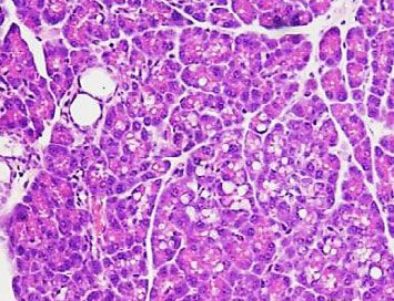
ARTICLE                                                                 NATURE COMMUNICATIONS | https://doi.org/10.1038/s41467-021-24506-w

P
       ancreatitis is an inflammatory disease of the exocrine                    For all parameters measured, pancreatitis was potentiated in
       pancreas which in most cases is initiated by bile acid reflux          Ins2Akita mice compared to corresponding WT control mice
       from gall stones or fatty acid/ethanol metabolites from               (Fig. 1). Specifically, caerulein induced a 69 ± 6 % increase in
excessive alcohol and fat consumption1. Severe acute pancreatitis            oedema (pancreas wet/dry weight ratio) in Ins2Akita mice
(SAP) is characterised by pancreatic necrosis and multiple organ             compared to 24 ± 4% increase in WT control mice (Fig. 1b).
failure, which increases mortality and prolongs critical care                Interestingly, in Ins2Akita mice without pancreatitis (injected with
occupancy. In the USA SAP accounts for 1.4 million hospital days             phosphate-buffered saline (PBS)), pancreas wet/dry weight ratio
at a cost of $2.6 billion every year2. There is no imminent cure             was lower (2.59 ± 0.19) compared to WT mice (3.13 ± 0.09;
and treatment is restricted to nutritional support and fluid                  Fig. 1b), suggesting the pancreas of Ins2Akita mice is more
resuscitation3,4. Clearly a radical approach to the treatment of SAP         dehydrated than WT pancreas. In addition, qPCR of pancreatic
is urgently required to improve survival and reduce critical care            tissue mRNA revealed that pancreatic tissue cytokine expression,
occupancy. Impaired metabolism and cytotoxic Ca2+ ([Ca2+]i)                  including TNFα (Fig. 1c) and IL-6 mRNA expression was
overload in pancreatic acinar cells are central events regardless of         markedly potentiated in Ins2Akita mice compared to WT mice
the causative factor1. Metabolism and [Ca2+]i are linked by the              (Fig. 1d). Histologically, numerous features of pancreatitis also
ATP-driven plasma membrane Ca2+ ATPase (PMCA), which                         appeared to be aggravated in Ins2Akita mice compared to WT
prevents cytotoxic Ca2+ overload. Therefore, restoration of acinar           mice. Haematoxylin and Eosin (H&E) staining of formalin-fixed
cell metabolism and protection of PMCA represents an attractive              paraffin-embedded (FFPE) pancreas tissue revealed that in WT
therapeutic strategy, which might be achieved by insulin.                    mice caerulein induced intralobular oedema with ‘patchy’ areas of
   Using cellular models of pancreatitis our previous studies                severe tissue injury interspersed by normal tissue (Fig. 1f
revealed that insulin protects acinar cells from cellular injury5,6.         compared to e). However, in Ins2Akita mice, caerulein induced
However, it is unclear from these studies in isolated pancreatic             more severe tissue oedema that was more widespread across the
acinar cells whether endogenous insulin release has any protective           tissue, rather than being “patchy” as was the case with the WT
effect in vivo and the specific molecular mechanism remains                   pancreas (Fig. 1j compared to i). There were clear signs of vacuole
unknown. Although pancreatitis can lead to type 3c diabetes, due             formation (red arrow, Fig. 1j) and inflammatory cell infiltration
to collateral pancreatic β-cell injury and the loss of insulin               (yellow arrow, Fig. 1j), which was confirmed using CD45
secretion7,8, emerging evidence from clinical9–14 and animal                 immunohistochemistry of pancreas tissue counterstained with
studies15–17 suggest that diabetes increases the severity of AP and          haematoxylin (Fig. 1g–l). CD45 is a non-specific cell surface
thus endogenous insulin may be protective.                                   marker expressed on haematopoietically-derived inflammatory
   However, it is very difficult to separate the systemic effects of          cells including, neutrophils, macrophages, eosinophils19. Consis-
hyperglycaemia, reduced insulin secretion (type-1 diabetes) or               tent with these observations, pancreatitis was further quantified
reduced insulin sensitivity (type-2 diabetes), from any putative             histologically using a well-validated histology injury score of H&E
direct protective effects of insulin on pancreatic acinar cells. This        stained slides20–22. These data revealed that cerulein induced a
was achieved in the current study by inducing experimental AP in             consistently higher injury score in diabetic Ins2Akita mice
Pancreatic Acinar cell-specific Conditional Insulin Receptor                  compared to WT mice for all four criteria (Fig. 1m–p).
Knock Out mice (PACIRKO) in which insulin receptors (IRs)
were deleted specifically in acinar cells using a tamoxifen-induced
Cre-lox-based gene deletion system18. Results show that pan-                 Functional characterization of PACIRKO mice. We next wan-
creatitis was more severe in both Ins2Akita mice (type-1 diabetic            ted to separate the confounding effects of hyperglycaemia, or
mouse model) and in PACIRKO mice. This was due to a loss of                  reduced systemic insulin, from a loss of any direct protection of
insulin-induced, Akt-mediated phosphorylation of the key gly-                insulin on acinar cells. This was achieved using PACIRKO mice,
colytic enzyme, 6-phosphofructo-2-kinase/fructose-2,6-bispho-                in which the insulin receptor (IR) was specifically deleted in
sphatase 2 (PFKFB2) in pancreatic acinar cells. This is sufficient            pancreatic acinar cells of adult mice using a tamoxifen-inducible
to drive glycolytic flux and maintain the glycolytic ATP supply to            gene deletion (Supplementary Information). Insulin receptor (IR)
the plasma membrane Ca2+ pumps (PMCA), thereby preventing                    expression was greatly reduced following tamoxifen administra-
cytotoxic Ca2+ overload and necrotic cell death even in the face             tion (daily oral gavage or tamoxifen feed for 4 consecutive days)
of impaired mitochondrial function. Collectively, this provides a            in pancreatic tissue (Fig. 2a, b) and acinar cells from PACIRKO
strong mechanistic link between diabetes and the severity of acute           mice compared to corresponding littermate control IRlox/lox mice
pancreatitis and suggests that the phosphorylation of PFKFB2, by             or the feeder Ela-CreER+/ mouse line (Fig. 2c; assessed by western
insulin or insulin-mimetics, and the preservation of acinar cell             blotting). Moreover, IR expression is specifically reduced in the
ATP, may represent a potential therapeutic strategy for the                  pancreas, as expression in the spleen, liver and kidney remains
treatment of AP.                                                             normal (Fig. 2c). This reduced IR expression occurred regardless
                                                                             of whether pancreatitis was induced with caerulein (Fig. 2a).
                                                                             Importantly, unlike Ins2Akita mice, plasma glucose was normal in
Results                                                                      PACIRKO mice (Fig. 2d).
Pancreatitis is worse in diabetic Ins2Akita mice. To test whether
endogenous insulin had any protective effect during AP, we first
induced experimental pancreatitis in parallel groups of age-                 Pancreatitis is worse in non-diabetic PACIRKO mice lacking
matched type-1 diabetic Ins2Akita mice vs control C57BL/6                    acinar IRs. Similar to Ins2Akita diabetic mice, pancreatitis was
wildtype mice (WT). This was done using the classical caerulein-             worse in PACIRKO mice, either induced by caerulein (Fig. 3) or
induced hyperstimulation model of AP. Ins2Akita mice were all                by fatty acid/ethanol20 (Fig. 4), compared to corresponding age-
confirmed to be hyperglycaemic, regardless of whether pancrea-                matched littermate control mice (IRlox/lox). Specifically, the
titis was induced (423 ± 47 mg/dl, caerulein treatment, n = 4; and           caerulein-induced increase in pancreas wet/dry weight ratio was
443 ± 82 mg/dl, PBS treatment, n = 3) compared to WT mice                    markedly potentiated in PACIRKO mice (51 ± 2 %) compared to
(125 ± 17 mg/dl, caerulein treatment, n = 6; and 145 ± 31 mg/dl,             IRlox/lox mice (18 ± 3 %, Fig. 3a). PACIRKO pancreas wet/dry
PBS treatment, n = 5; Fig. 1a).                                              weight ratio (2.61 ± 0.08) without pancreatitis (PBS-induced) was
                                                                             lower compared to IRlox/lox mice (2.96 ± 0.09). This suggests that

2                NATURE COMMUNICATIONS | (2021)12:4386 | https://doi.org/10.1038/s41467-021-24506-w | www.nature.com/naturecommunications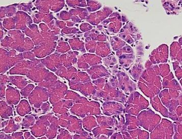
NATURE COMMUNICATIONS | https://doi.org/10.1038/s41467-021-24506-w                                                                                                                                                                     ARTICLE

                                 a           Blood [Glucose]                                      PBS                                        b Pancreas                            PBS
                                                                                                                                                                                                               p=0.065
                                             (mg/dl)                                              Caer                                             Wet/Dry                         Caer
                                                                                                                                                   Weight 5                                                      p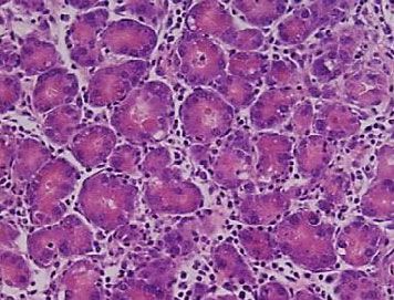
ARTICLE NATURE COMMUNICATIONS | https://doi.org/10.1038/s41467-021-24506-w
a b overload as a readout of cellular injury (Fig. 5). On average, 30
IRlox/lox PACIRKO IRlox/lox PACIRKO 1.0
µM POA induced a robust increase in [Ca2+]i by 600 ± 73 nM
Caer - + + + - + + - + + + - - + ++
Normalised IR
kDa (Fig. 5a–e), which was dramatically reduced to 103 ± 50 nM
IR 100 (Fig. 5b–e), in the presence of insulin (Fig. 5f; area under the
0.5 curve (AUC); 397 ± 53 µM.s with POA alone vs 100 ± 29 µM.s
Cyc-A with insulin (p < 0.05)). However, this insulin-mediated protec-
15 0 tion was completely abolished in acinar cells from PACIRKO
IRlox/lox PACIRKO mice (Fig. 5d; POA alone increased [Ca2+]i to 520 ± 56 nM
c d
Ela-CreER/+
(AUC; 315 ± 26 µM.s), while POA in the insulin-treated group
PACIRKO
PACIRKO
PACIRKO
PACIRKO
PACIRKO
IRlox/lox
IRlox/lox
IRlox/lox
increased [Ca2+]i to 625 ± 44 nM (AUC; 361 ± 36 µM.s; Fig. 5).
Blood [Glucose] (mg/dl)
kDa 600
PBS Collectively, these data clearly show that insulin protects mouse
IR 100
75 400
Caer pancreatic acinar cells against POA-induced cytotoxic [Ca2+]i
Acn overload, which is abolished in acinar cells from PACIRKO mice
37
200 lacking acinar IRs.
Pancreac Pancreas Liver
Spleen
Kidney
Acinar Tissue
Cells 0 Insulin-induced protection against POA-induced PMCA inhi-
IRlox/lox PACIRKO
bition is abolished in acinar cells from PACIRKO mice. To test
Fig. 2 PACIRKO mice that lack pancreatic acinar cell insulin receptors are whether the insulin protection of POA-induced Ca2+ overload in
normoglycaemic. a Insulin receptor (IR) expression (western blot using acinar cells was due to protection of PMCA activity we utilised a
anti-IRβ antibody) in pancreas tissue from double floxed insulin receptor similar in situ [Ca2+]i clearance assay to our previous
(IRlox/lox) mice vs Pancreatic Acinar Conditional Insulin Receptor Knock studies5,6,23. Under these conditions, 30 µM POA caused a dra-
Out (PACIRKO) mice following administration of tamoxifen for 4 days (oral matic inhibition of relative [Ca2+]i clearance, and thus PMCA
gavage or tamoxifen feed) with (+) or without (−) caerulein (Caer; 50 µg/ activity, in cells from both IRlox/lox mice (13 ± 6%; Fig. 6b,
Kg × 8 hourly IP injections over 2 days) to induce acute pancreatitis, or PBS. compared to time-matched control, 101 ± 2%, Fig. 6a) and
Western blotting with anti-cyclophilin-A was used as a loading control. IR PACIRKO mice (10 ± 3%; Fig. 6e compared to time-matched
expression was semi-quantified by normalizing band intensities to the control, 98 ± 1%, Fig. 6d). However, this POA-induced inhibition
corresponding cyclophilin-A band and further normalisation to the mean of of PMCA activity was completely abolished following pretreat-
IRlox/lox (b). c IR expression was also compared between lysates from ment with 10 nM insulin in cells from IRlox/lox mice (100 ± 6%;
pancreatic acinar cells and tissue from Ela-CreER/+ mice, IRlox/lox and Fig. 6c) but not PACIRKO mice (21 ± 5%; Fig. 6f). These data
PACIRKO and also liver, spleen and kidney tissue. Actin was used as a suggest that insulin protects the PMCA from inhibition by POA
loading control (lower panel, c). For each western blot of IR in a, c, the which is specifically mediated through IRs, as this protection is
corresponding loading control (cyclophilin-A in a and actin in b) were from abolished in PACIRKO mice lacking IRs.
the same gel (membrane cut and incubated with corresponding antibody)
and are representative of three separate experiments. d Blood glucose in Insulin-induced protection against POA-induced ATP deple-
IRlox/lox vs PACIRKO mice, with caerulein (grey bars) or PBS white bars). tion is abolished in acinar cells from PACIRKO mice. We next
Group sizes were: IRlox/lox PBS, n = 15; IRlox/lox Caer, n = 12; PACIRKO PBS, wanted to test whether insulin prevents POA-induced ATP
n = 13; PACIRKO Caer, n = 12. Data are presented as mean value ± SEM. depletion in IRlox/lox mouse pancreatic acinar cells, using the
firefly luciferase-based chemiluminescence assay5,6,23. In acinar
cells from IRlox/lox mice, POA caused a concentration-dependent
pancreatitis was more severe in PACIRKO mice vs IRlox/lox mice ATP depletion between 1–100 µM which generated an average
(Fig. 3n–q). IC50 of 12.2 ± 3.3 µM and Hill slope of −1.58 ± 0.3 (Fig. 7a–c).
We next tested the effect of the more aetiologically relevant Pre-treatment of cells with 10 nM insulin caused a rightward shift
palmitoleic acid/ethanol (POA/ETOH) model20. This model was in the concentration-response curve and significantly increased
further optimised using lower doses of both POA and ETOH, the average IC50 to 49.4 ± 7.4 µM (p < 0.05; Hill slope of −2.99 ±
which produced a more specific pancreatitis, and less peripheral 0.84; Fig. 7a–c). However, in acinar cells from PACIRKO mice,
organ injury independent of pancreatitis (Fig. S1 and Supple- POA caused a similar concentration-dependent ATP depletion
mentary Information). Similar results were obtained using the (IC50 of 20.6 ± 4.5 µM and Hill slope of −0.94 ± 0.08; Fig. 7b, c,
POA/ETOH-induced model of pancreatitis to those obtained black circles), which was unaffected by insulin pre-treatment
with caerulein. In PACIRKO mice, there was a potentiation of (IC50 of 14.6 ± 3.4 µM and Hill slope of −0.87 ± 0.04; Fig. 7b, c).
POA/ETOH-induced pancreas wet/dry weight ratio (58 ± 4% vs This suggests that insulin preserves ATP over the same POA
22 ± 2% in IRlox/lox mice; Fig. 4a), pancreatic tissue cytokine concentration range (3–100 μM) that induces cytotoxic Ca2+
expression (Fig. 4b–e), histological features of pancreatitis (H&E; overload and inhibition of PMCA.
Fig. 4f–k) and CD45-positive staining (Fig. 4h–m). Likewise,
there was a similar overall trend in the POA/ETOH-induced Effect of insulin on cellular bioenergetics in pancreatic acinar
histology injury scores in PACIRKO vs IRlox/lox mice (Fig. 4n–q). cells. To further explore the effect of insulin on acinar cell
metabolism we first measured NAD(P)H autofluorescence as an
Insulin-induced protection against POA-induced cytotoxic Ca2 indirect measure of glycolytic vs mitochondrial metabolism5. The
+ overload is abolished in acinar cells from PACIRKO mice. major source of cellular NAD(P)H is from the mitochondrial
Since pancreatitis was more severe in PACIRKO mice lacking Krebs cycle whereas a minor source comes from the glycolytic
acinar cell IRs, we next wanted to investigate the mechanism for enzyme, glyceraldehyde phosphate dehydrogenase (GAPDH).
the protective effect of insulin on acinar cells from IRlox/lox mice Therefore, treatment of cells with the protonophore and mito-
vs PACIRKO mice. This was achieved using ‘cellular models’ of chondrial uncoupler, CCCP (4 µM), causes rapid consumption of
pancreatitis in which isolated acinar cells (from IRlox/lox vs mitochondrial NAD(P)H, and thus a reduction in NAD(P)H
PACIRKO mice), were pre-treated with or without insulin (10 autofluorescence. CCCP acts by dissipating the mitochondrial
nM for 15 min) followed by POA to induce cytotoxic Ca2+ proton gradient and driving force for the ATP synthase activity.
4 NATURE COMMUNICATIONS | (2021)12:4386 | https://doi.org/10.1038/s41467-021-24506-w | www.nature.com/naturecommunications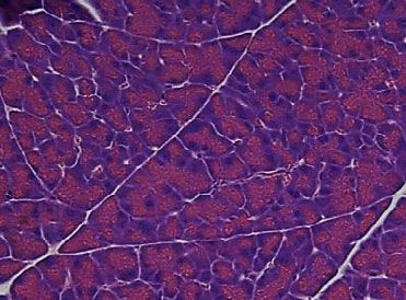
NATURE COMMUNICATIONS | https://doi.org/10.1038/s41467-021-24506-w                                                                                                                                                                                                ARTICLE

                                                                                                                                                                                                                                            p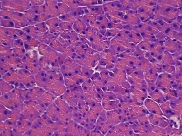
ARTICLE                                                                                                                                NATURE COMMUNICATIONS | https://doi.org/10.1038/s41467-021-24506-w

                                                                                                                                                p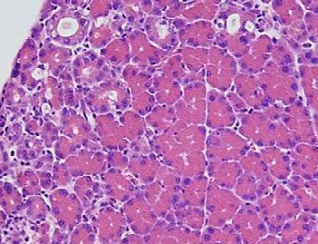
NATURE COMMUNICATIONS | https://doi.org/10.1038/s41467-021-24506-w ARTICLE
a IRlox/lox
30µM POA 30pM CCK
b IRlox/lox
30µM POA 30pM CCK
Insulin (10 nM) alone caused a marginal increase in ECAR
600 600 (127 ± 14% of untreated control). However, POA (30 μM)
[Ca2+]i [Ca2+]i + Insulin
(nM) 400 (nM) 400 markedly reduced ECAR to 54 ± 6% of untreated control cells
(Fig. S7), which was restored to similar levels of control (96 ± 5%)
200 200
by pre-incubation with insulin (10 nM). These data further
0
10 min
0
10 min
support the notion that insulin maintains glycolytic flux and thus
c PACIRKO d PACIRKO ATP production to fuel the PMCA, even in the face of POA-
30µM POA 30pM CCK 30µM POA 30pM CCK
induced metabolic crisis.
600 600 + Insulin
[Ca2+]i [Ca2+]i
(nM) 400 (nM) 400
200 200
The insulin-mediated increase in glycolysis is due to Akt-
mediated phosphorylation of phosphofructokinase fructose
0
0
10 min 10 min bisphosphatase-2 (PFKFB2). We next wanted to determine the
p=5x10⁻⁴ p=3x10⁻⁴
e 800 f AUC (µM.s)
500
p=0.001 p=0.004
specific molecular mechanism for the protective effects of insulin
[Ca2+]i (nM)
600 400 on acinar cells during pancreatitis. Since insulin preserved cellular
300
400
200
ATP and switched metabolism from mitochondrial towards gly-
200 100 colysis, we reasoned that the regulation of glycolytic enzymes may
0
POA (µM)
Insulin (nM)
0
30
0
30
10
30
0
30
10
POA (µM)
Insulin (nM)
30
0
30
10
30
0
30
10
be the primary mechanism. Specifically, we focussed on PI3K/Akt
IRlox/lox PACIRKO IRlox/lox PACIRKO as the major downstream signalling pathway of insulin, primarily
because in our previous study the PI3K inhibitor, LY294002,
Fig. 5 Insulin-mediated protection against palmitoleic-induced Ca2+
abolished most of insulin’s protection of POA-induced [Ca2+]i
overload is abolished in pancreatic acinar cells from PACIRKO mice.
overload in pancreatic acinar cells6. Therefore, we next tested
Representative traces showing POA-induced [Ca2+]i responses (a–d) in
whether insulin treatment led to the Akt-mediated phosphor-
untreated fura-2-loaded pancreatic acinar cells (a, c) and following pre-
ylation of key glycolytic enzymes that may be responsible for the
treatment with 10 nM insulin for 15 min (b, d) from IRlox/lox (a, b) and
upregulation of glycolysis. This was initially done using co-
PACIRKO (c, d). Cells were also subsequently treated with 30 pM CCK to
immunoprecipitation of all Akt-phosphorylated proteins using
test for recoverability and thus cell viability post-POA treatment. Mean
immobilised phospho-Akt-substrate antibody followed by wes-
(±SEM) maximum increase in resting [Ca2+]i above baseline (e) and mean
tern blotting with antibodies for pAkt substrate antibody (positive
(±SEM) area under the curve (AUC; f) over the treatment and recovery
control (Fig. S8a) and the key glycolytic enzymes, including
period in the absence (dark grey bars) or following treatment with 10 nM
hexokinase-I (HK-I, Fig. S8b), phosphofructokinase-1 (PFK-1,
insulin (light grey bars). Significance (specific p values as indicated) was
Fig. S8c), pyruvate kinase-M (PKM, Fig. S8d), pyruvate kinase-L
determined by one-way ANOVA with Sidak’s multiple comparisons. Data
(PK-L, Fig. S8e), and lactate dehydrogenase-A (LDHA, Fig. S8f).
were derived from individual values from multiple cells (6-36 cells per
However, none of these glycolytic enzymes tested were found to
experiment) in the field of view for each experiment. These values were
be phosphorylated by Akt following treatment with insulin
averaged giving the experimental mean, that were in turn averaged across
(Fig. S8). Although this is not an exhaustive list of Akt substrates,
multiple experiments (four separate experiments for each experimental
or even glycolytic enzymes and indeed there may be other
condition, except PACIRKO with POA, n = 5) giving the true mean ± SEM as
enzymes responsible for the shift towards glycolysis, we decided
indicated in (e, f).
to focus on the key glycolytic enzyme reported to be activated by
insulin via Akt-mediated phosphorylation, which is 6-phospho-
fructo-2-kinase/fructose-2,6-biphosphatase 2 (PFKFB2)24. Using
CCCP-induced and IAA-induced increases in MgGreen fluores- antibodies that specifically detect phospho-Akt (p-Akt(ser473)
cence were normalised to the total change in MgGreen and the Akt-mediated phospho-PFKFB2 (p-PFKFB2(ser483))
fluorescence (Fig. S6c). In agreement with the changes in NADH western blotting showed that treatment of acinar cells from IRlox/
autofluorescence, insulin treatment (10 nM for 15 min) reduced lox mice with insulin (10 nM) for 10 min induced phosphoryla-
the CCCP-induced increase in MgGreen fluorescence (70 ± 3.7% tion of Akt and PFKFB2, which was completely abolished by the
to 32 ± 1.6%; Fig. S6), a measure of mitochondrial ATP depletion, PI3K inhibitor LY294002 (10 μM) (Fig. 8). There was a surpris-
but enhanced the IAA-induced increase in MgGreen fluorescence ingly high basal Akt and PFKFB2 phosphorylation in untreated
(30 ± 3.7% to 68 ± 1.5%; Fig. S6), a measure of glycolytoc ATP acinar cells from PACIRKO mice compared to acinar cells from
depetion. These data show that insulin treatment switches IRlox/lox mice. Importantly, however, there was no further
pancreatic acinar cell metabolism towards glycolysis, which is increase in Akt or PFKFB2 phosphorylation following insulin
sufficient to preserve cellular ATP to fuel the PMCA and treatment of acinar cells from PACIRKO mice, suggesting that
maintain cytosolic [Ca2+]i homoeostasis. the loss of insulin receptors was responsible for this insulin
To investigate this further we next tested whether insulin insensitivity. In acinar cells from Ela-CreER/+ mice, that express
treatment was sufficient to preserve glycolytic flux during Cre (similar to PACIRKO) but also express IRs (similar to IRlox/lox),
pancreatitis. This was assessed in isolated mouse (C57BL/6) there was a lower basal Akt and PFKFB2 phosphorylation and
pancreatic acinar cells in response to the pancreatitis-inducing insulin-induced an increase in Akt and PFKFB2 phosphorylation
agent POA (30 μM) with or without insulin (10 nM) treatment similar to that observed in IRlox/lox mouse acinar cells (Fig. 8). This
using the pH-Xtra Glycolysis Assay (Agilent; see Supplementary suggests the high basal Akt and PFKFB2 phosphorylation in
Information). This measures extracellular acidification rate PACIRKO mice is due to loss of IRs rather than non-specific effects
(ECAR), due primarily to lactic acid efflux and thus is a of Cre expression.
convenient measure of glycolytic flux. The pH-Xtra assay utilises The high basal Akt and PFKFB2 phosphorylation in PACIRKO
a dual-read ratiometric time-resolved fluorescence lifetime mice was highly consistent and appeared specific for acinar cells
measurement (Fig. S7a), which is converted to extracellular pH from PACIRKO mice. We, therefore, investigated this further and
(Fig. S7b) and [H+], using the MARS data analysis software reasoned that the upstream insulin receptor substrates (IRS-1 and
(See Supplementary Methods) from which ECAR can be IRS-2) may be responsible. Indeed western blotting revealed that
determined and normalised to control (Fig. S7c, mean data). both IRS-1 and IRS-2 expression was upregulated in PACIRKO
NATURE COMMUNICATIONS | (2021)12:4386 | https://doi.org/10.1038/s41467-021-24506-w | www.nature.com/naturecommunications 7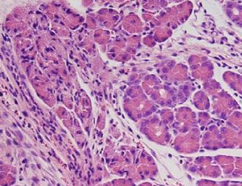
ARTICLE                                                                                                                                             NATURE COMMUNICATIONS | https://doi.org/10.1038/s41467-021-24506-w

                      a IRlox/lox                                                                        b IRloxlox                                                                         c IRlox/lox                            + Insulin
                                                       Time-matched control                                                                         30µM POA                                                                  30µM POA

                                                                                                                                                                                             0.5 ratio units
                                                                                                               0.5 ratio units
                                                                                                                                 CPA
                                         CPA

                                                                                                                                                                                                               CPA
                      0.5 ratio units

                                                                                                                                                                   0.2 units

                                                                                                                                                                                                                                         0.2 units
                                                                    0.2 units
                                                                                50 s                                                                                            50 s                                                                 20 s

                                              10 min                                                                                       10 min                                                                    10 min

                      d PACIRKO                                                                          e PACIRKO                                                                          f PACIRKO                               + Insulin
                                                       Time-matched control                                                                         30µM POA                                                                  30µM POA

                                                                                                                                                                                                               CPA
                                        CPA

                                                                                                                                 CPA
                                                                                                          0.5 ratio units

                                                                                                                                                                                            0.5 ratio units
                      0.5 ratio units

                                                                                                                                                                                                                                         0.2 units
                                                                    0.2 units

                                                                                                                                                                   0.2 units
                                                                                50 s                                                                                            50 s                                                                 50 s

                                              10 min                                                                                       10 min                                                                     10 min
                                                                                                                                            p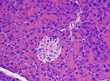
NATURE COMMUNICATIONS | https://doi.org/10.1038/s41467-021-24506-w                                                                                                                    ARTICLE

                                                                                                                                                           p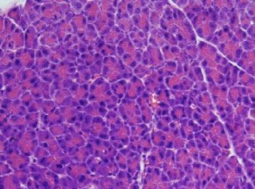
ARTICLE                                                                         NATURE COMMUNICATIONS | https://doi.org/10.1038/s41467-021-24506-w

                      a                                                               b
                                 IRlox/lox        PACIRKO                                    IRlox/lox        PACIRKO
                      kDa
                                                                                      kDa
                      100                                             IR               75
                                                                                                                           Total Akt
                       75                                                             50
                                                                      pAkt(S473)
                       50
                                                                                      37
                                                                                                                           actin
                       37
                                                                      actin
                       25                                                                   0 0 10 10 0          0 10 10 LY294002 (µM)
                             0 0 10 10            0     0 10 10 LY294002 (µM)               0 10 0 10 0         10 0 10 Insulin (nM)
                             0 10 0 10            0    10 0 10 Insulin (nM)

                      c
                            IRlox/lox PACIRKO
                      kDa
                      100
                                                      IR
                                                       pPFKFB2(S483)
                       50

                       37                             actin
                              0     10   0   10       Insulin (nM)
                      d                                     e         Ela-CreER/+                f
                                                                kDa
                             Ela-CreER/+                                                                  Ela-CreER/+
                      kDa
                                                                100                  IR
                                                                 75                              kDa
                                              pAkt(S473)        50                   Total Akt       50                 pPFKFB2(S483)
                       50
                       37                     actin              37                  actin           37
                                                                                                                        actin
                             0     0 10 10 LY294002 (µM)              0     0 10 10 LY294002 (µM)         0    0 10 10 LY294002 (µM)
                             0    10 0 10 Insulin (nM)                0    10 0 10 Insulin (nM)           0   10 0 10 Insulin (nM)

Fig. 8 Insulin increases Akt phosphorylation and the downstream Akt-mediated phosphorylation of the key glycolytic enzyme, PFKFB2, which is
abolished in acinar cells from PACIRKO mice. Pancreatic acinar cells from IRlox/lox (a–c), PACIRKO (a–c) or Elast-Cre (d–f) were treated with or without
10 nM insulin and/or the PI3K inhibitor, LY294002 (10 μM) for 10 min followed by cell lysis. Proteins were separated by SDS-PAGE and western blotted
using antibodies for the insulin receptor (IR, a, c and e), phospho-Akt (Ser473) (a, d), total Akt (b, e) and phospho-PFKFB2 (Ser483) (c and f), which
recognizes the specific Akt consensus phosphorylation site. For each representative experiment shown (a–f) separate gels were run and each membrane
cut and incubated with each corresponding antibody either in parallel or in series, including IR, Akt, pAkt(S473), pFKFB2 and the loading control actin.
These were all sufficiently separated so that they could be resolved on the same gel. Each experiment shown (a–f) is representative of at least three
independent experiments.

hyperinsulinaemic euglycaemic clamp significantly attenuated                         of acute pancreatitis12, diabetes is linked to mortality in patients
caerulein infusion induced pancreatic injury associated with the                    with chronic pancreatitis (CP)35,36 and ~50% of type-1 diabetic
early phase of pancreatitis.                                                        patients exhibit pancreatic exocrine lesions characteristic of CP11.
                                                                                    Moreover, the incidence of AP is higher among type-2 diabetics
                                                                                    compared to the normal population and the risk of AP is reduced
Discussion                                                                          among insulin-treated diabetic patients10.
The current study provides the first direct evidence that endo-                         The current data are also consistent with a previous study in
genously released insulin directly protects pancreatic acinar cell                  which caerulein-induced pancreatitis was aggravated in strepto-
injury during two mechanistically distinct experimental models of                   zotocin (STZ)-induced diabetic mice; both the acute phase of
acute pancreatitis (caerulein and POA/ethanol-induced).                             injury and regeneration of the pancreas (7 days later) was
Impaired insulin secretion (which occurs in type-1 diabetic                         delayed17. Moreover, this diabetic-induced severe pancreatitis
Ins2Akita mice) and deletion of IRs in pancreatic acinar cells                      phenotype was partially corrected by exogenous administration of
(which occurs in PACIRKO mice) both led to worse pancreatitis.                      insulin. Although generally specific for pancreatic β cells, STZ
Moreover, this study also provides the first evidence that exo-                      may induce collateral exocrine injury37 which may further
genous administration of insulin, using the hyperinsulinaemic                       aggravate caerulein-induced pancreatitis independent of reduced
euglycaemic clamp, reduces caerulein-induced increase in plasma                     insulin secretion. However, the mutation in Ins2Akita mice leads
amylase and thus acute pancreatic injury.                                           to a more specific pancreatic β cell death and is more relevant to
   Heterozygous Ins2Akita male mice exhibit a single point                          the clinical situation making this important when studying the
mutation in the Ins2 gene which gives rise to inappropriate                         diabetes-pancreatitis link.
folding of pro-insulin during its synthesis, leading to ER stress                      Nevertheless, regardless of the specific diabetes model, it is very
and the consequent pancreatic β-cell apoptosis32–34. This leads to                  difficult to separate the confounding effects of hyperglycaemia
the loss of endogenous insulin secretion and the mice sponta-                       [which facilitates more severe pancreatitis and sepsis38] or
neously develop early age onset non-obese type-1, insulin-                          reduced systemic effects of insulin [which exhibits anti-
dependent diabetes33. Ins2Akita mice exhibit no signs of col-                       inflammatory properties39] from a loss of direct insulin protec-
lateral injury or inflammation of neighbouring cells of the islets                   tion of acinar cells5,6. This can only be achieved using PACIRKO
(insulitis) or adjacent acinar cells making this strain a good model                mice. IR expression is greatly reduced specifically in the pancreas
for studying the effects of impaired insulin secretion on pan-                      and PACIRKO mice are normoglycaemic and insulin levels are
creatitis. Indeed, in the current study caerulein-induced pan-                      normal18, thereby removing the confounding systemic effects of
creatitis was more severe in type-1 diabetic Ins2Akita mice;                        hyperglycaemia and loss of insulin secretion that occurs in
characterised by pancreatic oedema, tissue injury (vacuoles),                       Ins2Akita mice. The PACIRKO mouse model is essential because
cytokine expression and inflammation19. This is consistent with                      global IR knockout mice develop early postnatal diabetes, die
clinical evidence that pre-existing diabetes increases the severity                 young due to severe ketoacidosis40 and pancreatic-specific IR

10                NATURE COMMUNICATIONS | (2021)12:4386 | https://doi.org/10.1038/s41467-021-24506-w | www.nature.com/naturecommunications
NATURE COMMUNICATIONS | https://doi.org/10.1038/s41467-021-24506-w ARTICLE
a b
Caerulein (50 μg/kg/h)
Glucose infusion rate
Glucose infusion rate
Blood Glucose (mg/dl)
Insulin (12mU/kg/min) 250 250 250
Blood Glucose (mg/dl)
250
200 200 200 Insulin (12mU/kg/min) 200
(mg/kg/min)
(mg/kg/min)
150 150 150 150
100 100 100 100
Glucose infusion Glucose infusion
50 50 50 50
0 0 0 0
0 1 2 3 4 5 0 1 2 3 4 5
c Time (h) d Time (h)
Glucose infusion rate
Glucose infusion rate
250 250 250 250
Blood Glucose (mg/dl)
Blood Glucose (mg/dl)
Caerulein (50 μg/kg/h) 200
Saline Control 200
200 200
(mg/kg/min)
(mg/kg/min)
150 150 150 150
100 100 100 100
50 no glucose infusion 50 50 50
no glucose infusion
0 0 0 0
0 1 2 3 4 5 0 1 2 3 4 5
Time (h) Time (h)
e f pARTICLE NATURE COMMUNICATIONS | https://doi.org/10.1038/s41467-021-24506-w
be translated to patients with severe AP (SAP). This is because the Ins2Akita
(T1-diabetes)
standard of care for SAP patients on critical care is fluid resus- PACIRKO
citation and supportive nutritional support frequently adminis-
tered via a parenteral IV line. This makes SAP patients on critical Diabetes Necrosis
care highly amenable to the hyperinsulinaemic euglycaemic Insulin
Protecon Severe
clamp as a therapeutic strategy. Pancreas
Insulin
Insulin therapy is routinely used to specifically treat hyper- Therapy
triglyceridaemia (HTG)-induced pancreatitis45,46, characterised
by plasma lipids exceeding 1,000 mg/dL (normal range, 101–150
mg/dL). Although HTG-induced pancreatitis is relatively rare Fig. 10 Cartoon depicting the putative molecular mechanism for insulin
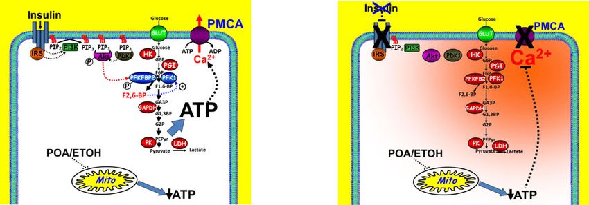
(~2–10% of all AP cases), HGT is a well-documented aetiological protection of pancreatic acinar cells during acute pancreatitis.
risk factor for severe disease47. The rationale for insulin treatment Pancreatitis inducing agents, such as fatty acid/ethanol metabolite
for HTG-induced SAP is to lower plasma triglycerides, thereby palmitoleic acid (POA) impair metabolism, most notably mitochondrial
limiting inflammation48. This is because insulin activates lipo- function, leading to ATP depletion and inhibition of Ca2+ clearance
protein lipase (converts triglycerides into free fatty acids) and pathways, such as the plasma membrane Ca2+ ATPase (PMCA), leading to
inhibits hormone-sensitive lipase (liberates triglyceride from cytotoxic Ca2+ overload. The net effect of ATP depletion and cytotoxic Ca2+
adipocyte). However, given the evidence presented in the present overload is rapid necrotic cell death, a major hallmark of acute pancreatitis.
study, it’s entirely possible that the beneficial effects of insulin Insulin binding to the insulin receptor leads to the downstream activation of
therapy in HTG-induced pancreatitis patients may also be due to the PI3K/Akt signalling pathway and the direct Akt-mediated
a direct protection of acinar cell injury. phosphorylation and activation of the key glycolytic enzyme,
Its also worth noting that intensive insulin therapy is the phosphofructokinase fructose bisphosphatase-2 (PFKFB2). This leads to the
standard of care for all critically ill patients, regardless of production of the key glycolytic metabolite fructose 2,6-bisphosphate, which
underlying disease, with the aim of reducing hyperglycaemia is an allosteric activator of the rate limiting phosphofructokinase-1 (PFK1),
associated with the acute phase of injury which facilitates which in turn drives glycolytic flux and thus glycolytic ATP production and
inflammation and sepsis49. Although several clinical studies and supply to the PMCA. This appears to be sufficient to maintain acinar cell
meta-analyses question the overall patient benefit50–54, careful ATP and prevent inhibition of the PMCA, even in the face of impaired
scrutinization of these studies reveal that fewer patients receiving mitochondrial function, thereby protecting against necrotic cell death and
insulin die from sepsis, but rather die from the complications of the self-perpetuating tissue injury and spiralling inflammatory response
inadvertent hypoglycaemia as a result of inadequate glucose characteristic of acute pancreatitis. During diabetes (Ins2Akita mice) and in
control. Moreover, it is unclear from any of these studies whether PACIRKO mice, this endogenous insulin protection is abolished, leading to
any SAP patients specifically benefited from intensive insulin more severe acute pancreatitis. Moreover, exogenous insulin administration,
therapy as patients were only identified within broad disease using the hyperinsulinaemic euglycaemic clamp to infuse high dose insulin
categories (e.g. GI diseases). Furthermore, the present study used while maintaining close glucose control, ameliorates or protects against
a relatively high dose insulin infusion (12 mU/kg/min—approx further pancreatic injury suggesting a potential therapy for severe acute
three times higher than the normal systemic concentration) with pancreatitis.
close glucose monitoring (every 10 min) to maintain tight blood
glucose. The rationale for such high dose insulin is because
in vivo pancreatic acinar cells receive a portal blood flow direct pancreatitis56. Glycolytic enzymes are also reported to be asso-
from the islets30 containing approximately ten times higher ciated with the plasma membrane of erythrocytes, smooth muscle
insulin concentration than the systemic circulation28,29. Fur- cells and pancreatic cancer cells where they provide a privileged
thermore, stress hormones, such as adrenaline, cortisol and glu- ATP supply to the PMCA57–64. Cutting off this ATP supply in
cagon, and inflammatory cytokines (TNFα and IL-1β) reduce the pancreatic cancer cells reduced numerous hallmarks of cancer
tissue sensitivity of insulin, so a higher dose of insulin may be and induced cytotoxic Ca2+ overload and cell death63,64. If
necessary to overcome this during the acute phase of injury. extrapolated to acinar cells, such insulin-mediated upregulation
The safety record of the hyperinsulinaemic euglycaemic clamp of this localized glycolytic ATP supply to the PMCA may be all
is well recognised as it is used routinely to test for insulin resis- the more critical when mitochondria, the major source of ATP,
tance. Moreover, high dose insulin infusion (8 mU/kg/min; are impaired and global ATP is severely depleted.
adjusted for surface area to weight ratio) with tight physiological PFKFB2 is a member of the PFKFB family of bi-functional
glucose control has been tested in healthy human volunteers glycolytic enzymes that convert F6P to F2,6BP, via their kinase
during endurance exercise studies with no reported adverse activity, and also catalyzes the reverse reaction by converting
effects55. Nevertheless, the use of high dose insulin, even with F2,6BP back to F6P, via their bisphosphatase activity24. F2,6BP is a
tight glycaemic control, would have to be approached with cau- potent positive allosteric activator of the rate-limiting irreversible
tion in SAP patients in critical care, given the potential problems glycolytic enzyme, PFK-1, which converts F6P to F1,6BP, thereby
of ketoacidosis, hypokalemia or other metabolic disturbances. maintaining glycolytic flux65. The relative kinase to bisphosphatase
The present study also showed, using cellular models of pan- activity, and thus the production of F2,6BP, varies between the
creatitis, that insulin signalling protects against POA-induced different PFKFB isoforms; with PFKFB1/4 expressed in the liver
ATP depletion, inhibition of the PMCA and the consequent exhibiting almost no kinase activity, which therefore acts as a break
cytotoxic Ca2+ overload. This was due to an insulin-mediated in glycolysis, promoting glycogen synthesis and flux through the
metabolic shift towards glycolysis, most likely due to Akt- pentose phosphate pathway important for nucleotide and amino
mediated phosphorylation of the rate-limiting glycolytic enzyme acid biosynthesis65. On the other hand, PFKFB3 (which is
PFKFB2. This preserves cellular ATP and fuels the PMCA, expressed in cancer cells) has a 700 fold higher kinase activity,
thereby maintaining low resting [Ca2+]i, even in the face of which means that glycolytic flux is essentially maximally activated
impaired mitochondria by pancreatitis-inducing agents (see and thus contributes to the Warburg effect in cancer cells66.
Fig. 10). A conceptually similar ‘boost’ in metabolism and pre- PFKFB2 is a tunable glycolytic enzyme with approximately equal
servation of ATP has been recently suggested to be responsible for kinase vs bisphosphatase activities, but its kinase activity is dra-
the protective effect of galactose supplementation during matically increased following phosphorylation by Akt, PKA and
12 NATURE COMMUNICATIONS | (2021)12:4386 | https://doi.org/10.1038/s41467-021-24506-w | www.nature.com/naturecommunicationsNATURE COMMUNICATIONS | https://doi.org/10.1038/s41467-021-24506-w ARTICLE
AMPK65. Therefore, phosphorylation of PFKFB2 acts as a ‘volume protein kinase-A, suggesting that AMPK activators or drugs that
control’ for glycolytic flux and thus ATP production and is thus elevate cAMP might mimic the protective effects of insulin.
likely to be the major molecular mechanism for the protective Interestingly, the anti-diabetic drug metformin is also an AMPK
effects of insulin during pancreatitis (see Fig. 10). activator, suggesting that this could be repurposed to treat acute
It is also interesting to note that the expression of amylase was pancreatitis. Such drugs might have a more specific protective
also reduced in pancreatic acinar cells of PACIRKO mice. effects without some of the more adverse systemic effects, tech-
Moreover, the pancreatitis-induced increase in plasma amylase, a nical difficulties or safety issues of insulin administration that
classical early clinical biomarker for pancreatitis, was blunted in could be used to treat all forms of acute pancreatitis from mild to
PACIRKO mice. This is particularly interesting because there severe disease.
have been numerous older studies that show that the expression
and synthesis of amylase is matched by the relative carbohydrate Methods
composition in the diet67–69, which in turn is controlled by Further information and requests for resources, reagents and access to data should
insulin (and insulin receptors within acinar cells)26,27,70. Speci- be directed to Jason Bruce (jason.bruce@manchester.ac.uk). A more comprehen-
fically, fasting reduces acinar amylase, which can be restored by sive description of the methods and list of reagents can be found in Supplementary
Information. All studies with research animals, including in vivo experiments and
glucose administration, but this is prevented by diazoxide which experiments using cells/tissues derived from research animals complied with all
inhibits glucose-dependent insulin secretion71–73. Conversely, relevant ethical regulations regarding the use of research animals (see below).
and consistent with the present study, acinar amylase content
(expression and synthesis) is severely reduced in type-1 diabetic Animal study approval. All animal procedures and in vivo experiments were
rat models (streptozotocin and alloxan), which is restored by approved by the University of Michigan Institutional Animal Care and Use
insulin administration26,27,70. Similarly, acinar amylase content is Committee (IACUC). The breeding of PACIRKO mice and feeder strains (Ela-
also reduced in hyperinsulinaemic insulin-resistant diabetes CreER/+ and IRlox/lox) at the University of Manchester, which includes the
administration of tamoxifen to induce insulin receptor deletion was approved by
models (Zucker rat) and restored by ciglitazone (reduces insulin the Home Office Project licence (PPL number, P08B76E2B; PPL holder, Michael
resistance and normalizes glucose)74. Therefore, during pan- Simonson-Jackson).
creatitis when acinar cells undergo necrosis and thus cell lysis
there is potentially less amylase available to be released into the Ins2Akita and PACIRKO mice. All experiments using Ins2Akita mice were per-
interstitium and thus plasma, which helps to reconcile these formed on male heterozygous mice at 6–9 weeks, as this was empirically deter-
results. mined to be the time of onset of consistent hyperglycaemia. Ins2Akita mice were a
It was also observed in Ins2Akita and PACIRKO mice that the kind gift from Peter Arvan (Department of Internal Medicine, Unversity of
Michigan). PACIRKO mice were originally generated from the feeder strains
wet/dry weight ratio was significantly lower in mice without Elastase-Cre-ER mice (Elast-CreER/−) double floxed insulin receptor mice (IRlox/
pancreatitis compared to the corresponding control mice (WT or lox) as previously described18. Both the experimental mice (PACIRKO) and age-
IRlox/lox, respectively). This suggests that the pancreas tissue is matched littermate control mice (IRlox/lox) were administered tamoxifen (75 mg/
more dehydrated in Ins2Akita and PACIRKO mice than control kg) by oral gavage daily for 4 days at 5–6 weeks and on day 7 pancreatitis was
mice, suggesting that endogenous insulin might regulate tissue induced using either caerulein or POA/ETOH. IR expression was confirmed and
western blotting with an anti-IRβ antibody (Cell Signaling). For additional details
water content. However, this is independent of systemic hyper- about breeding and genotyping of Ins2Akita and PACIRKO mice see Supplemen-
glycaemia, and thus osmotic diuresis, as this occurs in PACIRKO tary Information. All mice are kept in individually ventilated cages with 12 h light:
mice that are normoglycaemic. Therefore, this suggests that 12 h dark cycles maintained at 22 °C ± 2 °C and between 45 and 65 relative
insulin regulates acinar cell ion/volume regulation and thus tissue humidity with free access to food and water. These conditions are in accordance
fluid homoeostasis specifically mediated through activation of with the Codes of Practice for the care and accommodation of animals under
section 21 of the Animals (Scientific Procedures) Act 1986 as amended in 2012
acinar cell IRs. It is interesting to note that insulin, and its (‘ASPA’).
downstream signalling pathways, have been shown to regulate
numerous ion transport pathways, such as Na+/K+/2Cl− Caerulein-induced experimental pancreatitis. Mice received eight hourly intra-
cotransporter (NKCC)75–81 and Na+/K+-ATPase77,79,82,83 that peritoneal (IP) injections of 50 μg/Kg caerulein per day over two consecutive days
are critical for regulation of cell volume and thus fluid homo- and were euthanized by CO2 asphyxiation followed by cervical dislocation 2 h or
eostasis. It is also worth mentioning that the loss of NKCC 24 h after the last caerulein injection. Blood was immediately collected for
activity, for example during inhibition with the loop diuretic assessment of blood glucose and plasma amylase (Phadebas amylase test, Magle
Life Sciences). Whole pancreas tissue was rapidly dissected, weighed and cut into
bumetanide, reduces insulin sensitivity in hepatocytes84, sug- sections for processing for histology (10% formalin), RNA (RNA later, Thermo-
gesting that cell volume regulation is important for maintaining Fisher), protein (snap frozen in liquid nitrogen, prior to homogenisation in lysis
insulin effectiveness. Therefore, if extrapolated to pancreatic buffer) and wet/dry weight ratio (weighed before and after drying in an oven at 90 °
acinar cells, this suggests that the loss of NKCC activity and cell C for 24 h).
volume regulation during diabetes may further exacerbate acinar
cell injury and thus contribute to the severity of pancreatitis. POA/ETOH-induced experimental pancreatitis. Mice received one IP injection
In summary, the current study provides a mechanistic link containing PBS followed by two hourly IP injections of 100 µg/Kg POA, 0.8 g/Kg
ethanol (ETOH). This was a slightly lower dose of POA and ETOH used in the
between diabetes and the severity of acute pancreatitis (AP). study that originally characterised this model20 and was fully optimized in the
Moreover, this study provides evidence that exogenous ther- current study to produce the least collateral organ injury and a more specific
apeutic administration of insulin with tight moment-to-moment pancreatitis (Supplementary Information). Similar to the caerulein model, mice
glucose control, using the hyperinsulinaemic euglycaemic clamp were euthanized 2 h or 24 h after the last injection and tissue/blood were harvested
and processed in the same way.
reduces early pancreatic injury associated with AP. Therefore,
insulin infusion with the aim of reducing pancreatic injury may
prove effective for the treatment of SAP, as long as there is tight RNA extraction and quantitative real time PCR. Pancreatic tissue stored at 4 °C
in RNA later was transferred to TRIzol reagent (Ambion Life Technology) and
moment-to-moment glycaemic control (hyperinsulinaemic homogenised using a polytron for 5–10 s. RNA was isolated using chloroform/
euglycaemic clamp). Finally, the present study has identified isopropanol extraction and RNeasy spin column kit (Qiagen). Following quanti-
phosphorylation of PFKFB2 as a potential therapeutic strategy for fication and assessment of purity using the Nanodrop 280/260 nm optical density
the design of new drugs, or the repurposing of existing drugs, for ratio (OD280/OD260 ratios), isolated (200 ng) was reverse transcribed into cDNA
using TaqMan reverse transcription reagents (Thermofisher) with random hex-
the treatment of AP. In addition to Akt-mediated phosphoryla- amers as primers. Quantitative PCR reactions were carried out using the Absolute
tion, PFKFB2 can be phosphorylated at the exact same serine Blue SYBR Green ROX reagent (Thermo Scientific, Waltham, MA) with specific
residue (S483) by AMP-dependent kinase (AMPK) and/or primers (listed in Supplementary Table 1 and ref. 85).
NATURE COMMUNICATIONS | (2021)12:4386 | https://doi.org/10.1038/s41467-021-24506-w | www.nature.com/naturecommunications 13ARTICLE NATURE COMMUNICATIONS | https://doi.org/10.1038/s41467-021-24506-w
SDS PAGE and western blotting. Frozen tissue that had been snap-frozen in external Ca2+ (0 Ca2+, 1 mM EGTA) to depelete ER Ca2+ and activate store-
liquid nitrogen following dissection was homogenized in lysis buffer (in mM: 50, operated Ca2+ entry (SOCE). Addition of 20 mM external Ca2+, therefore, results
Tris-HCl; 50, NaCl; 5, EDTA, 0.2% triton X-100; 10 mM NF; 10, Na4P2O7; 25, in a rapid increase in [Ca2+]i which reaches a steady state and the subsequent
glycerophosphate, 1, DTT; 1, PMSF; 0.2, Na3VO4; 10 µg/ml leupeptin; 10 µg/ml removal of external Ca2+ (0 Ca2+, 1 mM EGTA) causes a rapid [Ca2+]i clearance
aprotinin). For phosphorylation assays (phospho-Akt and phosphor-PFKFB2), due predominantly to PMCA activity. Repeated Ca2+ influx-efflux phases allow
isolated pancreatic acinar cells were resuspended in a similar lysis buffer containing POA to be applied during the second phase and compared to the initial clearance
protease inhibitor cocktail tablets (Roche) and PhosSTOP (Roche). Lysates were phase. [Ca2+]i clearance rate (and thus PMCA activity) in the presence or absence
allowed to solubilize at 4 °C for 30 min followed by sonication and centrifugation at (time-matched control) of POA during this second clearance phase is quantified by
16,000 × g for 10 min to remove insoluble debris (pellet). Sample protein was measuring the linear rate from a standardised value of [Ca2+]i and normalised to
determined (Bradford assay, Bio-Rad Laboratories), denatured by boiling in SDS- the corresponding linear rate during the first clearance phase23.
Laemmli buffer for 5 min, separated using sodium dodecyl sulphate
electrophoresis-polyacrylamide gel electrophoresis (SDS-PAGE), transferred to
Measurement of cellular ATP. ATP depletion was assessed using fire-fly luci-
nitrocellulose and western blotted using specific antibodies at 1:1000 dilution
ferase (ViaLight® Plus kit; Lonza, Rockland, ME USA) as previously described5.
(unless otherwise stated) to insulin receptor (IRβ mAB), amylase (rabbit anti-α-
Cells were treated with or without 10 nM insulin for 20 min, followed by treatment
amylase, used at 1:15,000 dilution), Akt1 (C73H10) (rabbit mAb used at 1:14,000)
with or without various concentrations of POA (1–100 µM). In the same 96 well
phospho-Akt substrate, phospho-Akt (Ser473) (used at 1:15,000 dilution),
plate some wells were treated with an ATP depletion cocktail, consiting of 4 μM
phospho-PFKFB2 (Ser483) (used at 1:5,000 dilution), IRS-1 (rabbit MAb;), IRS-2
CCCP, 500 μM bromopyruvate, 10 μM oligimycin and 2 mM iodoacetate, for a
(mouse MAb), pan-phosphotyrosine (clone 4G10; used at 1:8,000 dilution), HKI,
further 20 min to induce maximum ATP depletion. The total luminescence count
PFK-1, PKM, PKL/R, LDH and actin (used at 1:100,000) or cyclophilin-A were
of the ATP-depletion cocktail was subtracted from each corresponding assay
used as a loading control (Supplementary Table 1, Supplementary Information).
condition prior to normalisation to the corresponding time-matched controls and
For immunoprecipitation, cells were lysed in Radioimmune Precipitation Assay
expressed as a % and data presented as sigmoidal concentration-response curves.
(RIPA) buffer (containing 50 mM Trizma® base, 1 mM EDTA, 1 mM EGTA, 0.1
mM vanadate, 1 mM NaF, 40 mM Na4P2O7, 1% Triton X-100, 0.1% SDS, and
supplemented with cOmplete EDTAfree protease inhibitors and PhosSTOP Imaging of NADH Autofluorescence. Pancreatic acinar cells were excited with
phosphatase inhibitors), sonicated and left to solubilize for 30 min on ice86. Protein light at 350 nm (500 ms exposure) and NADH autofluorescence collected through a
lysates containing around 1 mg/ml of protein were incubated with either IRS-1 or fura-2 400 nm dichroic filter without a band pass filter. Sequential background-
IRS-2 antibody (1 ml lysate; 1:100 dilution) overnight at 4 °C on a rocking plat- subtracted images were acquired every 5 sec and changes in NADH auto-
form. Protein lysates were incubated for a further 2 h at 4 °C with magnetic fluorescence quantified as raw fluorescence grey levels. To determine the relative
protein-G Dynabeads. Immunoprecipitates were washed five times with lysis mitochondrial and glycolytic contributions to NADH autofluorescence, cells were
buffer, denatured in SDS Laemmli sample buffer and boiled for 15 min at 100 °C. treated with 4 µM CCCP and then 2 mM iodoacetate (IAA), respectively.
Immunoprecipitated samples were then separated by SDS and western blotted.
Imaging magnesium green (MgGreen) fluorescence. Pancreatic acinar cells
Plasma amylase. Frozen plasma harvested from experimental mice was slowly isolated from IRlox/lox mice were loaded with 4 µM MgGreen acetoxymethyl ester
defrosted and assayed for amylase concentration using Phadebas reagent (Sup- (AM) for 30 min at room temperature (see Supplementary Information and
plementary Information). refs. 5,6).
Histological assessment of pancreatitis. Pancreatic tissue was formalin-fixed Measurement of glycolysis using the pH-Xtra Glycolysis Assay. Acinar cell
(10% formalin at 4 °C for 24 h, followed by 70% ethanol) and paraffin-embedded glycolysis was assessed by real-time, kinetic analysis of extracellular acidification
(FFPE), prior to cutting into 5 μm sections and mounting onto slides (Supple- rate (ECAR) using a pH-Xtra Glycolysis Assay kit (Agilent) and a plate reader with
mentary Information). Tissue sections were stained with haemotoxylin and eosin time-resolved fluorescence (TRF) capability (CLARIOstar, BMG Labtech). Extra-
(H&E) using a standard protocol by the University of Michigan Cancer Center cellular acidification is mainly due to lactic acid efflux, which results in a reduction
Histology Core Facility. All pancreatic tissue sections were imaged, archived and in assay buffer pH. The pH Xtra probe sensitively detects this reduction in pH as an
analysed using the 3D-Histech Pannoramic-250 microscope slide-scanner (Uni- increase in sensor signal. Briefly, isolated mouse pancreatic acinar cells were
versity of Manchester Bioimaging Facility) (Supplementary Information). Slides washed three times with respiration buffer and pre-incubated in the presence or
from all animal groups were graded by two independent, blinded observers absence of 10 nM insulin for 30 min followed by the addition of 30 μM POA. After
according to severity and extent of oedema, inflammatory cell infiltration and 15 min, 40 μl of cell suspension per condition was transferred to a 96 half area well
acinar necrosis using a well-validated histology injury score on a scale of 0–3 plate and 10 μl of pH-Xtra reagent (fluorescent probe) was added to each well.
(where 3 was the most severe). The total score was the sum of the oedema, Blank control wells were included and cell-free negative and positive (glucose
inflammation and necrosis scores for each slide (maximum score of 9)87. To fur- oxidase) wells were used as signal controls. Each condition was carried out in
ther assess pancreatic tissue inflammation, immunohistochemistry using anti- triplicate. Both the metabolic incubations and the measurements were carried out
CD45 antibody (1:50 dilution) was performed on FFPE pancreatic tissue sections at 37 °C.
(Supplementary Information). Dual read TRF lifetime detection mode was performed and the change in
fluorescent lifetime from 100 to 300 µs delay and a 30-µs read window (excitation
340 ± 50 nm, emission 615 ± 18 nm) was monitored over time. The two TRF
Pancreatic acinar cell isolation. Pancreatic acinar cells from IRlox/lox, PACIRKO intensity readings collected were used to calculate the ratio-metric fluorescence
and Ela-CreER/+ mice were isolated by collagenase-digestion which was adapted lifetimes using a data analysis software (MARS, BMG Biotech), which applies the
from previous methods23,88 and described in detail in Supplementary Information. following formula: Lifetime (μs)[τ] = (D2−D1)/ln(IW1/IW2) where IW1 and IW2
Mice were humanely killed by an approved method as set out in Schedule 1 of the represent the two (dual) measurement windows and D1 and D2 represent the delay
UK Animals Scientific Procedures Act 1986 (Certificate of Designation No 50/ time prior to measurement of W1 and W2, respectively. Lifetime values (μs) were
2506). Briefly, following rapid dissection the pancreas was chopped and incubated corrected based on the blank wells and represent the extracellular acidification in
in HEPES-buffered physiological saline (HEPES-PSS; for composition see Sup- each individual sample, then scaled to [H+] using the same software tool.
plementary Information) containing 0.15 mg ml collagenase P (~200 U/ml) and
0.15 mg/ml of soybean trypsin inhibitor (Sigma) for 25 min at 37 °C with
mechanical trituration every 5 min to break up the tissue into small cell clusters. Combined caerulein infusion-induced pancreatitis and hyperinsulinaemic
Cell clusters were resuspended in Dulbecco’s Modified Eagle Medium (DMEM) euglycaemic clamp. Mice (C57BL/6) were anesthetized with an intraperitoneal
containing 2.5% FBS, and 0.15 mg/ml of trypsin inhibitor and left to rest for 30 min injection of sodium pentobarbital (50–60 mg/kg). The ventral side of the neck and
at 37 °C prior to any experimentation. back of the head were shaved and the skin repeatedly scrubbed with iodine and
70% ethanol. Under aseptic conditions, a small incision at the right ventral side of
neck was made cephalic to the sternum at 30° to the midline, exposing the right
Imaging of fura-2 fluorescence. Pancreatic acinar cells were loaded with 4 μM
carotid artery and jugular vein. The right jugular vein was catheterized using silicon
fura-2-AM for 30 min at room temperature in HEPES-PSS and imaged using a
tubing (0.025′′OD). The right carotid artery was catheterized using a two-part
Nikon TE2000S microscope with ×40 oil immersion SFluor objective lens, Cool-
catheter constructed from polyurethane (0.010′′OD, with interior coated with
SNAP HQ CCD camera (Photometrics, Tucson, AZ), Cairn monochromator
heparin) and silastic tubing (0.025′′OD). The arterial catheter was inserted reaching
(Cairn Research, Kent, UK) and MetaFluor imaging software (Molecular Devices,
the level of the aortic arch and the venous catheter was extended to the level of the
Downington, PA)23. Background-subtracted fluorescence images were captured
right atrium. To allow free movement of the animal post-recovery, the free ends of
with 50 ms exposure and 5 × 5 binning every 5 s. The fura-2 fluorescence was
catheters were tunnelled subcutaneously and exteriorized at the back of the neck
calibrated into [Ca2+]i using the well-established Grynkiewicz method88. All
via a stainless steel tubing connector (coated with medical silicon) that were fixed
imaging experiments were carried out at room temperature (20–22 °C).
subcutaneously upon the closure of the incision. The catheters were flushed daily
after surgery with saline and sealed with heparin-saline (200 U/ml). Following
In situ Ca2+ clearance assay. Cells were treated with the sarco/endoplasmic surgery, mice were allowed to recover and acclimatize for 5 days prior to experi-
reticulum Ca2+-ATPase (SERCA) inhibitor, cyclopiazonic acid (CPA) in zero mentation and were housed individually and their body weight monitored daily.
14 NATURE COMMUNICATIONS | (2021)12:4386 | https://doi.org/10.1038/s41467-021-24506-w | www.nature.com/naturecommunicationsYou can also read