Ion Case Studies - Septodont
←
→
Page content transcription
If your browser does not render page correctly, please read the page content below
Case Studies
                      Septodont

No. 17 - September 2018
                                                  Collection
Alveolar preservation
with R.T.R
Dr Gabriela Vilar Pineda

Biodentine™: external
resorption management
Dr Maximiliano Casa

Biodentine™: root
reconstruction
Prof. Fernando Tenorio

                              Biodentine™:
Biodentine™: perforation
repair
                              retrograde apical
Dr Alberto Diaz Tueme         microsurgery
                              Dr Carlos Herrera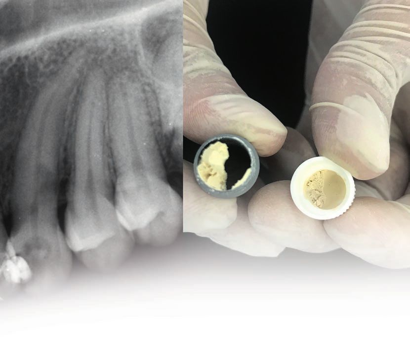
Editorial
     Since its foundation Septodont has developed, manufactured
     and distributed a wide range of high quality products for dental
     professionals.
     Septodont recently innovated in the field of endodontics, dentine
     care, bone grafting and gingival preparation with the introduction
     of BioRoot™ RCS, Biodentine™, RTR and Racegel which are
     appreciated by clinicians around the globe.

     Septodont created the “Septodont Case Studies Collection” - a
     series of case reports - in 2012 to share with you their experience
     and the benefits of using these innovations in daily practice.
     Over the past 6 years, authors from more than 15 countries have
     generously contributed to the success of our magazine that is
     now distributed on the 5 continents.
     Each new issue of the Case Studies Collection is the opportunity
     to discover new clinical challenges and their treatment solutions.

     This 17th issue features one RTR case and three Biodentine™
     cases:
        RTR Bone grafting aims at preserving bone dimensions

        especially when tooth removal is discussed. It is fully
        resorbable & osteoconductive. Its remarkable properties
        promote formation of patient’s new bone & pave the way for
        future successful treatment plans.
       Biodentine™, the first biocompatible and bioactive dentin
        replacement material. Biodentine™ uniqueness not only lies in
       its innovative bioactive and “pulp-protective” chemistry, but
       also in its universal application, both in the crown and in the
       root.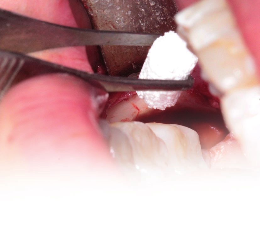
Content
Alveolar preservation with R.T.R
Dr Gabriela Vilar Pineda
                                   04

Biodentine™: retrograde apical
microsurgery
                                   13
Dr Carlos Herrera

Biodentine™: external resorption
management
                                   21
Dr Maximiliano Casa

Biodentine™: root reconstruction
Prof. Fernando Tenorio
                                   29

Biodentine™: perforation repair
Dr Alberto Diaz Tueme
                                   34

                                        3
Use of ß-tricalcium phosphate for
    alveolar preservation; a report on
    a series of cases.
    Authors: Medina Ramírez Jorge Michel*, Gabriela Vilar Pineda**,
    René García Contreras***.
    Corresponding author: Gabriela Vilar Pineda.

       Summary
    ß-tricalcium phosphate is a high purity material                        the quadrant with the R.T.R, favorable bone
    that helps to safely generate bone neoformation                         neoformation process was observed in a
    after tissue extraction or loss. Preservation                           shorter time compared to the left quadrant,
    treatment was performed using a ß-tricalcium                            and a progressive and total reabsorption of
    phosphate cone (R.T.R Cone, Septodont,                                  the R.T.R was observed after 3 months. The
    France) in the lower right quadrant, at the                             use of ß-tricalcium phosphate is a useful alter-
    lower third molar area (tooth 48), for research                         native for post-extraction alveolar preservation,
    purposes; it was kept under observation for                             improving the speed of bone regeneration.
    periods of 1 week, 1 month and 3 months. In

    *Student of Dentistry at the National School of Higher Studies of the National Autonomous University of Mexico, León Unit (ENES UNAM León Unit).
    **Specialist in Oral and Maxillofacial Surgery at the ENES of the UNAM, León Unit.
    ***Doctor of Health Sciences at ENES/UNAM, León Unit.
4
Introduction
Bone regeneration with alveolar preservation        biological process, the material is reabsorbed
following tooth extraction has been a topic of      and replaced by the recipient’s own bone.
major significance recently due to the use of       The interconnection between pores facilitates
dental implants as a method of esthetic and         osteoconduction. When the graft is placed
functional rehabilitation;(1) the improvement       at the receptor site, some serum proteins
of regeneration time is therefore a significant     are absorbed and retained on the surface of
issue, and for such purpose alveolar preser-        the particles, contributing to the subsequent
vation techniques using various materials have      cellular migration that will stimulate a neovas-
been proposed (hard and soft tissue grafts) in      cularization process in the porous structure.(3)
view of maximizing tissue preservation and          The R.T.R. (Resorbable Tissue Replacement),
minimizing defects.(2)                              is composed of ß-tricalcium phosphate, a
ß-tricalcium phosphate (ß-TCP) is a synthetic       material used for alveolar preservation after
ceramic bone graft material that has been in        a tooth removal when posterior prosthetic
use in medicine and dentistry for more than         rehabilitation is planned. The objective of
30 years, in the fields of orthopedics, perio-      the following series of clinical cases was to
dontology and maxillofacial surgery. Pore size      evaluate the alveolar preservation achieved
varies from 5 to 500 μm, and the porosity           with the use of R.T.R cones and without the
ranges from 20 to 90% depending on particle         use of R.T.R. by radiographic evaluations at
size. For dental use, particle size is gene-        three-month follow-up. Case reporting was
rally less than 100 μm.(3) Used as a graft          conducted in compliance with Case Report
material, ß-TCP stands in for the mechanism         Guidelines (CARE).
of osteoconduction; when it is used in the

  Report on a series of cases
Four patients were selected to conduct alveolar     Patients meeting the admission criteria for
preservation with the use of ß-tricalcium           the clinics and oral surgery department of the
phosphate (R.T.R) cones; these patients were        “Escuela Nacional de Estudios Superiores”, of
required to meet certain criteria. The inclusion    the UNAM, León Unit, were evaluated.
criteria used for this report were: patients of
both sexes, retained third molars Pell & Gregory    Auxiliary diagnostic studies were performed
class I and II subdivision A and B, bilateral,      (Panoramic radiographs, Periapical views),
age range between 18 and 22 years, no perio-        and no disorders being found in any patient, a
dontal or periapical disease in the lower molar     diagnosis was made in the full series of cases;
region, and willingness to perform the alveolar     retained third molars Pell & Gregory class I
preservation procedure for research purposes.       and II, subdivision A and B, bilateral.
The exclusion criteria used for this report were:
patients with uncontrolled systemic diseases,       The patients signed informed consent forms in
acute infectious processes, pregnant or             which they are made aware of the diagnosis,
lactating patients, patients with bone diseases,    treatment plan and possible complications
use of bisphosphonates, and poor oral hygiene.      during treatment.

                                                                                                       5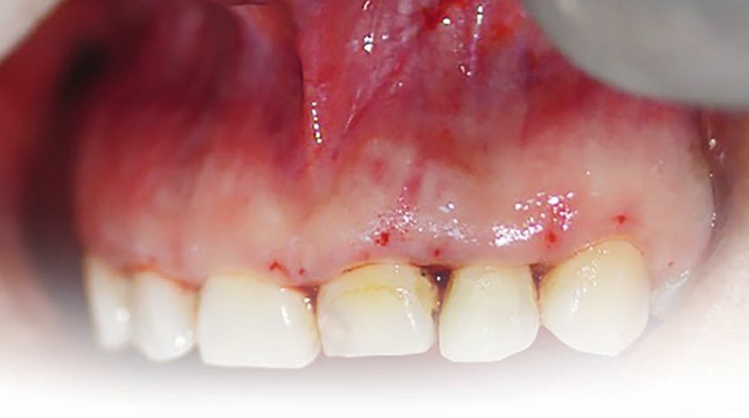
PATIENT 1

    Fig. 1: E
             xtraoral photographs   Fig. 2: Intraoral photographs
           (A: frontal B: lateral)          (A: right side B: top C: left side D: bottom)

    PATIENT 2

    Fig. 3: E
             xtraoral photographs   Fig. 4: Intraoral photographs
           (A: frontal B: lateral)          (A: right side B: top C: left side D: bottom)

    PATIENT 3

    Fig. 5: E
             xtraoral photographs   Fig. 6: Intraoral photographs
           (A: frontal B: lateral)          (A: right side B: top C: left side D: bottom)

6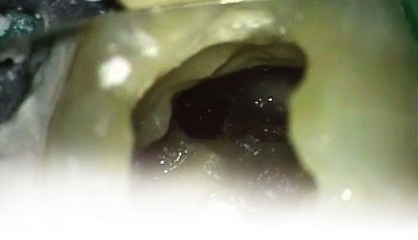
PATIENT 4

Fig. 7: E
         xtraoral photographs                      Fig. 8: Intraoral photographs
       (A: frontal B: lateral)                             (A: right side B: top C: left side D: bottom)

Fig. 9: Initial panoramic radiograph, patient 1.    Fig. 10: Initial panoramic radiograph, patient 2.

Fig. 11: Initial panoramic radiograph, patient 3.   Fig. 12: Initial panoramic radiograph, patient 4.

Surgical odontectomies of teeth 38 and 48 were      opposite side the lower left quadrant in the area
performed under local anesthesia by infiltration    of tooth 38 was left with nothing placed inside
with lidocaine and epinephrin 2%, 1:100,000; a      the alveolus, and both sides were sutured using
Newman’s incision was performed, the muco-          polyglactin 910 4/0; postoperative management
periosteal flap lifted, osteotomy and odon-         with Amoxicillin 500 mg, 1 every 8 hours for
tectomy conducted, the cavity washed, and a         5 days, and Ibuprofen 400 mg, 1 every 8 hours
cone of ß-tricalcium phosphate (R.T.R) placed       for 3 days; instructions for general postope-
in the residual alveolus of tooth 48; on the        rative measures were likewise provided.

                                                                                                           7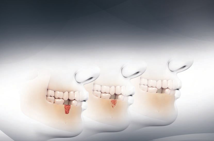
A B C
D E F
G H I
J K L
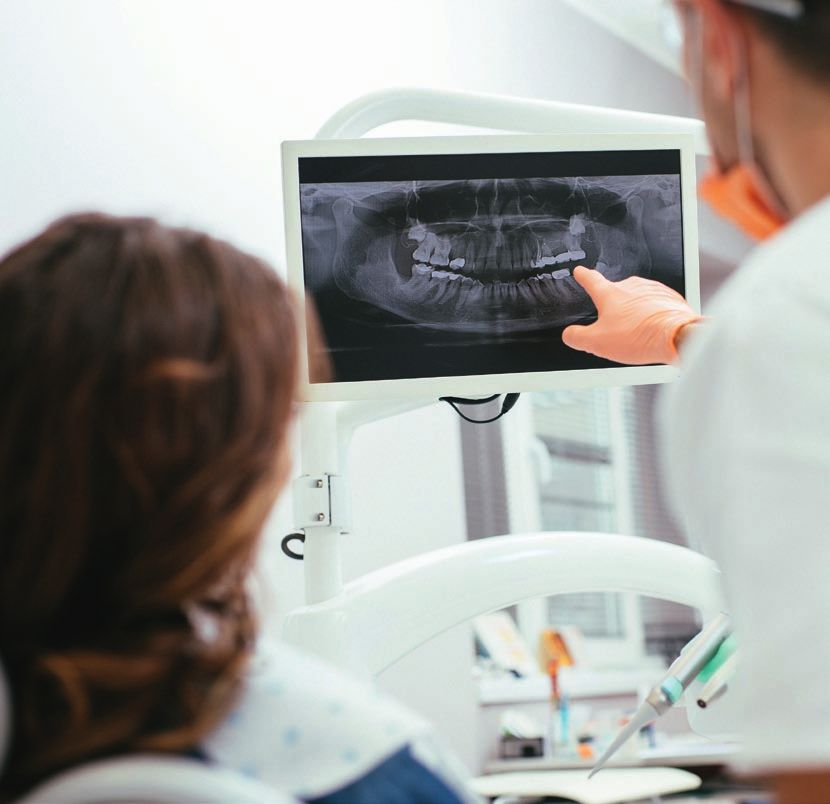
Fig. 13: Surgical procedure
(A) Anesthesia, (B) Incision, (C) Preparation of the flap, (D) Ostectomy, (E) Luxation,
(F) Extraction, (G) Extraction sample, (H) Residual Alveolus, (I) Septodont ß-tricalcium
phosphate, (J) ß-tricalcium phosphate Cone, (K) ß-tricalcium phosphate Cone transport,
(L) Application of ß-tricalcium phosphate Cone, (M) Syneresis.
M
Patients were evaluated 7 days after surgery; a radiolucent image was observed in tooth 38,
good evolution was observed, with an adequate corresponding to the residual alveolus; after a
healing process underway; radiographically, a month of evolution, we were able to observe
mixed image was observed (black and white improved healing. The area treated with R.T.R
radiographic image), interpreted as the ß-tri- showed larger areas of radiopacity, interpreted
calcium phosphate cone (R.T.R) in tooth 48, and as improved bone neoformation compared to the
8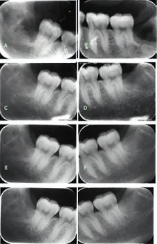
residual alveolar process area at tooth 38, where              a radiopaque image of the residual alveolus at
slower bone formation was observed, consi-                     tooth 48, in addition to total reabsorption of the
dering the greater areas of radiolucency. The                  material, as indicated by the manufacturer, and
third month of observation allowed us to verify                reduced bone trabeculation compared to the
the presence of improved bone regeneration,                    residual alveolar process area at tooth 38.

       Od 48                    Od 38                                 Od 48                    Od 38

                                                    1 week                                                         1 week

                                                    1 month                                                        1 month

                                                    3 months                                                       3 months

                                                    6 months                                                       6 months

Fig. 14: C
          ontrol periapical radiograph patient 1              Fig. 15: C
                                                                         ontrol periapical radiograph patient 2

       Od 48                    Od 38                                 Od 48                    Od 38

                                                    1 week                                                         1 week

                                                    1 month                                                        1 month

                                                    3 months                                                       3 months

                                                    6 months                                                       6 months

Fig. 16: C
          ontrol periapical radiograph patient 3              Fig. 17: C
                                                                         ontrol periapical radiograph patient 4

                                                                                                                              9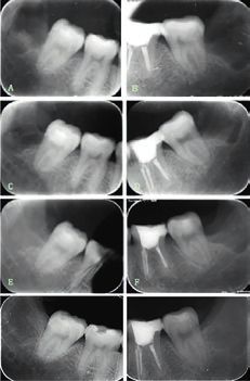
Fig. 18: Panoramic radiograph, 3 months, patient 1.   Fig. 19: Panoramic radiograph, 3 months, patient 2.

     Fig. 20: Panoramic radiograph, 3 months, patient 3.   Fig. 21: Panoramic radiograph, 3 months, patient 4.

        Discussion
     Classically, the ideal material considered for        When the ß-tricalcium phosphate is reabsorbed,
     bone regeneration has been autologous bone.           it is replaced by bone that is anatomically and
     However, in recent decades new materials of           functionally similar to the original bone, thus
     human, animal or synthetic origin have been           producing regenerated vital bone tissue, which
     incorporated into the arsenal, which have revo-       means that this bone remodeling and matu-
     lutionized alveolar preservation techniques.(4)       ration process, necessary for the functional
     The action of ß-tricalcium phosphate (R.T.R)          loading of implants, is not disturbed by the
     on alveolar preservation, in comparison to            material.(7) Residual elements may occasionally
     the natural bone healing process, has been            remain, which can be demonstrated clinically
     proven.(5) As for its regeneration mechanism,         and radiologically after 6 months.(8)
     ß-tricalcium phosphate (R.T.R) is a biocompa-
     tible material that would seem to have scaffold       The overall results of the study showed that
     action permitting osteoblasts to grow on its          at clinical follow-up, 1 year after the functional
     surface and invade its structure.(3)                  loading of implants (6 months after surgery), no
     It has proved to be an excellent biomaterial with     failures were observed in either the implants or
     high success in bringing about the bone rege-         the various different implant-supported pros-
     neration necessary to maintain adequate space         thodontic options.(9)
     for implant insertion.(6)

        Conclusion
     In light of the case reports discussed above          as compared to the residual alveolar process
     it was thus observed that after 3 months of           where no alloplastic material had been placed;
     observation the time of bone neoformation was         R.T.R is thus a choice material for the effective
     significantly improved where R.T.R was used,          post-odontectomy preservation of alveolar bone.

10
Author:
Dra. Gabriela Vilar Pineda
Dental Surgeon specialized in Oral and Maxillofacial Surgery by the Faculty of
Dentistry of the National Autonomous University of Mexico (UNAM) and recertified by
the Mexican Council of Oral and Maxillofacial Surgery.
Master of Education with focus on Innovation in Teaching Practice. She currently
works as a Career Researcher and Holder of the Degree in Dentistry of the National
School of Advanced Studies (ENES) at the UNAM Campus León. She actively
participates as an academic in several Continuing Education Programs, recently recognized for her
performance in the field of Oral and Maxillofacial Surgery with the distinction of participation in the
Scientific Commission of the Mexican Association of Oral and Maxillofacial Surgery.
She held the position of Associate Professor of the subject of Pharmacology in the Division of
Postgraduate Studies and Research by the UNAM from August 2010 to July 2011.
Coordinator of the University Diploma “Professional Update in Oral Surgery for the Dentist of
General Practice” in the period of August 2008 to July 201.
Coordinator of the Diploma “Training and Update for Dental Assistants” in the period of February
2010 to July 2011, both taught through the Continuing Education Coordination of the Faculty of
Dentistry of the UNAM.
At the bachelor's level teaches the theoretical-practical subjects of Anatomy-Physiology of the
Segment, Head and Neck, Problem-Based Learning, Introduction to Surgical Procedures, Exodontia
and Human Anatomy Physiology.
In addition, teaches the subject of Cardiopulmonary Resuscitation; within other subjects that have
been taught are Exodontia, Oral Surgery, Dental Medical Emergencies and Anesthesia in the period
of July 2005 to July 2011 at the Faculty of Dentistry of the UNAM.
She was Professor of Pharmacology, Toxicology and Clinical Analysis at the Faculty of Biological
Sciences of the Westhill University in the period of August 2008 to December 2009.
She also was Professor of Exodontia and Surgery Oral at the Faculty of Dentistry of the Autonomous
University of the State of Mexico (UAEM) in the period of September 2005 to March 2006.
Among the activities that he has carried out throughout his academic practice are the following:
Synodal of professional exams at bachelor´s degree, Tutor and Adviser of thesis to undergraduate
students of the Faculty of Dentistry at the UNAM.
Tutor of undergraduate students in the programs “BECALOS”, “PARA” and “PRONABES”, all of
these by the Faculty of Dentistry of the UNAM and of the National School of Advanced Studies of
the UNAM Campus León.
In her professional experience she has worked as a Physician assigned to the Oral and Maxillofacial
Surgery Service at the Social Security Institute of the State of Mexico and Municipalities (ISSEMYM)
from July 2005 to July 2011, simultaneously as a Physician attached to the Oral and Maxillofacial
Surgery Service of the Teachers' Union at the service of the State of Mexico in the period of January
2009 to July 2011.
She has also worked in a private practice in the “Hospital Angeles Mexico” in the period of October
2004 to April 2012 and currently a private practice in the “Hospital Angeles León”.
She has publications in national and international journals with topics with Craniomandibular
Dysfunction and Regenerative Medicine, including some studies on Health, Education and
Happiness.
She has participated as a national and international lecturer in conferences, courses and congresses
in the area of Maxillofacial Surgery, with topics such as Orthognathic Surgery, Dentoalveolar Surgery,
Implantology, Craniomandibular Dysfunction, Tissue Regeneration and Regenerative Medicine
among others.
Currently, she is responsible for projects PAPIME (Project Support Program for Innovation and
Improvement of Teaching), Multi-centric and International Research Projects and joint projects with
the area of Biomedical Engineering of the Autonomous University of Aguascalientes and develops
research projects in the area of Craniomandibular Dysfunction and Regenerative Medicine.
11References
1. Araujo MG, Lindhe J. Dimensional ridge alterations following tooth extraction. An experimental study in
the dog. J Clin Periodontol. 2005;32:212-8.
2. De, P., Ernesto, M., & Briseño, G. (2015). Materiales de injerto substitutos óseos. Fosfato tricálcico ß.
Presentación de casos clínicos. [Bone substitute graft materials. ß-tricalcium phosphate. Presentation of
clinical cases.]
3. Labanca M, Leonida A, Rodella, Natural synthetic biomaterial in Dentistry: Science and ethics as criteria
for their use. Implantologia. 2008; 1: 9-23
4. Jensen SS, Broggini N, Horting-hansen E, Schenk R, Buser D. Bone healing and graft resorption of
autograft, anorganic bovine bone and beta-tricalcium phosphate. A histologic and histomorphometric
study in the mandibles of minipigs. Clin Oral Impl Res 2006; 17: 237-43.
5. Salgado Castellanos, J., Zea del Rio, D. M., Gonzalez Miranda, J. M., & Velosa Porras, J. (2014).
Effectiveness of Alveolar Preservation Techniques over Post-Extraction Socket Compared with and
without Socket Preservation. Systematic Review of Literature. Universitas Odontologica, 33(70).
https://doi.org/10.11144/Javeriana.UO33-70.etpa.
6. Jensen OT, Garlini G, Bilk D, Peters F. Use of alloplasts for sinus floor grafting. In: Jensen OT. The sinus
bone graft (2nd ed.). Quintessence: Chicago 2006. pg.: 201-9.
7. Trisi P, Rao W, Rebaudi A, Fiore P. Histologic effect of pure-phase beta-tricalcium phosphate on bone
regeneration in human artificial jaw bone defects. Int J Perio Rest Dent 2003; 23: 69-77.
8. Suba Z, Takács D, Matusovits D, Barabás J, Fazekas A, Szabó G. Maxillary sinus floor grafting with
ß-tricalcium phosphate in humans: density and microarchitecture of the newly formed bone. Clin Oral
Impl Res 2006; 17: 102-108.
9. Zijderveld SA, Zerbo IR, van der Bergh JPA, Schulten EAJM, ten Bruggenkate CM. Maxillary sinus floor
augmentation using a ß-tricalcium phosphate (Cerasorb) alone compared to autogenous bone grafts.
Int J Oral Maxillofac Implants 2008; 20: 432-40.
R.T.R.
A complete solution
for your bone needs
12Retro-fillings with Biodentine™
in apical microsurgery
Dr Carlos Herrera
Introduction
The technique of microsurgery is a minimally Various retro-filling materials have been used in
invasive procedure that results in faster healing recent years, such as: amalgam, gold foil, zinc
and better patient response. The key stage of oxide eugenol cement, Diaket (ESPE GmbH,
microsurgery is inspection, which is entirely Seefeld, Germany), glass ionomer cements,
absent from older surgical techniques. Untreated composite resins, intermediate restorative
isthmuses often mean that treatment fails, so material (Putty / Dentsply, Milford, DE, USA),
they must be identified, cleaned, shaped and Super EBA (Bosworth, Skokie, IL, USA) and
filled as carefully as root canals (Floratos and mineral trioxide aggregate (MTA; ProRoot MTA;
Kim 2017). Dentsply, Tulsa, OK, USA). Although none of
Osteotomy in microsurgery is becoming more these meet every criterion for an ideal repair
and more conservative thanks to the improved material, MTA has been the preferred choice for
magnification and lighting offered by micros- root-end fillings (Torabinejad and Pitt 1996).
copes. The diameter of osteotomy is only 3 to In the present cases, we used Biodentine™ from
4 mm, enough to allow a 3 mm ultrasound tip to among the new biosilicate materials as a retro-
vibrate freely inside the bone cavity (Kim, Pecora filling material, developed by Septodont Research
and Rubinstein 2001) . Group as a new class of dental material which
Careful examination will identify the possible could combine excellent mechanical properties,
reason or reasons for the failure of non-sur- biocompatibility and clinical use, with a formula
gical treatment, the magnification and lighting based on calcium silicate (Ca3SiO5), a tried and
provided by the surgical microscope being tested replacement material where the dentin is
critical. (Kim, Pecora and Rubinstein 2001; damaged. (Biodentine™ Septodont 2010)
Cohen and Burns 1994 and 2002; Lubow, The following cases of microsurgery were retro-
Wayman and Cooley 1984). filled with Biodentine™ by Septodont.
13Clinical case study no.1
Female patient, 43 years of age, referred to in the periapical region of 2.1. The patient
assess apical microsurgery treatment in 2.1 and reported that she had previously received two
2.2 central and left upper incisors. The patient retreatments but the pain continued in both
displayed symptoms in both teeth; sensitivity teeth; retreatment was carried out to remove
tests revealed pain on tapping and palpation, the gutta-percha on tooth 2.1, which was not
with radiolucent lesion in both teeth, previously possible, obturation with biodentine™ with
treated pulp diagnoses and symptomatic peria- subsequent removal of the gutta-percha and
pical apical periodontitis, the radiographic apicoectomy; on tooth 2.2 microsurgery was
examination revealed extruded gutta-percha carried out with retro-filling with biodentine™.
Fig. 1 & 2: Initial photographs of 2.1 and 2.2. Fig. 3: Extruded gutta-percha in the Fig. 4: Initial radiography 06-24-16
periapical region of 2.1
Fig. 5: attempt to remove the gutta- Fig. 6: retreatment of tooth 2.1 Fig. 7: Apical microsurgery control
percha 2.1 Apicoectomy of 2.1 And retro-filling of
2.2 with Biodentine™.
Apicoectomy of tooth 2.1 and retro-filling with biodentine™ of tooth 2.2
14Fig. 8: First control 11-25-16 after Fig. 9: Second control 02-25-17 after Fig. 10: Third control 06-29-17 after 1 year
5 months 8 months
Fig. 11: First control 11-25-16 after Fig. 12 & 13: Retrograde cavity of 2.2
5 months
The patient currently presents no symptoms with no clinical
or radiographic findings at the 1-year follow-up, and shows
evidence of healthy and functional teeth.
Fig. 14: Condensation of retrofilling with
Biodentine™ in 2.2
Clinical case study no.2
Male patient, 44 years of age; referred to assess Apical Micro-
surgery of 2.2 upper left lateral incisor. Clinical and radiographic
findings: fistula at the mucosa of the tooth, evolution of the
wound: approximately 5 years; history of two retreatments in
that time; the patient reported having no symptoms; radiograph
revealed failure of endodontic treatment with placement of a
fiber glass post, light area in the periapical zone; Pulp Diagnosis:
previously treated; Periapical Diagnosis: chronic Apical Abscess.
Fig. 15: Initial Rx Microsurgery 11-07-2013
15Fig. 16: Initial Rx 11-07-2013. Fig. 17: Sinugraphy 11-07-2013. Fig. 18: End of Retro-filling with
Biodentine™.
Apical microsurgery was performed with retro- and removal of the damage confirming a peria-
filling with biodentine™ of 2.2; apicoectomy pical cyst through biopsy and histopathological
with removal of 3 millimeters of the apical root examination.
canal; irrigation with saline solution, curettage
Fig. 19: First control at one month after Fig. 20: Second control at 2 years and Fig. 21: Third control at 3 years and
treatment 12-15-2013. 5 months 04-07-2016. 1 month 12-12-2016.
At the control at 3 years and
10 months we can observe
full regeneration of the wound,
patient totally asymptomatic
at the date of control both
clinically and with evidence
of healing in the radiographic
examination.
Fig. 22: Fourth contro at 3 years and Fig. 23: Fifth control at 3 years and
4 months 04-11-2017. 10 months 10-10-2017.
16Clinical case study no.3
Male patient, 72 years of age, referred to assess radiolucent lesion of considerable size; sensi-
1.1 right upper central incisor; in his dental tivity tests were performed; the patient was
history the patient had received root canal asymptomatic with mild discomfort when biting,
treatment and final restoration 4 years earlier. positive response to tapping and palpation,
Clinical and radiographic findings: metal and previously treated pulp diagnosis and asymp-
porcelain crown, fiber glass post, periapical tomatic periapical Apical Periodontitis.
Fig. 24: Initial photograph. Fig. 25: Photograph during treatment.
Fig. 26: Initial radiography 09-25-14. Fig. 27: Final radiography retro-filling with Fig. 28: First control 11-20-14 at 2 months.
Biodentine™ 09-25-14.
Fig. 29: Second control 05-20-1 at Fig. 30: Third control 11-27-15 at 1 year Fig. 31: Fourth control 05-20-16 at 1 year
8 months. and three months. and 9 months.
17Fig. 32: Fifth control 12-06-16 at 2 years Fig. 33: Sixth control 03-31-17 at 2 years Fig. 34: Seventh control 05-05-17 at
and 4 months. and 7 months. 2 years and 8 months
Fig. 35: Eighth control 10-25-17 at 3 years Fig. 36: Ninth control 04-25-2018 at Fig. 37: Current photograph 04-25-2018 at
and 1 month. 3 years and 7 months. 3 years and 7 months.
At the control at 3 years and 7 months, tooth 1.1 showed complete regeneration of the wound.
Discussion
Amalgam has been used as a retro-filling 20% in weight of polymethyl methacrylate
material for many years. Its earliest use as a added to sodium and poly ethoxy benzonic acid
root filling after resection was recorded in 1884 (Super-EBA), which contains ethoxy benzonic
(Vasudev 2003). It has the advantages of being acid. Filtration in-vitro studies, animal studies
easily available, economical and easy to handle. and retrospective in vivo studies indicate that
Years ago, amalgam was therefore considered the materials containing ZOE are better than
as the material of choice for root fillings, and amalgam in terms of sealing and biocompati-
the clinical use of amalgam has been well bility (Dorn 1990; Kim 2006; King 1990).
documented in several clinical studies with a The shortcomings of cements currently avai-
success rate ranging from 50% to 80% (Dalal lable containing ZOE are mild to moderate
1983; Finne 1977; Grung 1990; Hirsch 1979; toxicity when freshly mixed together and their
Persson 1974; Rud 1972). radio-opacity which is similar to that of gutta-
In recent decades, amalgam has gradually percha (Johnson 1999).
given way to materials containing zinc oxide Mineral trioxide aggregate (MTA), developed at
eugenol (ZOE), such as IRM, which comprises Loma Linda University, California, USA (Tora-
18binejad 1993) has been the subject of much the production of cytokines (Koh, 1998) and
attention (Lee 1993). Its main constituents are encourages the proliferation and migration of
similar to those of Portland cement, a mixture of the parents followed by differentiation in odon-
dicalcium silicate, tricalcium silicate, tricalcium toblast cells (Kuratate 2008).
aluminate, gypsum and tetracalcium alumino- However, the average setting time for MTA is
ferrite (Camilleri 2005). 165 ± 5 minutes, which is longer than amalgam,
MTA offers major advantages including Super-EBA and IRM (Torabinejad 1995),
excellent biocompatibility (Camilleri 2006), something which is potentially problematic in
ideal adherence with cavity walls, low solubility endodontic surgery.
(Poggio 2007), and the ability to induce cemen- A new material, Biodentine™, possesses the
togenesis at the surface of the root, which is the reparative properties of dentin synthesis (Laurent
deposit of new cement on the exposed dentin 2012) with an initial setting time of 4 to 5 minutes
(Baek 2005). MTA is an excellent bioactive and a final setting time of 10 to 12 minutes; its
material. When placed in direct contact with biocompatibility with regards the periapical
human tissue, it forms calcium hydroxide which tissues makes it a very useful material with ideal
releases calcium ions for the union and proli- characteristics as a seal in retro-filling in apical
feration of cells (Takita 2006); it modulates microsurgery.
References
• Kim S, Floratos S. Modern Endodontic Microsurgery Concepts: A Clinical Update. Dent Clin 2017 N Am
(61)81–91.
• Kim S, Pecora G, Rubinstein R, editors. Color atlas of microsurgery in endodontics. Philadelphia: W.B.
Saunders; 2001
• Cohen S, Burns R, editors. Pathways of the pulp. 6th edition. St Louis (MO): Mosby; 1994.
• Cohen S, Burns R, editors. Pathways of the pulp. 8th edition. St Louis (MO): Mosby; 2002. p. 683–721.
• Lubow RM, Wayman BE, Cooley RL. Endodontic flap design: analysis and recommendations for current
usage. Oral Surg Oral Med Oral Pathol 1984;58(2): 207–12.
• Torabinejad M, Pitt Ford TR. Root end filling materials: a review. Endod Dent Traumatol 1996;12(4):161–78
• Biodentine™ - Publications and Communications 2005-2010. Research & Development Septodont, Paris
2010.
• Vasudev SK, Goel BR, Tyagi S. Root end filling materials A review. Endodontology 2003;15:12–8.
• Dalal MB, Gohil KS. Comparison of silver amalgam, glass ionomer cement & gutta percha as retrofilling
materials, an in vivo & in vitro study. Journal of the Indian Dental Association 1983;55(4):153–8.
• Finne K, Nord PG, Persson G, Lennartsson B. Retro-grade root filling with amalgam and Cavit. Oral
Surgery, Oral Medicine, and Oral Pathology 1977;43(4):621–6.
• Grung B, Molven O, Halse A. Periapical surgery in a Norwegian county hospital: follow-up findings of 477
teeth. Journal of Endodontics 1990;16(9):411–7.
• Hirsch JM, Ahlstrom U, Henrikson PA, Heyden G, Peterson LE. Periapicalsurgery. International Journalof
Oral Surgery 1979;8(3):173–85.
• Persson G, Lennartson B, Lundström I. Results of retrograde root-filling with special reference to amalgam
and Cavitas root-filling materials. Swedish Dental Journal 1974; 67(3):123–34.
• Rud J, Andreasen JO, Jensen JE. Radiographic criteria for the assessment of healing after endodontic
surgery. International Journal of Oral Surgery 1972;1(4):195–214.
• Dorn SO, Gartner AH. Retrograde filling materials: a retrospective success-failure study of amalgam, EBA,
and IRM. Journal of Endodontics 1990;16(8):391–3.
• Kim S, Kratchman S. Modern endodontic surgery concepts and practice: a review. Journal of Endodontics
2006;32(7): 601–23.
• King KT, Anderson RW, Pashley DH, Pantera EA Jr. Longitudinal evaluation of the seal of endodontic
retrofillings. Journal of Endodontics 1990;16(7):307–10.
19• Johnson BR. Considerations in the selection of a root-end filling material. Oral Surgery, Oral Medicine,
Oral Pathology, Oral Radiology, and Endodontics 1999;87(4):398–404
• Torabinejad M, Watson TF, Pitt Ford TR. Sealing ability of a mineral trioxide aggregate when used as a root
end filling material. Journal of Endodontics 1993;19(12):591–5. [PUBMED: 8151252]
• Lee SJ, Monsef M, Torabinejad M. Sealing ability of a mineral trioxide aggregate for repair of lateral root
perforations. Journal of Endodontics 1993;19(11):541–4.
• Camilleri J, Montesin FE, Brady K, Sweeney R, Curtis RV, Ford TR. The constitution of mineral trioxide
aggregate. Dental Materials 2005;21(4):297–303
• Camilleri J, Pitt Ford TR. Mineral trioxide aggregate: a review of the constituents and biological properties
of the material. International Endodontic Journal 2006;39(10): 747–54
• Poggio C, Lombardini M, Conti A, Rindi S. Solubility of root-end filling materials: a comparative study.
Journal of Endodontics 2007;33(9):1094–7.
• Baek SH, Plenk H Jr, Kim S. Periapical tissue responses and cementum regeneration with amalgam,
SuperEBA, and MTA as root-end filling materials. Journal of Endodontics 2005;31(6):444–9
• Takita T, Hayashi M, Takeichi O, Ogiso B, Suzuki N, Otsuka K, et al. Effect of mineral trioxide aggregate on
proliferation of cultured human dental pulp cells. International Endodontic Journal 2006;39(5):415–22
• Koh ET, McDonald F, Pitt Ford TR, Torabinejad M. Cellular response to Mineral Trioxide Aggregate. Journal
of Endodontics 1998;24(8):543–7.
• Kuratate M, Yoshiba K, Shigetani Y, Yoshiba N, Ohshima H, Okiji T. Immunohistochemical analysis of
nestin, osteopontin, and proliferating cells in the reparative process of exposed dental pulp capped with
mineral trioxide aggregate. Journal of Endodontics 2008;34:970
• Torabinejad M, Hong CU, McDonald F, Pitt Ford TR. Physical and chemical properties of a new root-end
filling material. Journal of Endodontics 1995;21(7):349–53.
Download the complete series
www.septodont.com
20Multidisciplinary management
of an external communicating
resorption caused by the ectopic
eruption of a maxillary canine
Maximiliano Casa. H; Alfredo Sierra, C.
Introduction
Root resorption is the decomposition, destruction Jacobs, Lambrechts, Loozen & Willems, 2009).
and subsequent loss of the dental root structure. The prevalence of ectopic eruption of maxillary
Root resorption caused by odontoclasts canines is 1 - 3%. It is more frequent in women
during the exfoliation of the deciduous teeth is than in men and it has been reported that 50%
a natural process that leads to the eruption of of the cases of root resorption of the maxillary
the permanent teeth. However, resorption can lateral and maxillary incisors occur as an
also occur in the permanent dentition due to adverse effect. (Alqerban, Jacobs, Fieuws &
trauma, excessive occlusal load, cysts, tumors, Willems, 2011). The canine, and the maxillary
orthodontic treatments and ectopic eruption of lateral and maxillary incisors are located in an
adjacent teeth (Hadler-Olsen et al., 2015). area of high aesthetic and functional demand, so
The ectopic eruption is defined as the abnormal it is important to carry out a diagnosis and early
position of the tooth, which alters its eruptive intervention, which allow us to generate thera-
trajectory and leads to its impaction against the peutic strategies that can improve the prognosis
adjacent tooth. After the third molars, the canines of affected parts and minimize possible sequels.
are the teeth that present the most anomalies in The root reabsorption of the lateral incisor can
their eruption, being trapped in the bone can be diagnosed radiographically at an early stage,
produce complications such as displacement of but the resorption process can remain asympto-
the teeth, loss of vitality of the adjacent incisors, matic, even in cases in which the pulp is affected
shortening of the dental arch, formation of folli- (Ericson & Kurol, 1987). When the diagnosis
cular cysts, canine ankylosis, recurrent infec- occurs at an advanced stage, it is difficult to
tions, recurrent pain, internal resorption, external define the treatment and prognosis, and it may
resorption of the canine and adjacent teeth, lead to the extraction of the affected tooth.
or combinations of these factors (Alqerban, For the sealing and restoration of root resorption,
21various materials have been used, such as: antibacterial action, of which no cytotoxicity,
calcium silicate-based cements such as mineral genotoxicity or mutagenicity have been
trioxide aggregate (MTA), glass ionomer cement, reported. When compared to the MTA it presents
calcium enriched mixture, etc. The MTA (ProRoot better physical and biological properties, better
MTA, Dentsply, Tusa, OK USA) developed by handling, adherence, fast setting time, greater
Torabinejad and Contributors, is a bioactive resistance to compression, early synthesis of
material that since the early nineties has been reparative dentin. Its powdered components
used for various applications in endodontics. are tricalcium silicate, dicalcium silicate, and
The MTA is indicated to restore defects of calcium carbonate as filler and zirconium oxide
internal and external resorptions, horizontal root acting as radiopacifier; on the other hand, the
fractures, sealing of perforations, pulp therapy in liquid component contains calcium chloride,
permanent and deciduous teeth, apical sealing of water and a reducing agent (Cernochova P et
the root canal in teeth with mature and immature al., 2011, Baranwal AK et al., 2016, Al-Haddad A
apices. It has been shown that MTA is biocom- et al., 2016, Eftekhar L et al., 2017, Sultana N et
patible, stimulates mineralization and promotes al., 2018).
crystalline deposits similar to apatite (Güzeler I In the present case we describe the mana-
& Uysal S., 2010, Tomás-Catalá CJ et al., 2018). gement of external communicating resorption
Taking as a reference the properties of the of the upper left lateral incisor, caused by the
MTA, Biodentine™ (Septodont, Saint-Maur-des- ectopic eruption of the left upper canine, by
Fosses, France) was developed, a material using Biodentine™ as a sealant for the sequel
based on calcium silicate, which may be a valid and subsequent rehabilitation with a fiberglass
option as it acts as a substitute for dentin, of post. and light curing resin.
Clinical case report
Female patient, 12 years old, ASA I. The patient
was referred by a pediatric dentist colleague.
Radiographically the tooth 1.3 is retained from
intra-osseous form, impacted on the distal
area of the root of the tooth 1.2, generating
an external cementum resorption, it can also
be observed that the periodontal ligament of
tooth 1.2 has slightly thickened (Fig.1). Clini-
cally, the patient presents absence of tooth 1.3
Fig. 1: Initial tomography, where it can be observed that external
and asymptomatic 1.2, which sensitivity tests cementum resorption shows a communication between the perio-
dontium and the pulp canal.
respond positively to both heat and cold and
negative to percussion and palpation. cleaning, shaping and sealing of the root canal
The following diagnoses were made: tooth system, the sealing of the perforations in the
number 1.2; vital pulp with external cementum root and later, positioning the tooth 1.3 inside the
resorption that shows a communication dental arch and thus return its functionality. We
between the periodontium and the pulp tissue divide it into the following stages:
and tooth number 1.3; impacted in the distal 1. Surgical resorption approach.
middle third of the root of tooth 1.2, being 2. Partial biopulpectomy 1.2.
retained by it intraosseously in the maxilla. 3. External resorption surgical seal.
A multidisciplinary Treatment Plan was proposed. 4. Rehabilitation of tooth 1.2.
The objectives of this proposal are firstly the 5. Orthodontic traction of tooth 1.3.
22Fig. 2: Surgical access to the area of resorption, Fig. 3: (a) Removal of the dental pulp, demonstrating that the dental organ was
where the passage of the file is observed when totally vital. (b) Odontometry, In this case it was not possible to perform it with the
removing the granulation tissue present in the LAE (electronic apex locator) due to the false positive that it was throwing in the area
resorption. of the resorption. Therefore, it was done using the radiographic method.
Fig. 4: (a) Once the channel was prepared with Protaper Next files, Fig. 5: (a) Once the apical third was sealed, the external resorption
the appropriate gutta-percha cone was chosen and then cut with the was sealed using a bioceramic (b), which on this occasion was the
Touch and Heat technique and thus sealing the apical third with the Biodentine™.
single cone technique and (b) BioRoot™ RCS Cement by Septodont.
The maxillo-facial surgeon, following the (Septodont, Saint-Maur-des-Fosses, France), a
previously established work plan, begins anes- bioceramic-based cement.
thetizing the area with an infiltrative technique, In this way, through the surgical access, the apical
to subsequently make a Newman type flap from third of the canal is sealed, dedicating efforts to
the distal side of tooth 1.4 to the mesial side of reconstruct with Biodentine™ the defect caused
tooth 1.1, accessing and clearing the area of by cemental resorption (Fig. 4, 5).
resorption for further treatment (fig. 2). Once the defect is reconstructed, the remaining
Then the endodontist, after absolute isolation, thirds and endodontic access are rehabilitated,
performs the endodontic access from the cementing a fiberglass pole (Relyx Fiber Post of
palatal tooth piece 1.2, linking the apical and 3M ESPE), with cement (RelyX U200), acid etching
cervical third, through the external cement (3M ESPE Scotchbond) and adhesive (3M) ESPE
reabsorption shown in figure 2. The channel Single Bond Universal) with light curing resin
is prepared chemically and mechanically, with (Filtex Z350 XT of 3M ESPE), giving the tooth the
the Protaper Next (Dentsply-Maillefer rotary necessary flexion to rejoin the stomatognathic
system, Ballaigues, Switzerland), irrigating system without problems (fig. 6).
with the technique of negative apical pressure Finally, the orthodontist intervenes, placing the
with abundant 2.5% sodium hypochlorite, then additives to begin with the orthodontic surgical
drying the canal with sterile paper tips, sealing detachment of the dental piece 1.3 towards the
the apical third with the technique of the single corresponding place in the arch, following the
cone (gutta percha 6%) and BioRoot™ RCS initial planning (Fig. 7).
23Fig. 6: (a) Taking advantage of
the control of the sterile area, it
is decided to permanently seal
the cervical and middle third
with (b) fiber post Relyx Fiber
Post, and cement RelyX U200,
acid etching with 3M ESPE
Scotchbond and application
of adhesive Single Bond
Universal. (c).
Fig. 7: (a) Placement of the
attachments, to begin with the
pulling of tooth 1.3. (b) closure
of the flap by the surgeon,
terminating the surgical
procedure.
Fig. 8: (a) Initial radiography.
(b) Immediate postoperative
radiograph. (c) X-ray of the
1-year control.
By way of summary in Figure 8 the radiographic
evolution of the case is shown. (Fig. 8a) Initial
X-ray, where it could be presumed that the
cementum resorption opened a communication
path between the periodontium and the pulp
canal. (Fig. 8b) Immediate postoperative image:
the apical third and the obturation of the external
cemental resorption are observed and (Fig. 8c)
controlled one year after the surgery, the sealing
of the canal is clearly observed in the apical third,
as well as the repair of the bone tissue and finally
the ongoing displacement of the canine. Fig. 9: (a) Clinical (b) and radiographic control 2-year follow-up of
the surgical therapy.
The favorable evolution was observed in the
last clinical and radiographic control completed
2 years after the surgical therapy (Fig. 9). Both
teeth involved are asymptomatic and fully
functional.
24Discussion
The treatment of cementum resorption gene- relate to the treatment that is approached from
rates, until today, different opinions among the periodontium, allowing the pulp to remain
endodontists, orthodontists, rehabilitators, healthy and undamaged.
implantologists, clinicians and scientists The planning and consensus among the
(Güzeler I & Uysal S., 2010, Alqerban, Jacobs, different professionals that should intervene
Fieuws & Willems., 2011, Umashetty G et al., in the treatment of these pathologies, is a
2015, Baranwal AK et al., 2016, Eftekhar L et challenge. Especially, when a few years ago,
al., 2017). the dentist was confined to his practice trying
Local and general factors are related to the to perform all procedures without any colla-
impact of higher permanent canines and to the boration. Currently this type of thinking would
reabsorption that this causes, such as; trauma- be untimely as the multidisciplinary work is
tisms, prolonged retention or premature loss of indicated so that each specialist develops
the primary canine, agenesis or alteration in the the treatment of their specialty, providing the
form of lateral incisors, chronic inflammation of patient with the quality of their treatment and
the pulp, periodontal tissues or both, abnormal the opportunity to keep the teeth in the mouth,
position of the dental germ, localized pathology functional and healthy.
such as cysts, neoplasms, odontomas and Surgical access to address the resorption zone
supernumeraries, ankylosis, idiopathic origin, is well described in the literature and depending
slow-growing tumors such as giant cell tumors, more on the clinical or anatomical characte-
osteosclerosis and other fibro-osseous lesions, ristics of the area to be treated, there are no
iatrogenic, naso-respiratory problems, variation major discrepancies (McDonald F & Yap WL.,
in the size of the tooth root, variation in the 1986, Caminiti MF et al., 1998, Chapokas AR et
time of root formation, sequence of abnormal al., 2012, Becker A & Chaushu S., 2015). These
eruption, systemic diseases (hyperparathy- begin at the moment in which we initiate the
roidism), narrow arch form, immune factors and endodontic treatment and we have to decide
inheritance, (Fuss., et al 2003, Alqerban, Jacobs, which concentration of hypochlorite, which
Fieuws & Willems., 2011, Gunraj MN., 1999). type of file and the last number we’ll use, which
It is very important then to foresee when there filling technique and material with which we will
will not be enough space in the arcade for the reconstruct the reabsorbed area (Brunson M et
eruption of the permanent teeth, since their al., 2010). Finally, the biggest question, will this
pressure on others will cause in a not lesser therapy succeed?
percentage, some degree of reabsorption. This With respect to irrigation, it is clear that
can cause their loss. The treatment plan that hypochlorite was used and that the lower its
will be carried out in a dental piece that contains concentration, the lower the effects, which
a communicating cementum resorption is very can be compensated with a frequent and
different from that in which it is not present abundant flush. (Regan JD & Fleury AA., 2006,
(Roscoe MG., Et al 2015). The biological Van Der Sluis L, 2006, Cohenca N et al., 2010,
consequences are diametrically different, the Haapasalo M., 2014). Therefore we could use
first being, the worst prognosis. Since it is 1.5% hypochlorite; 2.5% or 5.25% and get
not possible to treat only the communicating similar results. Regarding the last instrument to
cementum resorption without producing a be used, it is well documented that the larger
significant damage in the pulp tissue, forcing the diameters used, the greater the number
to plan a joint pulpal treatment. However, the of microorganisms removed, allowing an
evolution of those reabsorptive treatments adequate cleaning with sodium hypochlorite
that do not show communication between thanks to the access with irrigation and aspi-
the periodontium and the endodontium, only ration tips (LG Coldero., 2002, Card SJ.,
252002 Brito PRR., 2009). In this case, in which we seal the apical third and dedicate ourselves
the reabsorption occurs on the external face to reconstruct the space left by the cemental
of the root at the level of the curvature in the resorption.
apical third, there are anatomical and physical Again, we find different opinions about the
characteristics that prevent a common irri- material to be used for this purpose (Luo T et
gation as anatomically normal canals have al., 2018, Umashetty G et al., 2015, Baranwal
accustomed us. For that reason, it was chosen AK et al., 2016, Eftekhar L et al., 2017). With
in order to clean the canal, to divide it into two the advent of bioceramics, taking advantage of
portions. One, from the cameral access to the the fact that the canal was filled with a cement
cervical edge of the reabsorption (extra surgical with the same chemical characteristics, it was
portion) and the second, from the apical end decided to seal the resorption with another
of the reabsorption to the apex (intra-surgical bioceramic, Biodentine™. This one has ideal
portion). In both situations, the irrigation tech- chemical and physical characteristics, which
nique with negative apical pressure was used makes it the material of choice, durable, with
to minimize the possibility of extravasation of a flexural index similar to that of dentin, with
the irrigant to the periodontium (Goldman M et an adequate setting time, biocompatible and
al., 1998, Mehdipour O et al., 2007, Cohenca N bioactive among other characteristics (Cerno-
et al., 2010) chova P et al., 2011, Baranwal AK et al., 2016,
Taking into account the previously described Al-Haddad A et al., 2016, Eftekhar L et al., 2017,
and thanks to the location of the resorption Sultana N et al., 2018).
and anatomy of the root, it was possible to use Once the defect caused by the resorption
the Protaper Next rotary system to shape the was reconstructed, it was decided to rehabi-
canal. Properly protecting the alveolus from the litate the remaining third and the endodontic
inclemency of the different materials used, it access, cementing it with a fiberglass pole and
was decided to seal the canal with the use of a nanoparticle resin, providing the tooth with the
single gutta-percha cone with increased taper, flexion, resistance and esthetics needed to rein-
cemented with a bioceramic-based cement corporate it. Stomatognathic system without
such as BioRoot™ RCS, to be subsequently cut drawbacks.
with the Touch and Heat system. In this way,
Conclusion
There will always be criteria to take into account is always a radiographic finding with clinical
to decide the clinical procedures in these clues, therefore it is important to prevent and
therapies. However, there is no conclusive study avoid adverse sequelae. For this purpose, a
that supports a single treatment that gives us protocol for timely review should be imple-
success without exception in all cases. On the mented, including radiographic follow-up,
other hand, if there is scientific evidence that assessments of abnormal anatomical features
would support each of the steps we carry out such as the absence of canine eminence,
separately, leaving it up to us, to compromise to among others.
fill those missing spaces, rigorously complying Last, but not least, remember the importance
with the different protocols and deciding in of forming multidisciplinary work teams that
each case, the most appropriate to follow. provide the vision of each of the specialties in
The discovery of these traumatic sequelae decision making in cases of high complexity.
26Authors:
Maximiliano Casa Herzmann
Dentist, National University of La Plata, Buenos Aires, Argentina, 1996.
Dental surgeon. University of Chile, 2005.
Specialist in Endodontics 2006, (CONACEO)
21 years of experience as a teacher in the training of pre and post graduates in
the Department of Endodontics and Pediatric Dentistry in National and Interna-
tional Universities.
Author and co-author of practical work guides in the area of Endodontics and
Pediatric Dentistry.
Exhibitor of seminars, courses and congresses in national and international media.
Active member in the Endodontics Society of Chile.
Private professional practice 20 years ago.
Coordinator of the area of conservative treatments in Permanent Teeth Young,
in the department of Integral of the child I and II in the Autonomous University of
Chile.
Co-author of the book "Clinical Guide and Text of Endodontics", (in process of
edition 2018).
Contact; Orphans 1044, office 82, CP: 8320000,
telephone: +569226886979 / +56922334585 - mcasa@paisdental.cl
Alfredo Sierra Cristancho
Dentist - University of Cartagena (Colombia). Specialist in Endodontics Andrés
Bello University (Chile). Magister in Dental Sciences University of Chile.
References
1. Al-Haddad A, Che Ab Aziz ZA. Bioceramic-Based Root Canal Sealers: A Review. Int J Biomater. 2016.
Epub 2016 May 3.
2. Alqerban, A., Jacobs, R., Lambrechts, P., Loozen, G., & Willems, G. (2009). Root resorption of the maxillary
lateral incisor caused by impacted canine: a literature review. Clinical Oral Investigations, 13(3), 247-255.
3. Alqerban, A., Jacobs, R., Fieuws, S., & Willems, G. (2011). Comparison of two cone beam computed
tomographic systems versus panoramic imaging for localization of impacted maxillary canines and
detection of root resorption. The European Journal Of Orthodontics, 33(1), 93-102.
4. Baranwal AK. Management of external invasive cervical resorption of tooth with Biodentine™: A case
report. J Conserv Dent. 2016 May-Jun;19(3):296-9.
5. Becker A, Chaushu S. Surgical Treatment of Impacted Canines: What the Orthodontist Would Like the
Surgeon to Know. Oral Maxillofac Surg Clin North Am. 2015 Aug;27(3):449-58.
6. Brunson M, Heilborn C, Johnson J,Cohenca N. Effect of Apical Preparation Size and PreparationTaper on
Irrigant Volume Delivered by Using NegativePressure Irrigation System. Journal of Endodontics 2010; 36:
721-724.
7. Brito PRR, Souza LC, Machado de Oliveira JC, Alves FRF, De-Deus G, Lopes HP, et al. Comparison of
the effectiveness of three irrigation techniques in reducing intracanal Enterococcus faecalis populations:
an in vitro study. Journal of endodontics. 2009 Oct; 35(10):1422–7.
8. Caminiti MF, Sandor GK, Giambattistini C, Tompson B. Outcomes of the surgical exposure, bonding and
eruption of 82 impacted maxillary canines. J Can Dent Assoc. 1998 Sep;64(8):572-4, 576-9.
27References
9. Card SJ, Sigurdsson A, Orstavik D, Trope M. The effectiveness of increased apical enlargement in
reducing intracanal bacteria. J Endod. 2002 Nov;28(11):779-83.
10. Cernochova P, Krupa P, Izakovicova-Holla L. Root resorption associated with ectopically erupting
maxillary permanent canines: a computed tomography study. Eur J Orthod. 2011 Oct;33(5):483-91.
11. Cohenca N, Heilborn C, Johnson D, Silva D, Flores H, Yoko I, Bezerra L. Apical negative pressure
irrigation versus conventional irrigation plus triantibiotic intracanal dressing on root canal disinfection in
dog teeth. Oral Surg Oral Med Oral Pathol Oral Radiol Endod 2010; 109: 42- 46.
12. Coldero LG, McHugh S, MacKenzie D, Saunders WP. Reduction in intracanal bacteria during root canal
preparation with and without apical enlargement. Int Endod J. 2002 May;35(5):437-46.
13. Chapokas AR, Almas K, Schincaglia GP. The impacted maxillary canine: a proposed classification for
surgical exposure. Oral Surg Oral Med Oral Pathol Oral Radiol. 2012 Feb;113(2):222-8.
14. Ericson, S., & Kurol, J. (1987). Radiographic examination of ectopically erupting maxillary canines.
American Journal Of Orthodontics And Dentofacial Orthopedics, 91(6), 483-492.
15. Eftekhar L, Ashraf H, Jabbari S. Management of Invasive Cervical Root. Resorption in a Mandibular
Canine Using Biodentine™ as a Restorative Material: A Case Report. Iran Endod J. 2017
Summer;12(3):386-389.
16. F
uss Z, Tsesis I, Lin S. Root resorption--diagnosis, classification and treatment choices based on
stimulation factors. Dent Traumatol. 2003 Aug; 19(4):175-82.
17. G
oldman M, White RR, MoserCR, Tenca JI. A comparison ofthree methods of cleaning andshaping the
root canal in vitro. JEndodon 1988; 14:7-12.
18. G
unraj MN. Dental root resorption. Oral Surg Oral Med Oral Pathol Oral Radiol Endod. 1999 Dec;
88(6):647-53.
19. G
üzeler I, Uysal S, Cehreli ZC. Management of trauma-induced inflammatory root resorption using
mineral trioxide aggregate obturation: two-year follow up. Dent Traumatol. 2010 Dec;26(6):501-4.
20. H
adler-Olsen, S., Pirttiniemi, P., Kerosuo, H., Bolstad Limchaichana, N., Pesonen, P., Kallio-Pulkkinen,
S., & Lähdesmäki, R. (2015). Root resorptions related to ectopic and normal eruption of maxillary canine
teeth – A 3D study. Acta Odontologica Scandinavica, 73(8), 609-615.
21. H
aapasalo M, Shen Y, Wang Z, Gao Y. Irrigation in endodontics. Br Dent J. 2014 Mar;216(6):299-303.
22. L
uo T, Liu J, Sun Y, Shen Y, Zou L. Cytocompatibility of Biodentine™ and iRoot FS with human
periodontal ligament cells: an in vitro study. Int Endod J. 2018 Jan 19.
23. M
cDonald F, Yap WL. The surgical exposure and application of direct tractionof unerupted teeth.
Am J Orthod. 1986 Apr;89(4):331-40.
24. M
ehdipour O, Kleier D, Averbach R. Anatomy of sodium hypochlorite accidents. Compend Contin Educ
Dent 2007; 28: 544-6.
25. R
egan JD, Fleury AA. Irrigants in non-surgical endodontic treatment. J Ir Dent Assoc. 2006
Autumn;52(2):84-92.
26. R
oscoe MG, Meira JB, Cattaneo PM. Association of orthodontic force system and root resorption: A
systematic review. Am J Orthod Dentofacial Orthop. 2015. May;147(5):610-26.
27. S
ultana N, Singh M, Nawal RR, Chaudhry S, Yadav S, Mohanty S, Talwar S.Evaluation of
Biocompatibility and Osteogenic Potential of Tricalcium Silicate-based Cements Using Human Bone
Marrow-derived Mesenchymal Stem Cells. J Endod. 2018 Mar; 44(3):446-451.
28. Tomás-Catalá CJ, Collado-González M, García-Bernal D, Oñate-Sánchez RE, FornerL, Llena C, Lozano A.
29. M
oraleda JM, Rodríguez-Lozano FJ. Biocompatibility of NewPulp-capping Materials NeoMTA Plus, MTA
Repair HP, and Biodentine™ on Human DentalPulp Stem Cells. J Endod. 2018 Jan;44(1):126-132.
30. Umashetty G, Hoshing U, Patil S, Ajgaonkar N. Management of Inflammatory Internal Root Resorption
with Biodentine™ and Thermoplasticised Gutta-Percha. CaseRep Dent. 2015;2015:452609.
31. Van Der Sluis LWM, Gambarini G, Wu MK, Wesselink PR. The influence of volume, type of irrigant and
flushing method on removing artificially placed dentine debris from the apical root canal during passive
ultrasonic irrigation. International Endodontic Journal. 2006; 39(6):472–476.
28You can also read