BEST PRACTICES IN REAL-WORLD SITUATIONS - MANAGING DIABETIC MACULAR EDEMA: Retina ...
←
→
Page content transcription
If your browser does not render page correctly, please read the page content below
Supplement to                  October 2020                                                                                       Release Date: October 2020
                                                                                                                                  Expiration Date: October 2021

     MANAGING DIABETIC                                                                                                               Provided by

     MACULAR EDEMA:
     BEST PRACTICES IN
     REAL-WORLD SITUATIONS
                 SOPHIE J. BAKRI, MD                                                      BARUCH D. KUPPERMANN, MD, PHD
                 MODERATOR                                                               Roger F. Steinert Endowed Professor
                 Professor and Chair                                                     Chair, Department of Ophthalmology
                 Department of Ophthalmology                                             Director, Gavin Herbert Eye Institute
                 Mayo Clinic                                                             University of California, Irvine
                 Rochester, Minnesota                                                    Irvine, California

                  PETER K. KAISER, MD                                                     CHRISTINA Y. WENG, MD, MBA
                 Chaney Family Endowed Chair in Ophthalmology Research                   Associate Professor of Ophthalmology
                 Professor of Ophthalmology                                              Baylor College of Medicine
                 Cole Eye Institute                                                      Houston, Texas
                 Cleveland Clinic
                 Cleveland, Ohio

                                                                         A continuing medical education activity provided by Evolve Medical Education LLC.
                                                                               This activity is supported by an unrestricted educational grant from Allergan.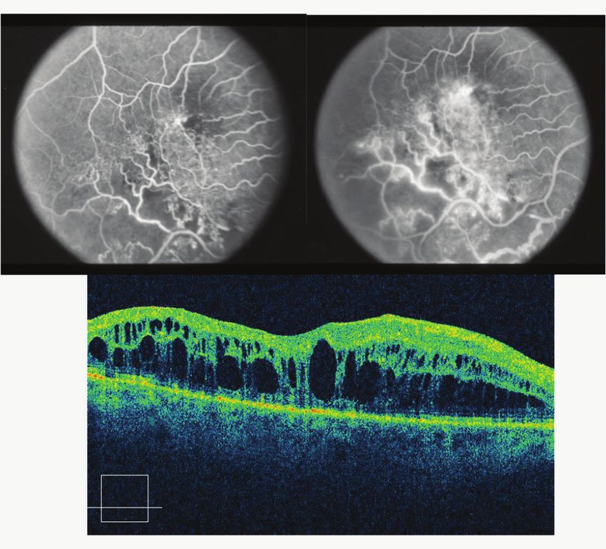
CONTENT SOURCE                                                                  The following faculty/staff members have the following financial
     This continuing medical education (CME) activity captures con-             relationships with commercial interests:
   tent from a recorded webinar and two live case discussions.                     Sophie J. Bakri, MD, and/or spouse/partner has had a financial
                                                                                agreement or affiliation during the past year with the following
   ACTIVITY DESCRIPTION                                                         commercial interests in the form of Consultant: Adverum, Alimera
      This supplement focuses on relevant and timely discussions                Sciences, Allegro, Allergan, EyePoint, Kala Pharmaceuticals, Oxurion,
   about treating patients with diabetic retinopathy (DR) and diabet-           Novartis, and Roche. Grant/Research Support: Lowy Medical
   ic macular edema (DME). The COVID-19 pandemic has resulted in                Foundation and National Institutes of Health (NIH).
   additional challenges for both patients and retina specialists. The             Peter K. Kaiser, MD, and/or spouse/partner has had a finan-
   esteemed faculty members examine clinical trial data and share               cial agreement or affiliation during the past year with the follow-
   their expertise on treatment options during these unprecedented              ing commercial interests in the form of Consultant: Aerie, Aerpio,
   times, methods for implementing treatment, and the role of                   Alcon, Allegro Ophthalmics, Allergan, Bayer, Bausch + Lomb, Biogen
   inflammation, among other topics.                                            Idec, Boerenger Ingelheim, Clearside Biomedical, Eyevensys, Galecto,
                                                                                Galimedix, iRenix, jCyte, Kanghong Biotech, Kodiak Sciences, NGM
   TARGET AUDIENCE                                                              Biopharmaceuticals, Novartis, Omeros, Opthea, Ophthotech,
     This certified CME activity is designed for retina specialists and         Oxurion, Regeneron, Regenexbio, Retinal Sciences, Santen, SciFluor,
   ophthalmologists involved in the management of retinal diseases.             Shire, Stealth Biotherapeutics, and Verena. Grant/Research Support:
                                                                                National Eye Institute, NIH, and Research to Prevent Blindness.
   LEARNING OBJECTIVES                                                          Speaker’s Bureau: Allegro Ophthalmics, Clearside Biomedical,
     Upon completion of this activity, the participant should be able to:       Ophthotech, Retinal Sciences, and Verena.
    • Identify the role of inflammation in patients with DME and DR.               Baruch D. Kuppermann, MD, PhD, and/or spouse/partner
    • Implement treatment regimens for patients with DME and DR.                has had a financial agreement or affiliation during the past year
    • Through real-world and post-hoc analyses, identify the                    with the following commercial interests in the form of Consultant:
   drawbacks to current use of anti-VEGFs in DME.                               Allegro, Allergan, Aprea, Cell Care, Dose, Eyedaptic, Galimedix,
    • Differentiate steroids in the management of DME while                     Genentech, Glaukos, Interface Biologics, Iveric Bio/Ophthotech,
   implementing strategies for treatment and patient outcomes.                  jCyte, Novartis, Oculis, Regeneron, Revana, Ripple Therapeutics,
                                                                                and Theravance Biopharma. Grants/Research Support: Alcon,
   GRANTOR STATEMENT                                                            Allegro, Allergan, Apellis, Clearside Biomedical, Genentech, GSK,
      This activity is supported by an unrestricted educational grant           Ionis, Iveric Bio/Ophthotech, jCyte, Novartis, and Regeneron.
   from Allergan.                                                                  Christina Y. Weng, MD, MBA, and/or spouse/partner has had
                                                                                a financial agreement or affiliation during the past year with the
   ACCREDITATION STATEMENT                                                      following commercial interests in the form of Consultant: Alcon,
     Evolve Medical Education LLC (Evolve) is accredited by                     Alimera Sciences, Allergan, and Novartis.
   the Accreditation Council for Continuing Medical Education
   (ACCME) to provide continuing medical education for physicians.              EDITORIAL SUPPORT DISCLOSURES
                                                                                  The Evolve staff and planners have no financial relationships
   CREDIT DESIGNATION STATEMENT                                                 with commercial interests. Michelle Dalton, writer, and Nisha
      Evolve Medical Education designates this enduring material for            Mukherjee, MD, peer reviewer, have no financial relationships with
   a maximum of 1 AMA PRA Category 1 Credit™. Physicians should                 commercial interests.
   claim only the credit commensurate with the extent of their par-
   ticipation in the activity.                                                  OFF-LABEL STATEMENT
                                                                                   This educational activity may contain discussion of published
   TO OBTAIN CREDIT                                                             and/or investigational uses of agents that are not indicated by the
      To obtain credit for this activity, you must read the activity in         FDA. The opinions expressed in the educational activity are those
   its entirety and complete the Pretest/Posttest/Activity Evaluation/          of the faculty. Please refer to the official prescribing information for
   Satisfaction Measures Form, which consists of a series of multiple-          each product for discussion of approved indications, contraindica-
   choice questions. To answer these questions online and receive               tions, and warnings.
   real-time results, please go to http://evolvemeded.com/online-
   courses/2017-supplement. Upon completing the activity and                    DISCLAIMER
   self-assessment test, you may print a CME credit letter awarding               The views and opinions expressed in this educational activity are
   1 AMA PRA Category 1 Credit™. Alternatively, please complete the             those of the faculty and do not necessarily represent the views of
   Posttest/Activity Evaluation/Satisfaction Form and mail or fax               Evolve, Retina Today, or Allergan.
   to Evolve Medical Education LLC, 353 West Lancaster Avenue,
   Second Floor, Wayne, PA 19087; Fax: (215) 933-3950.                          DIGITAL EDITION
                                                                                 To view the online version of the material, please go to
   DISCLOSURE POLICY                                                            http://evolvemeded.com/online-courses/2017-supplement.
     It is the policy of Evolve that faculty and other individuals who
   are in the position to control the content of this activity disclose
   any real or apparent conflicts of interest relating to the topics of this
   educational activity. Evolve has full policies in place that will identify
   and resolve all conflicts of interest prior to this educational activity.

2 SUPPLEMENT TO RETINA TODAY | OCTOBER 2020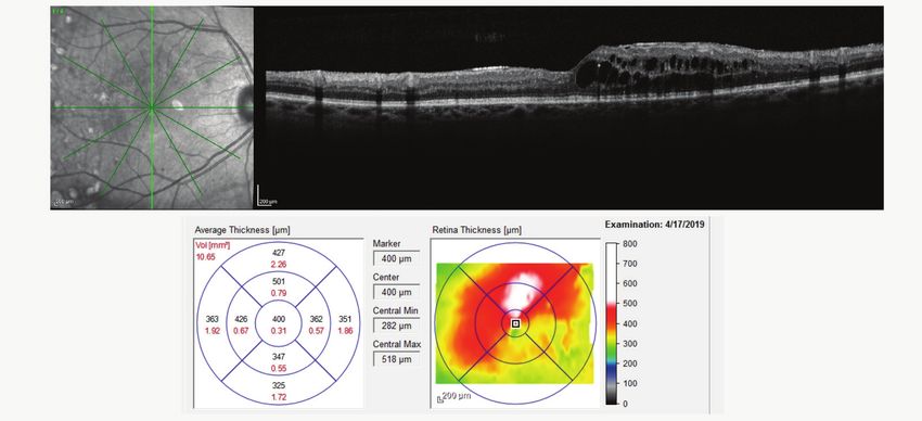
PRETEST QUESTIONS
                     Please complete prior to accessing the material and submit with Posttest/Activity Evaluation/Satisfaction
                                                      Measures Instructions for CME Credit.

1. Please rate your confidence in your ability to implement treatment           5. The international retina group real-life 24-month multicenter study
   regimens for patients with diabetic macular edema (DME) and diabetic             (IRGREL-DEX) demonstrated what percent of patients receiving a
   retinopathy (DR) (based on a scale of 1 to 5, with 1 being not at all            dexamethasone intravitreal implant needed subsequent intraocular
   confident and 5 being extremely confident).                                      pressure-lowering therapy?
       a. 1                                                                            a. ~5%
       b. 2                                                                            b. ~10%
       c. 3                                                                            c. ~15%
       d. 4                                                                            d. ~20%
       e. 5
                                                                                 6. Post-hoc analysis of randomized controlled trial data from DRCR.net
2. In the DRCR Protocol I subanalysis, what percentage of eyes with                 Protocol I identified the fact that ____% of patients were nonresponders
DME received early and consistent therapeutic benefits from anti-VEGF               or suboptimal responders to anti-VEGF treatment.
treatment?                                                                             a. 10%
       a. 25%                                                                          b. 20%
       b. 50%                                                                          c. 30%
       c. 75%                                                                          d. 40%
       d. 100%
                                                                                 7. Which of the following is true regarding the pathogenesis of DME
3. According to the literature, what impact does an intravitreal injection of      upregulation of anti-VEGF?
   bevacizumab have on concentrations of IL-6, IL-8, and other inflammatory            a. D
                                                                                           ME involves the upregulation of numerus inflammatory
   chemokines implicated in DME?                                                          mediators, including but not limited to anti-VEGF.
       a. T
           he concentrations of the inflammatory chemokines increase.                 b. DME is a process that is independent of anti-VEGF.
       b. The concentrations of the inflammatory chemokines decrease.                 c. DME does not involve anti-VEGF.
       c. T
           he concentrations of the inflammatory chemokines stay
          relatively stable.                                                     8. Which of the following drugs inhibits both VEGF-A and VEGF-B?
       d. The concentrations of some inflammatory chemokines decrease                a. Bevacizumab
           and the concentration of some inflammatory chemokines                      b. Ranibizumab
           increase.                                                                  c. Pegaptanib
                                                                                      d. Aflibercept
4. According to the “Shall we stay, or shall we switch” study, what outcome
   was noted in eyes with refractory DME?                                        9. Which of the following statements about the role of Ang1/Ang2 in
       a. E yes that received a dexamethasone implant were more likely to          diabetes is NOT true?
           gain at least 5 letters compared with eyes that were maintained             a. E levated Ang-2 (but not Ang-1) is associated with worse
           on anti-VEGF.                                                                   metabolic indices and endothelial dysfunction.
       b. Eyes that received anti-VEGF were more likely to gain at least              b. E levated Ang-2 (but not Ang-1) is associated with increased
           5 letters compared with eyes that received a dexamethasone                      HbA1c levels.
           implant.                                                                    c. D
                                                                                           ecreased levels of Ang-2 are found in the vitreous of patients
       c. E yes that received a dexamethasone implant were more likely to                with proliferative DR.
          lose at least 5 letters compared with eyes that were maintained              d. Elevated Ang-2/Ang-1 ratio is found in the vitreous of patients
          on anti-VEGF.                                                                    with nonproliferative DR and DME.
       d. Eyes that received dexamethasone implant and anti-VEGF
           performed equally with regard to letters gained.

                                                                                                                    OCTOBER 2020 | SUPPLEMENT TO RETINA TODAY 3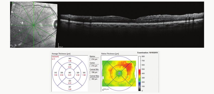
MANAGING DIABETIC MACULAR EDEMA: BEST PRACTICES IN REAL-WORLD SITUATIONS

   Managing Diabetic Macular Edema:
   Best Practices in Real-World Situations
      Diabetic macular edema (DME) and diabetic retinopathy (DR) are the primary ocular complications of diabetes.1 DR and DME not only cause vision
   impairment, but can cause irreversible blindness in a third of patients who develop them.2 Intensive intravitreal anti-VEGF injections have become
   the gold standard treatment for DME and DR and have been proven to reduce the risk of progression and further vision impairment. However, these
   treatments come with a significant treatment burden and aren’t uniformly effective, as some patients will not respond to anti-VEGF therapy regardless of
   treatment intensity. Further, anti-VEGF agents don’t address the inflammatory component of DME/DR pathogenesis, which may hinder their effectiveness.
   The following activity brings together thought leaders in the DME space to discuss how inflammation factors into DME development, how treatments can
   be improved for maximum effectiveness, and novel agents in the pipeline.
                                                                                                                           —Sophie J. Bakri, MD, Moderator

   THE ROLE OF INFLAMMATION AND STEROID USE                                     and begs the question: Why is anti-VEGF treatment effective in some
   IN DME TREATMENT                                                             disease states but not others?

   Q       SOPHIE J. BAKRI, MD: During our discussion, we will
           identify the role of inflammation in patients with DME and
                                                                                   A key reason for this could be inflammation, which is an impor-
                                                                                tant component of DME. Historically, we’ve focused on the role of
           DR, identify treatment regimens for patients with DME and            retino-capillary damage and anatomic changes in DME, but that
   DR, and review the drawbacks of our current use of anti-VEGF                 occurs quite late in the disease process. Inflammation, however,
   agents in DME. We’ll also touch on strategies to improve treatment           occurs almost immediately.
   and patient outcomes.
   Dr. Kuppermann, what is the role of inflammation in patients DME             Understanding How Inflammation Causes DME
   and DR?                                                                      Pathogenesis
      BARUCH D. KUPPERMANN, MD, PHD: Inflammation seems to be                   Q         DR. BAKRI: Specifically, what is known about
                                                                                          inflammation and DME pathogenesis?
   quite important in DME pathogenesis and may impact the effective-
   ness of our anti-VEGF agents. Anti-VEGF agents are the first-line               DR. KUPPERMANN: It’s a complicated process. Hyperglycemia
   therapy for patients with most retinal diseases, including wet age-          and oxidative stress cause local inflammation through the activation
   related macular degeneration (AMD), retinal vein occlusion, and              of microglial cells. The microglial cells migrate into the subretinal
   DME. However, the success rate of anti-VEGF agents in our patients           space where they accumulate, become trapped, and start to produce
   with DME is quite a bit lower than it is for other diseases.                 nitric oxide and a variety of cytokines and chemokines that lead to
      A 2012 subanalysis of the Diabetic Retinopathy Clinical Research          increased levels of inflammatory mediators.4-6 These inflammatory
   Network (DRCR.net) Protocol I by Bressler et al looked at factors            mediators and oxidative stress lead to dysfunction of the Mueller
   associated with changes in visual acuity (VA) and central subfield           cells, causing intracellular fluid accumulation, resulting in intracellular
   thickness (CST) at 1 year after intravitreal ranibizumab treatment for       edema. This cascade of events causes chronic inflammation, neuro-
   DME.3 The goal was to determine if certain factors predict anti-VEGF         degeneration, and then the subsequent vascular leakage and DME.
   treatment success or failure.                                                   Dong et al studied the relationship between 27 aqueous humor
      A total of 361 eyes were randomly assigned to either ranibizumab          cytokines and DR severity.7 Undiluted aqueous humor samples were
   with prompt or deferred laser, laser alone, or triamcinolone ace-            obtained from 102 nondiabetic patients and 136 diabetics who were
   tonide plus laser. Study eyes were differentiated into one of four           divided into nine groups according to the Early Treatment of Diabetic
   categories based on whether they had at least a 20% reduction from           Retinopathy Study (ETDRS) severity scale. The ETDRS score was very
   baseline CST at the 16-week, 32-week, and 1-year visit: early and con-       low, with about 20% of diabetics having a score of 10 (Table 1).
   sistent, early but inconsistent, slow and variable, and nonresponder.           The researchers measured cytokine levels, finding that diabetics
      When looking at the ranibizumab groups, only 50% had early                had significantly higher concentrations of interleukin (IL)-1ß, IL-6,
   and consistent responses. A surprisingly high number (23%) were              IL-8, monocyte chemoattractant protein-1 (MCP-1), interferon gam-
   considered nonresponders, with another 27% having intermediate               ma-induced protein-10, and VEGF in the aqueous humor compared
   responses. We know that this is suboptimal in many of our patients,          with non-diabetic controls. In the diabetic group, the VEGF level

4 SUPPLEMENT TO RETINA TODAY | OCTOBER 2020
MANAGING DIABETIC MACULAR EDEMA: BEST PRACTICES IN REAL-WORLD SITUATIONS

was about 1,000; in the control group, VEGF levels were below 100                        TABLE 1. RELATIONSHIP BETWEEN AQUEOUS HUMOR CYTOKINES AND
(Table 1). Based on these data, there seems to be a trigger related to                   SEVERITY OF DR 7
the onset of diabetes.
  Although patients develop more advanced retinopathy over time,                         ETDRS              N        Cytokine concentration (pg/mL)
Table 1 shows that VEGF levels don’t incrementally increase; they                        retinopathy                 VEGF          IL-1β IL-6 IL-8 MCP-1 IP-10
shoot up immediately in a binary fashion, as if a switch is thrown.                      severity
However, if you look at the other cytokines and interleukins in                          10                 28       967           10       32.1     22.8 252.2         2.1
Table 1, you see that they start low and increase over time. This may                    20                 23       952.8         11       33.5 20.6 303.6             2.5
help explain the variable responses we see from patients who have
DME getting VEGF inhibition.                                                             35                 26       956.4         9.2      33.1     22.7 339.5         5.6
  In summary, retinopathy progresses with time and is associated                         43                 18       1084.7        10.7     33.2 24.4         468.8     5.5
with changes in the amounts of multiple cytokines relative to VEGF,                      47                 13       1172.6        18.8     56.6 29.2 645.2             9.5
not just VEGF.
                                                                                         53                 8        1177.3        22.7     106.7 49.4 921.2            22.3
The Role of Steroids in Reducing Inflammation for                                        65                 7        1142.7        23.7     116.8 51          1215.1    31.3
DME Treatment                                                                            75                 8        1051.4        27.6     147      75.7     1286.6 34.3
Q         DR. BAKRI: How can steroids be used to address the
          inflammatory components of DME pathogenesis?
                                                                                         81                 5        1165.4        45.8     188.6 74.4        1630.8 29.2
                                                                                         P -value                    .733          .003     < .001 .001       < .001 < .001
   DR. KUPPERMANN: There are a lot of data from clinical trials                          Abbreviations: IL, interleukin; IP, interferon-inducible protein; MCP, monocyte che-
showing that if DME becomes chronic, it becomes unresponsive to                          motactic protein; VEGF, vascular endothelial growth factor; DR, diabetic retinopathy;
anti-VEGF therapy but is responsive to steroid therapy. For example,                     ETDRS, Early Treatment of Diabetic Retinopathy Score.
Sohn et al looked at the effects of a single injection of triamcinolone
acetonide or bevacizumab on cytokine levels in 11 DME patients                      could be because the other cytokines swamp the VEGF signal, caus-
with bilateral disease.8 Patients were treated in the simplest way pos- ing these patients to not be as responsive to VEGF inhibition alone.
sible: triamcinolone acetonide in one eye and bevacizumab in the                    These patients may need additional therapy, and it does seem that
other eye, followed by cytokine level measurement.                                  steroids are effective in treating both types of patients.11,12
   Bevacizumab only reduced VEGF levels, whereas triamcinolone                         For example, let’s take the FAME study, which looked at patients
acetonide reduced IL-6, MCP-1, platelet-derived growth factor-AA,                   with DME treated either with fluocinolone acetonide or sham.13 If
and VEGF (Table 2). We know that steroids are very effective at low-                patients didn’t have a good response in terms of optical coherence
ering the cytokine and VEGF levels. Table 2 shows that steroids are                 tomography (OCT) thickness after 6 weeks, then they could be treat-
great VEGF inhibitors, lowering VEGF by about 80%, but that bevaci- ed at will. Most of those patients received bevacizumab. Researchers
zumab is a fantastic VEGF inhibitor, lowering VEGF levels by 99%.                   looked at the median duration of DME prior to enrollment, which
   You would think that if you lowered something as important as                    was roughly 2 years, and then analyzed the outcomes. Fluocinolone
VEGF, there would be some collateral effects; upregulation, downreg- acetonide worked just as well on patients with chronic DME as
ulation, something from these other inflammatory
cytokines. However, they don’t appear to budge;              TABLE 2. STEROIDS ADDRESS THE MULTIFACTORIAL NATURE OF DME 8
they remain at the level they were prior to injection. Cytokine IVTA (n = 11)*                                             Bevacizumab (n = 11)*
It’s a bit of a mystery. It’s almost as though there are     Conc.,        Preinjection Postinjection P Value Preinjection Postinjection P Value
two parallel pathways: the VEGF-mediated pathway, pg/mL
and then the other inflammatory cytokine and che-            IL-6          29.9              13.8              < .01       26.7               24              .477
mokine pathway. The authors noted that a steroid
that could minimize adverse events and simultane-
                                                             IL-8          28.2              25.3              0.597       23.9               23.6            .374
ously address these components of pathogenesis               IP-10         366               249               0.013       401                433             .11
would be of great benefit.                                   MCP-1         3850              1090              0.01        3770               3840            .594
   Researchers have argued that there are two types
                                                             PDGF-AA 68.7                    37.1              0.016       81                 72.7            .722
of patients: (1) patients who have a low level of
VEGF but high levels of non-VEGF mediators, and              VEGF          55                10.5              0.05        61.5               0.1             < .01
(2) patients who have a high level of VEGF but who           Bilateral injection of patients with DME (1 eye IVTA*, 1 eye bevacizumab*)
have low levels of non-VEGF mediators.9-11 Patients          Abbreviations: IL, interleukin; IP, interferon-inducible protein; IVBe, intravitreal bevacizumab; IVTA, intra-
in group 2 are exquisitely sensitive to VEGF inhibi-         vitreal triamcinolone acetonide; MCP, monocyte chemotactic protein; PDGF, platelet-derived growth factor;
tion, while patients in group 1 tend to be unrespon- VEGF, vascular endothelial growth factor.
sive to anti-VEGF therapy. The unresponsiveness              *Not licensed for ophthalmic use; aWilcoxon signed rank test.

                                                                                                                              OCTOBER 2020 | SUPPLEMENT TO RETINA TODAY 5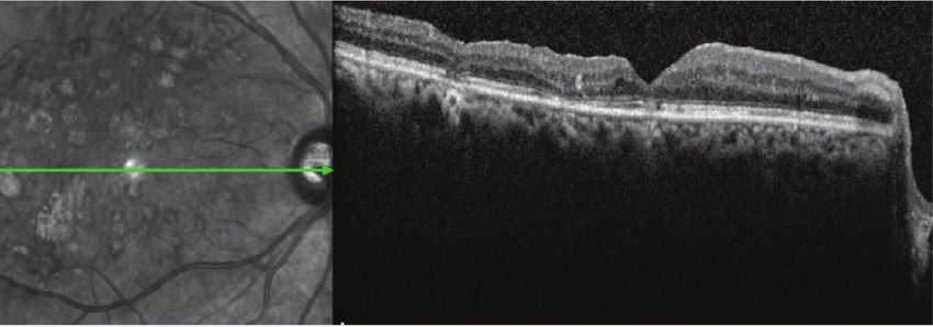
MANAGING DIABETIC MACULAR EDEMA: BEST PRACTICES IN REAL-WORLD SITUATIONS

   Figure 1. Fluocinolone acetonide implant study for macular edema: treatment effect seen in
   FAME by duration of DME at baseline (pooled data).13
                                                                                                Figure 2. RESTORE: Mean change in BCVA from baseline over time.15
   patients with less chronic disease (Figure 1). However, the patients
   with chronic disease treated with something other than steroids, like
   anti-VEGF therapy, had a much weaker response.

   Understanding How Disease Duration Impacts
   Anti-VEGF Response and Outcomes
   Q       DR. BAKRI: As you mentioned, there is some evidence
           that as DME becomes chronic, the responsiveness to VEGF
           inhibition is reduced. What does the literature tell us
   about this?

      DR. KUPPERMANN: In both the RISE and RIDE trials, the control
   populations received 24 months of sham therapy.14 After that time,
   investigators were finally able to give patients what they wanted—
   ranibizumab 0.5 mg. By then it was too late; the control populations                         Figure 3. Subanalysis of DRCR.net Protocol I.16
   demonstrated minimal response to monthly ranibizumab. This is
   because after 24 months of sham, the disease became chronic, result-                         received ranibizumab every 4 weeks and were evaluated at week 12
   ing in basically no response to VEGF inhibition. Ranibizumab was not                         for response. At the 12-week evaluation, they were placed into three
   effective at improving their vision. Early on, however, these eyes were                      groups: (1) patients who gained 5 or fewer letters (40%); (2) patients
   very sensitive to VEGF inhibition.                                                           who gained 5 to 9 letters (23%); and (3) patients who gained 10 or
      There are other examples of this as well. The RESTORE extension                           more letters (37%). Patients were then plotted over the next 3 years
   trial looked at 208 patients from the core study who received either                         for their response, and what happened was the famous swim lanes
   ranibizumab 0.5 mg (n = 83), ranibizumab 0.5 mg plus laser (n = 83),                         depicted in Figure 3.
   or laser alone (n = 74).15 Patients were eligible to receive individualized                     Eyes that gained fewer than 5 letters after three injections showed
   ranibizumab treatment as of month 12 guided by best-corrected visual                         limited additional improvement for the study duration. These
   acuity (BCVA) and disease progression criteria at the investigators’                         patients were unlikely to cross over to the 10-letter or more group; in
   discretion. Concomitant laser treatment was allowed according to the                         fact, only 30% of eyes from the 5-letter or fewer group crossed over
   ETDRS guidelines. In patients treated with ranibizumab in the core                           to the 10-letter or more group by the end of the study.
   phase, mean BCVA gain at month 12 was maintained to month 36. In                                This shows that early response with as few as three injections of
   patients treated with laser alone, mean BCVA progressively improved                          ranibizumab may be able to predict long-term outcomes. Upwards
   from month 12 to month 36 with ranibizumab (Figure 2).                                       of 70% of patients will not have an improved response with time if
      If you look at the left side of Figure 2, you see that there’s a green                    their initial response is limited. This should help inform clinicians on
   line that gradually builds over time, but there is clearly a blunted                         the choice to continue the same therapy or switch therapies as soon
   response compared to what you saw at baseline, represented by                                as after three injections, depending on the degree of response. This
   the yellow and orange lines. Again, even within 1 year, maybe even                           was partially corroborated by DRCR.net Protocol T.17,18 The outliers
   sooner, there’s an attenuation to response to VEGF inhibition as                             were bigger, and it wasn’t quite as clean, but that same concept was
   eyes develop more chronic inflammation and more chronic macu-                                true for ranibizumab, aflibercept, and bevacizumab.
   lar edema.                                                                                      In summary, we know that when anti-VEGF therapy is used in the
      Another example of this is an early sub-analysis of DRCR.net                              first-line setting, three intravitreal injections are frequently predictive
   Protocol I, which assessed 340 eyes from the primary trial.16 Patients                       of long-term outcomes.

6 SUPPLEMENT TO RETINA TODAY | OCTOBER 2020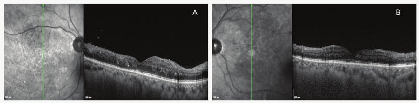
MANAGING DIABETIC MACULAR EDEMA: BEST PRACTICES IN REAL-WORLD SITUATIONS

Dexamethasone Implant Versus Anti-VEGF Therapy                              center-involved DME.28 The primary outcome was mean change in
Q       DR. BAKRI: The dexamethasone implant is approved by
        the US Food and Drug Administration for the treatment of
                                                                            BCVA from baseline to week 24. There was no significant difference in
                                                                            VA between the two treatment arms at 24 weeks. Why do you think
        DME among other indications. We know that                           the results of Protocol U differ from the real-world studies?
dexamethasone is six times stronger than triamcinolone
acetonide.19 How does the dexamethasone implant compare to                     DR. KUPPERMANN: Protocol U was a flawed study. It was intend-
anti-VEGF therapy?                                                          ed to be in pseudophakic eyes, but included phakic eyes because,
                                                                            in reality, including only pseudophakic eyes was more difficult to
   DR. KUPPERMANN: If you look at real-life outcomes with the dexa-         enroll than anticipated. If you look at the pseudophakic subset,
methasone implant, response was quite robust with 6 to 9 letters            there was a trend toward better outcomes in the dexamethasone
gained with two to three injections in the first year. This is further      implant plus ranibizumab arm compared to the ranibizumab alone
corroborated by a host of studies showing similar responses in both         arm. This was a nominal difference and not statistically significant
treatment-naive and refractory patients.11,20-24                            because it was underpowered. There were better OCT outcomes as
   More recently, a study from Escobar-Barranco et al showed good           well in the combo group, which was statistically significant. There
response in patients with both treatment-naive or refractory dif-           was good anatomic evidence but allowing phakic eyes blunted the
fuse DME. Patients with treatment-naive disease gained an average           response in vision.
of 12 letters with a median reinjection time of 5 months; refrac-
tory patients gained 8 letters with a median reinjection time of              DR. BAKRI: What adverse events should clinicians look for with
4 months.11 The IRGREL-dexamethasone study from Iglicki et al told          the dexamethasone implant?
a similar story.25 Refractory eyes gained about 7 letters, while treat-
ment-naive eyes gained 11.                                                     DR. KUPPERMANN: There’s a lot of evidence that after poor
   One of the more interesting studies is from Busch et al, the "Shall we   responsiveness to three to six injections of anti-VEGF therapy, switch-
stay, or shall we switch" study. Patients were treated with three month-    ing to the dexamethasone implant or to steroids seems to be an
ly anti-VEGF injections and then evaluated.26 If they were deemed to        effective option. However, there are some concerns. Briefly, as with
have refractory DME, they were randomly assigned to either switching        any steroid, there is the risk of elevated intraocular pressure (IOP).
to the dexamethasone implant or continuing anti-VEGF therapy. They          About 40% of dexamethasone patients in the MEAD study needed
were then evaluated for BCVA and OCT thickness change.                      IOP-lowering medications.12 In real-world studies, this occurs less fre-
   Eyes that were switched to the dexamethasone implant after three         quently (15 to 20%).25
anti-VEGF injections and then deemed to be refractory showed a sig-            Additionally, steroids cause cataracts. The good news is that
nificant benefit with the switch compared with continuing anti-VEGF         postcataract surgery, when there can be a lot of inflammation, the
therapy, both in terms of vision and in OCT thickness. On average,          dexamethasone implant does a wonderful job managing inflam-
eyes continuing with anti-VEGF therapy received 4.0 injections versus       mation. In fact, one of the relative recommendations for using the
1.4 injections in dexamethasone implant eyes. Eyes switched to the          dexamethasone implant is in the perioperative period surrounding
dexamethasone implant gained a mean of 6.1 letters after 12 months,         cataract surgery.
compared with –0.4 letters for anti-VEGF-treated eyes. They were               As mentioned earlier, we know that three anti-VEGF injections
also more likely to gain at least 5 letters and at least 10 letters after   can be predictive of long-term outcomes. Therefore, when I think
12 months than eyes maintained on anti-VEGF therapy. The authors            about patient selection for the dexamethasone implant, in addi-
concluded that in a real-world setting, switching patients with refrac-     tion to pseudophakic eyes, my biggest indication is the suboptimal
tory DME to the dexamethasone implant results in better visual and          response after three to six anti-VEGF injections regardless of phakic
anatomic outcomes at 1 year.                                                status. I also consider if the fellow eye had a poor response to anti-
   This was further corroborated in a metaanalysis from Khan et             VEGF therapy. If one eye was unresponsive, should we try it again in
al, who looked at a number of studies with more than 3,800 total            the other eye? I usually do, but more and more I’m considering going
patients, all of whom had at least six prior anti-VEGF treatments           straight to the dexamethasone implant when the fellow eye was
and then were either switched to dexamethasone implant or con-              poorly responsive to anti-VEGF therapy.
tinued on anti-VEGF therapy.27 All studies favored switching to                To conclude, there’s a significant subset of patients who have poor
the dexamethasone implant compared to staying with anti-VEGF                responsiveness to anti-VEGF therapy. The dexamethasone implant
therapy.                                                                    appears effective in treating treatment-naive patients and eyes with
                                                                            chronic disease.

Q        DR. BAKRI: These real-world studies have great
         outcomes. The DRCR.net Protocol U phase 2 study,                   Determining When—and if—to Switch Therapy
         however, had different results. Protocol U evaluated the
efficacy of combination dexamethasone and ranibizumab versus                Q        DR. BAKRI: The swimming lanes from the subanalysis of
                                                                                     DRCR.net Protocol I clearly showed that patients start off
ranibizumab monotherapy in 236 patients with persistent                              in one lane and after 12 weeks you know what lane

                                                                                                            OCTOBER 2020 | SUPPLEMENT TO RETINA TODAY 7
MANAGING DIABETIC MACULAR EDEMA: BEST PRACTICES IN REAL-WORLD SITUATIONS
they’re going to end up in for visual outcomes. Have Protocol I
data altered your decision on when to switch therapies?
DR. KUPPERMANN: There’s not a one-size-fits-all approach for
these patients. When to switch depends on many patient-specific
variables such as their vision and response on OCT. I look at response
patterns over time. For example, after one injection, was there a
20% reduction of excess macular thickness? After the second injec-
tion, was there another 20% reduction? If so, then I’m satisfied with
the direction we’re going and want it to continue. However, if there
was a modest response of 20% on the first injection and no further Figure 4. Case 1: 63-year-old male with insulin-dependent diabetes 20 weeks LTFU.
response on injections two or three, then I’ll consider switching the
patient to the dexamethasone implant.
DR. BAKRI: Do you think it’s important to monitor the OCT
monthly after each injection within the loading dose?
DR. KUPPERMANN: I prefer to do an OCT each time because it
helps me assess responsiveness early on. That said, I understand an
OCT during each appointment isn’t efficient. Some clinicians skip
the OCT after they’ve committed to the loading doses. Although I
wouldn’t argue with someone taking that approach, I do think you
Figure 5. Case 1: 7 weeks after fourth aflibercept injection.
lose important data that will help guide you in subsequent manage-
ment . Therefore, I argue that it’s worth doing. From what we know so far on this patient, aflibercept was effective
up to 7 weeks, but we weren’t able to assess any longer duration.
SWITCHING AGENTS: REAL-WORLD CASE STUDIES Throughout the 20 months with this patient, there were two adverse
Case 1: Insulin-dependent Diabetic circumstances that caused a delayed follow-up and fluid recurrence.
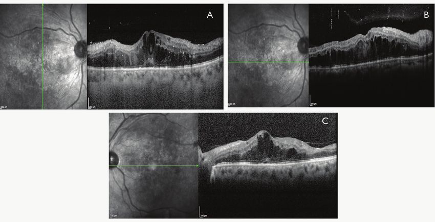
DR. BAKRI: Our first case is a 63-year-old man with insulin-depen- In hindsight, should I have done anything differently?
dent diabetes, high cholesterol, hypertension, and a 10.2 HbA1c. He
has an ocular history of focal laser and panretinal photocoagulation PETER K. KAISER, MD: The issue with this patient isn’t response
(PRP) in both eyes. VA was 20/40 in the right eye and hand motions to anti-VEGF injections, it’s delayed follow-up. If the patient was fol-
in the left. He presented with DME in the right eye and vitreous and lowed up regularly and weren’t responding to aflibercept, then we
subhyaloid hemorrhages in the left. We will focus on the right eye for could consider switching them to a long-term steroid injection. I
this case discussion; the left eye was managed with vitrectomy. would not be comfortable switching to the dexamethasone implant
Before presenting to us, he had received multiple injections of in this patient because of these follow-up issues. It is very important
bevacizumab in the right eye, the last of which was 9 weeks prior. than after using a dexamethasone implant, we check the patient’s
Nine weeks after the bevacizumab injection, vision was 20/30 in the IOP at around 6 to 8 weeks after injection. Given this patient’s his-
right eye, but macular edema was present. We proceeded with beva- tory, I’m not confident they would return.
cizumab every 4 weeks for three more injections. After that series
of injections, the patient still had macular edema on the OCT. We DR. KUPPERMANN: I’d also defer any consideration of the dexa-
switched him to monthly aflibercept for three more injections. After methasone implant in this case, given that the patient is showing a
those injections, his VA was 20/25; the macula looked pretty good response to anti-VEGF injections with 7-week durability. Yes, that’s
with only a trace of edema. shorter than the dexamethasone implant, but I’m also concerned the
We then decided to extend the interval to 6 weeks for two more patient won’t return for the required follow-up visits to assess their
aflibercept injections. The patient was lost-to-follow-up (LTFU) for IOP. If there are no pressure increases after the first couple of implant
20 weeks due to transportation and weather issues. Figure 4 shows injections, then I’d be more comfortable with the LTFU potential. But
his images after 20 weeks. We gave an aflibercept injection that day, there has to be a very clear commitment from the patient that they
then shortened the interval to 4 weeks. Figure 5 shows the results will show up to the initial follow-up appointments for me to feel
after four additional aflibercept injections, 7 weeks after the last injec- comfortable about that switch. VEGF inhibition seems to be effective.
tion; there’s no macular edema. We kept the patient at 7-week inter- I’m not sure this is a candidate for steroid therapy.
vals for three more injections.
There was another LTFU of 12 weeks due to COVID-19. We Case 2: Significant Edema
went back to aflibercept every 4 weeks for a few more injections. DR. KAISER: This is a 54-year-old man with type 2 insulin-depen-
8 SUPPLEMENT TO RETINA TODAY | OCTOBER 2020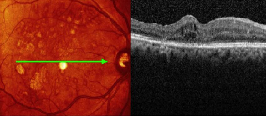
MANAGING DIABETIC MACULAR EDEMA: BEST PRACTICES IN REAL-WORLD SITUATIONS

dent diabetes with a VA of 20/80 who has significant DME, especially        Case 3: Poorly Controlled DR
outer retinal cystic and intraretinal changes, and ischemia. This is his       DR. KUPPERMANN: This is a patient who ended up with bilat-
first visit to a physician in years and he denies previous treatment.       eral dexamethasone implants after a limited amount of anti-VEGF
There are also some hyper-reflective foci on the OCT, as shown in           exposure. I was referred this patient after a cataract surgery in the
Figure 6. How would you treat this patient?                                 right eye with decreased vision. He has a history of bilateral DR and
                                                                            hypertensive retinopathy that is very poorly controlled. His systolic
                                                                            blood pressure is 180 to 190. He has chronic kidney failure and poorly
                                                                            controlled HbA1c of 9.8%.
                                                                               His right eye is 20/100 VA with central thickening. I gave an afliber-
                                                                            cept injection, and he came back 6 weeks later. His vision is margin-
                                                                            ally better at 20/80, but the macular edema is far worse than it was at
                                                                            the beginning.
                                                                               In his treatment-naive, left eye, which was phakic, he had a lot of
                                                                            edema that came on suddenly, all in the 6-week time frame after the
                                                                            initial aflibercept injection in the fellow eye. He felt as though he was
                                                                            losing vision in both eyes and was imploring me for help. I decided to
                                                                            give a dexamethasone implant in the left eye. I already started afliber-
                                                                            cept in his right eye, which had no response after the initial injection,
                                                                            but I decided to try another aflibercept injection.
                                                                               Five weeks later, the right eye was not any better after two afliber-
                                                                            cept injections. Meanwhile, the left eye demonstrated an excellent
                                                                            anatomic response to dexamethasone implant (Figure 7).
                                                                               I decided to inject the right eye with the dexamethasone implant
                                                                            after two aflibercept injections, given the strong response to dexameth-
                                                                            asone implant in the fellow eye. This was unusual for me, as I would
                                                                            typically give at least three anti-VEGF injections before considering
Figure 6. Case 2: Patient with 20/80 VA and significant edema.              switching, even in a pseudophakic eye. He had a beautiful response
                                                                            9 weeks out with his vision continuing to improve (Figure 8).
   DR. KUPPERMANN: There appears to be some vein occlusion as
well. I start almost every case with anti-VEGF inhibition, typically          DR. KAISER: Are you going to continue the steroids in this patient or
aflibercept. The diffuse nature of the edema makes me think a steroid       are you going to go back to anti-VEGF now that you’ve flattened them?
may be useful, but I would first start with VEGF inhibition.

   DR. BAKRI: Although it is tempting to go straight to a steroid, we
usually start with anti-VEGF agents because of pressure increases and
the other issues that come with steroid use. I do wonder whether a
steroid may be a more effective. We know that steroids are neuro-
protective as well.

   DR. KUPPERMANN: I would first want to get the edema under
control and see if there’s any disease state modification. I’d start with
at least three anti-VEGF injections before considering moving to a
steroid. I would certainly not delay switching to the dexamethasone
implant if the patient was nonresponsive after three injections.            Figure 7. Case 3: Patient 5 weeks post second aflibercept injection OD (A, B) and dexamethasone
                                                                            implant OS ( C ).
   DR. KAISER: We treated this patient with bevacizumab. It didn’t
work well, so we switched him to aflibercept. That didn’t work well
either, so we switched to the dexamethasone implant. The edema
improved, but the vision didn’t improve as much as we’d like. That’s
one of the problems we have with these chronic cases. The patient’s
outer retina looks ratty, which is an indication that anti-VEGF isn’t
going to work as well as we hope. Anatomically, we can do well, but         Figure 8. Case 3: Patient 4 weeks after dexamethasone implant OD (A); 9 weeks post dexametha-
VA wise, the response isn’t there.                                          sone implant OS (B).

                                                                                                                       OCTOBER 2020 | SUPPLEMENT TO RETINA TODAY 9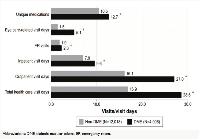
MANAGING DIABETIC MACULAR EDEMA: BEST PRACTICES IN REAL-WORLD SITUATIONS

     DR. KUPPERMANN: I’ve been continuing the dexamethasone
   implants. He keeps needing them, and he continues to show a good
   response to dexamethasone implant with good IOP.

   DRAWBACKS TO CURRENT ANTI-VEGFS IN
   DIABETIC MACULAR EDEMA
   Q        DR. BAKRI: Dr. Weng, what are some of the drawbacks of
            our current anti-VEGF treatments in patients with DME?

      CHRISTINA Y. WENG, MD, MBA: There’s no doubt that anti-                  Figure 9. VA outcomes in the RISE and RIDE Trials.29
   VEGFs have revolutionized the way we treat retinal disease, includ-
   ing DME. Although anti-VEGFs remain the gold standard, there are
   well-known downsides such as cost, potential systemic side effects,
   and patient discomfort during administration. More importantly,
   anti-VEGFs have limited durability, thereby conveying a heavy treat-
   ment burden on our patients. This limited durability also means that
   patients are subjected to injection-related risks like endophthalmitis
   and retinal detachment. Although these complications are rare, it’s
   important to consider that these risks do compound over one’s life-
   time with repeated injections.
      The second larger issue is that anti-VEGF agents are not univer-
   sally effective because they only target VEGF; they don’t target other      Figure 10. VA outcomes in VISTA and VIVID.30
   inflammatory mediators that may be involved in DME pathogenesis.
   There will be some patients who have an incomplete response to              course of the study (Figure 9).
   anti-VEGF treatment or may be refractory to treatment altogether.              VIVID and VISTA, the phase 3 registration trials that compared
                                                                               aflibercept given every 4 or 8 weeks versus laser for DME, also met
   Real-World Patients Versus Clinical Trial Outcomes                          their primary endpoint. Greater BVCA gains and anatomic improve-
      DR. WENG: In focusing on the issues of durability and lack of uni-       ments were seen at week 52 in patients treated with aflibercept ver-
   versal efficacy, here are two real-life examples of my own patients         sus those treated with laser.30 Figure 10 shows that patients treated
   who illustrate these concepts.                                              with aflibercept gained anywhere between 10 and 12 letters.
      The first patient is a 69-year-old man with insulin-dependent dia-          Finally, Protocol T from the Diabetic Retinopathy Clinical Research
   betes and bilateral DME. He is 20/50 and 20/60 in his right and left        Research Network compared aflibercept, bevacizumab, and ranibi-
   eyes, respectively. He’s done very well on anti-VEGF monotherapy            zumab head-to-head in the treatment of DME. From baseline to
   for the last 2.5 years. However, he has required injections every 4 to      1 year, the mean VA letter score improved by 10 to 13 letters with all
   8 weeks. He has a heavy treatment burden, especially since he prefers       three agents.17,18
   not to have same-day bilateral injections.                                     The commonality between these last three trials are double-
      The second patient is a 43-year-old man with noninsulin-depen-           digit VA gains. Where are the double-digit gains in the real world?
   dent diabetes. He has severe bilateral DME with profound amounts            Interestingly, the 5-year extension results of Protocol T showed that
   of subretinal and intraretinal fluid. He’s 20/50 in both eyes. After four   between years 2 and 5, when patients are managed at clinician dis-
   monthly anti-VEGF injections, there’s slight improvement, but signifi-      cretion rather than trial protocol, patients will lose a few letters.17
   cant fluid remains. I transitioned him to dexamethasone, and he had         Although the overall VA still improved from baseline by about
   a remarkable response, both anatomically and visually. This patient         7.4 letters, it did decrease by 4.7 letters from the 2-year timepoint
   is a great example that not everyone may respond completely to              despite stable OCT. Of note, there was a median of four injections
   anti-VEGF monotherapy, likely because of a significant inflammatory         given in the extension phase.
   component to his DME.                                                          These are important points because they illustrate what can
      How do these examples compare with the results from our major            happen even in our “best” patients. We know that those who are
   DME trials? Let’s start off with RISE and RIDE, the landmark phase 3        enrolled in our clinical trials tend to have a higher level of compli-
   trials that evaluated ranibizumab versus sham in the treatment of           ance, they tend to have a greater level of motivation, and even in
   DME.29 Both studies met their primary endpoint, showing that more           these “best” patients, they may not achieve those double-digit gains
   patients gained 15 or more letters from baseline to 24 months in the        long-term. Although the reason for that is likely multifactorial,
   treatment groups versus sham. Patients treated with ranibizumab             potential undertreatment is a factor to be considered.
   also had greater reduction in macular thickening, and patients treat-
   ed with ranibizumab gained between 11 to almost 13 letters over the

10 SUPPLEMENT TO RETINA TODAY | OCTOBER 2020
MANAGING DIABETIC MACULAR EDEMA: BEST PRACTICES IN REAL-WORLD SITUATIONS
were managed at the 28- and 365-day timepoints after diagnosis, and
Undertreatment, Noncompliance Drives found that 75% of patients received no treatment within 28 days of
Mediocre Outcomes their DME diagnosis. This database included all-comers regardless of
DR. WENG: If you look at real-world studies, this gap in the visual baseline VA. Even 1 year out, 60% were still observed. Among those
outcomes becomes even more evident. For example, a real-world who were treated with anti-VEGF, they tended to have a lower mean
study looking at approximately 15,000 eyes from the Vestrum data- VA and also achieved greater levels of 1-year VA improvement, espe-
base showed much more modest visual gains of only 4 to 5.5 letters cially if they received six injections or more. This drives home the
at 12 months among eyes treated with anti-VEGF.31 This is a whole point that more frequent injections leads to better visual outcomes.
line worse than the outcomes from Protocol T. Interestingly, under- We can’t talk about undertreatment without talking about
treatment might not have been solely to blame here because the compliance. Two studies address this. Gao et al found that 25% of
mean number of injections was seven versus nine in Protocol T. patients with nonproliferative diabetic retinopathy (NPDR) and
Another large database study of nearly 30,000 eyes showed similar DME were LTFU.34 They defined that as no subsequent visits for
findings.32 At 1 year, a mean of 6.4 anti-VEGF injections were given, 12 months following an injection. The factors that were associated
leading to a VA gain of 4.2 letters. More injections led to greater VA with a greater risk of LTFU were lower income, lower baseline VA,
improvements. That’s a recurrent theme; the more injections you severe NPDR, and Hispanic, American Indian, or Pacific Islander race.
receive, the better your visual outcomes in general. Another study looked at the differences in compliance between
Figure 11 summarizes the results of a recently published study that AMD and DME patients, finding that DME patients were more than
included more than 13,000 treatment-naive patients with DME from twice as likely to have at least one break-off, which was defined as
the IRIS database.33 The authors analyzed how patients with DME tardiness in follow-up of greater than 100 days.35 Not surprisingly,
there was a significant correlation between the number of break-offs
and change in VA.
Figure 12 looks at the use of medical services based on one’s DME
status, illustrating that diabetics with DME tend to be on more med-
ications, have more emergency room visits, and spend more time
in the hospital as inpatients compared to diabetics without DME.36
Diabetics with DME have 29 health care visit days in a year, which is
more than two visits a month. Given that many of these patients are
working-age people with families, it’s no wonder they struggle with
the heavy treatment burden required in the management of DME
with anti-VEGF.
Making Sense of Suboptimal Responders Despite
Aggressive Therapy
DR. WENG: We know that despite aggressive anti-VEGF therapy,
Figure 11. IRIS Registry data analysis.33 a significant portion of patients continue to have persistent DME.
Some data suggest that persistent DME may limit visual gains. In a
subanalysis of Protocol I,16 which showed that despite receiving simi-
lar numbers of ranibizumab injections, patients’ mean BCVA through
year 3 was within 5 letters of their response at week 12 (Figure 13).
We can interpret the Protocol I data in two ways. First, it could mean
that some patients are destined to be poor or super-responders and
not much can be done to alter that course. However, it could also
suggest that someone’s early response has predictive value, and thus
if someone isn’t responding as well as they should, there may be an
opportunity early on to switch or combine therapy to generate a
greater response.
We know that limited treatment durability and undertreat-
ment limits the visual potential for some DME patients. However,
even with monthly anti-VEGF injections, a significant proportion of
patients may have incomplete drying or persistent fluid. For example,
let’s take a look at the 24-week post-hoc analysis of Protocol T.37
Figure 12. Utilization of medical services by DME status.36 The treatment algorithm followed in Protocol T is perhaps differ-
Abbreviations: DME, diabetic macular edema; ER, emergency room. ent from the way some of us practice in the real world. All patients
OCTOBER 2020 | SUPPLEMENT TO RETINA TODAY 11MANAGING DIABETIC MACULAR EDEMA: BEST PRACTICES IN REAL-WORLD SITUATIONS
of patients had persistent edema for 12 to 14 visits over year 1, and
more than half had edema at eight or more visits over the course of
that year.
Next, the investigators looked at mean BCVA change from
baseline at 12 months based on their categories. After 12 months,
patients with edema at 12 to 14 visits gained significantly fewer let-
ters than those with edema at three or fewer visits (Figure 14). This
observation held true at years 2 and 3, although it lost statistical
significance at year 3.
Strategies to Improve DME Treatment in the
Figure 13. Post-hoc analysis of Protocol I.38 Real World
received six monthly injections unless they were 20/20 with normal Q DR. BAKRI: Dr. Weng, given the data and the issues you
covered, how could the management of DME be improved
OCT after three injections. After the six injections, anti-VEGF was in the real-world setting?
only given if there was a significant improvement or worsening, and
no injections were given if there was persistent, stable DME. The DR. WENG: We need therapies that work better and last lon-
researchers divided the patients into three cohorts based on letter ger. There are several promising candidates in the pipeline. More
gains and CST decrease at week 12. durable agents may also mitigate these frequent anatomic fluctua-
Persistent DME was noted in 31.6% (aflibercept), 65.6% (beva- tions, which some have suggested may be harmful. One way to treat
cizumab), and 41.5% (ranibizumab) of eyes.37 Among these, rates suboptimal response to anti-VEGF may be to target other disease
of chronic persistent DME through 2 years were 44.2% to 68.2%, mediators in addition to VEGF. We also need agents that can offer
depending on treatment group. The data suggest that in the afliber- improved safety and comfort. As Dr. Kuppermann said, it would be
cept and ranibizumab groups, visual outcomes were slightly worse great to have reliable biomarkers that can help optimize treatment
for eyes with persistent DME compared to eyes without persistent or drug regimens for our patients. I’d also like to be able to better
DME. However, there wasn’t much difference seen between the identify patients at risk for noncompliance so that appropriate safe-
groups at 2 years. This points to an opportunity to get these patients guards could be implemented. Lower cost therapeutics would also
drier, which potentially could lead to better VA gains. Could these meaningfully impact the field from a societal standpoint.
patients have done better if managed with a different agent or per-
haps a combination of agents, rather than continuing on with their DR. BAKRI: In reviewing Protocol T extension data, you men-
monotherapy? The authors concluded that despite DME persistence tioned that the CSTs were stable during that time period, yet the
through 2 years in a subgroup of patients, meaningful gains in VA vision declined. Why is that?
continue to be achieved. Furthermore, there were very few patients
with 10 or more letter losses. However, it’s important to remember DR. WENG: It’s interesting because the decline that you see in the
that this is only a 2-year follow-up, and there are data that suggest extension also differs from what we saw in the longer-term follow-
that persistent fluid in DME may have detrimental effects in terms of up of studies like RISE/RIDE, VIVID/VISTA, and Protocol I.16,29,30,38 It’s
visual prognosis. hard to say why that is. Patients were not followed or treated on a
One such example is a post-hoc analysis of Protocol I by Sadda standardized regimen. It’s possible that patients were undertreated,
et al.38 Patients in Protocol I were stratified based on the number as they received a median of four injections in that extension period,
of visits over the course of a year where there was edema present, between years 2 and 5. There was persistent fluid in a not-insignif-
defined as a central retinal thickness that exceeded 250 µm based on icant proportion of patients as well, but we also see similar things
time-domain OCT. Despite intense anti-VEGF treatment, one-third with the Protocol I follow-up. Protocol I patients received a similar
number of injections in their extension phase and they also had a
significant proportion with persistent fluid. There has to be more
going on.
Perhaps the patients in Protocol T extension phase weren’t moni-
tored frequently enough. In Protocol I, patients had about 20 visits
between years 3 and 5, whereas they only were seen about 14 times
on average in the extension phase from years 2 to 5 in Protocol T.
It is also important to note that only two-thirds of patients in
Protocol T completed the extension phase.
The bottom line is that regimented clinical trial outcomes
Figure 14. Case 4: OCT 4 weeks post anti-VEGF injection #4. don’t seem to carry over to the real-world setting. We do see
12 SUPPLEMENT TO RETINA TODAY | OCTOBER 2020MANAGING DIABETIC MACULAR EDEMA: BEST PRACTICES IN REAL-WORLD SITUATIONS
improvement, but there are other things, such as ischemia and dis-
ease progression, that could be negatively impacting visual acuity
gains. Patients with diabetes also tend to acquire more comorbidities
over time. All of that could influence what we are seeing.
DR. BAKRI: One thing you mentioned is that there’s often a delay
in treatment of DME. We also know that many retina specialists
watch DME for longer, even when patients are getting treated and
just not responding. Why do you think these delays exist? Figure 15. Case 4: OCT following dexamethasone implant after LTFU for 13 weeks.
Four weeks after his fourth anti-VEGF, his fluid had recurred in the
DR. WENG: The IRIS database study was really eye-opening for me temporal macula (Figure 14). We discussed introducing the dexa-
in showing how many people with DME we’re observing. Protocol methasone implant into his therapy, and he was more than happy to
V showed us that observation is a valid and safe option for patients try it. I told him he needed to come back in 6 weeks so I could check
with very good VA of 20/20 to 20/25.39 However, this has not been for an efficacy and IOP response. This was early March when travel
shown for lower levels of vision. Providers seems to feel less urgency restrictions due to COVID-19 were just beginning. The week after his
with DME than with AMD. It might be because DME is slower-acting; dexamethasone injection, he went home to the Middle East to visit
we don’t see the acute tumbling of VA that you do with wet AMD. his family for one week, but got stuck there for 13 weeks, missing his
That said, it’s very important that we don’t become complacent. follow-up appointment. I didn’t see him again in the clinic until July.
If you wait and observe the patient too long, you may be leaving Figure 15 shows his OCT when he returned to the clinic. He looked
potential for VA improvements on the table. excellent. There were a couple of cysts in the perifoveal area, just tem-
poral to the foveal center, but he was 20/20 -2 with normal IOP.
Case Study: The Dexamethasone Implant in a This case is very pertinent to the times we’re in right now. Our
Patient LTFU diabetic patients have unique challenges with compliance, and
DR. WENG: This patient is a 54-year-old man with insulin-depen- COVID-19 has added even more. Part of me takes comfort in putting
dent diabetes and DME in his right eye. He has a moderately good in these longer-durability steroid products because you know that
level of control, with an HbA1c of 8.3. He is from the Middle East the patient will be covered even if they can’t or don’t come in. On
but is working long-term in the United States. In his home country, the flip side, I understand the concerns that some of my colleagues
he received multiple anti-VEGF injections and had pretty aggressive have regarding IOP and the consequences of LTFU if there is an issue.
focal laser. He’s pseudophakic in both eyes. When he first came to Finally, it’s important to note that this patient was vitrectomized.
me, he had blurry vision and metamorphopsia. His VA was down In my experience, anti-VEGFs don’t seem to last as long in patients
to 20/40 -2, and his IOP was normal. His silt-lamp examination was without vitreous. I am quicker to pull out steroids for these patients
unremarkable, with the exception of a posterior chamber IOL and because I think they’re very helpful.
absent vitreous. His DFE revealed focal laser scars and some manifes-
tations of NPDR.
His cup-to-disc ratio was about 0.5 with no evidence of prolifera- Q DR. BAKRI: Do you change your technique for steroid
implant injection in vitrectomized patients?
tive disease. Because he said that the anti-VEGF had been working so
well for him at home, he requested specifically that we continue the DR. WENG: Studies have been done in model eyes to show that the
same treatment here. pellet of dexamethasone can be ejected with rapid velocity without the
His presenting OCT showed some thickening, especially nasal to dampening that vitreous usually provides. In rare circumstances, it can
the foveal center. Four weeks after his first anti-VEGF injection, his VA cause mechanical traumatic injury to the retina or lens. In patients who
improved by about 1 line. Anatomically, he responded slightly, but are vitrectomized, I still bevel my entry while stabilizing the globe with a
there was still some thickening, especially nasal to the foveal center. pair of forceps on the eye, but I depress the injector button much more
We brought him back 4 weeks after his second anti-VEGF, and slowly than I would for a patient who is not vitrectomized. This slows
while he has responded nasal to his fovea, it seems like he has wors- down the speed with which the pellet is injected and allows it to be
ened temporal to his fovea. His VA has decreased to 20/50 +2. more safely deposited into the vitreous cavity.
I gave him a third anti-VEGF injection and brought him back 4
weeks later. There was not much change in his VA or anatomy. I DR. BAKRI: Do you have any concerns about wound closure?
asked him if he felt that the injections were helping, and he respond-
ed enthusiastically that they were, and said that he enjoyed good DR. WENG: There’s a thought that vitreous helps plug the injec-
vision one week after an injection which seemed to wane by week 3. tion entry site, and I do believe that’s probably true. So, in patients
I gave him his fourth anti-VEGF injection and brought him back who have a very thorough vitrectomy as this man did, I am careful to
2 weeks later to confirm that he was responding. His VA was 20/25 roll over the entry site with a cotton-tipped applicator as soon as I’m
+1 and his fluid was essentially gone. withdrawing the injector.
OCTOBER 2020 | SUPPLEMENT TO RETINA TODAY 13You can also read