Cancer Association of South Africa (CANSA)
←
→
Page content transcription
If your browser does not render page correctly, please read the page content below
Cancer Association of South Africa (CANSA)

                                                                                           Fact Sheet
                                                                                               on
                                                                                         Prostate Cancer

Introduction
The prostate gland is about the size of a walnut. It is situated
between the base of the penis and the rectum immediately
below the bladder. It is a compound muscular alveolar gland and
is part of the male reproductive system. It secretes a slightly
alkaline fluid during ejaculation – the alkalinity assists in
neutralising the normal acidic environment in the female vagina
prolonging the lifespan of sperm. It also contains smooth muscle
which assists in ejaculation. The prostate gland surrounds a
portion of the urethra, a tube that carries urine from the bladder
to the tip of the penis. It is an important part of the male
reproductive system.

                                                 [Picture Credit: Prostate Gland]

Prostate Cancer
Excluding melanoma of the skin and non-melanoma skin cancers, prostate cancer is the most common
cancer of men.

Normally all human cells grow and divide to form new cells as the body needs them. As cells grow old or
become damaged, they die (apoptosis), and new cells are required to take their place. In the case of prostate
cancer, this orderly process of cell division breaks down and cells start to divide in an uncontrolled manner
to form growths called tumours. Cancerous tumours are malignant – they can spread into, or invade, nearby
tissues. Apart from this, as the tumours grow, cancer cells can break off from the original cancer and spread
to distant parts of the body through the blood or lymphatic system to form new tumours. This is referred to
as metastasis.

Important Information for Men Who are at High Risk of Prostate Cancer
Recent research recommends that that because men with BRCA2 gene mutations have such a high risk of
aggressive prostate cancers that they should be offered annual prostate screening inclusive of PSA testing.

Researched and Authored by Prof Michael C Herbst
[D Litt et Phil (Health Studies); D N Ed; M Art et Scien; B A Cur; Dip Occupational Health; Dip Genetic Counselling; Dip Audiometry and
Noise Measurement; Diagnostic Radiographer; Medical Ethicist]
Approved by Ms Elize Joubert, Chief Executive Officer [BA Social Work (cum laude); MA Social Work]
April 2021                                                                                                                       Page 1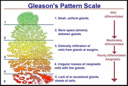
Any man of 40 years of age or older, to whom any of the undermentioned apply, should be referred for
genetic counselling and if identified with BRCA2 gene mutation should receive annual full screening for
prostate cancer:

•    Has a first- or second-degree relative (male or female) diagnosed with Breast Cancer
•    Has a first- or second-degree relative diagnosed with Prostate Cancer
•    Is aware of a first- or second-degree relative (male or female) who has tested positive for BRCA2 gene
     mutation

A first-degree relative is defined as a close blood relative which includes the individual's parents, full
siblings, or children.

A second-degree relative is defined as a blood relative which includes the individual's grandparents,
grandchildren, aunts, uncles, nephews, nieces or half-siblings.

Incidence of Prostate Cancer in South Africa
According to the outdated National Cancer Registry (2017) known for under reporting the following number
of prostate cancer cases was histologically diagnosed in South Africa during 2017:

Group - Males                      No of Cases             Lifetime Risk         Percentage of
2017                                                                              All Cancers
All males                              8 937                    1:16                22.37%
Asian males                             229                     1:25                23.27%
Black males                            4 102                    1:21                29.34%
Coloured males                         1 018                    1:14                21.34%
White males                            3 588                    1:10                16.78%

N.B.       ‘Histologically diagnosed’ means that a biopsy (removal of a specimen of tissue) was performed and
           that a diagnosis of Prostate Cancer was confirmed by a qualified pathologist.

The frequency of histologically diagnosed cases of prostate cancer in South Africa for 2016 was as follows
(National Cancer Registry, 2017):

Group - Males                     0 – 19         20 – 29         30 – 39        40 – 49       50 – 59       60 – 69       70 – 79         80+
2017                              Years           Years           Years          Years         Years         Years         Years         Years
All males                           1               4              20             255          1 655         3 557         2 671          774
Asian males                         0               0               0              4             32           100            78            15
Black males                         1               4              18             145           828          1 692         1 979           66
Coloured males                      0               0               1              29           206           408           306            68
White males                         0               0               1             227           589          1 357         1 217          347
N.B. In the event that the totals in any of the above tables do not tally, this may be the result of uncertainties as to the age, race or sex of the
individual. The totals for ‘all males’ and ‘all females’, however, always reflect the correct totals.

Bleyer, A., Spreafico, F. & Barr, R. 2019. Prostate cancer in young men: an emerging young adult and older
adolescent challenge. Cancer. DOI: 10.1002/cnr.32498.
Background: Recent observations suggest that prostate cancer is an increasing disease among older
adolescents and young adults.

Researched and Authored by Prof Michael C Herbst
[D Litt et Phil (Health Studies); D N Ed; M Art et Scien; B A Cur; Dip Occupational Health; Dip Genetic Counselling; Dip Audiometry and
Noise Measurement; Diagnostic Radiographer; Medical Ethicist]
Approved by Ms Elize Joubert, Chief Executive Officer [BA Social Work (cum laude); MA Social Work]
April 2021                                                                                                                       Page 2
Method: Incidence, mortality, and survival data were obtained from the United States National Cancer
Institute Surveillance, Epidemiology, and End Results program and the Institute for Health Metrics and
Evaluation Global Burden of Disease database.
Results: worldwide, the incidence of prostate cancer has increased in all groups between ages 15 and 40
years and increased globally at a steady rate averaging 2% per year since 1990 (P< .01). In the United States,
this age group was >6 times more likely than older men to have distant disease at diagnosis. Stage for stage,
their survival rate improved less than in older men. Whereas the overall 5-year relative survival rate in the
United States for men diagnosed between ages 40 and 80 years was between 95% and 100%, it was 30% in
those aged 15 to 24 years, 50% in those aged 20 to 29 years, and 80% in those aged 25 to 34 years.
Conclusion: Prostate cancer in older adolescents and young adult men has increased in most countries.
There is some evidence that this may be caused in part by underdiagnosis, Prostate-specific Antigen
screening, and overdiagnosis. It also may be caused by trends in obesity, physical inactivity, HPV infection,
substance exposure, environmental carcinogens, and/or referral patterns. How the biology of these cancers
differs from that in older men and how these etiologies vary from country to country remain to be
determined. Cancer. 2019;0:1-12.

Cancer of the Prostate
Prostate cancer is a malignant tumour that begins in the prostate gland. Some prostate cancers grow very
slowly and may not cause symptoms or problems for years. However, most prostate cancer cells make
excessive amounts of a protein called prostate specific antigen (PSA). PSA is also found in higher-than-
normal levels in men with various other prostate conditions, such as benign prostatic hyperplasia (BPH) and
prostatitis, in addition to prostate cancer.

Differential Diagnosis
The following table provides an overview of the signs and symptoms of prostate problems. Individuals with
any of the symptoms listed below should contact a medical professional:

                                                                       Acute         Chronic         Benign       Prostate
                                                                     Prostatitis    Prostatitis     Enlarged       Cancer
Symptom                                                                                             Prostate
Pain or burning sensation when urinating (dysuria)                        ӿ              ӿ              ӿ
Difficulty urinating, such as dribbling or hesitant urination             ӿ              ӿ              ӿ             ӿ
Frequent urination, particularly at night (nocturia)                      ӿ              ӿ              ӿ             ӿ
Urgent need to urinate                                                    ӿ              ӿ              ӿ             ӿ
A urinary stream that starts and stops                                    ӿ              ӿ              ӿ             ӿ
Pain in abdomen, groin or lower back                                      ӿ              ӿ              ӿ             ӿ
Pain or discomfort of the penis or testicles                              ӿ              ӿ                            ӿ
Pain in the area between the scrotum and rectum (perineum)                ӿ
Painful orgasms (ejaculation)                                             ӿ              ӿ                            ӿ
Flulike symptoms (with bacterial prostatitis)                             ӿ              ӿ
Feels like bladder does not empty completely                                             ӿ              ӿ
Erectile dysfunction                                                      ӿ              ӿ              ӿ             ӿ
Decreased urinary stream                                                  ӿ              ӿ              ӿ             ӿ
Blood in semen                                                            ӿ              ӿ                            ӿ
Blood in urine                                                            ӿ              ӿ              ӿ             ӿ
Raised Prostate Specific Antigen (PSA)                                    ӿ              ӿ              ӿ             ӿ
Bone pain                                                                                                             ӿ

Researched and Authored by Prof Michael C Herbst
[D Litt et Phil (Health Studies); D N Ed; M Art et Scien; B A Cur; Dip Occupational Health; Dip Genetic Counselling; Dip Audiometry and
Noise Measurement; Diagnostic Radiographer; Medical Ethicist]
Approved by Ms Elize Joubert, Chief Executive Officer [BA Social Work (cum laude); MA Social Work]
April 2021                                                                                                                       Page 3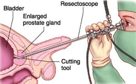
Types of Prostate Cancer
It is said that by the age of about 50, around half of all men have small changes in the size and shape of the
cells in the prostate. This is called prostatic intraepithelial neoplasia (PIN). Some research has indicated these
cellular changes may eventually develop into prostate cancer. This is controversial and preventive treatment
is not recommended.

If PIN is present, then careful follow-up screening with a PSA blood test and digital rectal examination (DRE)
is usually recommended.

More than 9 out of 10 prostate cancers (90%) are a type called acinar adenocarcinoma. It starts from gland
cells in the prostate. There are other types of adenocarcinoma, which include atrophic, foamy, colloid and
signet ring carcinoma.

The remaining prostate cancers include the following types:

•   Ductal adenocarcinoma
•   Transitional cell (or urothelial) cancer
•   Squamous cell cancer
•   Carcinoid
•   Small cell cancer
•   Sarcomas and sarcomatoid cancer

Because these cancers are so rare, there is sometimes very little information about which treatments work
best.

Ductal adenocarcinoma - this type of prostate cancer starts in the cells that line the ducts of the prostate
gland. It tends to grow and spread more quickly than acinar adenocarcinoma. This is why some men have an
advanced prostate cancer when they are diagnosed. This type of cancer is usually less sensitive to hormone
therapy than acinar adenocarcinoma.

Transitional cell (urothelial cancer) - this type of prostate cancer also starts in the cells that line the urethra.
More commonly, this type of cancer may start in the bladder and spread into the prostate.

Squamous cell cancer - squamous cell prostate cancer starts from the squamous cells covering the prostate
gland. Squamous cell prostate cancer tends to grow and spread more quickly than adenocarcinoma of the
prostate. This may be why some men have an advanced prostate cancer when they are diagnosed.

Carcinoid of the prostate - carcinoid tumours start from cells of the neuroendocrine system, which is made
up of specialised nerve and gland cells. These tumours are very rare and seem to be slow growing, although
some of them may be more aggressive. They may not cause any symptoms for many years.

Small cell cancer - this is a type of neuroendocrine tumour and is made up of small round cells. This type of
cancer often cause a raised prostate specific antigen (PSA) test. Many men are diagnosed when it is already
advanced. Small cell prostate cancer tends to grow and spread more quickly than adenocarcinoma of the
prostate. Hormone therapy usually does not work for this type of prostate cancer.

Sarcoma and sarcomatoid cancer - sarcomas start from muscle cells. They often grow quite quickly. The
most common type of prostate sarcoma in adult men is leiomyosarcoma. It tends to occur in men between
the ages of 35 and 60. Sarcomatoid cancers have a mixture of sarcoma and adenocarcinoma cells.
Researched and Authored by Prof Michael C Herbst
[D Litt et Phil (Health Studies); D N Ed; M Art et Scien; B A Cur; Dip Occupational Health; Dip Genetic Counselling; Dip Audiometry and
Noise Measurement; Diagnostic Radiographer; Medical Ethicist]
Approved by Ms Elize Joubert, Chief Executive Officer [BA Social Work (cum laude); MA Social Work]
April 2021                                                                                                                       Page 4

Oligometastatic Prostate Cancer
Cancer progresses in a stepwise fashion. Oligometastatic prostate cancer is defined as up to five extrapelvic
lesions on conventional imaging. There are controversies surrounding the management of this malignancy,
but retrospective and population-based studies suggest a role for radical prostatectomy. Despite insufficient
data to draw conclusions regarding the effectiveness of aggressive therapies on overall or cancer-specific
survival of patients with oligometastatic prostate cancer, current studies suggest that surgery decreases
tumour burden, disease-related morbidity, and the need for palliative surgical intervention, while increasing
the period of time to development of castration-resistant disease.

Risk Factors for Prostate Cancer
Age is the strongest risk factor for prostate cancer. Prostate cancer is very rare before the age of 40, but the
chance of having prostate cancer rises rapidly after age 50.

Other possible risk factors include:
• Family history: Prostate cancer seems to run in some families, and scientists have found several
   inherited genes that seem to raise prostate cancer risk

                                                                                                      Relative Risk for
                                                                                                       Prostate Cancer
           Risk Group                                                                                 (95% Confidence
                                                                                                          Index) *
           Brother(s) with prostate cancer diagnosed at any age                                       3.14 (2.37 – 4.15)
           Father with prostate cancer diagnosed at any age                                           2.35 (2.02 – 2.72)
           One affected first-degree relative diagnosed at any age                                    2.48 (2.25 – 2.74)
           Affected first-degree relatives diagnosed
•   High alcohol intake – Alcohol was declared a Group 1 carcinogen by the International Agency for
    Research on Cancer (IARC) in the 1980s
•   Use of tobacco products

According to Harvard Health, 2019.
The Harvard ejaculation study
The Health Professionals Follow-Up Study has been collecting information about a large group of volunteers
since 1986. All the men are health care providers, including dentists, pharmacists, veterinarians,
optometrists, ophthalmologists, and podiatrists. Most are white. In 1992, 29,342 men between the ages of
46 and 81 provided information about their average number of ejaculations per month in young adulthood
(age 20–29), middle age (40–49), and in the most recent year. Ejaculations included sexual intercourse,
nocturnal emissions, and masturbation. The volunteers provided comprehensive health and lifestyle data
every two years until the study concluded in 2000.
The scientists found no evidence that frequent ejaculations mark an increased risk of prostate cancer. In
fact, the reverse was true: High ejaculation frequency was linked to a decreased risk. Compared to men who
reported 4–7 ejaculations per month across their lifetimes, men who ejaculated 21 or more times a month
enjoyed a 31% lower risk of prostate cancer. And the results held up to rigorous statistical evaluation even
after other lifestyle factors and the frequency of PSA testing were taken into account.

Ejaculation: data from Down Under
An Australian study of 2,338 men examined the impact of sexual factors on the occurrence of prostate
cancer before the age of 70. Like the Harvard research, the Australian investigation evaluated total
ejaculations rather than sexual intercourse itself. Like the American men, the Australians who ejaculated
most frequently enjoyed a reduced risk of prostate cancer. The effect was strongest for the frequency of
ejaculations in young adulthood, even though prostate cancer was not diagnosed until many decades later.
Even so, the apparent protection extended to all age groups. In all, men who averaged 4.6–7 ejaculations a
week were 36% less likely to be diagnosed with prostate cancer before the age of 70 than men who
ejaculated less than 2.3 times a week on average.

Husby, A., Wohlfahrt, J. & Melbye, M. 2020.
BACKGROUND: A man's risk of prostate cancer has been linked to his prior reproductive history, with low
sperm quality, low ejaculation frequency, and a low number of offspring being associated with
increased prostate cancer risk. It is, however, highly controversial whether vasectomy, a common
sterilization procedure for men, influences prostate cancer risk.
METHODS: We established a cohort of all Danish men (born between 1937 and 1996) and linked
information on vasectomy, doctor visits, socioeconomic factors, and cancer from nationwide registries using
unique personal identification numbers. Incidence risk ratios for prostate cancer by time since vasectomy
and age at vasectomy during the follow-up were estimated using log-linear Poisson regression.
RESULTS: Overall, 26 238 cases of prostate cancer occurred among 2 150 162 Danish men during 53.4
million person-years of follow-up. Overall, vasectomized men had an increased risk of prostate
Researched and Authored by Prof Michael C Herbst
[D Litt et Phil (Health Studies); D N Ed; M Art et Scien; B A Cur; Dip Occupational Health; Dip Genetic Counselling; Dip Audiometry and
Noise Measurement; Diagnostic Radiographer; Medical Ethicist]
Approved by Ms Elize Joubert, Chief Executive Officer [BA Social Work (cum laude); MA Social Work]
April 2021                                                                                                                       Page 6
cancer compared with nonvasectomized men (relative risk = 1.15, 95% confidence interval = 1.10 to 1.20).
The increased risk of prostate cancer following vasectomy persisted for at least 30 years after the procedure
and was observed regardless of age at vasectomy and cancer stage at diagnosis. Adjustment for the number
of visits to the doctor and socioeconomic factors did not explain the association.
CONCLUSIONS: Vasectomy is associated with a statistically significantly increased long-term risk of prostate
cancer. The absolute increased risk following vasectomy is nevertheless small, but our finding supports a
relationship between reproductive factors and prostate cancer risk.

Signs and Symptoms of Prostate Cancer
Signs and symptoms of prostate cancer may include the following:

Urinary symptoms of prostate cancer - because of the proximity of the prostate gland in relation to the
bladder and urethra, prostate cancer may be accompanied by a variety of urinary symptoms. Depending on
the size and location, a tumour may press on and constrict the urethra, inhibiting the flow of urine. Some
prostate cancer signs related to urination include:
• Stranguria - a slow and painful discharge of urine, drop by drop, produced by spasmodic muscular
    contraction of the urethra and bladder
• Dysuria
• Pollakiuria - abnormally frequent urination
• Haematuria
• Trouble starting and stopping while urinating
• Nocturia - frequent urges to urinate at night
• Loss of bladder control
• Decreased flow or velocity of urine stream

Other prostate cancer signs & symptoms - prostate cancer may metastasise to nearby tissues or bones.
Other prostate cancer symptoms include:
• Blood in semen
• Erectile dysfunction
• Painful ejaculation
• Swelling in legs or pelvic area
• Numbness or pain in the hips, legs or feet
• Bone pain that does not go away, or leads to fractures

Diagnosis of Prostate Cancer
Caught in its early stages whilst still confined to the prostate gland prostate increases effective treatment.
Testing through a Prostate Specific Antigen (PSA) blood test and Digital Rectal Examination (DRE) and
subsequent prostate biopsy are currently mostly employed to detect the presence of prostate cancer.
Early detection is key to enabling better outcomes and potential cure of prostate cancer. Accordingly, it is
recommended that men over age 50, or 40 with a family history of prostate cancer, should talk to a doctor
about testing for prostate cancer using the PSA test and DRE as part of their annual health check-up. Men
should make an individual informed decision about testing based on the latest available evidence on the
benefits and potential harms of testing and subsequent treatment for prostate cancer.

Researched and Authored by Prof Michael C Herbst
[D Litt et Phil (Health Studies); D N Ed; M Art et Scien; B A Cur; Dip Occupational Health; Dip Genetic Counselling; Dip Audiometry and
Noise Measurement; Diagnostic Radiographer; Medical Ethicist]
Approved by Ms Elize Joubert, Chief Executive Officer [BA Social Work (cum laude); MA Social Work]
April 2021                                                                                                                       Page 7
It can be life threatening to wait for symptoms to appear before seeking assessment.

Most men seek testing for prostate cancer for the following reasons:
• As part of a general check-up - usually after 50 years of age
• Due to a recent experience with a relative or friend who has suffered from prostate cancer
• A family history of prostate cancer
• A recent onset of urinary symptoms

Screening for Prostate Cancer
Carlsson, S.V. & Vickers, A.J. 2020.
“This article gives an overview of the current state of the evidence for prostate cancer early detection with
prostate-specific antigen (PSA) and summarizes current recommendations from guideline groups. The article
reviews the global public health burden and risk factors for prostate cancer with clinical implications as
screening tools. Screening studies, novel biomarkers, and MRI are discussed. The article outlines 7 key
practice points for primary care physicians and provides a simple schema for facilitating shared decision-
making conversations.”

The Prostate Specific Antigen (PSA) Test
The PSA blood test looks for the presence in the blood of a protein that is produced specifically by prostate
cells called Prostate Specific Antigen (PSA). The presence of an elevated PSA does not necessarily mean
prostate cancer is present as there are other medical conditions that can lead to a PSA result outside the
normal range.

The result of a PSA test needs expert evaluation by an experienced doctor. As a general rule, the higher the
PSA result the greater the chance that prostate cancer is present. Where cancer is present, the PSA can
predict the volume of disease. Where the PSA is less than 10, the cancer is commonly confined to the
prostate. If the PSA is above 30, it is very likely the cancer has spread beyond the prostate and is, therefore,
less likely to be curable.

If the PSA test reveals a slightly elevated PSA, a doctor should recommend the test be repeated from time to
time to establish the rate of change, if any, before recommending a biopsy.

                                                                                                                  [Picture Credit: DRE]
The Digital Rectal Examination (DRE)
The DRE involves the insertion a gloved finger in the anus, where it is
possible to feel part of the surface of the prostate. Irregularities include
swelling or hardening of the prostate, or lumps on the surface that may
indicate development of a tumour, or other problems. The drawback to
this test is that one can feel only part of the prostate, so may miss
irregularities beyond reach. There are views that a DRE is not a
requirement in the diagnosis of prostate cancer.

Biopsy
A Biopsy is a small tissue sample taken with a spring loaded needle. This is normally conducted by a
urologist. A small probe containing an ultrasound generator and sampling needles (known as Trans Rectal
Ultra Sound or TRUS) is inserted in the anus.

Researched and Authored by Prof Michael C Herbst
[D Litt et Phil (Health Studies); D N Ed; M Art et Scien; B A Cur; Dip Occupational Health; Dip Genetic Counselling; Dip Audiometry and
Noise Measurement; Diagnostic Radiographer; Medical Ethicist]
Approved by Ms Elize Joubert, Chief Executive Officer [BA Social Work (cum laude); MA Social Work]
April 2021                                                                                                                       Page 8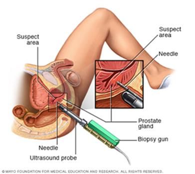
The ultrasound generates an image of the prostate on a computer screen and guides the urologist to insert
the sampling needle into selected areas of the prostate. The biopsy samples are analysed by a pathologist to
determine the stage and grade of the cancer.

There are four likely results of a prostatic biopsy:
• The tissue is normal benign prostate tissue
• A condition called atypia or dysplasia where the cells do not look typical of either normal or cancerous
   cells
• Prostatic intraepithelial neoplasia (PIN) where the cells appear to be in the transitional stage between
   normal and cancer
• Prostate cancer - which are currently graded on a numerical scoring system call the Gleason Score and
   the Stage of cancer

PET/CT for Prostate Cancer
Improvements in cancer care have been closely
linked to advances in imaging technologies, as
these have allowed for more accurate diagnosis,
staging,      and     surveillance    of     the
disease. Prostate cancer (PCa) has been unique
in its reliance on a serum biomarker, prostate-
specific antigen (PSA), as the major player for
disease diagnosis, treatment, and surveillance.
Until recently, the use of imaging in PCa was
limited to transrectal ultrasonography (TRUS)
for guidance of prostate biopsies and computed
tomography (CT) and 99Tc bone scans for staging
of the disease. Over the last 10 years, advances
in magnetic resonance imaging (MRI)
technology and the development of novel
nuclear medicine radiotracers have revolution-
nised PCa management.                                                                [Picture Credit: PET Radio Tracers]

Trabulsi, E.J., Rumble, R.B., Jadvar, H., Hope, T., Pomper, M., Turkbey, B., Rosenkrantz, A.B., Verma, S.,
Margolis, D, J., Froemming, A., Oto, A., Purysko, A., Milowsky, M.M., Schlemmer, H-P., Eiber, M., Morris,
M.J. Choyke, P.L., Padhani, A., Oldan, J., Fanti, S., Jain, S., Pinto, P.A., Keegan, K.A., Porter, C.R., Coleman,
J.A., Bauman, G.S., Jani, A.B., Kamradt, J.M., Sholes, W. & Vargas, H.A. 2020.
PURPOSE: Provide evidence- and expert-based recommendations for optimal use of imaging in advanced
prostate cancer. Due to increases in research and utilization of novel imaging for advanced prostate cancer,
this guideline is intended to outline techniques available and provide recommendations on appropriate use
of imaging for specified patient subgroups.
METHODS: An Expert Panel was convened with members from ASCO and the Society of Abdominal
Radiology, American College of Radiology, Society of Nuclear Medicine and Molecular Imaging, American
Urological Association, American Society for Radiation Oncology, and Society of Urologic Oncology to
conduct a systematic review of the literature and develop an evidence-based guideline on the optimal use of
imaging for advanced prostate cancer. Representative index cases of various prostate cancer disease states
are presented, including suspected high-risk disease, newly diagnosed treatment-na¨ıve metastatic disease,
suspected recurrent disease after local treatment, and progressive disease while undergoing systemic
Researched and Authored by Prof Michael C Herbst
[D Litt et Phil (Health Studies); D N Ed; M Art et Scien; B A Cur; Dip Occupational Health; Dip Genetic Counselling; Dip Audiometry and
Noise Measurement; Diagnostic Radiographer; Medical Ethicist]
Approved by Ms Elize Joubert, Chief Executive Officer [BA Social Work (cum laude); MA Social Work]
April 2021                                                                                                                       Page 9
treatment. A systematic review of the literature from 2013 to August 2018 identified fully published English-
language systematic reviews with or without meta-analyses, reports of rigorously conducted phase III
randomized controlled trials that compared $ 2 imaging modalities, and noncomparative studies that
reported on the efficacy of a single imaging modality.
RESULTS: A total of 35 studies met inclusion criteria and form the evidence base, including 17 systematic
reviews with or without meta-analysis and 18 primary research articles.
RECOMMENDATIONS: One or more of these imaging modalities should be used for patients with advanced
prostate cancer: conventional imaging (defined as computed tomography [CT], bone scan, and/or prostate
magnetic resonance imaging [MRI]) and/or next-generation imaging (NGI), positron emission tomography
[PET], PET/CT, PET/MRI, or whole-body MRI) according to the clinical scenario. J Clin Oncol 38. © 2020 by
American Society of Clinical Oncology

The Gleason Score
The Gleason score is used to help determine how quickly a tumour may grow or spread. It may seem
confusing because with the Gleason system (the most common system used), there’s both a ‘grade’ and a
‘score’.

The Gleason grade                uses
numbers 1 to 5.

       [Picture Credit: Gleason Score]

A number is assigned to two of
the areas of the prostate that
have the most cancer (based
on biopsy core samples that
are taken). This is because the
cancer may look different in
each of those two areas.

Once those two numbers are
determined, they are added
together to come up with the
Gleason score, which ranges
from 2 to 10.

What the Gleason grades mean :

Grade 1: The cells look almost like normal cells (called well differentiated) and are uniformly spaced in a tight
mass.
Grade 2: The cancer cells are still well differentiated, but are arranged more loosely, are more irregular in
shape, and some cells have spread to other prostatic tissue.

Grade 3: The cancer is moderately differentiated; cells vary in size from small to large; and more cells have
invaded other prostatic tissue.

Researched and Authored by Prof Michael C Herbst
[D Litt et Phil (Health Studies); D N Ed; M Art et Scien; B A Cur; Dip Occupational Health; Dip Genetic Counselling; Dip Audiometry and
Noise Measurement; Diagnostic Radiographer; Medical Ethicist]
Approved by Ms Elize Joubert, Chief Executive Officer [BA Social Work (cum laude); MA Social Work]
April 2021                                                                                                                     Page 10
Grade 4: The cancer cells are irregular, distorted, and look less like normal cells (called poorly differentiated),
and there is considerable spread (called invasion) to other prostatic tissue.
Grade 5: The cancer cells do not look anything like normal cells and have spread in haphazard ‘clumps’ of all
different shapes and sizes through the prostate.
What the Gleason scores mean:
• If the score is less than 6, the cancer may be considered to be well-differentiated or low-grade cancer
• A score of 7 may be considered to be moderately differentiated or intermediate-grade cancer
• A score of 8 to 10 may be considered to be poorly differentiated or high-grade cancer
According to the American Urological Association, the lowest Gleason score that is usually found after a
biopsy is 5. The cancer is considered to be more aggressive as the score rises. Scores of 8 to 10 are
considered to be the most aggressive, which means that the cancer is more likely to grow and spread more
quickly.
The biopsy results (called the pathology report) will contain other important information that helps to assess
how aggressive the cancer may be.
This includes:
• How many biopsy core samples were positive for cancer
• How much cancer was in each core sample (this is given as a percentage)
• Whether cancer was found in just one side of the prostate gland or in both sides (which is referred to
as bilateral)
Biopsy of the Prostate
A PSA test on its own cannot tell if a patient has prostate cancer (many patients with prostate cancer in fact
may not have an abnormal PSA level) – it is only an indicator, although generally a reliable one. As a result,
further investigation is required. This involves tests in the form of an MRI scan and/or a biopsy, which are
usually performed to give the oncologist/urologist a much clearer picture and firm data about the presence
of any possible cancer.
An MRI scan on its own is also not necessarily sufficient. Although MRI scanning is getting close to being able
to reliably predict the presence of a significant (Gleason grade 7 or higher) prostate cancer, it is still only 80%
accurate. One can, therefore, not rely on it on its own. Only prostate cancer seen under a microscope by a
trained pathologist can be taken as incontrovertible proof of the presence of cancer within a prostate gland,
and this requires a biopsy to be performed.
There are two methods of performing a prostate biopsy, namely a transrectal biopsy and a transperineal
prostate biopsy.
Transrectal biopsy method - prostate biopsy has traditionally been done via the transrectal route – i.e. via
the rectum. This is because before the advent of transrectal ultrasound (where an ultrasound probe is placed
in the rectum to provide a visual of the prostate as the needles were inserted into it), the biopsies were
Researched and Authored by Prof Michael C Herbst
[D Litt et Phil (Health Studies); D N Ed; M Art et Scien; B A Cur; Dip Occupational Health; Dip Genetic Counselling; Dip Audiometry and
Noise Measurement; Diagnostic Radiographer; Medical Ethicist]
Approved by Ms Elize Joubert, Chief Executive Officer [BA Social Work (cum laude); MA Social Work]
April 2021 Page 11finger-guided. This means that the biopsy needle was placed alongside a gloved finger placed in the rectum,
which allowed the operator to feel which part of the prostate to guide the needle into. The advent of the
transrectal ultrasound probe allowed urologists to see exactly where the needle was within the prostate
before the biopsy was taken, allowing a much greater degree of accuracy than with the finger-guided
approach.
However, even with the probe to help, problems remain with transrectal prostate biopsies. These may
include:
• An infection rate of 10-20% due to needles penetrating the rectal wall, and faecal matter contaminating
other parts of the body.
• A serious infection (septicaemia or blood poisoning) rate of 1-2%.
• The development of antibiotic-resistant strains of bacteria causing infection.
• Pain during biopsy.
• Inability to accurately place the biopsy needle within the prostate in 3 dimensions.
In addition, perhaps the biggest downside of the transrectal approach is that it is harder/impossible to reach
the part of the prostate (the anterior, or frontal aspect) that is furthest away from the biopsy needle. This
leads to poor ‘sampling’ – in that the tissue samples collected by the needles may miss any cancer found in
this part of the prostate, leading to an inaccurate biopsy.
A transrectal approach might miss the cancer in the anterior, with the biopsy coming back clear from the
other parts of the prostate. In essence, all parts of the prostate need to be sampled completely for the most
accurate biopsy result.
Transperineal biopsy method - the answer to the above problems lies
in using a transperineal approach to inserting the needles – meaning
that the needles go through the perineum (the area behind the
scrotum and in front of the anus) instead of through the rectum. A
brachytherapy grid is placed over the perineum to allow accurate
placement of the biopsy needle within the prostate, together with
the use of an ultrasound probe to direct the urologist. This allows
better sampling of all parts of the prostate, including the anterior
section.
[Picture Credit: Transrectal Prostate Biopsy]
[Picture Credit: Transperineal Prostate Biopsy]
Targeting the biopsy accurately using MRI - by doing an MRI
scan before a transperineal biopsy, one can identify the
suspicious areas to be specifically targeted during the
biopsy – increasing the accuracy of the biopsy. This can
either be done by comparing the MRI scan with the
transrectal ultrasound view during the biopsy process
(cognitive targeting) or by overlaying the 2 images in real-
time using computer software: fusion biopsy. Because
Researched and Authored by Prof Michael C Herbst
[D Litt et Phil (Health Studies); D N Ed; M Art et Scien; B A Cur; Dip Occupational Health; Dip Genetic Counselling; Dip Audiometry and
Noise Measurement; Diagnostic Radiographer; Medical Ethicist]
Approved by Ms Elize Joubert, Chief Executive Officer [BA Social Work (cum laude); MA Social Work]
April 2021 Page 12patients are asleep under a general anaesthetic for transperineal biopsy, discomfort during the biopsy
process is not an issue, allowing many more samples to be taken and so increasing the detection rate. And
because the biopsies are not taken via the rectum, infection-related complications are much less common.
In summary - the point of a biopsy is to accurately determine the grade of prostate cancer, and so one must
be able to access the whole of the prostate to do so. A quick overview of the main points of comparison is
found below.
Eggener, S.E., Rumble, R.B., Armstrong, A.J., Morgan, T.M., Crispino, T., Cornford, P., van der Kwast, T.,
Grignon, D.J., Rai, A.J., Agarwal, N., Klein, E.A., Den, R.B. & Beltran, H. 2020.
Purpose: This guideline provides recommendations for available tissue-based prostate cancer biomarkers
geared toward patient selection for active surveillance, identification of clinically significant disease, choice
of postprostatectomy adjuvant versus salvage radiotherapy, and to address emerging questions such as the
relative value of tissue biomarkers compared with magnetic resonance imaging.
Methods: An ASCO multidisciplinary Expert Panel, with representatives from the European Association of
Urology, American Urological Association, and the College of American Pathologists, conducted a systematic
literature review of localized prostate cancer biomarker studies between January 2013 and January 2019.
Numerous tissue-based molecular biomarkers were evaluated for their prognostic capabilities and potential
for improving management decisions. Here, the Panel makes recommendations regarding the clinical use
and indications of these biomarkers.
Results: Of 555 studies identified, 77 were selected for inclusion plus 32 additional references selected by
the Expert Panel. Few biomarkers had rigorous testing involving multiple cohorts and only 5 of these tests
are commercially available currently: Oncotype Dx Prostate, Prolaris, Decipher, Decipher PORTOS, and
ProMark. With various degrees of value and validation, multiple biomarkers have been shown to refine risk
stratification and can be considered for select men to improve management decisions. There is a paucity of
prospective studies assessing short- and long-term outcomes of patients when these markers are integrated
into clinical decision making.
Recommendations: Tissue-based molecular biomarkers (evaluating the sample with the highest volume of
the highest Gleason pattern) may improve risk stratification when added to standard clinical parameters, but
the Expert Panel endorses their use only in situations in which the assay results, when considered as a whole
with routine clinical factors, are likely to affect a clinical decision. These assays are not recommended for
routine use as they have not been prospectively tested or shown to improve long-term outcomes-for
example, quality of life, need for treatment, or survival. Additional information is available at
www.asco.org/genitourinary-cancer-guidelines.
Item: Transrectal Prostate Transperineal Prostate
Biopsy Biopsy
Anaesthetic Local General
Hospital stay Outpatient Day case
Discomfort during procedure Moderate None
Discomfort after procedure Ache Ache
Infection risk 10 – 20% 1 – 2%
Serious infection risk 1 – 2% Less than 1%
Ability to sample anterior prostate Limited Full
Allows systematic sampling (prostate mapping) No Yes
Researched and Authored by Prof Michael C Herbst
[D Litt et Phil (Health Studies); D N Ed; M Art et Scien; B A Cur; Dip Occupational Health; Dip Genetic Counselling; Dip Audiometry and
Noise Measurement; Diagnostic Radiographer; Medical Ethicist]
Approved by Ms Elize Joubert, Chief Executive Officer [BA Social Work (cum laude); MA Social Work]
April 2021 Page 13Staging of Prostate Cancer
Like other forms of cancer, the prognosis for prostate cancer depends on how far the cancer has spread at
the time it is diagnosed.
A system of staging is used to describe prostate cancer spread.
Accurately identifying the prostate cancer stage is extremely important.
Prostate cancer staging helps to determine the optimal treatment as well as prognosis.
Treatment of Prostate Cancer
The main treatments for prostate cancer are surgery, radiotherapy and hormone therapy. Chemotherapy is
also sometimes used.
A number of different factors needs to be taken into consideration when deciding on treatment. The most
important of these are how fast the cancer is likely to grow and how far it has already grown.
It is generally recommended that treatment is considered individually for every man with prostate cancer.
Patients often query their treatment when they come across other men with prostate cancer who are having
different treatment to what they are receiving. They should be informed that it is because they have a
different stage or grade of cancer.
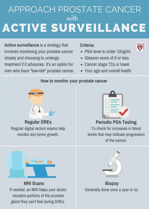
• Active Surveillance -The concept of active surveillance, or watchful waiting, has increasingly emerged in
recent years as a viable option for men who decide not to undergo immediate surgery or radiation
therapy.
During active surveillance, prostate cancer is carefully monitored for signs of progression. A PSA blood
test and digital rectal exam (DRE) are usually administered periodically along with a repeat biopsy of the
prostate at one year and then at specific intervals thereafter. If symptoms develop, or if tests indicate
the cancer is growing, treatment might be warranted.
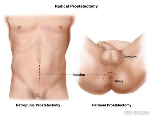
• Surgery –
A surgical approach to treating prostate cancer will remove all or part of the prostate. Typically, men
with early-stage disease or cancer that’s confined to the prostate will undergo radical prostatectomy -
removal of the entire prostate gland, plus some surrounding tissue. Other surgical procedures may be
performed on men with advanced or recurrent disease.
The most common types of prostatectomy include:
Researched and Authored by Prof Michael C Herbst
[D Litt et Phil (Health Studies); D N Ed; M Art et Scien; B A Cur; Dip Occupational Health; Dip Genetic Counselling; Dip Audiometry and
Noise Measurement; Diagnostic Radiographer; Medical Ethicist]
Approved by Ms Elize Joubert, Chief Executive Officer [BA Social Work (cum laude); MA Social Work]
April 2021 Page 14Radical Retropubic Prostatectomy - an incision is made in the abdomen and the prostate is removed
from behind the pubic bone. The surgeon then stitches the urethra directly to the bladder so urine is
able to flow.
[Picture Credit: Radical Prostatectomy Incision]
Radical Perineal Prostatectomy - A perineal
prostatectomy is done through a cut in the
area between the testicles and back passage,
the perineum.
Transurethral prostatectomy – transurethral
resection of the prostate (TURP) is a type of
prostate surgery done to relieve moderate to
severe urinary symptoms caused by an
enlarged prostate, a condition known as benign prostatic hyperplasia (BPH).
Researched and Authored by Prof Michael C Herbst
[D Litt et Phil (Health Studies); D N Ed; M Art et Scien; B A Cur; Dip Occupational Health; Dip Genetic Counselling; Dip Audiometry and
Noise Measurement; Diagnostic Radiographer; Medical Ethicist]
Approved by Ms Elize Joubert, Chief Executive Officer [BA Social Work (cum laude); MA Social Work]
April 2021 Page 15[Picture Credit: Transurethral Prostatectomy]
During TURP, a combined visual and surgical
instrument (resecto-scope) is inserted through
the tip of the penis and into the tube that
carries urine from the bladder (urethra). The
urethra is surrounded by the prostate. Using the
resectoscope, the doctor trims away excess
prostate tissue that is blocking urine flow and
increases the size of the channel that allows one
to empty one’s bladder.
TURP is one of the most effective options for
treating urinary symptoms caused by BPH. To determine whether TURP or another treatment is the
right choice, the doctor will consider how severe the patient’s symptoms are, what other health
problems he has, and the size and shape of the prostate.
• Radiation therapy - it involves the killing of cancer cells and surrounding tissues with directed radioactive
exposure. The use of radiation therapy as an initial treatment for prostate cancer is described below.
Some forms of radiation therapy can also be used in men with advanced or recurrent prostate cancer.
Although technological advancements have made radiation therapy (RT) treatment more specific to the
prostate, surrounding structures such as the bladder and rectum can still be affected. Close to 70% of all
cancers are treated with radiation; depending on the location of the involved organ, radiation colitis can be a
common complication outside of prostate cancer. Yet up to 20% of patients with prostate cancer treated
with RT (external or internal via brachytherapy) will develop radiation colitis.
Radiation colitis can present in 2 ways: acute (within 6 weeks of RT) and chronic (within 9 months after RT
and for up to 30 years thereafter). Patients with acute and chronic radiation colitis have similar symptoms,
although chronic can cause more gastrointestinal bleeding. A patient's bowel movements will contain more
mucus and will be less formed; they may experience diarrhoea as well as urgency. Tenesmus is another
common symptom.
Chronic inflammation of the rectum over many years can lead to stricturing of the left side of the colon,
which may present with changes in stool calibre or constipation.
[Picture Credit: Brachytherapy]
• External Beam Radiation Therapy - this is the most common type of radiation therapy. CT scans and MRIs
are used to map out the location of the tumour cells, and X-rays are targeted to those areas. With 3-D
conformal radiotherapy, a computerised program maps out the exact location of the prostate tumours
so the highest dose of radiation can reach the cancer cells within the gland.
Intensity-modulated radiation therapy (IMRT) allows the radiation doctors to modulate, or change, the
intensity of the doses and radiation beams to better target the radiation delivered to the prostate, while
simultaneously delivering lower doses to the tumour cells that are immediately adjacent to the bladder
and rectal tissue. These techniques are always improving, including the use of guidance markers (fiducial
markers), which may be able to reduce the risks to the bowel and bladder over time.
Researched and Authored by Prof Michael C Herbst
[D Litt et Phil (Health Studies); D N Ed; M Art et Scien; B A Cur; Dip Occupational Health; Dip Genetic Counselling; Dip Audiometry and
Noise Measurement; Diagnostic Radiographer; Medical Ethicist]
Approved by Ms Elize Joubert, Chief Executive Officer [BA Social Work (cum laude); MA Social Work]
April 2021 Page 16Because the treatment planning with these types of radiation therapy is far more precise, higher—and
more effective—doses of radiation can be used with less chance of damaging surrounding tissue. Also,
because radiation works slowly, toxicities to the normal surrounding tissues can also develop slowly.
• Proton Therapy
The advantage of using protons over other external beam sources is precision. Protons of energetic
particles can hit a targeted prostate cancer tumour without affecting surrounding tissue. This direct
attack on cancerous cells ultimately causes their death, as the cells are particularly vulnerable to attack
due to their rapid division.
Proton treatment is notably valuable for treating localized, isolated, solid tumours before they spread to
other tissues and the rest of the body. However, to date, proton beam therapy has never been
compared directly to standard IMRT techniques, so we do not truly know if this offers an advantage over
standard approaches.
• Brachytherapy
With brachytherapy, tiny metal pellets containing
radioactive iodine or palladium are inserted into
the prostate via needles that enter through the
skin behind the testicles. As with 3-D conformal
radiation therapy, careful and precise maps are
used to ensure that the seeds are placed in the
proper locations.
Over the course of several months, the seeds give off radiation to the immediate surrounding area, killing
the prostate cancer cells. By the end of the year, the radioactive material degrades, and the seeds that
remain are harmless.
Compared with external radiation therapy, brachytherapy is less commonly used, but some patients
prefer this option primarily because it doesn’t require daily visits to the treatment centre. Side
effects can include erectile dysfunction, urinary frequency and obstruction, and rarely rectal injury.
• Hormone Therapy - Prostate cancer cells are like other living organisms—they need fuel to grow and
survive. Because the hormone testosterone serves as the main fuel for prostate cancer cell growth, it’s a
common target for therapeutic intervention in men with the disease.
Hormone therapy, also known as androgen-deprivation therapy or ADT, is designed to stop testosterone
from being released or to prevent it from acting on the prostate cells. Although hormone therapy plays
an important role in men with advancing prostate cancer, it is increasingly being used before, during, or
after local treatment as well.
• Orchidectomy - About 90% of testosterone is produced by the testicles. So orchidectomy (the surgical
removal of the testicles) is an effective solution to blocking testosterone release. This approach has been
used successfully since the 1940s. Because it is permanent and irreversible, most men opt for drug
therapy instead.
Researched and Authored by Prof Michael C Herbst
[D Litt et Phil (Health Studies); D N Ed; M Art et Scien; B A Cur; Dip Occupational Health; Dip Genetic Counselling; Dip Audiometry and
Noise Measurement; Diagnostic Radiographer; Medical Ethicist]
Approved by Ms Elize Joubert, Chief Executive Officer [BA Social Work (cum laude); MA Social Work]
April 2021 Page 17• LHRH Agonists - LHRH, or luteinizing-hormone releasing hormone, is one of the key hormones released
by the body before testosterone is produced. (Note that LHRH is sometimes called GnRH, or
gonadotropin-releasing hormone.)
• Chemotherapy - The term ‘chemotherapy’ refers to any type of therapy that uses chemicals to kill or halt
the growth of cancer cells. The drugs work in a variety of ways, but are all based on the same simple
principle: stop the cells from dividing and you stop the growth and spread of the tumour.
Cha, J-R., Lee, J.H. & Ponnazhagan, S. 2020.
“Therapeutic interventions to harness the immune system against tumor cells have provided mixed results
in the past for several solid tumors and hematologic malignancies. However, immunotherapy has advanced
considerably over the last decade and is becoming an integral combination for treating patients with
advanced solid tumors. In particular, prostate cancer immunotherapy has shown modest efficacy for
patients in the past. With several key discoveries on immune mechanisms and advanced molecular
diagnostic platforms recently, immunotherapy is re-emerging as a viable option for prostate cancer,
especially castration-resistant prostate cancer (CRPC), to stimulate antitumor immunity. Combination of
patient-tailored immunotherapy and immune checkpoint blockers with conventional cytotoxic agents and
androgen receptor-targeted therapies should move the field forward. With a recent adaptation that the
application of immune checkpoint inhibitors has been successful in the treatment of more than a dozen solid
tumors, including melanoma, lymphoma, liver, cervical, gastrointestinal, and breast cancers, it is a timely
endeavor to harness immunotherapy for prostate cancer. Here, we provide an account on the progression of
immunotherapy with new discoveries and precision approaches for tumors, in particular CRPC, from
mechanistic standpoint to emerging limitations and future directions.”
Albertsen, P.C. 2020. Prostate cancer screening and treatment: where have we come from and where are
we going? BJU Int. 2020 Aug;126(2):218-224.
Objective: To evaluate the current prostate cancer screening and treatment paradigm in light of recently
published long-term results of major screening and treatment trials.
Methods: Historical review of the evolution of the diagnosis and treatment of prostate cancer followed by a
detailed summary of the findings and differences among the three major screening trials and the three
major treatment trials.
Results: Prostate-specific antigen (PSA) testing can identify clinically significant prostate cancer and has
produced a significant stage shift and is the likely explanation for the decline in prostate cancer mortality.
Unfortunately, PSA testing predominantly identifies low-grade disease that is unlikely to progress during a
patient's lifetime leading to substantial diagnosis of indolent disease. Treatment with radical prostatectomy
(RP) appears to benefit primarily younger men (agedpathway, and the increasing use of prostate-specific membrane antigen positron emission tomography for
early stratification in the salvage setting for failure of primary treatment in localised disease. In addition,
upfront combinations of androgen deprivation therapy with other systemic treatments have yielded
significant gains in overall survival for patients with metastatic disease. There has also been an increasing
recognition of the association between germline DNA repair defects and progressive disease, and interest in
the potential to identify patients for therapies that target these defects. There have been significant changes
in how prostate cancer is diagnosed and managed in the past five years, with the introduction of new clinical
pathways that were unprecedented just a decade previously.
Gourdin, T. 2020.
Purpose of review: Summarize recent advances in the treatment of advanced prostate cancer.
Recent findings: Recent randomized data suggest a survival advantage to early use of novel androgen
receptor inhibitors in combination with androgen deprivation therapy both in the setting of hormone-
sensitive metastatic prostate cancer and nonmetastatic castration-resistant disease. While ongoing analyses
examine optimal sequencing of existing agents for treatment of advanced prostate cancer, additional
research focuses on expanding treatment options through development of novel genomically targeted
therapies, antibody-drug conjugates, and immune therapy combinations.
Summary: In this review, we summarize the recent data supporting the early use of novel androgen receptor
inhibitors in advanced prostate cancer and aim to also frame how these drugs may fit within the existing
context of docetaxel and abiraterone. We present recent series examining sequencing of approved therapies
while searching for predictive biomarkers. Finally, we examine trials evaluating novel agents that target
certain biological pathways to highlight the likely future directions for progress in the clinical management of
advanced prostate cancer.
• Other treatment options - Surgery and radiation therapy remain the standard treatment for localised
prostate cancer, but other, less popular treatment options might be beneficial as well. As time goes on
and the benefits of these treatment options are further explored, it’s possible that they will move more
into the mainstream. For now, though, none are seen as standard treatments for localized prostate
cancer.
• Cryotherapy - Cryotherapy, also known as cryosurgery or cryoablation, has been around for years, but
until a few years ago, it was rarely used. With this approach, probes are inserted into the prostate
through the perineum (the space between the scrotum and the anus), and argon gas or liquid nitrogen is
delivered to the prostate, literally freezing to death the prostate cells and any prostate tumours.
Over the years, a number of modifications were made to avoid freezing damage to the nearby
structures, but the rates for both erectile and urinary dysfunction remain high, and data on long-term
outcomes are limited.
• Primary Hormone Therapy - Prostate cancer cells are like other living organisms—they need fuel to grow
and survive. Because the hormone testosterone serves as the main fuel for prostate cancer cell growth,
it’s a common target for therapeutic intervention in men with the disease. Hormone therapy, also
known as androgen-deprivation therapy or ADT, is designed to stop testosterone from being released or
to prevent it from acting on the prostate cells.
Although hormone therapy plays an important role in men with advancing prostate cancer, it is also
increasingly being used before, during, or after local treatment. In some cases, hormone therapy may be
used in conjunction with radiation therapy. If so, treatment with ADT is gernally given before, during and
Researched and Authored by Prof Michael C Herbst
[D Litt et Phil (Health Studies); D N Ed; M Art et Scien; B A Cur; Dip Occupational Health; Dip Genetic Counselling; Dip Audiometry and
Noise Measurement; Diagnostic Radiographer; Medical Ethicist]
Approved by Ms Elize Joubert, Chief Executive Officer [BA Social Work (cum laude); MA Social Work]
April 2021 Page 19after radiation therapy in the form of an LHRH agonist. LHRH, or luteinizing hormone releasing hormone,
is one of the key hormones involved in the production of testosterone. This medicine works through a
complicated feedback loop to lower the body’s testosterone. Note that LHRH is sometimes called GnRH,
or gonadotropin-releasing hormone. Although there is little, if any, data to show that hormone therapy
alone is an effective treatment strategy for men with localized prostate cancer, it is increasingly being
used in this setting.
Because it is not invasive, it is possible that the therapy is seen as a middle ground between active
surveillance and local therapy. For men who are not good candidates for surgery or radiation, and who
require immediate therapy, primary hormonal therapy is a reasonable option. However, hormonal
therapy has a long list of side effects, and thus, the main question is whether therapy can be safely
deferred in men who are not candidates for immediate surgery or radiation. Primary hormonal therapy
is also a reasonable option in men who have metastatic disease (cancer spread beyond the prostate)
when the diagnosis of prostate cancer is made. In these men, hormonal therapy will shrink the prostate
gland and cancer and may delay any need for local therapy.
• Targeted Therapies - Chemotherapy drugs can play an important role in improving the lives of men with
advanced prostate cancer, but they often don’t distinguish between tumour cells and healthy cells to a
high degree and can kill off some normal cells along the way. So-called targeted therapies, by contrast,
are drugs that are specifically designed to interfere with the way cancer cells grow, with the way cancer
cells interact with each other, and/or with the way that the immune system interact with the cancer
without damaging a man’s normal cells.
• Emerging Therapies - In laboratories around the world, researchers are busy identifying new drugs and
treatment approaches that might prove beneficial to men with prostate cancer. Most of these
investigational agents are being tested in men with advanced prostate cancer: therapy options for men
at this stage of disease may not be effective enough to halt progression of the disease, and men are
typically affected by side effects from the disease and/or the medications that they’re taking. It’s
therefore the perfect stage at which to test out new drugs because any improvement will likely be
rapidly noticed and much appreciated.
Questions that Patients should ask their Health Professional
It is important for health professionals to have honest, open discussions with their patients about prostate
cancer. Some questions from patients pertaining to prostate cancer that every health professional should be
able respond to:
• What type of prostate cancer do I have?
• What are the chances that the cancer has spread beyond my prostate? If so, is it still curable?
• What further tests (if any) do you recommend, and why?
• Are there other types of doctors I should talk to before deciding on treatment?
• What is the clinical stage and Gleason score (grade) of my cancer? What do those mean to me? Does this
make me a low-risk, intermediate-risk or high-risk patient?
• What is my expected survival rate based on clinical stage, grade, and various treatment options?
• Should I consider active surveillance as an option? Why or why not?
• Do you recommend a radical prostatectomy or radiation therapy? Why or why not?
• Should I consider laparoscopic or robot-assisted prostatectomy?
• What types of radiation therapy might work best for me?
Researched and Authored by Prof Michael C Herbst
[D Litt et Phil (Health Studies); D N Ed; M Art et Scien; B A Cur; Dip Occupational Health; Dip Genetic Counselling; Dip Audiometry and
Noise Measurement; Diagnostic Radiographer; Medical Ethicist]
Approved by Ms Elize Joubert, Chief Executive Officer [BA Social Work (cum laude); MA Social Work]
April 2021 Page 20You can also read