Breast cancer? What is Let us explain it to you.
←
→
Page content transcription
If your browser does not render page correctly, please read the page content below
Breast Cancer

                        What is
                     breast cancer?

                                        Let us explain
                                           it to you.

www.anticancerfund.org   www.esmo.org

                                                ESMO/ACF Patient Guide Series
                                            based on the ESMO Clinical Practice Guidelines
BREAST CANCER: A GUIDE FOR PATIENTS

     PATIENT INFORMATION BASED ON ESMO CLINICAL PRACTICE GUIDELINES
This guide for patients has been prepared by the Anticancer Fund as a service to patients, to help
patients and their relatives better understand the nature of breast cancer and appreciate the best
treatment choices available according to the subtype of breast cancer. We recommend that patients
ask their doctors about what tests or types of treatments are needed for their type and stage of
disease. The medical information described in this document is based on the clinical practice
guidelines of the European Society for Medical Oncology (ESMO) for the management of primary
breast cancer and for the management of locally recurrent or metastatic breast cancer. This guide for
patients has been produced in collaboration with ESMO and is disseminated with the permission of
ESMO. It has been written by a medical doctor and reviewed by two oncologists from ESMO including
the lead author of the clinical practice guidelines for professionals. It has also been reviewed by
patient representatives from ESMO’s Cancer Patient Working Group.

More information about the Anticancer Fund: www.anticancerfund.org

More information about the European Society for Medical Oncology: www.esmo.org

For words marked with an asterisk, a definition is provided at the end of the document.

Breast Cancer: a guide for patients - Information based on ESMO Clinical Practice Guidelines - v.2013.1               Page 1
This document is provided by the Anticancer Fund with the permission of ESMO.
The information in this document does not replace a medical consultation. It is for personal use only and cannot be modified,
reproduced or disseminated in any way without written permission from ESMO and the Anticancer Fund.
Table of contents

Definition of breast cancer ...................................................................................................................... 3

Is breast cancer frequent?....................................................................................................................... 4

What causes breast cancer?.................................................................................................................... 5

How is breast cancer diagnosed? ............................................................................................................ 7

Common misconceptions about breast cancer treatment ..................................................................... 8

What is it important to know to get the optimal treatment? ................................................................. 9

What are the treatment options? ......................................................................................................... 13

What are the possible side effects of the treatment? .......................................................................... 23

What happens after adjuvant treatment has been terminated?.......................................................... 28

Definitions of difficult words ................................................................................................................. 31

The first version of this guide was published in 2011 and was written by Dr. Gauthier Bouche (the Anticancer Fund) and
reviewed by Prof. Martine Piccart (ESMO), Prof. Bernhard Pestalozzi (ESMO) and Prof. Raphael Catane (ESMO’s Cancer
Patient Working Group).

This is the third update of this guide. Updates reflect changes in the successive versions of the ESMO Clinical Practice
Guidelines.

The second update was done by Dr. Gauthier Bouche and was reviewed by Dr. Svetlana Jezdic (ESMO), Prof. Bernhard
Pestalozzi (ESMO), Stella Kyriakides (Europa Donna) and Dr. Gabriella Kornek (ESMO’s Cancer Patient Working Group).

The third update was done by Dr. Gauthier Bouche (the Anticancer Fund) and was reviewed by Dr. Svetlana Jezdic (ESMO).
Pr. Gabriella Kornek (ESMO’s Cancer Patient Working Group) and Pr. Raphael Catane (ESMO’s Cancer Patient Working
Group) approved the changes included in this update.

Breast Cancer: a guide for patients - Information based on ESMO Clinical Practice Guidelines - v.2013.1                                     Page 2
This document is provided by the Anticancer Fund with the permission of ESMO.
The information in this document does not replace a medical consultation. It is for personal use only and cannot be modified,
reproduced or disseminated in any way without written permission from ESMO and the Anticancer Fund.

DEFINITION OF BREAST CANCER
This definition comes from and is used with the permission of the National Cancer Institute (NCI) of the United States of America.

Cancer that forms in tissues of the breast, usually the ducts (tubes that carry milk to the nipple) or
lobules (glands that make milk). It occurs in both men and women, although male breast cancer is
rare.

                                 Anatomy of the breast, showing lymph nodes and lymph vessels

Breast Cancer: a guide for patients - Information based on ESMO Clinical Practice Guidelines - v.2013.1                              Page 3
This document is provided by the Anticancer Fund with the permission of ESMO.
The information in this document does not replace a medical consultation. It is for personal use only and cannot be modified,
reproduced or disseminated in any way without written permission from ESMO and the Anticancer Fund.
IS BREAST CANCER FREQUENT?

Breast cancer is the most common of all cancers in women and is the leading cause of death from
cancer in European women. It is estimated that one in every 9 European women will develop breast
cancer at some point in her life but this estimates varies by country. In the European Union, about
332,000 women were diagnosed with breast cancer in 2008.

Breast cancer occurs more frequently in older women but 1 in 4 breast cancers is diagnosed in
women under the age of 50. Less than 5% of all breast cancers are diagnosed in women younger than
35.

In most Western countries, fewer and fewer women have died of breast cancer in recent years
(especially in younger age groups) because of improved treatment and earlier detection.

Breast cancer can also occur in men, but is rare, accounting for less than 1% of all breast cancers.
Every year, one out of 100,000 men is diagnosed with breast cancer.1

There are different types of breast cancer, which will be explained in this guide.

1
 Even if the management of breast cancer in men shares some elements with the management of breast cancer in women,
the explanations given in this summary do not fully apply to men. Frequency and risk factors for men are different than for
women, as well as some treatments. To get more information on the management of breast cancer in men we advise you
to look here.

Breast Cancer: a guide for patients - Information based on ESMO Clinical Practice Guidelines - v.2013.1               Page 4
This document is provided by the Anticancer Fund with the permission of ESMO.
The information in this document does not replace a medical consultation. It is for personal use only and cannot be modified,
reproduced or disseminated in any way without written permission from ESMO and the Anticancer Fund.

WHAT CAUSES BREAST CANCER?
Today, it is not clear why breast cancer occurs. Some risk factors* have been identified. A risk factor*
increases the risk that cancer occurs, but is neither necessary, nor sufficient, to cause cancer by itself.
Some women with these risks factors will never develop breast cancer and some women without
any of these risk factors* will develop breast cancer.
The majority of breast cancers need estrogens to grow. Without estrogens they stop growing or grow
more slowly. This is why, with a few exceptions, risk factors* for breast cancer are linked to
estrogens.
The main risk factors* for breast cancer in women are:
Aging: the risk of breast cancer increases as women get older.
Genes: mutations of certain genes that are inherited from the mother or the father increase
the risk of breast cancer. Current knowledge suggests that these abnormal genes cause less
than 10 percent of breast cancers.
Family history of breast cancer: having a first-degree relative (mother, sister, daughter,
brother and father) who had breast cancer increases the risk of developing breast cancer,
especially if this relative was under 45 years of age at the time of the diagnosis. When
multiple family members have been affected by breast and/or ovarian cancer at a young age,
a genetic predisposition must be suspected. BRCA1 and BRCA2 are the 2 main genes involved
in familial forms of breast cancer. The lifetime risk of breast cancer in a BRCA1 mutation
carrier is 80–85%, with a 60% chance that the cancer will be bilateral. The risk of both
subsequent breast cancer occurrence and mortality is reduced by prophylactic surgery*.
Careful genetic assessment and psychological counselling are mandatory before undertaking
such surgery.
Personal history of breast cancer: having had breast cancer increases the risk of having
breast cancer in a different part of the breast or in the other breast.
Lifetime exposure to estrogen and progesterone:
o Women whose menstrual periods began before the age of 12 and ended after the
age of 55 are at an increased risk of developing breast cancer.
o Women who have not had children or had their first child after the age of 30 are at
an increased risk of developing breast cancer.
History of certain benign* breast conditions: the risk of breast cancer occurring is
particularly high for women with two conditions called atypical lobular hyperplasia* and
atypical ductal hyperplasia*.
Geographic and social factors: women living in western countries and women with a higher
level of education are at an increased risk of developing breast cancer.
Use of medications containing estrogens and progesterone:
o Use of the oral contraceptive pill, especially before the first pregnancy, increases the
risk of breast cancer. If a woman has not taken the oral contraceptive pill for a period
of 10 years, the increased risk of breast cancer from such medication is no longer
present.
o Use of hormone replacement therapy after the menopause* increases the risk of
developing breast cancer. A higher risk of breast cancer has been confirmed for
Breast Cancer: a guide for patients - Information based on ESMO Clinical Practice Guidelines - v.2013.1 Page 5
This document is provided by the Anticancer Fund with the permission of ESMO.
The information in this document does not replace a medical consultation. It is for personal use only and cannot be modified,
reproduced or disseminated in any way without written permission from ESMO and the Anticancer Fund.
hormone replacement therapy combining estrogen and progesterone, and to a lesser
extent for hormone replacement therapy with estrogen alone. The increased risk of
breast cancer is present in current or recent users. Among users who stopped
hormone replacement therapy at least five years ago, the risk is no greater than that
for someone who has never received hormone replacement therapy.
Radiotherapy* of the breast during childhood or adolescence: having received radiotherapy
in childhood or adolescence (usually for the treatment of lymphomas*) increases the risk of
developing breast cancer in adulthood.
Overweight and obesity: being overweight or obese increases the risk of developing breast
cancer, especially after menopause*. This is probably due to the production of estrogens in
fat tissues - the main source of estrogens after menopause*.
Alcohol consumption and smoking: the risk of breast cancer increases with alcohol
consumption and with smoking, but the mechanisms are unclear.
Other factors have been suspected to be associated with an increased risk of breast cancer, but the
evidence is inconsistent. Unfortunately, the factors that have the highest influence on the risk of
developing breast cancer like age, genes, personal and familial history of breast cancer and history of
atypical hyperplasia* cannot be changed.
Breast Cancer: a guide for patients - Information based on ESMO Clinical Practice Guidelines - v.2013.1 Page 6
This document is provided by the Anticancer Fund with the permission of ESMO.
The information in this document does not replace a medical consultation. It is for personal use only and cannot be modified,
reproduced or disseminated in any way without written permission from ESMO and the Anticancer Fund.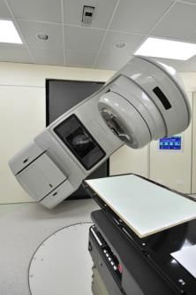
HOW IS BREAST CANCER DIAGNOSED?

Breast cancer can be suspected under different circumstances. The main
circumstances are a positive screening mammography*, the discovery by
palpation* of a mass in the breast, any modification of the skin of the breast
noticed by the patient or her doctor, or any fluid leaking from the nipple of one
single breast.
The diagnosis of breast cancer is based on the three following examinations:

    1. Clinical examination*. The physical examination of the breasts and
       neighbouring lymph nodes* includes inspection and palpation*.

    2. Radiological examination*. This includes conducting an X-ray*,
       (mammography*), and an ultrasound* examination of the breasts
       and neighbouring lymph nodes*. Magnetic resonance imaging*
       (MRI) of the breast may be needed in some patients, especially
       young women with dense breast tissue, women with BRCA gene
       mutations, and women with silicone gel implants. MRI can also be
       considered when tumor cells have been found in a suspicious lymph
       node in the armpit but no tumor can be seen in the breast on
       mammography, or when several tumors are suspected. Additional
       investigations such as chest X-ray*, abdominal ultrasound* and bone
       scintigraphy* may be performed to exclude distant spread of the disease,
       also known as metastasis*.

    3. Histopathological* examination. This is the laboratory examination of the breast and tumor
       tissue after removing a sample from the tumor. This is called a biopsy*. This laboratory
       examination will confirm the diagnosis of breast cancer and will give more information on
       the characteristics of the cancer. The biopsy* is obtained manually
       by the doctor with a needle, often with the help of ultrasound* to
       guide the needle into the tumor. Once the needle is introduced
       into the tumor, a sample is removed. Depending on the needle
       used, this is called either a fine needle aspiration or a core
       biopsy*. A second histopathological examination will be
       performed later when examining the tumor and the lymph nodes*
       removed by surgery.

Breast Cancer: a guide for patients - Information based on ESMO Clinical Practice Guidelines - v.2013.1               Page 7
This document is provided by the Anticancer Fund with the permission of ESMO.
The information in this document does not replace a medical consultation. It is for personal use only and cannot be modified,
reproduced or disseminated in any way without written permission from ESMO and the Anticancer Fund.
COMMON MISCONCEPTIONS ABOUT BREAST CANCER TREATMENT
According to Prof. Martine Piccart, expert in breast cancer treatment:
Breast cancer does not develop within days or weeks! There is always time to seek a second
opinion about treatment options.
The multidisciplinary consultation before starting treatment is very important and should not
be underestimated. The treating physician and the general practitioner should be provided
with the written report of this consultation.
The importance of the pathological examination of the tumor is often underestimated. The
entire treatment strategy depends on a carefully conducted, well standardized pathological
examination in a well-experienced laboratory. Asking for a second, independent pathological
examination is a good idea if the testing has been done in a laboratory with limited
experience in breast cancer diagnosis.
Access to new agents or strategies in the context of well-designed and carefully conducted
clinical trials has more benefits than risks at all stages of the disease. Patients should ask
their doctors which clinical trials are relevant for them.
Pregnancies after breast cancer are possible, especially if the ovaries are not damaged by the
use of certain chemotherapy* drugs which are toxic to them. This needs to be discussed
upfront with young women who want to preserve their fertility. For women who become
pregnant following completion of breast cancer therapy, neither pregnancy nor subsequent
breastfeeding increase the likelihood of relapse*.
Breast Cancer: a guide for patients - Information based on ESMO Clinical Practice Guidelines - v.2013.1 Page 8
This document is provided by the Anticancer Fund with the permission of ESMO.
The information in this document does not replace a medical consultation. It is for personal use only and cannot be modified,
reproduced or disseminated in any way without written permission from ESMO and the Anticancer Fund.
WHAT IS IT IMPORTANT TO KNOW TO GET THE OPTIMAL TREATMENT?
Doctors will need to consider many aspects of both the patient and the cancer in
order to decide on the best treatment.
Relevant information about the patient
Personal medical history
History of cancer in relatives, especially breast cancer and ovarian cancer
Status regarding menopause*, which in some cases requires taking a
blood sample to measure the level of some hormones in the blood
(estradiol* and FSH*)
Results from the clinical examination* by the doctor
General well-being
Results from blood tests performed to assess the white blood cells*, the red blood cells* and
the platelets*, and tests performed to exclude any problems in the liver, the kidneys and the
bones.
Relevant information about the cancer
Staging*
Doctors use staging* to assess the risks and prognosis* associated with specific characteristics of the
patient and the type of cancer involved. The TNM staging* system is commonly used. The
combination of size of the tumor (T) and invasion of nearby tissue, involvement of lymph nodes* (N),
and metastasis* or spread of the cancer (M) to other organs of the body, will classify the cancer into
one of the following stages.
The stage of cancer is fundamental for decisions regarding treatment. The less advanced the stage,
the better the prognosis is. Staging* is usually performed twice: after clinical and radiological
examination* as well as after surgery. If surgery is performed, staging* may also be influenced by the
laboratory examination of the removed tumor and lymph nodes*.
Additional radiological examinations* such as chest X-ray*, abdominal ultrasound* or CT and bone
scintigraphy* can be performed to be sure that there is no metastasis* in the lung, the liver and the
bones. CT and/or MRI of the brain should only be performed if there are symptoms pointing in that
direction. All of these examinations are usually only recommended for stages II or higher (see below).
They are also considered for patients for whom a pre-surgery therapy is planned. Conversely, for
patients with small tumors and no suspicious lymph nodes* (stage I), there is no reason to do all
these exams.
The table below presents the different stages for breast cancer. The definitions are very technical so
it is recommended to ask doctors for more detailed explanations.
Breast Cancer: a guide for patients - Information based on ESMO Clinical Practice Guidelines - v.2013.1 Page 9
This document is provided by the Anticancer Fund with the permission of ESMO.
The information in this document does not replace a medical consultation. It is for personal use only and cannot be modified,
reproduced or disseminated in any way without written permission from ESMO and the Anticancer Fund.Stage Definition
Stage 0 The abnormal cells are still contained in the duct where they initially appeared.
Stage I The tumor is less than 2 cm in diameter and small clusters of cancer cells may
be found in the lymph nodes*.
Stage I breast cancer is divided into stages IA and IB.
Stage II The tumor is either smaller than 2 cm in diameter and has spread to the lymph
nodes* in the armpit or the tumor is between 2cm and 5cm in diameter, but has
not spread to the lymph nodes in the armpit.
Stage II breast cancer is divided into stages IIA and IIB.
Stage III The tumor maybe of any size, but:
- has spread either to the chest wall and/or the skin of the breast
- has spread to at least 10 lymph nodes* in the armpit or the lymph
nodes in the armpit are attached to each other or to other structures
- has spread to lymph nodes near the sternum (breastbone).
- has spread to lymph nodes below or above the clavicle (collar bone).
Stage III breast cancer is divided into stages IIIA, IIIB, and IIIC
Stage IV Cancer has spread to other organs of the body, most often the bones, lungs,
liver, or brain. Such distant tumor deposits are called metastases*.
Results of the biopsy*
Tumor obtained by biopsy* will be examined in the laboratory. The method and the result of such an
examination are called histopathology*. A second histopathological examination is performed on the
tissues obtained by surgical removal of the tumor and the lymph nodes*. This is very important to
confirm the results of the biopsy* and to provide further information on the cancer. Results of the
examination of the biopsy* should include:
o Histological type*
Assignment of histological type is based on the type of cells that compose the tumor.
Breast cancers form in tissues of the breast, usually the ducts or the lobules. The main
histological types of breast cancer are ductal carcinomas* and lobular carcinomas*. The
histopathological examination will also classify the cancer as invasive* or non-invasive*.
Non-invasive* cancers are also called cancer in situ
o Grade*
Assignment of grade* is based on the heterogeneity of tumor cells, the architectural
structure of the tissue they form and the frequency of mitosis* (cell division) of tumor
cells. A well differentiated tumor (grade 1) has low heterogeneity of cells, preserved
architectural structure and few mitoses. An undifferentiated tumor (grade 3) has high
heterogeneity, loss of architecture and many mitoses. A moderately differentiated tumor
(grade 2) is in between grade 1 and grade 3. The lower the grade*, the better the
prognosis*.
When systemic treatment is planned before surgery, the biopsy results should include hormone
receptor* status and HER2* status. When no systemic treatment is planned before surgery, these
can be determined in the tumor (and/or the lymph nodes*) after their removal by surgery.
o Hormone receptor* status for estrogen and progesterone
Tumor cells can present receptors to estrogen and receptors to progesterone on their
surface or inside the cell. Cells of some tumors present a high level of receptors. This
means that their growth and multiplication are stimulated by hormones. Tumors with a
Breast Cancer: a guide for patients - Information based on ESMO Clinical Practice Guidelines - v.2013.1 Page 10
This document is provided by the Anticancer Fund with the permission of ESMO.
The information in this document does not replace a medical consultation. It is for personal use only and cannot be modified,
reproduced or disseminated in any way without written permission from ESMO and the Anticancer Fund.high level of estrogen receptors (ER+)* and/or progesterone receptors* (PR+) have a
better prognosis* than tumors lacking estrogen receptors (ER-) and/or lacking
progesterone receptors (PR-).
o HER2* status
HER2* is a cell surface protein* present in about 20% of breast cancer cases. HER2 is
involved in the growth and migration of cells.*. HER2* status of tumor tissue can be
analysed by various tests in the laboratory: by Immunohistochemistry* (IHC), by
Fluorescence In Situ Hybridization* (FISH) or by Chromogenic In Situ Hybridization*
(CISH). A cancer is HER2* positive when the result of the IHC test is 3+ or the result of a
FISH or CISH test is positive as stated in the pathology report. Otherwise, the HER2*
status is negative. Before anti-HER2 directed therapy was available, HER2* positive
cancers had more aggressive behaviour than other cancers.
o Multigene expression profiles*
The quantification of the expression of distinct sets of genes expressed by the tumor can
also be performed on the biopsy*. Such multigene signature analyses are not routinely
performed, but can help to predict the risk of recurrence* and the likelihood of benefit
from chemotherapy*.
o Ki-67 labelling index
Ki-67 is a protein* found in the nucleus* of cells when they are dividing but not when
they rest. Ki-67 labelling index indicates the percentage of cells in which Ki-67 can be
found. Analysing the proportion of dividing cells is a method to determine the level of
proliferation* of the tumor. Highly proliferating tumors grow faster and have a worse
prognosis than slowly proliferating tumors, but at the same time highly proliferating
tumors are more sensitive to chemotherapy*.
It is important to know that tests used to define hormone receptor* status and HER2* status may
give an incorrect result. No test used to assess HER2 status today is 100% reliable. Moreover, it is also
possible that the piece of tissue examined classifies the tumor as HER2 negative but examination of
another piece of the tumor would have classified it as HER2 positive. That is why, whenever possible,
these analyses should be performed on both the biopsy* material and on the tumor tissue removed
by surgery.
Another very important part of the histopathological examination after tumor removal by surgery is
to check whether the tumor has been completely removed. This is done by analyzing if the
microscopic edges of the tumor are surrounded completely by normal tissue. It is reported either as
negative margins* of resection, (meaning that it is very likely that the whole tumor has been
removed) or as positive margins* of resection (meaning that it is very likely that the tumor has not
been removed completely).
Hormone responsiveness
Based on the analysis of the biopsy and/or of the tumor removed by surgery, tumors are classified
into three groups according to their hormonal receptor status:
o Hormone responsive* (ER+ and/or PR+) when estrogen or progesterone receptors*
have been detected on cancer cells.
o Hormone non-responsive (ER- and PR-) when no estrogen and no progesterone
receptors have been detected on cancer cells.
o A third in-between group with uncertain hormone responsiveness*.
Breast Cancer: a guide for patients - Information based on ESMO Clinical Practice Guidelines - v.2013.1 Page 11
This document is provided by the Anticancer Fund with the permission of ESMO.
The information in this document does not replace a medical consultation. It is for personal use only and cannot be modified,
reproduced or disseminated in any way without written permission from ESMO and the Anticancer Fund.Based on this analysis the decision is made whether to add hormone treatment. A hormonal
treatment will usually stop or slow the growth of hormone responsive* tumors because these
tumors need hormones to grow, but will have no effect on the growth of hormone non-responsive
tumors.
Intrinsic subtypes of breast cancer
The combination of the results regarding hormone receptor status, HER2 status and Ki-67 labelling
index is used to classify breast cancer in 5 subtypes. This is also important in order to know which
therapies are most likely to be effective. The 5 subtypes are presented in the table below. This
classification is rather technical and it is recommended to ask a doctor for a more detailed
explanation.
Subtype of breast cancer Hormone receptor status HER2 status Ki-67 status
Luminal A ER+ and/or PR+ HER2 negative Low (WHAT ARE THE TREATMENT OPTIONS?
Planning of the treatment involves an inter-disciplinary team of medical
professionals. This usually implies a meeting of different specialists, called
multidisciplinary opinion* or tumor board. In this meeting, the planning of
treatment will be discussed based on the relevant information summarized
above.
The treatment will usually combine intervention methods that:
act on the cancer locally, such as surgery or radiotherapy*
act on cancer cells all over the body with systemic therapy* such as chemotherapy*,
hormone therapy *and/or HER2-directed therapy.
The extent of the treatment will depend on the characteristics of the tumor cells and on the stage of
the cancer, as well as on the age, the menopausal status and the co-morbidity of the patient.
Treatments listed below have their benefits, their risks and their contraindications. It is
recommended to ask an oncologist about the expected benefits and risks of every treatment in order
to be informed of the consequences of the treatment. For some treatments, several options are
available. The choice should be discussed according to the balance between benefits and risks.
Treatment plan for non-invasive* cancer (Stage O)
A non-invasive* cancer has not spread outside the duct (ductal carcinoma in situ). Treatment options
include the following two possibilities of local therapies.
Either the tumor or a part of the breast is removed, but not the whole breast. This is called
breast-conserving surgery. This is usually followed by whole breast irradiation except in
patients with very low risk of recurrence where radiation may be omitted. Additional
irradiation (called a boost) of the area from which the tumor has been removed can be
considered for patients who have a high risk of local recurrence, for instance in very young
patients.
Or the whole breast is removed by mastectomy, without muscles and skin surrounding the
breast. When mastectomy is performed, additional radiation treatment is not necessary for
non-invasive cancer.
In addition, treatment with tamoxifen*, a drug which counteracts the action of estrogens on the
breast, can be considered if the tumor is estrogen receptor positive*, since it lowers the risk of
recurrence, i.e. that the cancer comes back in the breast. Tamoxifen also lowers the risk of
developing contralateral breast cancer, i.e. cancer in the opposite breast.
Lobular neoplasia*, which was formerly called lobular carcinoma in situ, is now regarded as a risk
factor for future development of breast cancer in both breasts. It therefore requires a discussion with
the doctor whether to pursue a surveillance strategy by close follow-up and/or a treatment strategy.
Breast Cancer: a guide for patients - Information based on ESMO Clinical Practice Guidelines - v.2013.1 Page 13
This document is provided by the Anticancer Fund with the permission of ESMO.
The information in this document does not replace a medical consultation. It is for personal use only and cannot be modified,
reproduced or disseminated in any way without written permission from ESMO and the Anticancer Fund.Treatment plan for invasive* cancer (Stage I to III)
An invasive* cancer has spread outside the duct (invasive ductal carcinoma) or outside the lobule
(invasive lobular carcinoma). The treatment will target the cancer locally as well as cancer cells
potentially spread in the body.
In most cases, the treatment will consist of surgery, radiotherapy* and systemic therapy*. Treating
cancer cells that have spread to other parts of the body can be done with the help of hormone
therapy, chemotherapy* or HER2-directed therapy.
For tumors of more than 2 cm in diameter, systemic therapy* is sometimes preferred as the first
treatment since shrinkage of the tumor with drugs can facilitate local therapy and might permit
breast conservation. Surgery is preceded by chemotherapy* for most cases of stage IIIA and IIIB
cancers. This is called neo-adjuvant* chemotherapy. It is also indicated to reduce tumor size so as to
permit breast-conserving surgery. Trastuzumab* is added in cases with HER2*-positive tumors.
Surgery
The surgery will be performed under general anaesthesia*. The surgeon will
remove the tumor and some lymph nodes* during the same operation by
one of two methods.
Removal of the tumor or a part of the breast including the tumor,
but not the whole breast. This is called breast-conserving surgery.
Removal of the whole breast but not muscles and skin surrounding
the breast. This is called total mastectomy.
The choice between breast-conserving surgery and total mastectomy
depends on the characteristics of the tumor, on the size of the breast and on the patient’s
preference. Some patients require a mastectomy because of tumor size, multiple locations of the
tumor(s) in the breast or other reasons. This has to be discussed with doctors. Currently in Western
Europe, breast-conserving surgery can be performed in 2 out of 3 women with breast cancer.
For some patients, a treatment is given before surgery (neo-adjuvant) with the intent of reducing the
size of the tumor and allowing for breast-conserving surgery. Once the neo-adjuvant treatment has
produced its effect, the doctor will ask for an MRI to check whether it will indeed be possible to
conserve the breast without decreasing the chances of cure. In some cases, the complete removal of
the breast will still be necessary.
For women requiring mastectomy, a breast reconstruction may be recommended. This
reconstruction can be immediate or delayed (for medical reasons or for personal preference). It is
not necessary that patients should wait 2 years after mastectomy before being offered
reconstruction. It is also not true that reconstruction of the affected sites makes detection of
recurrence* of cancer more difficult.
One or several lymph nodes* in the armpit will also be removed
Breast Cancer: a guide for patients - Information based on ESMO Clinical Practice Guidelines - v.2013.1 Page 14
This document is provided by the Anticancer Fund with the permission of ESMO.
The information in this document does not replace a medical consultation. It is for personal use only and cannot be modified,
reproduced or disseminated in any way without written permission from ESMO and the Anticancer Fund.This removal is very important to know whether the cancer has spread to lymph nodes, but it has a
limited effect in treating the cancer. Two types of surgery of the lymph nodes can be performed:
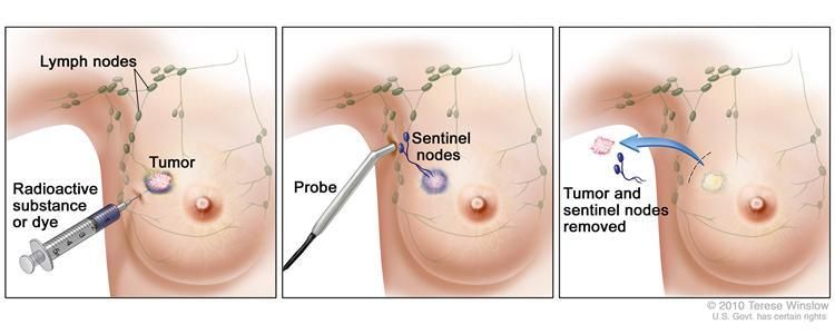
The surgeon performs a sentinel lymph node* biopsy*. After injection of a marker near the
tumor, the marker will naturally be led to lymphatic vessels and then to lymph nodes. With
the help of a probe, the surgeon will be able to identify in which lymph node(s) the marker is
located. He/she will remove the lymph node(s) to check if cancer cells are present. A rapid
examination of the lymph nodes will be made while the patient is still in surgery. If cancer
cells are found in the lymph node(s), the surgeon will usually perform an axillary dissection*
(see below). For patients with tumors of less than 5 cm in diameter, axillary dissection might
not be necessary if the examination shows that only 1 or 2 sentinel lymph nodes contain
cancer cells.
The surgeon performs an axillary dissection*. The surgeon makes an incision under the arm
and removes the axillary soft tissue where lymph nodes are located. These lymph nodes will
be checked for the presence of cancer cells.
Sentinel lymph node* biopsy* causes less arm swelling (lymphedema) and shoulder stiffness than
axillary dissection*. Sentinel lymph node biopsy is recommended in stage I and stage II breast cancer,
unless involved lymph nodes can be detected preoperatively on physical examination or by
ultrasound*. In higher stages, an axillary dissection will be performed.
Laboratory examination of the tumor and lymph nodes* removed by surgery
Once the tumor and lymph nodes have been removed, they will be examined in the laboratory to:
Confirm the results of the biopsy* regarding histological type*, grade*, hormone receptor*
status, HER2* status and possibly multigene expression profile*.
Measure the size of the tumor and see if it has spread to surrounding tissues.
Check whether cancer cells have entered lymphatic vessels or blood vessels, which would
make it more likely that they have spread outside of the breast.
Check whether the whole tumor has been removed and margins are free of tumor tissue*.
Check whether cancer cells have spread to the lymph nodes and count the number of lymph
nodes affected.
Second surgery
Some patients may be operated on a second time. The two main reasons are:
The margins* of resection were positive; the tumor is not completely surrounded by normal
tissue. The new operation should remove the rest of the tumor.
After a more thorough examination of the lymph node(s)* from the sentinel lymph node
biopsy*, it turns out that they contain cancer cells. An axillary dissection* will usually be
performed. For patients with tumors of less than 5 cm in diameter, axillary dissection might
not be necessary if the examination shows that only 1 or 2 sentinel lymph nodes contain
cancer cells.
Adjuvant therapy
Breast Cancer: a guide for patients - Information based on ESMO Clinical Practice Guidelines - v.2013.1 Page 15
This document is provided by the Anticancer Fund with the permission of ESMO.
The information in this document does not replace a medical consultation. It is for personal use only and cannot be modified,
reproduced or disseminated in any way without written permission from ESMO and the Anticancer Fund.An adjuvant therapy* is a therapy given in addition to surgery. For patients with stage I to III breast
cancer, possible adjuvant therapies are radiotherapy*, chemotherapy*, hormone therapy and
targeted therapy*. In this setting, radiotherapy is a local treatment whereas chemotherapy, hormone
therapy and targeted therapy can reach cancer cells that may have spread to other parts of the body.
These latter treatments are called systemic therapies.
Radiotherapy*
Radiotherapy is the use of radiation to kill cancer cells. Generally, cancer cells are less capable of
recovery from radiation damage than normal cells.
Radiotherapy is recommended for almost all invasive* breast cancers. A limited number of patients
may not benefit from radiotherapy, which could therefore be omitted. This concerns patients of
more than 70 years of age who have a tumor of less than 2 cm in diameter that is hormone-
responsive. In addition, one should be sure that the whole tumor has been removed by surgery with
negative margins.
Radiotherapy in breast cancer aims to destroy cancer cells locally using high-energy radiation
produced by a radiotherapy device.
After breast-conserving surgery radiotherapy is strongly recommended for all patients:
radiotherapy of the whole breast, followed by additional irradiation (called a boost) of the
area from which the tumor has been removed.
After mastectomy radiotherapy is recommended or should be considered for patients with
large tumors and/or for whom cancer cells have been found in axillary lymph nodes. The
radiotherapy will target the chest wall and sometimes the regional lymph node areas as well.
When there is a clear and extensive spread of cancer cells to the lymph nodes over the collar
bone or behind the breastbone (sternum), the field of irradiation can be extended to include
these areas.
The dose of radiation to be delivered is between 45 and 50 Grays (Gy). A Gray is the unit used to
measure the quantity of radiation delivered in radiotherapy. This total dose is divided into fractions.
Each fraction is given during one session of radiotherapy. Typically for breast cancer, 25 to 28
fractions are planned, but a shorter treatment using 16 fractions has shown the same efficacy
without increased side effects. When a boost is planned, an additional 10 to 16 Gy is administered in
fractions of 2 Gy. The goal of giving the treatment in fractions is to lower the risk of significant
damages to normal tissues and to increase the probability of long-term tumor control.
In order to shorten the time of treatment duration and to avoid the patient
having to come between 16 and 35 times to the radiotherapy unit, attempts
have been made to deliver radiotherapy during surgery. This has been called
accelerated partial breast irradiation. Research is ongoing but preliminary
results suggest that this could be considered for patients of at least 50 years of
age, with a single tumor of less than 3 cm in diameter and resection margins of
more than 2 mm, and no spread to the lymph nodes*. In addition, the tumor
should have specific histological characteristics (non-lobular histology without
any intraductal component and no lymphovascular invasion). This type of
radiotherapy requires specific devices that are not available in many centres
because research is still on-going.
Systemic therapy*
Breast Cancer: a guide for patients - Information based on ESMO Clinical Practice Guidelines - v.2013.1 Page 16
This document is provided by the Anticancer Fund with the permission of ESMO.
The information in this document does not replace a medical consultation. It is for personal use only and cannot be modified,
reproduced or disseminated in any way without written permission from ESMO and the Anticancer Fund.The goal of systemic therapy* is to act on cancer cells that might have reached other parts of the
body.
The characteristics of the tumor tissue identified by laboratory examination of the biopsy* and of
surgically removed tumor are essential to decide which therapy or combination of therapies is most
appropriate. These tumor characteristics include tumor size, histological type*, grade*, margins* of
resection, number of lymph nodes* involved, hormone receptor* status, HER2* status and, if
available, multigene expression profile*. Age, menopausal status and medical conditions of the
patient are the patient factors important for making an informed decision regarding adjuvant
systemic treatment.
For each individual, the choice must take into account the potential benefits, the possible side effects
and the patient’s preference.
Three types of treatment can be used for systemic therapy*: hormonal therapy, chemotherapy* and
HER2-directed therapy.
Tumors are classified into three groups according to hormonal receptor status: hormone responsive*
(ER+ and/or PR+), hormone non-responsive (ER- and PR-) and a third in-between group with
uncertain hormone responsiveness*. A hormonal treatment will usually stop or slow the growth of
hormone responsive* tumors because these tumors need hormones to grow, but will have no effect
on the growth of hormone non-responsive tumors.
Patients with hormone responsive* tumors may receive either hormone therapy alone or a
combination of hormone therapy and chemotherapy*.
Patients with tumors of uncertain hormone responsiveness* may receive a combination of
hormone therapy and chemotherapy.
Patients with hormone non-responsive tumors should receive chemotherapy, but no
hormone therapy.
Hormone therapy
This therapy consists of one or possibly a combination of two of the following treatments:
A drug called tamoxifen* which counteracts the action of estrogens on the breast andis
active in both premenopausal and in postmenopausal patients
A drug from the aromatase inhibitor* family like anastrozole, exemestane or letrozole which
inhibit the production of estrogens in post-menopausal women
A drug from the gonadotropin*-releasing hormone analogues family that lower the level of
estrogens in pre-menopausal women
Ovariectomy - the removal of the ovaries in premenopausal women
The choice of hormone therapy is based on the menopausal status of the patient.
For patients in whom menopause has not yet begun (pre-menopausal patients), tamoxifen* alone for
5 years, or the combination of a bilateral ovariectomy or a drug from the gonadotropin*-releasing
Breast Cancer: a guide for patients - Information based on ESMO Clinical Practice Guidelines - v.2013.1 Page 17
This document is provided by the Anticancer Fund with the permission of ESMO.
The information in this document does not replace a medical consultation. It is for personal use only and cannot be modified,
reproduced or disseminated in any way without written permission from ESMO and the Anticancer Fund.hormone analogue family plus tamoxifen for 5 years, are the usual treatments. Tamoxifen should not be used simultaneously with chemotherapy. For patients after their menopause (post-menopausal patients), aromatase inhibitors* for 5 years is preferred for women at higher risk, but for patients treated with tamoxifen, a switch after 2-3 years to aromatase inhibitor for 2-3 years could be considered. Patients treated with aromatase inhibitors are at higher risk of developing osteoporosis*. This should be counteracted by sufficient intake of calcium and vitamin D*. Other examinations such as measurement of bone mineral density and treatments such as bisphosphonates* are available to deal with osteoporosis. Tamoxifen slightly increases the risk of blood clots and should be stopped if a surgical intervention is planned. It also doubles the risk of developing endometrial cancer (a cancer of the uterus). Chemotherapy* Chemotherapy for early-stage breast cancer consists of combining two or three anti-cancer drugs, which are given according to a precise protocol. For breast cancer, the treatment is generally given for 4 to 8 cycles, a cycle being a time period of 2 to 4 weeks with a precise dosage, duration and sequence of drugs including a resting period before a new cycle is started. It is not clear which combination of drugs is best, but it is recommended that it contains doxorubicin* or epirubicin*, which are anti-cancer drugs from the anthracycline family*. Assessment of heart function is important before therapy with anthracyclines. However, regimens without any anthracycline have been shown to be as effective, for instance the combination of docetaxel* and cyclophosphamide*. Treatments are often named with acronyms using the initial letter of each drug name (e.g. FEC, stands for the combination of Fluorouracil, Epirubicin and Cyclophosphamide). For frail or elderly patients the CMF (Cyclophosphamide, Methotrexate and Fluorouracil) regimen may still be appropriate. Another option, especially for women in whom tumor cells have spread to the lymph nodes, is to combine an anthracycline* (doxorubicin* or epirubicin*) with a taxane* drug (paclitaxel*), preferably given in sequence rather than in combined fashion. HER2*-directed therapy HER2-directed systemic treatments are used for HER2* positive cancers, i.e. when the result of the laboratory examination reports that the IHC* test is “3+” or the FISH* or CISH test is “positive”. Trastuzumab is* a drug effective in patients with HER2* positive tumors, regardless of the size of the tumor and of its hormonal status. In the studies performed to evaluate its efficacy as an adjuvant therapy, trastuzumab was always given in combination with chemotherapy. It is not clear, whether the adjuvant use of trastuzumab without chemotherapy has a positive effect. * The standard recommended duration of adjuvant treatment with trastuzumab is 1 year. Results from studies comparing this standard duration to shorter or to longer durations are pending. Trastuzumab can be given together with paclitaxel* or carboplatin* but should not be given together with doxorubicin* or epirubicin*. The latter two drugs and trastuzumab are both toxic to the heart. Trastuzumab cannot be given to patients whose heart function is abnormal. If there is doubt about the heart function, it should be assessed before trastuzumab treatment. Breast Cancer: a guide for patients - Information based on ESMO Clinical Practice Guidelines - v.2013.1 Page 18 This document is provided by the Anticancer Fund with the permission of ESMO. The information in this document does not replace a medical consultation. It is for personal use only and cannot be modified, reproduced or disseminated in any way without written permission from ESMO and the Anticancer Fund.
Treatment plan for metastatic cancer (Stage IV)
A metastatic* breast cancer is one that has spread to other parts of the body. The most frequent
locations of metastases in breast cancer are bone, the liver, the lungs and the brain. Since tumor cells
have spread to other parts of the body, systemic therapy* is the mainstay of treatment. About 5% of
women with breast cancer have metastases at the time of diagnosis.
For the treatment of patients with metastatic breast cancer:
The main treatment goal is to maintain or improve quality of life. Patients should be offered
appropriate psychological, social and supportive care.
The realistic treatment goals should be discussed with the patient and her family and the
patient should be encouraged to actively participate in all decisions. The patient’s
preferences should always be taken into account, including preferences relating to the
practicalities of the treatment (for instance, oral or intravenous*).
In many hospitals, specialist breast nurses can provide crucial support to patients and should be
available to all patients.
Surgery and radiotherapy*
Some patients exhibiting metastases*may benefit from having the primary breast tumor removed by
surgery or treated by radiotherapy. In some rare cases, surgery can also be used to treat patients
with a single or very few metastases e.g. in the liver, in the lung or in the brain. Radiotherapy is also
used in the management of bone and brain metastases.
Systemic therapy*
The goal of systemic therapy* is to simultaneously act on cancer cells in various organs involved in
metastases*. Systemic therapy* options are the same as for invasive* cancer without metastasis
(hormone therapy, chemotherapy* and HER2-directed therapy) with a few additional targeted
biological agents such as bevacizumab* or everolimus*. If chemotherapy is used, its composition and
duration should be tailored to the individual patient.
The choice of the systemic therapy* depends on the hormone receptor* status, on the HER2* status,
on the urgency of obtaining a response and on prior therapies and their effectiveness.
Hormone therapy
Hormone therapy is the treatment of choice for patients with hormone responsive* (ER+ and/or PR+)
metastatic* breast cancer. The choice of the hormone therapy depends on the menopausal status
and on previous hormone therapy applied.
For patients before their menopause
o If there has been no prior treatment with tamoxifen* or if the use of
tamoxifen has been discontinued for more than 12 months, tamoxifen with
either gonadotropin*-releasing hormone analogues or an ovariectomy is the
preferred option.
o Otherwise, aromatase inhibitors* like anastrozole, exemestane or letrozole in
combination with either gonadotropin-releasing hormone analogues or an
Breast Cancer: a guide for patients - Information based on ESMO Clinical Practice Guidelines - v.2013.1 Page 19
This document is provided by the Anticancer Fund with the permission of ESMO.
The information in this document does not replace a medical consultation. It is for personal use only and cannot be modified,
reproduced or disseminated in any way without written permission from ESMO and the Anticancer Fund.ovariectomy. Calcium and vitamin D
supplements are recommended in addition to
this treatment.
For patients after their menopause
o If there has been no prior treatment with
aromatase inhibitors* like anastrozole,
exemestane or letrozole or treatment with them has been discontinued for
more than 12 months, they are the preferred option. Calcium and vitamin D*
supplements are recommended in addition to this treatment.
o Otherwise, tamoxifen, fulvestrant*, megestrol* or androgens* can be used.
o When there are signs that the cancer progresses or comes back despite
treatment with anastrozole or letrozole, an option is to use a combination of
exemestane and everolimus*. Combining tamoxifen* and everolimus* may
also be an option, but cannot yet be proposed in Europe.
Cancers change over time and it is possible that an ER+ cancer becomes ER- or that an ER+ cancer
becomes otherwise resistant to hormone therapy.
Patients with clear evidence of resistance to hormone therapy should be offered chemotherapy or
participation in clinical trials.
HER2-directed therapy
HER2*-directed therapy, such as trastuzumab* or lapatinib* should be offered early to all patients
with HER2* positive metastatic disease, in addition to chemotherapy*, to hormone therapy, or
alone. This should be the case in patients who didn’t receive such therapy in the adjuvant treatment
and who do not have contraindications for it (e.g. heart insufficiency). If the cancer continues to
expand and to progress under a trastuzumab treatment, trastuzumab may be continued with a
different chemotherapy. Lapatinib*, an oral drug that also targets the HER2* receptor, can be given
in combination with the oral chemotherapy drug capecitabine*. The choice of treatment should be
discussed with an oncologist. Two new drugs, namely pertuzumab* and ado-trastuzumab
emtansine*, may soon be available in Europe for patients with HER2*-positive tumors.
Chemotherapy*
Chemotherapy should be offered to:
Patients with fast-growing tumors involving vital organs (e.g. extensive liver
involvement), where an immediate response to systemic treatment is necessary.
Patients with cancers that are both hormone non-responsive and HER2* negative.
Such cancers are called “triple negative” (ER-, PR- and HER2*-) and for these
chemotherapy is the main treatment option.
Patients with hormone-responsive* cancers that do not respond to hormone
therapy, or that have ceased to respond to hormone therapy.
If patients have previously received chemotherapy with anthracyclines* (epirubicin* or
doxorubicin*), they should be offered chemotherapy including a taxane* (paclitaxel* or docetaxel*).
Single drug chemotherapy is mostly preferred over a combination of drugs because it is associated
with a better quality of life without a decrement in survival duration. Duration of chemotherapy
Breast Cancer: a guide for patients - Information based on ESMO Clinical Practice Guidelines - v.2013.1 Page 20
This document is provided by the Anticancer Fund with the permission of ESMO.
The information in this document does not replace a medical consultation. It is for personal use only and cannot be modified,
reproduced or disseminated in any way without written permission from ESMO and the Anticancer Fund.should be tailored to the individual patient. In general, in patients with triple-negative tumors, metastases* may be more frequent and disease progression more rapid. Therefore, the combination with chemotherapy* may be offered. Continuing chemotherapy after the patient has received 3 different types of regimens is possible for patients who are in good general condition and whose tumor has “responded” (shown shrinkage) to previous chemotherapy. Other biologic therapies Bevacizumab* is a drug that is thought to limit the development of new vessels around the tumor. In Europe, it is now available only for patients with metastatic* breast cancer in combination with first- line chemotherapy* (paclitaxel* or capecitabine*). This combination could be considered in selected patients with limited treatment options, but only upon evaluation of possible side effects and expected benefits. Bevacizumab* is not anymore authorized for patients with breast cancer in the USA. Other therapies Radiotherapy* can be used as a palliative therapy for the management of bone metastases*, brain metastases or other local tumor masses such as fungating soft tissue* lesions. Bisphosphonates* should be used for the treatment of hypercalcemia* and when bone metastases are present. The goal is to relieve pain and to prevent consequences of bone metastases, such as fractures. Bisphosphonates* exist in oral or intravenous* forms. They are generally well tolerated, but in rare instances they can induce a complication called osteonecrosis* of the jaw. These are lesions of the upper or lower jaw bones with bone denudation that take a long time to heal. This complication occurs more often in patients with poor dental conditions. It is therefore recommended to have a dental check-up prior to a bisphosphonate* treatment. Denosumab is a new therapy used for bone metastases. It seems to be slightly more efficacious than bisphosphonates in preventing bone complications, and has also less kidney toxicity. Like bisphosphonates, denosumab can also cause osteonecrosis of the jaw. Clinical trials Clinical trials of new drugs are often proposed to patients with metastatic cancer. Participation in clinical trials should be encouraged since they are the only way to make progress in a context where cure remains extremely rare. Response evaluation The response to treatment has to be evaluated in order to weigh the benefit of the treatment against the adverse events experienced. This response evaluation is recommended after 2-3 months of hormone therapy and after 2- 3 cycles of chemotherapy*. The evaluation relies on clinical and symptom evaluation, assessment of quality of life, blood tests and repeating the initially abnormal radiological examinations* with comparative measurements. Breast Cancer: a guide for patients - Information based on ESMO Clinical Practice Guidelines - v.2013.1 Page 21 This document is provided by the Anticancer Fund with the permission of ESMO. The information in this document does not replace a medical consultation. It is for personal use only and cannot be modified, reproduced or disseminated in any way without written permission from ESMO and the Anticancer Fund.
You can also read