Ovarian cancer? What is Let us explain it to you.
←
→
Page content transcription
If your browser does not render page correctly, please read the page content below
Ovarian Cancer

                        What is
                    ovarian cancer?

                                        Let us explain
                                           it to you.

www.anticancerfund.org   www.esmo.org

                                                ESMO/ACF Patient Guide Series
                                            based on the ESMO Clinical Practice Guidelines
The ESMO / Anticancer Fund Guides for Patients are designed to
                   assist patients, their relatives and caregivers to understand the nature
                   of different types of cancer and evaluate the best available treatment
                   choices. The medical information described in the Guides for Patients
                   is based on the ESMO Clinical Practice Guidelines, which are
                   designed to guide medical oncologists in the diagnosis,
                   follow-up and treatment in different cancer types.
                   These guides are produced by the Anticancer Fund in
                   close collaboration with the ESMO Guidelines Working
                   Group and the ESMO Cancer Patient Working Group.

                   For more information please visit www.esmo.org
                   and www.anticancerfund.org

www.anticancerfund.org      www.esmo.org
OVARIAN CANCER: A GUIDE FOR PATIENTS

     PATIENT INFORMATION BASED ON ESMO CLINICAL PRACTICE GUIDELINES
This guide for patients has been prepared by the Anticancer Fund as a service to patients, to help
patients and their relatives better understand the nature of ovarian cancer and appreciate the best
treatment choices available according to the subtype of ovarian cancer. We recommend that
patients ask their doctors about what tests or types of treatments are needed for their type and
stage of disease. The medical information described in this document is based on the clinical practice
guidelines of the European Society for Medical Oncology (ESMO) for the management of ovarian
cancer. This guide for patients has been produced in collaboration with ESMO and is disseminated
with the permission of ESMO. It has been written by a medical doctor and reviewed by two
oncologists from ESMO including the lead author of the clinical practice guidelines for professionals.
It has also been reviewed by patient representatives from ESMO’s Cancer Patient Working Group.

More information about the Anticancer Fund: www.anticancerfund.org

More information about the European Society for Medical Oncology: www.esmo.org

For words marked with an asterisk, a definition is provided at the end of the document.

Ovarian cancer: a guide for patients - Information based on ESMO Clinical Practice Guidelines - v.2014.1              Page 1
This document is provided by the Anticancer Fund with the permission of ESMO.
The information in this document does not replace a medical consultation. It is for personal use only and cannot be modified,
reproduced or disseminated in any way without written permission from ESMO and Reliable Cancer Therapies.
Table of contents

Definition of ovarian cancer .................................................................................................................... 3

Is ovarian cancer frequent? ..................................................................................................................... 3

What causes ovarian cancer? .................................................................................................................. 5

How is ovarian cancer diagnosed? .......................................................................................................... 8

What is it important to know to define the optimal treatment? .......................................................... 12

What are the treatment options? ......................................................................................................... 19

What are the possible side effects of the treatments? ......................................................................... 24

What happens after the treatment? ..................................................................................................... 28

Definitions of medical terms ................................................................................................................. 32

This text was written by Dr.An Billiau, Celsus Medical Writing LLC (for the Anticancer Fund) and reviewed by Dr. Svetlana
Jezdic (ESMO), Dr. Nicoletta Colombo (ESMO), Pr. Cristiana Sessa (ESMO) and Ruth Payne (Ovacome UK).

This is the third update of this guide. Updates reflect changes in the successive versions of the ESMO Clinical Practice
Guidelines. The update was done by Dr. Gauthier Bouche (Anticancer Fund) and was reviewed by Dr. Svetlana Jezdic (ESMO)
and by Pr. Cristiana Sessa (ESMO) and by Ruth Payne (Ovacome UK).

Ovarian cancer: a guide for patients - Information based on ESMO Clinical Practice Guidelines - v.2014.1                                   Page 2
This document is provided by the Anticancer Fund with the permission of ESMO.
The information in this document does not replace a medical consultation. It is for personal use only and cannot be modified,
reproduced or disseminated in any way without written permission from ESMO and Reliable Cancer Therapies.
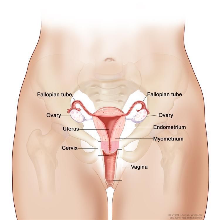
DEFINITION OF OVARIAN CANCER

Ovarian cancer is a cancer that forms in tissues of the ovary. The ovaries are the female reproductive
organs in which the egg cells (ova) are formed and female hormones are produced. Ovarian cancers
either arise from the cells of the surface of the ovary (the ovarian epithelium*), a form called
epithelial ovarian cancer (also referred to here as ovarian carcinoma) or from other tissues within
the ovary (non-epithelial ovarian cancer). Both terms refer to a very diverse group of different
ovarian cancer subtypes.
The most frequent type of ovarian cancer is epithelial, which accounts for approximately 90% of
primary ovarian tumours. In the group of uncommon non-epithelial cancer, two subtypes appear
more frequently, namely malignant* germ cell tumours and sex cord stromal tumours.

Anatomy of the female reproductive system showing the ovaries, the Fallopian tubes, the uterus and the vagina. The
ovaries are the organs where the egg cells are formed and female hormones are produced. Egg cells migrate through the
Fallopian tubes to the uterus, where the foetus develops during pregnancy.

IS OVARIAN CANCER FREQUENT?

Ovarian cancer is the seventh most common cancer and cause of cancer deaths in women in the
world.
The worldwide incidence* of ovarian cancer shows strong geographical variation. Developing
countries have the lowest incidence. In the European Union, it was estimated that, in 2008, a total of
45,300 women were diagnosed with ovarian cancer. The overall probability for a woman in the
European Union in developing ovarian cancer during her lifetime lies between 0.64% and 1.6%.
Approximately half of the women diagnosed with ovarian cancer are aged 60 or older. However,
younger women can be affected, mainly by two rare types of non-epithelial ovarian cancer called
germ cell and sex cord-stromal tumours. Germ cell tumours are diagnosed principally in the first two
decades of life, whereas sex cord-stromal tumours are more common in adult women (granulosa

Ovarian cancer: a guide for patients - Information based on ESMO Clinical Practice Guidelines - v.2014.1              Page 3
This document is provided by the Anticancer Fund with the permission of ESMO.
The information in this document does not replace a medical consultation. It is for personal use only and cannot be modified,
reproduced or disseminated in any way without written permission from ESMO and Reliable Cancer Therapies.

adult type has an average age at diagnosis of 50 years, 90% of juvenile type occurs in pre-pubertal
girls and Sertoli*-Leydig*occurs mainly in women younger than 40 years).

Ovarian cancer: a guide for patients - Information based on ESMO Clinical Practice Guidelines - v.2014.1              Page 4
This document is provided by the Anticancer Fund with the permission of ESMO.
The information in this document does not replace a medical consultation. It is for personal use only and cannot be modified,
reproduced or disseminated in any way without written permission from ESMO and Reliable Cancer Therapies.

WHAT CAUSES OVARIAN CANCER?

Today, the cause of ovarian cancer is not well understood. Importantly, the term ovarian cancer
refers to a very diverse group of different types of malignant* ovarian tumours, and the cause of the
different tumour types may not be the same. A number of risk factors for ovarian cancer have been
identified. Some of these risk factors are specific for selected subtypes of ovarian cancer.
In many cases, however, none of the risk factors are apparently present. A risk factor increases the
risk of cancer occurring, but is neither necessary nor sufficient to cause cancer. A risk factor is not a
cause in itself.

Some people who have one or more risk factors will never develop ovarian cancer and some
people without any of these risk factors may nonetheless develop ovarian cancer.

Up to 90% of all ovarian cancer cases are sporadic ovarian cancer. This means that they are not
associated with inherited genetic mutations*.
The risk of developing a sporadic form of ovarian cancer principally relates to the total number of
ovulatory cycles that have taken place in the ovaries during the reproductive years of a woman. An
ovulatory cycle is the monthly stimulation of the ovary leading to the release of an egg cell
(ovulation). The total number of ovulatory cycles taking place in a woman’s ovaries between
menarche* and menopause* (the so-called reproductive years) constitute the ‘lifetime number of
ovulatory cycles’. It is thought that repetitive stimulation of the ovarian tissues during ovulatory
cycles increases the risk of damage to the DNA* of the cells, which may lead to cancer.

According to this understanding, risk factors for this type of ovarian cancer are:

    -    Aging. It is thought that as a woman grows older, changes in the
         DNA* within the ovarian tissues may accumulate, increasing the
         risk of developing ovarian cancer. Overall, the incidence* of
         ovarian cancer increases with each age decade. The average
         age of women diagnosed with ovarian cancer is approximately
         60 years. The incidence declines slightly after the age of 80
         years.

    -    Family history of ovarian cancer or breast cancer is another important risk factor of
         developing ovarian cancer. This is explained by the fact that up to 10% of patients with
         ovarian cancer have inherited a genetic mutation* that may cause cancer in the ovary.
         A genetic mutation is a variation of the normal DNA* structure of a gene. Certain mutations
         produce a faulty gene that may cause cancer. Mutations in cells that are destined to become
         egg cells or sperm cells (these cells are called germ cells), are transmitted by a parent to his
         or her off-spring. For epithelial* ovarian cancer, certain subtypes are associated with well-
         known mutations, for example the mutations called BRCA1 and BRCA2. These mutations are
         also associated with an increased risk of breast cancer.
         In general, it is assumed that a woman who has one first-degree relative (mother, daughter
         or sister) with ovarian cancer, runs a three-fold higher risk of developing ovarian cancer. The
         risk further increases if there is more than one first- degree relative with a history of ovarian
         cancer.

Ovarian cancer: a guide for patients - Information based on ESMO Clinical Practice Guidelines - v.2014.1              Page 5
This document is provided by the Anticancer Fund with the permission of ESMO.
The information in this document does not replace a medical consultation. It is for personal use only and cannot be modified,
reproduced or disseminated in any way without written permission from ESMO and Reliable Cancer Therapies.
Jewish women of Ashkenazi ancestry have a particularly high chance of carrying an inherited
         mutation that predisposes ovarian cancer: amongst all ovarian cancer patients, up to 40% of
         Ashkenazi Jewish women have the BRCA1 or BRCA2 mutation, whereas in the overall
         population of women with ovarian cancer this is only 10%.
         In general, for women carrying the BRCA1 mutation, the estimated lifetime risk of developing
         ovarian cancer lies between 26 and 54%, and for those carrying the BRCA2 mutation this risk
         lies between 10 and 23%.

    -    Personal history of breast cancer prior to the age of 50, or a family history (broader than
         first-degree relatives) of ovarian cancer, breast cancer, endometrial or colon cancer is also
         associated with a higher risk of developing ovarian cancer.

    -    The number of children a woman has given birth to. Women who have never had children
         run a 2-fold higher risk of developing ovarian cancer than those who have given birth. The
         risk of developing ovarian cancer decreases with each live birth; however, beyond the
         number of 5 live births, the risk does not decrease any further. During pregnancy, ovulation
         is temporally halted and the resulting reduction in the lifetime number of ovulatory cycles is
         thought to reduce the risk of ovarian cancer. In addition, it is thought that pregnancy may
         help ovaries to shed premalignant* cells.

    -    Race. Caucasian women have a 30 to 40% higher risk of developing ovarian cancer than black
         or Hispanic women. This racial difference is not understood. It is thought that differences in
         parity (see below) and frequency of gynaecological surgical interventions (see below)
         between the races may play a role.

There are factors associated with a reduced risk of developing ovarian cancer, these are:

    -    Strong reproductive history. As explained above, the risk of developing ovarian cancer
         decreases with the number of times a woman has given live birth, the effect being maximal
         at 5 births. The reduction in the total number of ovulatory cycles, as well as enhanced
         shedding of premalignant* cells, is thought to explain the risk
         reduction.
    -    Breastfeeding has a protective effect on the development of
         ovarian cancer. This is presumed to relate to the fact that
         breastfeeding suppresses ovulation, thereby reducing the
         lifetime number of ovulatory cycles.
    -    Combined oral contraceptives suppress ovulation and
         therefore exert a protective effect. Long-term use of oral contraceptives reduces the risk of
         developing ovarian cancer by up to 50%. Moreover, the protection lasts for more than 30
         years after the last use of the contraceptive.
    -    Gynaecological surgery. Both tubal ligation* and hysterectomy* are associated with a
         reduction in the risk of developing ovarian cancer. The reason for this is not well understood,
         but it is thought that these surgical procedures disrupt the blood supply to the ovaries,
         thereby also disrupting their function (ovulation), and reducing the lifetime number of
         ovulatory cycles and the risk of developing ovarian cancer.
    -    Oophorectomy. The surgical removal of the ovaries significantly reduces the risk of
         developing ovarian cancer.

Ovarian cancer: a guide for patients - Information based on ESMO Clinical Practice Guidelines - v.2014.1              Page 6
This document is provided by the Anticancer Fund with the permission of ESMO.
The information in this document does not replace a medical consultation. It is for personal use only and cannot be modified,
reproduced or disseminated in any way without written permission from ESMO and Reliable Cancer Therapies.
Some factors have been suspected to be associated with an increased risk of ovarian cancer, but the
evidence is inconsistent:

    -    Certain fertility drugs* have been suggested to play a role in causing ovarian cancer, but the
         evidence is conflicting.
    -    Studies have suggested that hormone replacement therapy* with oestrogens* in
         postmenopausal women, when given for periods longer than 10 years, may be associated
         with a higher risk for ovarian cancer. This evidence, however, needs to be confirmed. The
         heightened risk is thought to wane when replacement therapy is interrupted.
    -    The use of talcum powder in the genital area has been suggested to be related to the
         development of ovarian cancer. Talc may reach the ovaries through the reproductive tract
         and may irritate the ovarian epithelium*. However, the evidence of the relationship between
         the use of talcum powder and ovarian cancer is inconsistent.

Ovarian cancer: a guide for patients - Information based on ESMO Clinical Practice Guidelines - v.2014.1              Page 7
This document is provided by the Anticancer Fund with the permission of ESMO.
The information in this document does not replace a medical consultation. It is for personal use only and cannot be modified,
reproduced or disseminated in any way without written permission from ESMO and Reliable Cancer Therapies.
HOW IS OVARIAN CANCER DIAGNOSED?
Ovarian cancer may be suspected during a routine physical check-up, when a clinical examination
shows a mass in the pelvis*, or on the basis of specific symptoms.
The main symptoms of ovarian cancer are related to the presence of a mass in
the abdomen and may include:
Pelvic or abdominal discomfort, pressure or pain
Abdominal fullness or abdominal swelling
Eating difficulties: early satiety (being rapidly satisfied), dyspepsia*
(stomach upset)
Bowel habit changes, for example constipation
Changes in voiding pattern, for example increased frequency of voiding
Pain during sexual intercourse
When disease is advanced, the aforementioned symptoms may be more pronounced, and may also
include:
Nausea (feeling sick) and anorexia (loss of appetite)
Abdominal distension due to fluid accumulating in the abdominal cavity (ascites*)
Bowel obstruction due to a mass in the abdomen
Shortness of breath due to fluid accumulating around the lungs (pleural effusion*)
These symptoms, however, are not specific for ovarian cancer and can also occur in various non-
malignant* conditions.
Malignant* ovarian tumours may produce hormonal substances that cause specific symptoms or
signs. Such tumours are called functional tumours. This is particularly common for sex cord stromal
tumours. Excess production of estradiol* and/or androgens* may cause sexual precocity (premature
onset of puberty) in prepubertal girls. Excess estradiol may cause irregular menstrual cycles (menses)
in a premenopausal patient, or postmenopausal uterine bleeding in a postmenopausal woman.
Excess production of testosterone*, a male hormone, may cause virilization*. Excess production of
cortisol* may produce Cushing syndrome*, a condition characterized by weight gain, thinning of the
skin and excess hair growth.
Besides asking about the aforementioned symptoms, the doctor will
perform a general physical examination and ask for blood tests to
evaluate blood cell counts as well as the liver and kidney function.
Young prepubescent girls who develop an ovarian tumour may have
dysgenetic gonads*, meaning that they have an inborn growth
disturbance of the ovaries due to a variation in the chromosomes*. In
such patients, a blood test should be performed to identify the number
and size of the chromosomes, also called the karyotype*.
If a postmenopausal patient presents symptoms of an ovarian tumour and postmenopausal bleeding,
a hysteroscopy* (examination of the interior of the uterus using a small camera) may be indicated to
document endometrial hyperplasia*. Endometrial hyperplasia refers to excessive growth of the inner
lining of the uterus (the endometrium), which may give rise to abnormal uterine bleeding.
Ovarian cancer: a guide for patients - Information based on ESMO Clinical Practice Guidelines - v.2014.1 Page 8
This document is provided by the Anticancer Fund with the permission of ESMO.
The information in this document does not replace a medical consultation. It is for personal use only and cannot be modified,
reproduced or disseminated in any way without written permission from ESMO and Reliable Cancer Therapies.The diagnosis of ovarian cancer is based on the following specific examinations:
o Clinical examination
Clinical pelvic examination*
As part of the general gynaecological examination, the doctor
will perform a bimanual pelvic examination* to evaluate the
presence of a mass, as well as its size and its possible fixation
to surrounding tissues. During this exam the gynaecologist will
palpate the ovaries simultaneously via the abdomen and the
vagina.
General physical examination
In advanced stages of disease doctors will search for signs of the presence of ascites*, bowel
obstruction, pleural effusion*, and enlarged lymph nodes* or solid organs (e.g. liver) due to
metastases*.
Radiological investigation
o Transvaginal ultrasonography*
The doctor performs an ultrasound examination imaging of the organs in the pelvis* using a
probe that is inserted vaginally. This exam is well tolerated.
The objective of ultrasound imaging is to detect the
presence of a tumour in the ovaries and Fallopian tubes
(also called adnexal mass). The objective is also to
distinguish, on the basis of how the mass looks, benign*
lesions from lesions that require further evaluation
(histopathology) for malignancy*. Ultrasound imaging using
the transvaginal route allows very good visualization of the
adnexall structures.
A pelvic mass is to be considered suspicious for malignancy* if it shows a solid component (and
not only fluid), irregular margins and the presence of many blood vessels. In that case, it is
necessary to refer the patient to an experienced ultrasound examiner. Transvaginal
ultrasonography may also reveal ascites* (accumulation of fluid in the abdominal cavity) or
peritoneal* metastases* (metastases on the peritoneum*, the tissue lining the abdominal
cavity), which are also indicative of malignancy.
Other imaging techniques may provide additional information, but are not routinely necessary
in the preoperative evaluation. The goal of imaging in ovarian cancer detection is to distinguish
benign* adnexal mass from those requiring further histopathological evaluation for
malignancy*.
o Magnetic Resonance Imaging* (MRI)
MRI examination of the pelvis* may provide additional information on the nature of an ovarian
mass, particularly if ultrasonography could not provide evidence on the benign* or malignant*
aspect. It is helpful for staging and planning of treatment.
Ovarian cancer: a guide for patients - Information based on ESMO Clinical Practice Guidelines - v.2014.1 Page 9
This document is provided by the Anticancer Fund with the permission of ESMO.
The information in this document does not replace a medical consultation. It is for personal use only and cannot be modified,
reproduced or disseminated in any way without written permission from ESMO and Reliable Cancer Therapies.o Computed tomography* (CT)
CT features could raise suspicion on certain types of ovarian cancer. It is helpful for staging and
planning of treatment.
o Positron-emission tomography* – computed tomography* (PET-CT)
PET-CT is an imaging technique that visualizes the anatomy of a tissue as well as the metabolic
activity of the cells in that tissue. PET-CT is not recommended for primary cancer detection. It
may be useful in the staging of tumours that are metabolically active, meaning that the tumour
produces substances that induce changes in the chemical composition of the fluids in the
patient’s body. The small cell carcinoma of the ovary is an example of a tumour that may be
metabolically active.
Tumour markers*
Certain types of ovarian cancer produce factors that can be measured using a blood test. These so-
called tumour markers may help in establishing the diagnosis of ovarian cancer. It is important to
note that unless ovarian tumours produce substances that cause specific symptoms or signs of
disease, they are not often recognized. Therefore, cancer antigen 125 (CA125) is a tumour marker
usually measured in the primary assessment of a suspicious adnexal mass, and other tumour markers
are used if there is a suspicion of a certain type of non-epithelial ovarian cancer. Some tumour
markers may also be used during or after treatment to monitor response to treatment and/or to
monitor recurrence* of tumour disease (see chapter ‘What happens after the treatment’). Whether
or not the tumour marker is clinically useful depends on many tumour- and patient-specific factors,
and needs to be carefully determined for each individual patient.
It is important to note that, although tumour markers may be helpful, the diagnosis of ovarian cancer
is essentially based on imaging and histopathology.
CA125 - The doctor usually asks to determine the blood level of the protein* CA125. Most ovarian
cancer cells produce CA125 at higher levels than non-malignant cells do. Combining the results of
ultrasonography* and CA125 level is more accurate for the diagnosis of primary ovarian cancer than
transvaginal ultrasound alone. It is important to note that whereas an elevated level of CA125 may
support the diagnosis, it is by itself not diagnostic. Elevated CA125 levels can also be found in various
benign* conditions such as menstruation, benign* cysts*, uterine fibroids*, pelvic inflammatory
disease*, adenomyosis*, endometriosis* and peritoneal* inflammation.
Non-epithelial ovarian cancers are rare and may generate difficulty in establishing diagnosis. Review
of tumour markers* and clinical findings may indicate some of these tumours.
If younger women present symptoms of pelvic mass, their age should raise suspicion for germ cell
tumours. Elevated levels of proteins* called human chorionic gonadotropin* (hCG), α-fetoprotein*
(AFP) and lactate dehydrogenase* (LDH) may be found in patients with these tumours. Otherwise
these markers* are measured in the blood when a diagnosis of such tumour type is already
established.
In androgen*- and cortisol*-secreting ovarian tumours that present signs of virilization* or Cushing
syndrome*, these substances may be measured and useful, especially in follow-up.
Estradiol* and testosterone* are reproductive hormones that may be measured and may be useful
in the follow up of granulosa cell tumours* (estradiol) and Sertoli*-Leydig* cell tumours
(testosterone).
Ovarian cancer: a guide for patients - Information based on ESMO Clinical Practice Guidelines - v.2014.1 Page 10
This document is provided by the Anticancer Fund with the permission of ESMO.
The information in this document does not replace a medical consultation. It is for personal use only and cannot be modified,
reproduced or disseminated in any way without written permission from ESMO and Reliable Cancer Therapies.Inhibin* is a hormone secreted by granulosa cell tumours* and may be measured as a marker* for the disease. Neuron-specific enolase (abbreviated NSE) is a protein* that may be elevated in some ovarian neuro-endocrine* tumours. Histopathological examination This is the laboratory investigation of the tumour cells and is performed on tissue from the ovarian tumour. The histopathological information will confirm the diagnosis of ovarian cancer and will reveal the specific characteristics of the tumour, allowing the doctor to determine the histological type* of ovarian cancer according to the established criteria of the World Health Organization (WHO). Histopathological examination is also performed on tissues from other organs, such as from the pelvis* or the abdomen to which the ovarian tumour has spread, or may have spread. This is part of a process called surgical staging. Staging means that the doctor defines the extent to which the ovarian tumour has invaded other organs. In ovarian cancer, staging involves a laparotomy*. This is a surgical procedure in which the surgeon makes an incision in the abdominal wall to inspect the abdominal cavity and organs, and to perform resections or biopsies* from (potentially) affected organs. The histological type* of the tumour and the stage of the disease provide very important information on the cancer. The histological types are explained in ‘What is important to know to define the optimal treatment’ section of this document. Ovarian cancer: a guide for patients - Information based on ESMO Clinical Practice Guidelines - v.2014.1 Page 11 This document is provided by the Anticancer Fund with the permission of ESMO. The information in this document does not replace a medical consultation. It is for personal use only and cannot be modified, reproduced or disseminated in any way without written permission from ESMO and Reliable Cancer Therapies.
WHAT IS IT IMPORTANT TO KNOW TO DEFINE THE OPTIMAL
TREATMENT?
Doctors will need to consider many aspects of both the patient and the cancer in
order to decide on the best treatment. In some patients, this information can be
used to predict the risk of recurrence* of the cancer.
Relevant information about the patient
Age
Reproductive history and menopausal* status in adult women
Pubescent status in pre-adolescent girls
Family history of ovarian cancer, breast cancer or other cancer
Personal medical history, previous illnesses and treatments
General well-being and specific physical complaints
Results of the clinical examination
Results of laboratory tests on blood counts, kidney and liver function
Results of possible other specific laboratory tests, such as initial value of tumour markers*
that might be important for monitoring response to treatment.
Relevant information about the cancer
Information about the cancer that is important to direct the treatment include the stage of the
cancer, the histological type* and the grade of the tumour; in selected tumour types the gene
expression profile of the tumour cells may be relevant.
STAGING
Determining the stage of the cancer means that doctors assess the extension of the cancer and the
prognosis* of the patient. The lower the stage, the better the prognosis. The size of the tumour and
invasion* of nearby tissue, the involvement of lymph nodes*, and the absence or presence of
metastasis* are taken into account for determining the stage of the disease. The stage is
fundamental in order to make the right decision about the treatment.
In ovarian cancer, staging is complete when the following examinations are performed: clinical
examination, radiological investigations, a surgical exploration of the abdomen (called surgical
staging), and the histopathological examination of tissue from the primary tumour and from
biopsies* from possibly affected other organs.
A critical element of the staging procedure in ovarian cancer is performing laparotomy* under
general anaesthesia*. This so-called ‘surgical staging procedure’ allows the surgeon to visually
determine the presence and the spread of the ovarian cancer, and to
obtain tissues from the tumour (and from other possibly affected
abdominal organs) for histopathological examination. In addition to
its use for staging, the laparotomy* is also the first step (and in some
cases the final step) in the treatment, since it allows the surgeon to
remove the primary tumour and visibly affected organs.
Surgical staging is performed according to guidelines issued by the
Ovarian cancer: a guide for patients - Information based on ESMO Clinical Practice Guidelines - v.2014.1 Page 12
This document is provided by the Anticancer Fund with the permission of ESMO.
The information in this document does not replace a medical consultation. It is for personal use only and cannot be modified,
reproduced or disseminated in any way without written permission from ESMO and Reliable Cancer Therapies.International Federation of Gynaecology and Obstetrics (abbreviated: FIGO). The surgeon performs
an incision in the abdominal wall, and carefully examines the abdominal cavity and all abdominal
organs for the presence of a primary tumour and the possible spread of the tumour to other organs.
Supported by the findings from clinical and radiological examinations, this will enable the surgeon to
determine the stage of the disease.
The surgeon will remove the tumour and selected organs, and will perform biopsy* sampling
(surgical removal of small pieces of tissue for histopathological examination) of other organs to
which the cancer may have spread. The exact protocol of necessary interventions depends on the
stage of the disease as diagnosed during the procedure. The spread of the disease (through
histopathological examination of tissues and biopsies) can be assessed and affected tissues may be
removed by these interventions. The interventions during surgical staging may include:
- a total abdominal hysterectomy* (resection of the uterus) and bilateral salpingo-
oophorectomy* (resection of the ovaries and Fallopian tubes), a complete or selected
lymphadenectomy (resection of lymph nodes*) of pelvic and para-aortic lymph nodes*
(lymph nodes in the pelvis* and alongside the main artery called aorta), a partial or complete
omentecomy (resection of the omentum*, a large fold of the peritoneum* that lines the
bowel), and resection of any other organ to which the tumour has spread.
- Biopsy* sampling from the peritoneum (tissue that lines the abdominal cavity) of the
diaphragm, the pelvis* and the spaces between the abdomen and the large bowel (called
paracolic gutters*)
- washing of the abdominal cavity with salt-water to detect presence of malignant* cells (also
called peritoneal* washing*)
- for selected tumour types, resection of the appendix* (appendectomy)
In selected cases, the surgical staging procedure can be performed using a laparoscopy* rather than
a laparotomy*. Whether or not this is possible needs to be evaluated for each patient individually.
The table below presents the different stages for ovarian cancer according to the guidelines of the
International Federation of Gynaecology and Obstetrics (FIGO)1. The definitions are somewhat
technical. Therefore, it is recommended to ask doctors for more detailed explanations.
1
It should be noted that a new FIGO classification, with only minor modifications, is proposed for use from
January 2014 on. However, decisions about treatment still rely on the previous classification. This will change
progressively in the coming years but has no consequence on the treatment recommendations indicated in his
guide.
Ovarian cancer: a guide for patients - Information based on ESMO Clinical Practice Guidelines - v.2014.1 Page 13
This document is provided by the Anticancer Fund with the permission of ESMO.
The information in this document does not replace a medical consultation. It is for personal use only and cannot be modified,
reproduced or disseminated in any way without written permission from ESMO and Reliable Cancer Therapies.STAGE DEFINITION CATEGORY
Stage I The tumour is confined to the ovaries
Stage IA The tumour is confined to the interior of one ovary: there is no tumour on
the outer side of the ovary and the capsule surrounding the ovary is intact.
There are no ascites* containing malignant* cells.
Stage IB There is tumour growth in both ovaries but the tumours are confined to
the interior of the ovaries. There is no tumour on the outer sides of the
ovaries and the capsules surrounding the ovaries are intact. There are no
ascites* containing malignant* cells.
Stage IC There is tumour growth in one or both ovaries (stage IA or stage IB), and
early
there is one or more of the following elements:
disease
- tumour growth on the outer side of one or both ovaries
- tumour growth through the capsule of one or both ovaries, or
rupture of the capsule during the operation
- ascites* with malignant* cells
- peritoneal* washing* fluid showing the presence of malignant
cells
Stage II The tumour involves one or both ovaries and has extended beyond the
ovaries, into the pelvic organs
Stage IIA The tumour involves one or both ovaries and has extended to the uterus
and/or the Fallopian tubes
Stage IIB The tumour involves one or both ovaries and has extended to pelvic
tissues other than the uterus and the Fallopian tubes.
Stage IIC The tumour involves one or both ovaries and has extended to the uterus
or Fallopian tubes (stage IIA) or other pelvic organs (stage IIB). In addition,
there is one or more of the following: Advanced
- tumour growth on the outer side of one or both ovaries disease
- tumour growth through the capsule of one or both ovaries
- ascites* with malignant* cells
- peritoneal* washing* fluid showing the presence of malignant
cells.
Ovarian cancer: a guide for patients - Information based on ESMO Clinical Practice Guidelines - v.2014.1 Page 14
This document is provided by the Anticancer Fund with the permission of ESMO.
The information in this document does not replace a medical consultation. It is for personal use only and cannot be modified,
reproduced or disseminated in any way without written permission from ESMO and Reliable Cancer Therapies.Stage III The tumour involves one or both ovaries. To the naked eye, the tumour
growth seems limited to the pelvis*, but histopathological examination
shows that it has extended beyond the pelvis to one or more of the
following:
- the peritoneum* outside the pelvis
- the lymph nodes* in the region of the pelvis
- the surface layers of the liver
- small bowel or omentum*.
Stage IIIA The tumour involves one or both ovaries. To the naked eye, the tumour
growth seems limited to the pelvis*, but histopathological examination
shows tumour growth -at the microscopic level- in the small bowel or the
mesentery*, or in the peritoneal* membranes outside the pelvis (including Advanced
the omentum*). There is no tumour spreading to lymph nodes*. disease
Stage IIIB The tumour involves one or both ovaries. Histopathological examination of
biopsies* from the peritoneal* membranes outside the pelvis* shows
metastases* smaller than 2 cm in diameter. There is no tumour spreading
into the lymph nodes*.
Stage IIIC The tumour involves one or both ovaries. The tumour growth has caused
metastasis* in the peritoneal* membranes larger than 2 cm in diameter
and/or has spread into the lymph nodes* of the pelvis*.
Stage IV The tumour involves one or both ovaries and has caused
- liver metastasis* in the deep liver tissues (parenchymal metastasis)
- metastasis in organs at a distance of the pelvis*
- fluid around the lungs (pleural effusion*) containing malignant* cells
Ovarian cancer: a guide for patients - Information based on ESMO Clinical Practice Guidelines - v.2014.1 Page 15
This document is provided by the Anticancer Fund with the permission of ESMO.
The information in this document does not replace a medical consultation. It is for personal use only and cannot be modified,
reproduced or disseminated in any way without written permission from ESMO and Reliable Cancer Therapies.RESULTS OF THE EXAMINATION OF THE TUMOUR TISSUE The surgical specimen of the tumour is examined in the laboratory by a pathologist*. This examination is called histopathology and will provide information on the histological type* and the grade of the tumour. The histopathological investigation of the tumour tissue provides critical information on the cancer. The results of the examination of the tumour tissue thus include the histological type, and the grade. Some tumours produce characteristic proteins* that can be identified in tumour tissue using a special laboratory method called immunohistochemistry*. In selected forms of ovarian cancer, this additional investigation on the surgical specimen of the tumour may help in identifying the histological type of the tumour. Histological type* The histological type of a tumour refers to the type of cells that compose the tumour. The histological type is defined according to established criteria of the World Health Organization (WHO). Approximately 90% of malignant* ovarian tumours arise from the epithelium* of the ovary or from the epithelium of the outer end of the Fallopian tube. These tumours are referred to as’ epithelial ovarian cancer’ or ‘ovarian carcinomas’. About 10% of ovarian cancers derive from other ovarian tissues other than the epithelium. These tumours are referred to as ‘non-epithelial ovarian cancer’. Both epithelial ovarian cancer and non-epithelial ovarian cancer constitute a mixed group of different types of tumours. These two groups are discussed separately below. If a malignant tumour of ovarian type is found in the peritoneum*, it is considered a primary ovarian tumour. OVARIAN CARCINOMA or EPITHELIAL OVARIAN CANCER Amongst ovarian carcinomas, several histological types* are recognized, each representing distinct entities with distinct process of development (carcinogenesis). Every histological type is classified in one of three categories that reflect the prognosis*: - Benign* tumours are composed of non-malignant cells. Benign tumours may grow larger, but do not spread to other parts of the body. - Malignant* tumours are composed of cancer cells. Malignant tumours grow in an unlimited manner, and may invade and destroy the surrounding tissue. They may also spread to other parts of the body (metastases*). - Borderline tumours* are composed of cells that are neither considered benign*, nor malignant*, but rather show a low malignant potential. These lesions are also known as tumours of intermediate malignancy*, tumours of low malignant potential, and atypical proliferative tumours. Borderline tumours are usually serous carcinomas, less frequently mucinous carcinomas and rarely endometrioid carcinomas. The six major histological types* of epithelial ovarian cancer are described below. The definition of these tumours and their different subtypes is very specialized and technical; it is therefore recommended to consult your doctor for more information. Ovarian cancer: a guide for patients - Information based on ESMO Clinical Practice Guidelines - v.2014.1 Page 16 This document is provided by the Anticancer Fund with the permission of ESMO. The information in this document does not replace a medical consultation. It is for personal use only and cannot be modified, reproduced or disseminated in any way without written permission from ESMO and Reliable Cancer Therapies.
-    Serous carcinomas account for approximately 80-85% of all ovarian carcinomas in Western
     countries. Most serous carcinomas are high-grade; low-grade serous carcinomas are rare. Up to
     95% of patients with FIGO stage III-IV disease have serous carcinomas, while serous carcinomas
     of FIGO stage I are very uncommon. Low-grade and high-grade serous carcinomas are
     considered distinct types of tumours.
-    Endometrioid carcinomas account for approximately 10% of all ovarian carcinomas. Most
     carcinomas of this type are FIGO stage I or II.
-    Clear-cell carcinomas make up approximately 5% of all ovarian carcinomas. They are common in
     Japanese women only. Most carcinomas of this type are FIGO stage I or II, and they are the most
     common tumour amongst FIGO stage I tumours.
-    Mucinous carcinomas consist of two subgroups. The intestinal-type mucinous tumour is the
     most common. The endocervical-type (seromucinous or Mullerian) mucinous tumour is usually
     a borderline tumour*, and is similar to the borderline serous tumours.
-    Transitional cell carcinomas are common, and most are high-grade tumours with histological
     features similar to those seen in serous carcinomas.
-    Squamous carcinomas
In addition to these six major histological types*, other ovarian carcinomas include:
-    Undifferentiated carcinomas that behave similarly to high-grade serous carcinomas.

NON-EPITHELIAL OVARIAN CANCER
Non-epithelial ovarian cancer also consists of a mixed group of malignant* tumours.
Characteristically, these are all uncommon tumours. Six major histopathological types, often with
several distinct subtypes are recognized. A comprehensive overview of the classification is given
below. The definition of these tumours, in particular the different subtypes, is very specialized and
technical; it is therefore recommended to consult doctors for more information.

-    Germ cell tumours arise from the egg cells within the ovary. Overall, germ cell tumours
     constitute only 5% of all ovarian tumours, but they make up more than 75% of all the
     malignant* ovarian tumours that are diagnosed in pre-adolescent girls. Several different types of
     germ cell tumours are recognized. Since the distinction between these subtypes is very
     technical, it is recommended to ask doctors for more information.
     The so-called dermoid* cyst*(or mature cystic teratoma*) is a subtype of germ cell tumours that
     may account for 20% of all ovarian tumours, and is usually benign*.
-    Sex cord stromal tumours arise from the ovarian stroma (the soft tissue that forms the
     supportive structure of the ovary) or from the sex cords (the structures that during development
     of the reproductive organs give rise to specific cell types such as Leydig cells*, Sertoli cells*,
     granulosa cells* and thecal cells*).
     Sex cord stromal tumours make up 5% of all ovarian tumours and 7% of all ovarian malignant*
     tumours. They occur mostly in women of adult age. These tumours usually produce hormonal
     substances, producing distinct clinical symptoms such as virilization*, or endometrial
     hyperplasia* which may cause irregular menses or postmenopausal bleeding.
     Several types of sex cord stromal tumours are recognized. The distinction between these
     subtypes is very technical and it is recommended to ask doctors for more information.

Ovarian cancer: a guide for patients - Information based on ESMO Clinical Practice Guidelines - v.2014.1             Page 17
This document is provided by the Anticancer Fund with the permission of ESMO.
The information in this document does not replace a medical consultation. It is for personal use only and cannot be modified,
reproduced or disseminated in any way without written permission from ESMO and Reliable Cancer Therapies.
The most common type of malignant* tumour in the group of sex cord stromal tumours is the
granulosa cell tumour*. These tumours can occur in adults but are most frequent in juveniles,
namely in females younger than 20 years of age. In this age group they often present with signs
of sexual precocity (premature onset of puberty).
Immunohistochemistry* may help in the differentiating these tumours from other types of
ovarian cancer since these tumours typically show tissue expression of the proteins* CD99* and
melanA* (in the adult form) and α-inhibin* and calretinin* (in the adult and juvenile forms).
The Sertoli cell*, Leydig cell* and Sertoli-Leydig cell tumours form a subtype of sex cord
stromal tumours that may typically produce male hormones. The Leydig cell tumour (also called
hilus cell tumour) is always benign* and typically presents virilization* due to secretion of
androgen*. The Sertoli-Leydig cell tumour also presents itself in younger patients and may also
produce hormones.
Also in these tumours, immunohistochemistry* may help in their identification since they
typically show expression of the specific proteins* α-inhibin and low molecular weight
cytokeratin.
- Carcinosarcomas* make up 2 to 4% of all ovarian tumours. Typically these tumours consist of
malignant* cells arising from the ovarian epithelium* as well as from the ovarian stroma.
- Small cell and neuro-endocrine* tumours of the ovary characteristically consist of cells that are
smaller than normal cells. Neuro-endocrine tumours of the ovary consist of cells that typically
occur in endocrine and nervous systems*.
This category of tumours, which also consists of different subtypes, makes up approximately 1%
of all ovarian cancers and each subtype has a characteristic clinical presentation. These tumours
are rare but most of them are very aggressive, in particular when they are diagnosed beyond
FIGO stage I. The distinction between the different subtypes is very technical, and it is
recommended to consult their doctor for more information.
- Squamous cell carcinoma arising within a dermoid* cyst*/teratoma* is a malignant* tumour
that arises within a dermoid cyst: this so-called ‘malignant transformation’ is uncommon and
occurs in only 1 to 2% of dermoid cysts. These tumours typically occur in postmenopausal
women and are usually diagnosed in a late stage when the large tumour size causes discomfort
or when twisting of the tumour (called torsion) causes pain. The diagnosis is also often made
when a patient undergoes surgery for a presumed dermoid cyst.
- Struma ovarii malignum (or strumal carcinoid) is a malignant* tumour that arises within a
teratoma* and that consists of more than 50% of tissue that is typically found in the thyroid
gland*. Struma ovarii malignum is very uncommon and is usually diagnosed as an incidental
finding in 50- to 60-year old women. It rarely produces metastases*. Very rarely, the ovarian
tumour represents a metastasis from a primary thyroid malignant tumour, and this possibility
should therefore be investigated in patients presenting strumal carcinoid.
Grade
The so-called grade of a malignant* tumour reflects the presence of atypical characteristics of the
cells and/or the atypical architecture of the tumour. The grade is considered to provide information
on the rate at which the tumour will grow and the degree to which it will be invasive*.
For ovarian cancer, numerous grading systems can be used. The grading system may differ according
to the histological type* of the tumour. A number (usually from 1 to 3) or an adjective (low or high) is
attributed to the grade. A general rule is that the lower the grade is, the better the prognosis* is.
Ovarian cancer: a guide for patients - Information based on ESMO Clinical Practice Guidelines - v.2014.1 Page 18
This document is provided by the Anticancer Fund with the permission of ESMO.
The information in this document does not replace a medical consultation. It is for personal use only and cannot be modified,
reproduced or disseminated in any way without written permission from ESMO and Reliable Cancer Therapies.WHAT ARE THE TREATMENT OPTIONS? The planning of the treatment for the patient involves a multidisciplinary team* of medical professionals. This usually implies a meeting of different specialists, called multidisciplinary opinion* or tumour board review. In this meeting, the treatment planning will be discussed according to the relevant information mentioned before. The treatment will usually combine surgery, and systemic chemotherapy*, which acts on the cancer cells wherever they are located in the body. The extent of the treatment will depend on the stage of the cancer, on the characteristics of the tumour and on the risks for the patient. The treatments listed below have their benefits, their risks and their contraindications*. It is recommended that patients ask their doctors about the expected benefits and risks of every treatment in order to be informed about the consequences of the treatment. For some treatments, several possibilities are available and the choice should be discussed according to the balance between benefits and risks. In general, the treatment of ovarian cancer follows a standard treatment plan. This is given below. However, ovarian cancer represents a very diverse group of tumours and for selected subtypes of non-epithelial ovarian cancer, the recommended treatment varies. It should be noted that most non-epithelial cancers are very rare tumours, and the treatment options given below are regimens based on the current clinical experience in a limited number of cases. Apart from the standard treatment, it is possible to participate in a clinical trial* at all stages of the disease. In a clinical trial, new treatments or strategies are proposed with the intent to learn more about the possible benefits and risks. If a patient wants to participate in a clinical trial, it is recommended to discuss ongoing clinical trial(s) with the doctor. STANDARD TREATMENT PLAN FOR OVARIAN CANCER Treatment plan for early disease (FIGO stage I and IIA) At these stages, the tumour is confined to the ovaries (stage I) or to the ovaries, the uterus and/or the Fallopian tubes (stage IIA). Since there is no extension beyond the pelvis*, the main goal of the treatment is to surgically remove the tumour as well as the organs to which the tumour has spread, if that were the case. For patients with selected risk profiles, however, additional treatment (called adjuvant* chemotherapy*) is recommended since it lowers the risk of the tumour to progressing and/or returning. Surgical resection of the tumour and the affected organs during surgical staging Surgical resection of the tumour and the organs affected by the tumour constitute the first (and in selected cases the definitive) step in the treatment of early ovarian cancer. The surgical resection takes place during the surgical staging procedure, a critical element of the diagnostic work-up for ovarian cancer (see chapter on ‘Staging’). By means of a laparotomy* under general anesthesia*, the surgeon visually determines the presence and the spread of the ovarian cancer; in other words, the surgeon will determine the ‘stage’ of the cancer. Depending on the stage, the surgeon Ovarian cancer: a guide for patients - Information based on ESMO Clinical Practice Guidelines - v.2014.1 Page 19 This document is provided by the Anticancer Fund with the permission of ESMO. The information in this document does not replace a medical consultation. It is for personal use only and cannot be modified, reproduced or disseminated in any way without written permission from ESMO and Reliable Cancer Therapies.
will then perform a standardized protocol of resections and biopsy*
sampling. This not only provides tissues for histopathological
examination, but thus also constitutes the local treatment of the tumour.
In the case where surgical staging shows early ovarian cancer, the
surgeon will perform
a total abdominal hysterectomy* (resection of the uterus)
bilateral salpingo-oophorectomy* (resection of the ovaries and Fallopian tubes on both
sides)
omentectomy (resection of a large fold of the tissue that lines the bowel)
In addition, as part of the staging procedure, the entire abdominal cavity is evaluated and includes
assessment of the pelvic and para-aortic* retroperitoneum (the space behind the tissue that
lines the abdominal cavity in the region of the pelvis* and the aorta)
biopsy* sampling of the peritoneum* (the tissue that lines the abdominal cavity)
peritoneal* washing* (salt-water is used to wash the abdominal cavity and then examined
for the presence of malignant* cells)
In selected patients, so-called fertility-sparing surgery can be performed. This means that one
ovary and Fallopian tube, as well as the uterus, are preserved. Fertility-sparing surgery may be
considered in patients who wish to preserve the ability to conceive and bear children, provided
comprehensive surgical staging is performed, the preserved organs are healthy, and the patient is
appropriately counseled. Fertility-sparing surgery may be considered as the only treatment for
certain types of ovarian cancer, such as germ cell tumours.
Adjuvant* Chemotherapy*
In patients with stage I ovarian cancer, grade is the strongest indicator
for the risk of recurrence*.
Low risk: stage IA and IB, grade 1
Medium risk: stage IA and IB, grade 2
High risk: stage IA grade 3, stage IC all grade 1, 2 or 3; stage IB grade 2 or 3, and clear cell
histology
Other clinical factors that determine the risk of recurrence are:
the occurrence of tumour rupture before surgery or during surgery
the presence of a tumour on both ovaries
age at time when tumour presents itself.
Therefore for those patients in whom surgical staging and histopathological examination of the
tumour tissue indicate the presence of intermediate-risk or high-risk early disease, it is
recommended to give 6 cycles of chemotherapy* with carboplatin*, given intravenously*.
Adjuvant* chemotherapy may reduce the risk of recurrence and/or progression.
Treatment plan for advanced disease (FIGO stages IIB to IIIC)
At these stages, the tumour has extended into pelvic tissues other than the uterus and Fallopian
tubes; the tumour has extended into upper abdominal tissues and/or has caused distant metastasis*.
The tumour has spread significantly and it is difficult or impossible to surgically remove all tumour
growth. Therefore, the goal of the initial treatment for these patients is to maximally remove tumour
Ovarian cancer: a guide for patients - Information based on ESMO Clinical Practice Guidelines - v.2014.1 Page 20
This document is provided by the Anticancer Fund with the permission of ESMO.
The information in this document does not replace a medical consultation. It is for personal use only and cannot be modified,
reproduced or disseminated in any way without written permission from ESMO and Reliable Cancer Therapies.tissue through surgery, and to subsequently target remaining tumour cells using chemotherapy*. Chemotherapy is given through a vein and therefore acts systemically on the tumour cells. More and more, part of the chemotherapy is administered before surgery to reduce the extent of the tumour, thus allowing maximal removal of the tumour during surgery. Debulking surgery* As for all patients suspected of ovarian cancer, a laparotomy* is performed for comprehensive surgical staging (see chapter on ‘Staging’). If surgical staging shows and/or confirms advanced disease, the initial surgical intervention aims to remove the bulk of the tumour, meaning that the surgeon will try to surgically remove all visible tumour. This procedure is also referred to as maximal (or optimal) cytoreduction. The extent of the surgical interventions in this procedure, and the likelihood that the surgeon can achieve optimal cytoreduction, depends on the stage of the individual patient. The goal of this procedure is to remove as much as possible of the primary tumour mass, and the term optimal cytoreduction refers to the absence of residual disease (no tumour left). Standard elements of the procedure are a total abdominal hysterectomy*, bilateral salpingo- oophorectomy*, omentectomy, lymphadenectomy and peritoneal* washing*; the procedure may further include (partial) resections of the peritoneum*, liver, spleen, stomach, gallbladder, pancreas, bowel and urinary bladder. If optimal cytoreduction is not possible, and the patient subsequently either responds or shows stable disease under chemotherapy*, so-called ‘interval debulking* surgery’ should be considered. This means that after three cycles of chemotherapy, patients undergo debulking surgery, which is followed by another three cycles of chemotherapy. This strategy is becoming more widely accepted and more frequently offered to patients, especially when there is extensive tumour dissemination. Chemotherapy* After primary surgical tumour reduction, the standard treatment for patients with advanced ovarian cancer is platinum-based therapy*, in particular a combination of carboplatin* and paclitaxel*, given intravenously* in six cycles. Some patients with extensive tumour dissemination may receive 3 cycles before surgery in order to decrease the extent of the tumour, then undergo surgery and afterwards receive the remaining 3 chemotherapy cycles. For patients who develop an allergy to or do not tolerate paclitaxel, the combination of docetaxel and carboplatin or of pegylated liposomal doxorubicin* and carboplatin can be considered an alternative. In the USA, it has been suggested that intraperitoneal chemotherapy - the administration of chemotherapy through a surgically implanted flexible tube that allows passage of fluids into the abdomen – should be offered to patients with very limited or no residual disease after surgery. However, a combination of intraperitoneal and intravenous* chemotherapy cannot be considered as standard of care in Europe. Targeted therapy* Bevacizumab* is an antibody* that binds to vascular endothelial growth factor* (VEGF), a growth factor for blood vessels. Ovarian cancer cells produce high amounts of VEGF, which stimulates the formation of new blood vessels in and around the tumour. Blocking VEGF using bevacizumab therefore may prevent this from occurring. Ovarian cancer: a guide for patients - Information based on ESMO Clinical Practice Guidelines - v.2014.1 Page 21 This document is provided by the Anticancer Fund with the permission of ESMO. The information in this document does not replace a medical consultation. It is for personal use only and cannot be modified, reproduced or disseminated in any way without written permission from ESMO and Reliable Cancer Therapies.
You can also read