YB-1 UNWINDS MRNA SECONDARY STRUCTURES INVITRO AND NEGATIVELY REGULATES STRESS GRANULE ASSEMBLY IN HELA CELLS
←
→
Page content transcription
If your browser does not render page correctly, please read the page content below
Published online 1 September 2021                                             Nucleic Acids Research, 2021, Vol. 49, No. 17 10061–10081
                                                                                                       https://doi.org/10.1093/nar/gkab748

YB-1 unwinds mRNA secondary structures in vitro
and negatively regulates stress granule assembly in
HeLa cells
Karina Budkina1,2 , Krystel El Hage 1 , Marie-Jeanne Clément1 , Bénédicte Desforges1 ,
Ahmed Bouhss1 , Vandana Joshi1 , Alexandre Maucuer1 , Loic Hamon 1 ,
Lev P. Ovchinnikov2 , Dmitry N. Lyabin 2,* and David Pastré 1,*

                                                                                                                                                              Downloaded from https://academic.oup.com/nar/article/49/17/10061/6362110 by guest on 30 November 2021
1
 SABNP, Univ Evry, INSERM U1204, Université Paris-Saclay, 91025 Evry, France and 2 Institute of Protein Research,
Russian Academy of Sciences, Pushchino, 142290, Russian Federation

Received June 25, 2021; Revised August 12, 2021; Editorial Decision August 13, 2021; Accepted August 16, 2021

ABSTRACT                                                                        an increase in non polysomal mRNA in the cytoplasm
                                                                                (stalled translation initiation complexes, mRNPs without
In the absence of the scanning ribosomes that un-                               ribosomes). The association of non polysomal mRNA with
wind mRNA coding sequences and 5 UTRs, mRNAs                                   self-adhesive RNA-binding proteins such as G3BP-1 sub-
are likely to form secondary structures and inter-                              sequently promotes the assembly of liquid-like mRNA-
molecular bridges. Intermolecular base pairing of                               rich condensates in the cytoplasm called stress granules
non polysomal mRNAs is involved in stress granule                               (SGs) (3–6). In addition to the important contribution of
(SG) assembly when the pool of mRNAs freed from                                 self-adhesive RNA-binding proteins (RBPs), intermolecu-
ribosomes increases during cellular stress. Here, we                            lar RNA–RNA interactions also contribute to SG assem-
unravel the structural mechanisms by which a major                              bly (7). Indeed, the non polysomal mRNA concentration
partner of dormant mRNAs, YB-1 (YBX1), unwinds                                  is higher in SG condensates than in the surrounding cyto-
mRNA secondary structures without ATP consump-                                  plasm. The confinement of mRNAs in SGs then increases
                                                                                the occurrence of intermolecular base pairing between mR-
tion by using its conserved cold-shock domain to
                                                                                NAs. Furthermore, intramolecular base-pairing may direct
destabilize RNA stem/loops and its unstructured C-                              self-adhesive RBPs in SGs owing to their high affinity for
terminal domain to secure RNA unwinding. At en-                                 mRNA secondary structures (8). In agreement with the crit-
dogenous levels, YB-1 facilitates SG disassembly                                ical role of RNA:RNA interactions in SG assembly, the
during arsenite stress recovery. In addition, over-                             overexpression of the RNA helicases, eIF4A and DDX19A,
expression of wild-type YB-1 and to a lesser extent                             was shown to negatively regulate SG assembly (9).
unwinding-defective mutants inhibit SG assembly in                                 In this study, we considered the putative role of YB-1,
HeLa cells. Through its mRNA-unwinding activity,                                an abundant mRNA-binding protein, in SG assembly. YB-
YB-1 may thus inhibit SG assembly in cancer cells                               1 is a member of the Y-box binding protein family in mam-
and package dormant mRNA in an unfolded state,                                  mals (10,11). The other members of this family, namely,
thus preparing mRNAs for translation initiation.                                YB-2 and -3, share a single cold-shock domain (CSD), a
                                                                                high level of identity and, most likely, common functions.
                                                                                Albeit YB-1 is not an ATP-dependent helicase, it forms
INTRODUCTION                                                                    a linear nucleoprotein filament in the presence of mRNA
                                                                                in vitro (12,13), which requires mRNA-unwinding activ-
In proliferating cells such as cancer cells, mRNAs are
                                                                                ity. In addition, the single CSD in YB-1 (10,14) has been
mostly associated with scanning ribosomes to synthesize
                                                                                inherited from cold-shock proteins (CSPs) that originated
proteins, and only a small fraction of mRNAs remain dor-
                                                                                in bacteria and plants. The chaperone activity of CSPs is
mant (1). However, during many forms of cellular stresses
                                                                                known to disrupt secondary mRNA structure and preserve
such as oxidative stress, hypoxia, and osmotic stress, spe-
                                                                                translation at low temperatures (15). In mammals, YB-1
cific kinases are activated to block translation initiation, no-
                                                                                is extensively studied because of its link with cancer pro-
tably by preventing cap-dependent translation (2). As most
                                                                                gression and resistance (10,11) and its role in cell develop-
mRNAs rely on cap-dependent translation in mammalian
                                                                                ment (16). YB-1 putative functions related to nucleic acids
cells, polysome dissociation after stress induction leads to

* To
   whom correspondence should be addressed. Email: lyabin@vega.protres.ru
Correspondence may also be addressed to David Pastré. Tel: +33 1 69 47 03 23; Email: david.pastre@univ-evry.fr


C The Author(s) 2021. Published by Oxford University Press on behalf of Nucleic Acids Research.
This is an Open Access article distributed under the terms of the Creative Commons Attribution License (http://creativecommons.org/licenses/by/4.0/), which
permits unrestricted reuse, distribution, and reproduction in any medium, provided the original work is properly cited.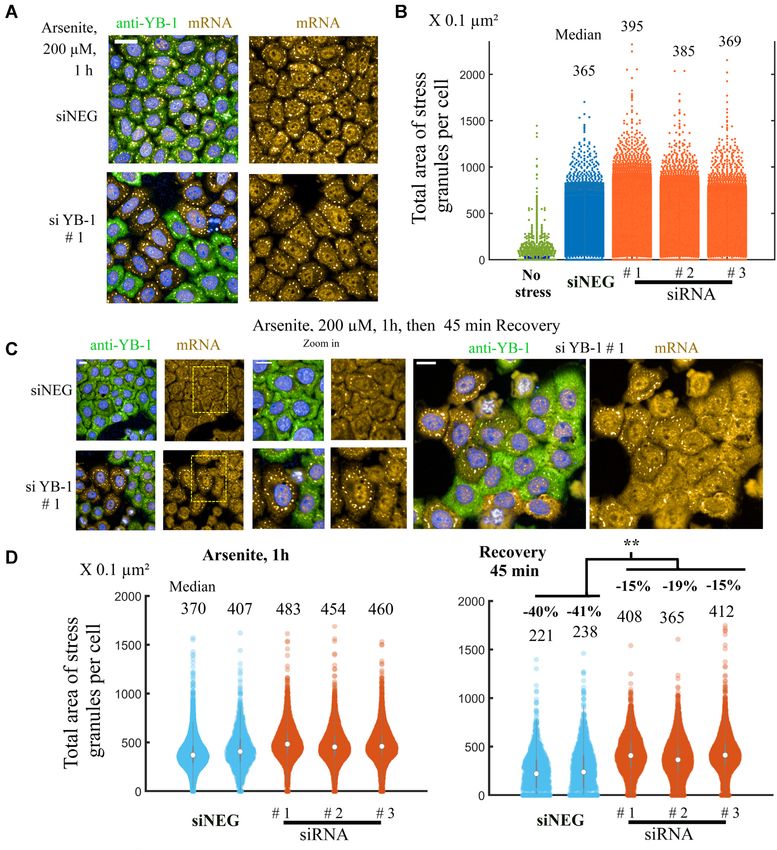
10062 Nucleic Acids Research, 2021, Vol. 49, No. 17

encompass mRNA splicing (17), transcription (18), the            sion was silenced, supporting the idea that YB-1 negatively
processing of noncoding RNA (19–22), and DNA repair              regulates SG assembly (43).
(23,24). The secretion of YB-1 from mammalian cells has             In addition to experiments based on silencing YB-1 ex-
also been reported and may participate in intercellular in-      pression, the overexpression of YB-1 inhibited SG assem-
teractions (25). YB-1 is generally a cytoplasmic protein and     bly in NRK and NG108-15 cells (40,43). The expression of
a core protein partner of non polysomal mRNA in mam-             most RBPs such as G3BP-1, TIA-1 (44), TDP-43 (45), and
malian cells (26) but it can also be found in the nucleus        FUS, is either neutral or beneficial to SG assembly in most
in cancer cells, a phenotype generally associated with a         cases. As overexpressed RNA-helicases such as EIF4A were
bad outcome (18). In agreement with the association of           reported to inhibit SG assembly after arsenite stress (9). An
YB-1 with non-polysomal mRNAs in the cytoplasm (27),             RNA-unwinding activity mediated by YB-1 may thus pro-
crosslinking immunoprecipitation coupled to sequencing           vide a rational explanation for this specific phenotype.
(CLIP) results have indicated a significant binding of YB-1         To explore putative mRNA-unwinding YB-1 activity, we
to coding sequences (CDS) in glioblastoma cancer cell lines.     investigated the interaction of YB-1 with short RNA and

                                                                                                                                   Downloaded from https://academic.oup.com/nar/article/49/17/10061/6362110 by guest on 30 November 2021
Although a consensus motif has been proposed (UYAUC),            DNA stem/loops through a combined NMR spectroscopy
the cross links are dispersed across most transcribed cod-       and molecular dynamics analysis. The results showed clear
ing genes, in agreement with YB-1 being a general mRNP           RNA-unwinding activity due to the loop 3 located in the
factor (26). More recently, in a large scale CLIP map of         cold-shock domain, which is long and positively charged.
multiple RBPs (28), YB-3 was classified as a CDS-binding         We also identified two YB-1 mutants with decreased abil-
protein (YB-1 was not analyzed in this study). While YB-1        ity to unwind RNA in vitro. Then, focusing on the role
binds to non polysomal mRNAs, its role in mRNA transla-          of YB-1 in SG assembly after arsenite-induced stress, we
tion remains debated. Given the inhibition of mRNA trans-        observed that the overexpression of the RNA unwinding-
lation by recombinant YB-1 in rabbit reticulocyte lysates        defective YB-1 mutants less efficiently prevented the forma-
(29), YB-1 may be considered a translation repressor. On         tion of SGs than wild type YB-1 in HeLa cells. A possible
the other hand, the mRNA-unwinding activity of YB-1              role of YB-1-RNA-unwinding activity in the inhibition of
has already been advanced to explain the YB-1-dependent          SG assembly thus appears to be plausible, although alterna-
translation activation of specific transcripts, such as Snail1   tive interpretations can be proposed notably due to overex-
(30) and HIF-␣ (31) mRNAs. In proliferating cells, YB-1          pression conditions. However, in agreement with a negative
is generally abundant (32,33) which may contradict the no-       impact of YB-1 in SG assembly, we found that endogenous
tion that it is a global inhibitor of mRNA translation. Cellu-   YB-1 globally increases translation and facilitates SG disso-
lar signaling, YB-1 post-translational modification (34,35),     ciation during stress recovery in HeLa cells. Interestingly, a
and/or protein partners such as Lin28 (36) may either fa-        strong translation inhibition mediated by the unstructured
cilitate translation in proliferating cells or maintain YB-1-    and self-adhesive C-terminal domain (CTD) of YB-1 was
packaged mRNA in a dormant state, as evidence in testis          reported in vitro (29) but the removal of most of the CTD
(37).                                                            residues decreased the capacity of YB-1 to prevent SG as-
   Here, we formulated the hypothesis that a putative RNA-       sembly. In addition, the YB-1 CTD further facilitates the
unwinding activity of YB-1 prevents mRNAs from forming           inhibition of SG assembly by YB-1.
SGs. However, contradictory results have been reported re-          We therefore propose a model in which YB-1 prepares
garding the role of YB-1 in SG assembly. Most of the data        non polysomal mRNA for translation through its RNA-
have been obtained in cancer cells upon arsenite treatment,      unwinding activity. After cellular stress is induced, the large
which is the most robust means of driving SGs assembly. In       pool of non polysomal mRNA increases the occurrence of
U2OS cells and under certain conditions, YB-1 may posi-          RNA:RNA intermolecular interactions in the cytoplasm
tively regulate G3BP-1 expression to promote indirectly SG       thereby promoting SG assembly. This occurs when YB-1
assembly after arsenite-induced stress (38). In an indepen-      is outnumbered by non polysomal mRNA. However, when
dent study including U2OS cells, a positive regulation of        YB-1 expression is sufficiently high, non polysomal mR-
G3BP-1 expression levels by YB-1 was not confirmed. YB-          NAs no longer form SGs after arsenite stress. Preventing
1 was instead considered dispensable for SG assembly after       SG assembly may be important to sustain the high prolifer-
arsenite treatment but possibly important after ER-stress        ative rate of cancer cells and help cells to cope with stress.
was induced (39). Interestingly, in the same study (39), a de-   Accordingly, a recent multitissue transcriptome analysis at
tailed analysis reported a critical role of YB-1 when SGs        the single cell level revealed YB-1 as a key gene in the adap-
are formed following the stress-induced cleavage of tRNA         tation of cells to caloric restriction in aging Rattus Norvegi-
into small fragments. Small tRNA fragments can sequester         cus (46).
YB-1 to promote the formation of SGs (39) but YB-1 se-
questration may in turn also lead to decreased solubility        MATERIALS AND METHODS
of non polysomal mRNA. In two other studies, arsenite-
                                                                 Computational methods
induced SG assembly was also not significantly affected in
cells treated with siRNA to decrease YB-1 levels (40,41).        System preparation and MD simulations setup. The follow-
More recently, a recent large-scale analysis seeking to iden-    ing systems were considered for MD simulations: WT YB-1
tify positive SG regulators with a CRISPR screen in which        protein alone, WT YB-1:RNA/DNA stem loop complexes,
YB-1 was included again did not show YB-1 to be among            where two types of stem loops were considered ACU-
the proteins found important for SG assembly (42). Finally,      4G/ACT-4G and ACU10/ACT10, WT CSD:RNA/DNA
a study reported increased SG assembly after YB-1 expres-        stem loop complexes with both stem loop types as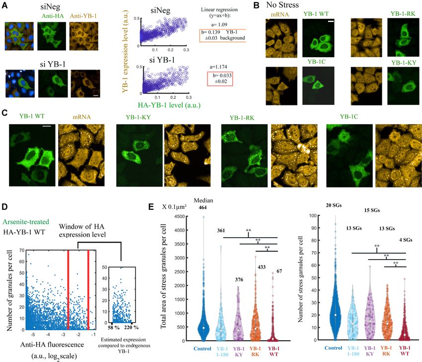
Nucleic Acids Research, 2021, Vol. 49, No. 17 10063
above, R97A/K98A YB-1:RNA/DNA stem loop com- Protein purification
plexes, with ACU-4G/ACT-4G, And K137A/Y138A YB-
Cells carrying plasmids encoding for wild type and mutant
1:RNA/DNA stem loop complexes, with ACU-4G/ACT-
YB1-C (1–180), CSD (50–129), FUS-RRM (275-385) and
4G.
TDP-43-RRM1-2 (101–277) were grown at 37◦ C in 2YT-
Protein sequence used for YB-1 is from A45 to Q165
ampicillin medium (1-l culture) (non-labeled proteins) or in
(length 121 a.a.) and for CSD is from A45 to G129 (length
minimal medium M9 supplemented with 15 NH4 Cl (labeled
85 a.a.). The length of all RNA/DNA stem loops is of 24
proteins). When the optical density of the culture reached
nucleotides.
0.7, IPTG was added at a final concentration of 1 mM,
and growth was continued for 3 h. Cells were harvested and
Molecular modeling. The starting 3D coordinates of YB- washed with 20 ml of cold 25 mM Tris–HCl buffer, pH 7.4,
1 stem loop/RNA models were constructed using as a tem- containing 1 mM TCEP, 1 mM PMSF and EDTA-free pro-
plate the X-ray structure of human Lin28A in complex with tease inhibitor Cocktail (Roche) and 1.5 M KCl (buffer A).
Downloaded from https://academic.oup.com/nar/article/49/17/10061/6362110 by guest on 30 November 2021
let-7f-1 micro RNA pre-element (PDB ID 5UDZ, resolu- The cell pellet was suspended in 10 ml of the same buffer,
tion 2 Å) (47,48). YB-1 structure (A45-Q165) bound to and cells were disrupted by sonication on ice (Bioblock Vi-
polyC RNA was taken from the trimer complex of YB- bracell sonicator, model 72412). The resulting suspension
1:ssRNA (16-nt C), which is a linear nucleoprotein filament was centrifuged at 4◦ C for 30 min at 150 000 × g in a TL100
recently published by our group (12) that used the human Beckman centrifuge. The supernatant was used for purifica-
WT YB-1 NMR structure (position 52–129) as a building tion experiments.
block for the cold shock domain (PDB ID: 1H95). Mod- YB-1 proteins were purified under native conditions as
els were constructed by extracting the middle YB-1 struc- previously described (12). The cells were harvested and re-
ture and superimposing it to Lin28 on the alpha carbons. suspended in 20 ml of lysis buffer (20 mM Tris–HCl, pH
Lin28 was then removed and the RNA was extended in or- 7.6, 10 mM imidazole, 2M KCl, 0.5 mM DTT, protease in-
der to form a total of 24 nucleotides (4 nucleotides form- hibitor tables (Roche)). The cell lysate obtained by sonica-
ing the loop and 10 bp for the stem). Then the nitroge- tion was cleared by centrifugation at 70 000 × g for 1 h at
nous bases were substituted with the stem loop sequences 4◦ C. The supernatant was added to Ni-NTA agarose slurry
studied here (ACU-4G, ACT-4G, ACU10 and ACT10). for 30 min at 4◦ C under agitation and then loaded onto a
In a next step, the CSD:RNA/DNA stem loop complexes gravity column. The column was washed with buffer (20
were modeled from the YB-1:RNA/DNA structures previ- mM Tris–HCl, pH 7.6, 10 mM imidazole, 500 mM KCl, 0.5
ously generated by truncating the CTD part (residues 130 to mM DTT, 0.5 mM PMSF) and eluted with the same buffer
165). The mutants R97A/K98A and K137A/Y138A were supplemented with 250 mM imidazole. The eluate was then
also generated but only for the ACU-4G/ACT-4G stem dialyzed against 20 mM Tris–HCl, pH 7.6, 500 mM KCl.
loops. Then RNase A treatment was applied to remove all traces
of RNA contaminants for 1 h at room temperature under
MD set up. All MD simulations were carried out with agitation. In a second purification step, the eluate was di-
GROMACS software package version 2018.2 using the ff03 alyzed against 50 mM phosphate buffer, pH 6.8, 1 M KCl
Amber ‘all atom’ force field with associated nucleic acid and concentrated with Spin-X UF concentrators (Corning).
parameters and periodic boundary conditions. The proto- Note that we controlled that the structure of His-tagged and
nation states of the residues were adjusted to the PH used non his-tagged protein with or without DNA were similar.
in our NMR experiments (6.8). The systems were centered The final preparations were stored at –80◦ C.
and solvated in a triclinic box of TIP3P water model with TDP-43 fragments were purified basically following the
1.4 nm distance between the boundary of the box and the manufacturer’s recommendations (Qiagen). Imidazol (10
macromolecular complex. A [KCl] of 25mM was used and mM) was added to soluble fractions described above and
counter-ions were added to neutralize the system. Each sys- incubated for 2 h at 4◦ C with Ni2+ -NTA-agarose (Qiagen)
tem was first energy minimized using 50000 steps of steep- (20 mg of proteins/ml of resin) pre-equilibrated in buffer A.
est descent, then heated from 0 to 298 K at constant volume After incubation, the resin was transferred to an Econo-Pac
for 500 ps and equilibrated in the NPT ensemble at p = 1 chromatography column (Bio-Rad). The polymer was then
atm for 500 ps which was followed by 200 ns of NPT pro- washed extensively with buffer A containing 20 mM imida-
duction run. The Velocity Rescaling (with = 0.1 ps) and zole. The elution of the protein was obtained by increasing
Parrinello-Rahman methods were used for temperature and step by step the concentration of imidazole, from 40 to 250
pressure control, respectively. The equations of motion were mM, in buffer A. Pure protein-containing fractions (100–
propagated with the leap-frog algorithm and the time step 250 mM imidazole) were pooled and incubated with a His6 -
was t = 2 fs. The particle mesh Ewald (PME) method [was tagged TEV protease to cleave off the His6 -tag peptide from
used for electrostatic interactions, with grid spacing of 1.6 the target protein. The protease (15 g) was mixed with 1
Å, a relative tolerance of 10−5 , an interpolation order of 4 mg of target protein (0.5 M TEV to ∼30 M protein) in
for long-range electrostatics, and a cutoff of 14 Å together buffer A containing of 1 mM DTT and 1 mM EDTA. All
with a 12 Å switching threshold for LJ interactions. All co- digestions were conducted for 16 h at room temperature.
valent bond lengths were constrained with LINCS. Trajec- A PD-10 column (GE Healthcare) was used to remove im-
tories and geometries have been visualized and represented idazole and to exchange buffer. Then, TEV protease and
using VMD. The MD simulations in this work were done His6 -tag peptide from target protein were trapped on Ni-
using NVIDIA GPU resources owned by Synsight. NTA agarose column and target protein was recovered in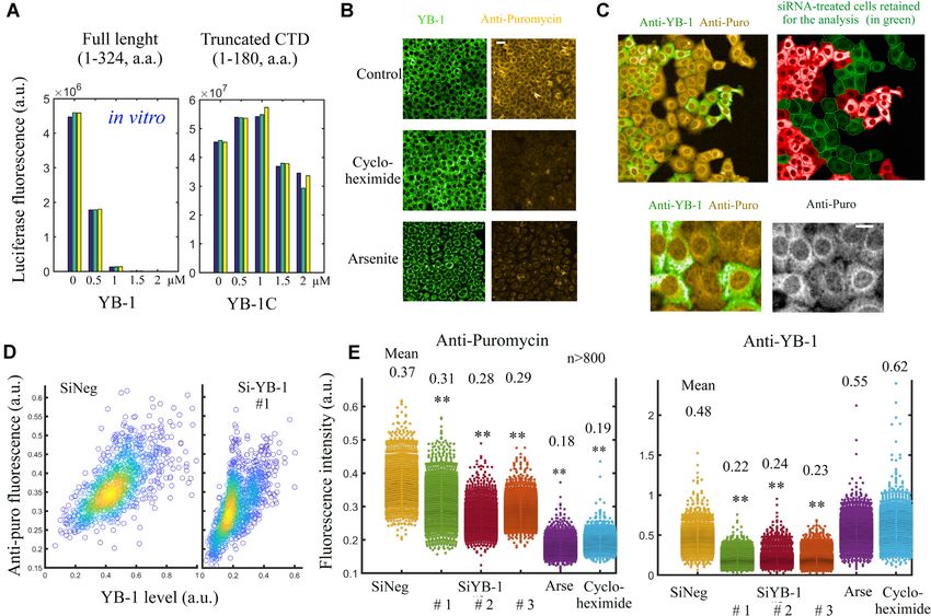
10064 Nucleic Acids Research, 2021, Vol. 49, No. 17
pass-through (nonbinding) fraction. The protein was con- Gels mobility shift assays
centrated to 2 ml and conserved in 20 mM Tris–HCl buffer,
Indicated amounts of YB-1, YB-1-RK and YB-1-KY (1–
pH 7.4, containing 25 mM KCl and 1 mM TCEP by using
180, a.a.) were incubated with 0.16 pmol of 2Luc mRNA
a PD-10 column. For NMR experiments, the 15 N-labeled
(3000 nt) in 20 l of binding buffer (20 mM HEPES, pH
proteins were stored in phosphate buffer 15 mM pH 6.8
7.5, 60 mM KCl) at room temperature for 5 min. Complexes
containing 25 mM KCl and 1 mM TCEP by using a PD-
were separated in 0.65% agarose gel in 0.5× TAE buffer at 5
10 column (GE Healthcare).
V/cm for 20 min and were stained with 0.5 g/ml ethidium
bromide.
Nuclear magnetic resonance
Purified 15 N-labeled protein fragments were incubated with Cell culture and Transfections
indicated DNA/RNA oligonucleotides (Eurogentec) dur- HeLa and U2OS cell lines (American Type Culture Collec-
ing 10 min at 25◦ C. Free and oligonucleotide-bound pro- tion, USA) were cultured at 37◦ C in a humidified atmo-
Downloaded from https://academic.oup.com/nar/article/49/17/10061/6362110 by guest on 30 November 2021
tein samples were prepared in NMR buffer (25 mM phos- sphere with 5% CO2 and maintained in the high glucose
phate, pH 6.8, containing 25 mM KCl) supplemented with formulation of DMEM (Life Technologies) supplemented
SUPERase·In RNase inhibitors (ThermoFisher Scientific) with penicillin G 100 U/ml, streptomycin 100 g/ml and
for RNA samples. All samples were prepared in a final vol- fetal bovine serum (FBS) 5% (10% for HeLa cells; Thermo-
ume of 60 l using 1.7 mm diameter capillary tubes (Bruker) Fisher).
and 2,2-dimethyl-2-silapentane-5-sulfonic acid as external
reference in pure D2 O (Eurisotop) for chemical shift refer-
encing. Plasmid transfection, siRNA treatment and addback experi-
NMR spectra were acquired on a Bruker AVIII HD ments
600 MHz spectrometer equipped with a triple-resonance The cells were grown in 24- or 96-well plates and tran-
cryoprobe at 298 K. The binding of CSD, YB-1C and TDP- siently transfected with plasmids, carrying the studied pro-
43 to DNA or RNA oligonucleotides was investigated using tein gene, at a final concentration of 1 g using lipofec-
2D 1 H–15 N SOFAST-HMQC recorded on 50 or 100 M tamine 2000 (Thermofisher) transfection reagent for 24 h.
protein samples at a 1:1 molar ratio. The number of dummy Three siRNAs targeting YB-1 expressions were used.
scans and scans was respectively set to 16 and 512. Data Two of them target the coding sequence: siRNA-1: [sense
were acquired with 2048 points along the direct dimension 5 -(CCACGCAAUUACCAGCAAA)dTdT-3 , anti-sense
and with 128 t1 increments with a relaxation delay of 0.2 s. 5 -(UUUGCUGGUAAUUGCGUGG)dTdT-3 ]; siRNA-
Shaped pulse length and power were calculated by consid- 2:[sense 5 -AGUGUAGGAGAUGGAGAAAdTdT-3 ,
ering an amide 1 H bandwidth of 4.5 ppm and a chemical antisense 5 -(UUUCUCCAUCUCCUACACUdTdT-3 ].
shift offset of 8.25 ppm. SiRNA-3 which targets the 3 UTR of YB-1 mRNA
NMR assignment: 1 H and 15 N chemical shifts of YB- was used for the addback experiments [sense
1(1–180) and TDP-43 RRM1-2 residues were assigned us- 5 -(GAUUGGAGCUGAAGACCUA)dTdT-3 , anti-
ing our previous results (12) (YB-1) or previous assignments sense 5 -(UAGGUCUUCAGCUCCAAUC)dTdT-3 ]. The
obtained for the unbound TDP-43 RRM1 and RRM2 negative siRNA (1027310, Qiagen), SiNeg, was applied in
(BMRB Entries: 18765 and 19922, 19290). the same concentration as YB-1 siRNA. The mix of 1 g
siRNA or siNeg in 300 l optiMEM with 0.8 l lipofec-
Transcription in vitro tamine was left for 20 min at room temperature and added
to cells for 3 h, after that the solution was removed and the
Reporter mRNA for was transcribed with a SP6- usual media was added to the well. Control of the efficiency
Scribe Standard RNA IVT Kit (CellScript). Polyadeny- of was performed by immunofluorescence (Figures 8E).
lated BTF3 Fluc mRNA was transcribed from pSP36T- We obtained clusters of cells expressing endogenous YB-1
5 UTR BTF3-FLuc-A50 linearized with HpaI. mRNAs coexisting in the same sample with clusters of cells that
were capped using the ScriptCap m7 G Capping System and displayed a significantly reduced expressing of endogenous
the ScriptCap 2 -O-methyltransferase enzyme (CellScript) YB-1. Only the cells with a low YB-1 expression were
according to the manufacturer’s manual. retained in the analyzes (Figure 8C).
To add-back the expression of YB-1 (Figure 7, Supple-
In vitro translation assays mentary Figures S8 and S10), HeLa cells were first treated
by siRNA-3 targeting the 3 UTR of endogenous YB-1 for
The translation mixture (10 l) consisted of 5 l of 36 h. Then, HeLa cells were transfected with plasmids to
nuclease-treated rabbit reticulocyte lysate, 1 l 10× transla- express wild type or mutant YB-1, as indicated. Wild type
tion buffer (200 mM HEPES–KOH, pH 7.6, 10 mM DTT, or mutant exogenous YB-1 expressions obtained by using
5 mM spermidine–HCl, 80 mM creatine phosphate, 10 mM plasmid transfections are not the targets of siRNA-3.
ATP, 2 mM GTP and 250 M of each amino acid), 100 mM
KAc, 1 mM Mg(Ac)2 , 0.15 pmol reporter Fluc mRNA, and
In situ hybridization
recombinant YB-1/YB-1C protein. Reaction mixtures were
incubated for 25 min at 30◦ C, and then the luciferase activ- To visualize mRNA using the red channel, after fixation
ities were measured using the OneGlo Luciferase Assay kit HeLa cells were incubated with oligo-dT-[Cy3], diluted in
(Promega). Raw data are shown in Supplementary Table S3. SSC 2×, 1 mg/ml yeast tRNA, 0.005% BSA, 10% dextran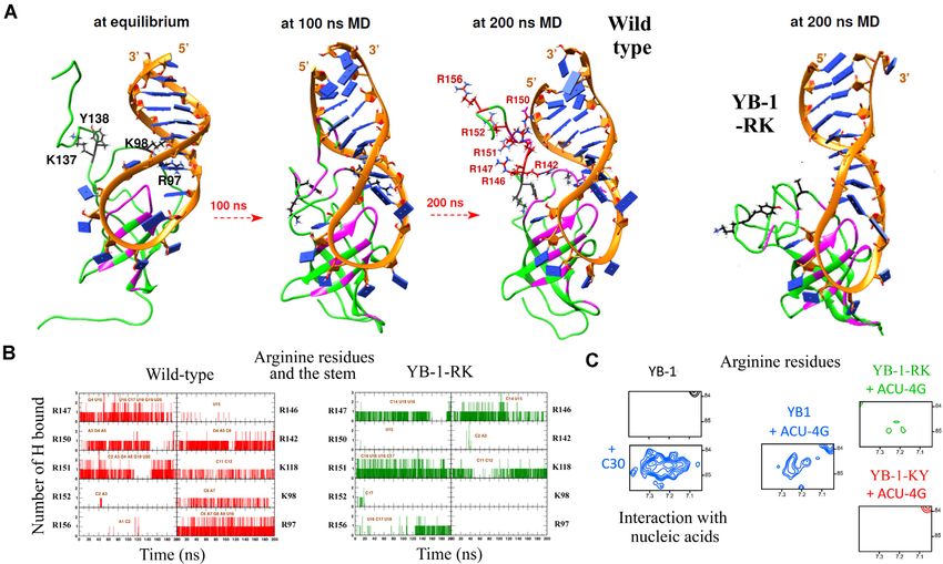
Nucleic Acids Research, 2021, Vol. 49, No. 17 10065
sulfate, 25% formamide, for 2 h at 37◦ C. Wash steps were DNA-melting activity assay
performed using 4× and then 2× SSC buffer (0.88% sodium
Before assay DNA oligonucleotide 5 FAM-GGGGGGG
citrate, 1.75% NaCl, pH 7.0). To visualize mRNA in blue
GTTAACCCCCCCC-BHQ1-3 (DNA1) was incubated 5
color for SGs experiments, the oligo-dT with digoxigenin
min at 98◦ C and slowly chilled to 20◦ C. The reaction mix-
was used after cells fixation with the same incubation pro-
ture of a 20 l final volume contained 130 mM KCl, 10 mM
cedure as oligo-dT-Cy3. Then the primary anti-digoxigenin
HEPES–KOH (pH 7.6), 20 pmol of the DNA1 oligonu-
antibodies (mouse, ab420, Abcam) and secondary antibod-
cleotide and 200 pmol of protein (YB-1, or YB-1 fragment
ies (goat anti-mouse, Alexa 350, Invitrogen) were applied to
(1–180), or YB-1 fragment (1–180) mutant forms). The con-
cells according to supplier’s protocol.
trol experiment was performed without proteins. All reac-
tion mixtures were incubated at 30◦ C in a DTlite real-time
SG assay PCR system (DNA-Technology, Russia) to detect fluores-
cence every 5 s for 5–25 min. To plot the graph, the first flu-
Downloaded from https://academic.oup.com/nar/article/49/17/10061/6362110 by guest on 30 November 2021
HeLa cells, transfected with corresponding plasmids for 24 orescence value detected by the real-time PCR system was
h, were subjected to oxidative stress using arsenite during subtracted from the subsequent ones as background.
1 h at 37◦ C. The cells were fixed with methanol for 20 min
at −20◦ C, followed with 4% paraformaldehyde for 30 min
at 37◦ C. Immunofluorescence was performed using anti- RESULTS
HA (against overexpressed YB-1, mouse, sc-7392, Santa YB-1 unwinds DNA/RNA stem/loops in vitro
Cruz Biotechnology), anti-YB-1 (against endogenous pro-
tein, rabbit polyclonal, Bethyl Laboratories, Montgomery, YB-1 possesses three distinct domains (10,14): (i) an un-
USA), anti-FMRP (anti-rabbit, ab 17722, Abcam), anti- structured alanine/proline-rich domain which may interact
G3BP-1 (anti-rabbit, G6046, Sigma)). with protein partners, (ii) a highly conserved -barrel struc-
Quantifications were performed with Opera Phenix® ture, i.e., the cold-shock domain (CSD) that binds to RNA
Plus High Content Screening System (PerkinElmer) in con- and DNA and (iii) a long unstructured C-terminal domain
focal mode. The Harmony v4.8 software was used to detect (CTD) that harbors well separated clusters of negatively
and measure the number or total area of SGs per cell or the and positively charged residues that may be involved in self-
number of SGs per cell (These values are directly accessible adhesive interactions (49). The melting of RNA secondary
by selecting them in the ‘spot analysis’ parameters). structures (50) and the separation of DNA strands by YB-1
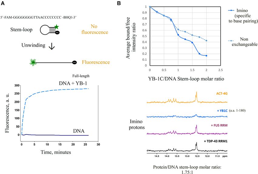
have been observed in vitro (51). To confirm that YB-1 un-
winds the secondary structures of nucleic acids in vitro, a
Screening of the effect of RBP overexpression in SG assembly DNA stem/loop with a quencher and a fluorophore at each
extremity and a short loop that could accept only one pro-
To obtain the results presented in Supplementary Figure S6, tein was used to probe a putative unwinding activity (Figure
HeLa cells were transfected with corresponding plasmids 1A). The results of the fluorescence assay clearly showed a
for 24 h and were subjected to oxidative stress using 300 significant increase in fluorescence intensity in the presence
M arsenite during 1 h at 37◦ C. The cells were fixed with of YB-1 revealing an opening of the DNA stem/loop by
methanol for 20 min at −20◦ C, followed with 4% PAF for full-length YB1.
30 min at 37◦ C. The staining was performed using anti-HA To examine the molecular interactions critical for the un-
(YB-1, mouse, sc-7392, Santa Cruz Biotechnology), anti- winding of RNA secondary structure, we performed an ex-
myc (to detect myc-tagged IGF2BP3) primary antibod- tensive NMR analysis with a truncated form of YB-1 (1–
ies and then secondary antibody (goat anti-mouse/donkey 180, a.a., also called YB-1C). With its N-terminus (1–50,
anti-rabbit, Alexa 594, Invitrogen). Other RBPs were visu- a.a.), CSD (51–128, a.a.), and short CTD (129–180, a.a.) in-
alized with a GFP-tag. cluding a cluster of arginine residues, YB-1C was to date the
Quantifications were performed by recoding the percent- longest YB-1 fragment amenable to NMR spectroscopy. We
age of cells harboring SGs from fluorescence images. then analyzed the unwinding activity of YB-1C by NMR
spectroscopy at different YB-1C concentrations. The peak
intensities of imino protons that are specific to DNA/RNA
Cellular translation assays
base pairing were probed using a short stem/loop (AC
The cells were treated with puromycin (10 g/ml) for AGACAGAACCCCTTCTGTCTGT, called ACT-4G, Tm
10 min prior to fixation after washing out puromycin. ∼ 60◦ C). A marked decrease in peak intensities was ob-
Cell were fixed with 4% PAF for 30 min at 37◦ C and served at high YB-1C concentrations (Figure 1B). In addi-
subjected to immunoblotting using puromycin antibody tion, the imino protons displayed a steeper slope than non-
(Merck, MABE343) or beta-actin antibody (Sigma, A2228) exchangeable protons which are not specific to base pairing.
as described previously. For the negative control, cells These results therefore indicated a destabilization of DNA
were treated with cycloheximide (100 g/ml) prior to stem/loop in addition to an increase in molecular weight
the addition of puromycin. The anti-puromycin fluores- (Supplementary Figure S1a, b). The peak intensities of the
cence in the cytoplasm was detected automatically using imino protons were also measured in the presence of two
the Opera Phenix® Plus High Content Screening System different RNA-recognition Motifs (RRMs), the RRM of
(PerkinElmer). The cell cytoplams were detected automati- FUS and RRM-1 of TDP-43. Despite the presence of CSPs
cally using the Harmony v4.8 software. indicating the binding of the RRMs to the DNA stem/loop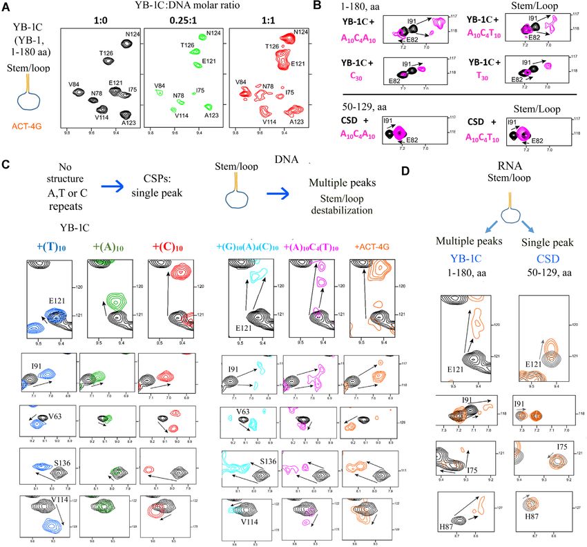
10066 Nucleic Acids Research, 2021, Vol. 49, No. 17
Downloaded from https://academic.oup.com/nar/article/49/17/10061/6362110 by guest on 30 November 2021
Figure 1. YB-1 destabilizes DNA stem/loops in vitro. (A) Full-length YB-1 melts short DNA duplex. DNA oligonucleotide 5 FAM-GGGGGGGGTTA
ACCCCCCCC-BHQ1-3 (Tm ∼ 70◦ C) was incubated 5 min at 98◦ C and slowly chilled to 20◦ C. After YB-1 protein (dashed line) or buffer (solid line)
addition, reaction mixtures were incubated at 30◦ C in a real-time PCR system to detect fluorescence every 5 s for 26 min. (B) Upper panel: NMR analysis
of the average bound:free intensity ratio of the ACT-4G stem/loop (ACAGACAGAACCCCTTCTGTCTGT, Tm ∼ 60◦ C) imino and non-exchangeable
protons as a function of protein:DNA concentration ratio. 1D 1 H NMR spectra at 25◦ C of 50 M ACT-4G DNA stem free (in orange) and in presence of
YB-1C (in blue). The decrease of peak intensity with increasing concentration of YB-1 is more marked for imino than non-exchangeable protons. Lower
panel: Imino region of the 1D 1 H NMR spectra in the presence of YB-1C (in blue), FUS RRM (in purple) or TDP43 RRM1 (in black) at a protein:DNA
concentration ratio of 1.75:1. The decrease of imino signals with YB-1C corresponds to an increase of imino proton exchange in agreement with a stem/loop
opening.
(data not shown), the peak intensities were not significantly results were obtained in the presence of an RNA stem/loop
affected (Figure 1B). (ACAGACAGAACCCCUUCUGUCUGU, Tm ∼ 60◦ C)
According to this analysis, the DNA stem/loop destabi- (Figure 2D). To test whether more than one YB-1C
lization was more pronounced at high YB-1 concentrations can interact with the same stem/loop, we analyzed the
(Supplementary Figure S1b). The binding of several YB-1 interaction of YB-1C at a low YB-1C:DNA molar ratio
proteins was probably required to secure the opening of the (at 0.25:1) when the occurrence of multiple bindings to
4 C-G stem:loop. However, even at a YB-1C:DNA molar the ACT-4G stem/loop should decrease. Multiple peaks
ratio lower than 1:1, YB-1C has the capacity to form a mul- indeed disappeared at a low YB-1C:DNA molar ratio
timer because of its cooperative binding to ssDNA (12,52). (0.25:1, Figure 2A). We also analyzed the interactions of
We then considered the chemical shift perturba- YB-1 with two other DNA stem/loops that had a short
tions (CSPs) of 15 N-labeled YB-1 residues induced by loop, G10 –A4 –C10 (Tm ∼ 85◦ C), A10 –C4 –T10 (Tm ∼ 49◦ C).
DNA/RNA stem/loops. In the presence of the ACT-4G Multiple peaks were again detected at a 1:1 YB-1C:DNA
stem/loop, we noticed the presence of multiple peaks and molar ratio.
peak broadenings (Figure 2A, B)). Multiple peaks are To further test whether the appearance of multiple peaks
typical of a slow exchange regime between different states is due to stem/loop destabilization, we performed a se-
suggesting an interaction of at least two YB-1 proteins per ries of experiments. First, we controlled the presence of
stem/loop. The destabilization of stem/loops generates single peaks when YB-1C interacted with unstructured A,
additional ssDNA binding sites presumably available for T or C homo-oligonucleotides (A10 , T10 , C10 ; Figure 2C,
CSD. Accordingly, multiple peaks were detected in the D). G10 was not considered because of the formation of
conserved CSD residues interacting with nucleic acids G-quartets. In addition, single peaks were detected when
(V63, I91 and E121 (Supplementary Figure S2b)). Similar multiple YB-1C proteins oligomerized in the presence of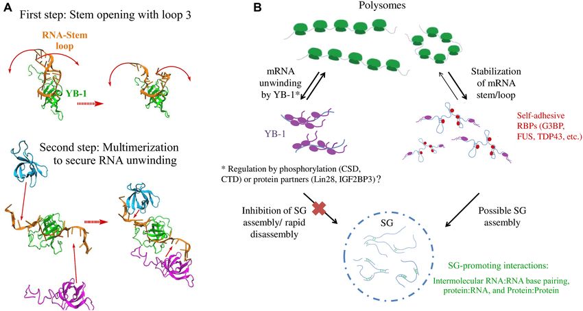
Nucleic Acids Research, 2021, Vol. 49, No. 17 10067

                                                                                                                                                          Downloaded from https://academic.oup.com/nar/article/49/17/10061/6362110 by guest on 30 November 2021
Figure 2. YB-1C destabilizes RNA/DNA stem loop but not CSD alone. (A) Two-dimensional 1 H–15 N HSQC spectra of YB-1C (1–180 a.a.) in free
state or in presence of the ACT-4G stem/loop (ACAGACAGAACCCCTTCTGTCTGT, Tm ∼ 60◦ C) at different YB-1C:DNA molar ratios. Note the
appearance of multiple peaks at a 1:1 molar ratio. (B) As observed with I91, a representative residue, multiple peaks are observed when YB-1C interacts
with a stem/loop or an unstructured hetero-nucleotide ssDNA mimicking an open stem/loop but not with a long homo-nucleotide ssDNA. Only single
peaks were also detected with CSD alone (50–129 a.a.) that cannot multimerize like YB-1C. (C) NMR spectra of YB-1C (1–180 aa) in free state or in
presence of indicated unstructured or structured DNA oligonucleotides. Representative residues experiencing CSPs with multiple peaks in the presence of
DNA stem/loops are shown. The experiments were performed at 25◦ C, a temperature well below the Tms of the DNA stem/loop (ACT10, Tm ∼ 49◦ C;
ACT-4G, Tm ∼ 60◦ C). (D) Two-dimensional 1 H–15 N HSQC spectra of YB-1C (1–180 a.a.) and the CSD alone (50–129 a.a.) in free state or in presence of
the ACU-4G RNA stem/loop (Tm ∼ 60◦ C).

30 nt-long homo-nucleotides such as poly(C) or poly(T),                       nucleotides, in contrast with YB-1 interactions with long
as shown previously (12), probably because each YB-1C                         homo-oligonucleotides (Figure 2B, C).
protein interacted with the same nucleotides in the multi-                       We also performed a similar experiment with an iso-
mer (Figure 2C). However, an unstructured A10 –C4 –A10 ss-                    lated CSD (50–129, a.a.) that was unable to form multimers
DNA mimicking an open A10 –C4 –T10 stem/loop induced                          with ssDNA/RNA under our experimental conditions (12).
the appearance of multiple peaks (Figure 2B, C, Supple-                       Multiple peaks in NMR spectra failed to appear in CSD
mentary Figure S2a). The binding of multiple YB-1 pro-                        residues (Figure 2B, D). One CSD per RNA stem/loop
teins to A10 –C4 –A10 may explain this behavior since YB-1                    most probably binds to the RNA loop without opening the
proteins in the multimer should not interact with the same                    stem. Single peaks were also detected in the presence of the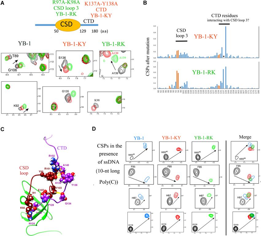
10068 Nucleic Acids Research, 2021, Vol. 49, No. 17
A10–C4–T10 stem/loop with the tandem RRM1-2 of TDP- of wild type YB-1, which indicates that the double mu-
43 (Supplementary Figure S2c). tation, K137A/Y138A, most probably disrupts the inter-
Altogether, the disappearance of imino protons from action between the CTD segment with CSD loop 3 (Fig-
DNA stem/loops, the CSPs of YB-1C residues and, com- ure 3A, B). The CTD residues involved in the interaction
plementary fluorescence assays showing the DNA-melting with CSD loop 3 are also highly conserved across species
activity of YB-1 support a destabilization of nucleic acid (Supplementary Figure S4), in contrast to the remainder
stem/loops by YB-1. of CTD sequences. Only the presence of positively charged
residues is conserved. This residue conservation is an addi-
tional argument for an interplay between CSD loop 3 and
CSD Loop 3 and CTD residues interact and thus recruit many
the CTD, which could also initiate an interaction between
positively charged residues near stem in a YB-1:stem/loop
the additional arginine residues in the CTD (R142, R146-
complexes
R147, R150-153) with the stem (Figure 3A, B), thus further
While seeking to determine how YB-1 may unwind RNA, contributing to the destabilization of base pair interactions
Downloaded from https://academic.oup.com/nar/article/49/17/10061/6362110 by guest on 30 November 2021
we noticed the presence of 4 positively charged residues in (Supplementary Table S1).
the CSD loop 3 (KKNNPRKYL; 92–100, a.a.) in Y-Box
protein family members, but not in other cold-shock pro-
Identification of two mutants, YB-1-RK and -KY with a re-
teins in humans (such as Lin28 or CSDE1) which retained
duced efficiency in destabilizing DNA/RNA stem/loops
our attention. In addition, we noticed that loop 3 in Y-box
proteins is the longest among the cold-shock proteins (Sup- To investigate the molecular mechanisms by which CSD
plementary Figure S4). To understand the role of a long loop 3 and the CTD promote the unwinding of nucleic
CSD loop 3, we examined the crystal structure of Lin28, acids, we performed a combined NMR spectroscopy and
complexed with let-7 (47) a micro-RNA with a stem/loop MD analysis with different DNA/RNA stem/loops in in-
structure, and noticed the location of the CSD loop 3 with teraction with wild type YB-1C or YB-1C mutants, YB-
respect to the stem. Assuming a similar binding of YB- 1-RK and YB-1-KY. Prior to this analysis, the capac-
1 and Lin28 CSDs on basis of their high structural ho- ity of YB-1C mutants to bind unstructured ssDNA was
mology, the additional positively charged residues found controlled (Figure 3D). We noticed that all the conserved
in the CSD loop 3 of YB-1 may interfere with the base- residues in the -barrel display strong chemical shifts that
pair stability between complementary strands in the stem, are very similar to those observed with wild type YB-1
possibly leading to the possible destabilization of the RNA (H87, F85, W65 and G119) in the presence of unstructured
stem/loop. ssDNA. CSPs for residues located near loop 3 or in the
To determine whether CSD loop 3 plays a key role in CTD residues also showed similar shifts, except for the mu-
RNA-unwinding, we generated a mutant, YB-1-RK, by tated residues or those near the mutated residues. In sum-
substituting two positively charged residues (R97 and K98) mary, the results indicate unaffected binding of YB-1-RK
with alanine residues. Given the central location of R97 and -KY CSD residues to unstructured ssDNA compared
and K98 in loop 3, the chances of altering the stable - to wild type YB-1 (Figure 3D).
barrel structure through their mutation into alanine are lim- We then analyzed the interactions of YB-1-RK and -KY
ited. To analyze the impact of these mutations in the YB- with an RNA stem/loop, ACU-4G, by NMR spectroscopy.
1 structure, 15 N-labeled wild type YB-1 (1–180, a.a.) and Significant differences in the CSPs were detected as multi-
the R97A/K98A mutant were investigated by NMR spec- ple peaks were no longer present for the conserved RNA-
troscopy in liquid at 25◦ C (Supplementary Figure S3b). interacting residues in YB-1-RK and -KY in contrast with
Unexpectedly, the double mutation, R97A/K98A, had a wild type YB-1 (Figure 4A, V84, H87, I91, E121, G106,
marked impact on the chemical shifts (CSPs) of the residues G135, S136). The absence of multiple peaks indicates a sin-
located in a segment of the CTD (130–140, a.a.), although gle environment for the CSD residues of YB-1-RK and -KY,
the spatial proximity of these residues with CSD loop 3 is most probably because of their interactions with the RNA
not intuitive at first sight (Figure 3A, B and Supplemen- loop that do not destabilize the stem. To verify this suppo-
tary Figure S3c). We then generated a molecular dynam- sition, we analyzed the opening of DNA stem/loop with a
ics (MD) model of YB-1C alone (Figure 3C, 1–180 a.a.), florescence assay. While YB-1C significantly increased the
as described in the Methods. Several interaction pairs dis- fluorescence intensity due to the opening of the stem/loop
played a low free energy (G106-Q134, E107-Q134, R101- (as observed with full length YB-1, Figure 1A), both YB-
R152, Y99-Y138, Y99-A139, Table 1) suggesting a puta- 1-RK and -KY increased the fluorescence intensity to a
tive interaction between CSD loop 3 and the CTD segment lesser extent than YB-1C, in agreement with an impaired
(135–140, a.a.). Consistent with this finding, an interaction unwinding of the DNA stem/loop by the YB-1 mutants
between CSD loop3 and the CTD in a shorter YB-1 mu- (Figure 4B). We also analyzed the fluorescence of ethid-
tant (51–140 a.a.) has been recently proposed (53), although ium bromide (EtBr) in mRNA:protein complexes formed
this mutant is probably too short to fully capture the dy- in the presence of a long mRNA (Figure 4C, 2Luc mRNA,
namics of the CTD segment. To disrupt the putative inter- 3000 nt). The intercalation of EtBr in double stranded nu-
action between CSD loop 3 and CTD, we prepared a new cleic acids results in a high-fluorescence yield. Accordingly,
mutant (YB-1-KY) with two mutations, K137A/Y138A, in the unwinding of mRNA secondary structures would re-
a central location in the CTD segment that interacts with duce EtBr fluorescence. We previously showed that YB-
CSD loop 3. Strikingly, the CSPs observed for the YB-1- 1C (1–180, a.a.), but not HuR, an RNA-binding protein
KY and YB-1- RK mutants were very similar to the CSPs with RRM domains, significantly reduced the intensity of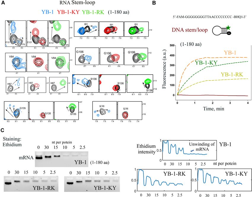
Nucleic Acids Research, 2021, Vol. 49, No. 17 10069

                                                                                                                                                         Downloaded from https://academic.oup.com/nar/article/49/17/10061/6362110 by guest on 30 November 2021
Figure 3. The CSD loop 3 interacts with residues located in the CTD to orient the CTD. (A) CSPs of some YB-1 residues located in the CTD or near
the CSD loop 3 for the two mutants, YB-1-RK (R97A/ K98A), YB-1-KY (K137A/Y138) in the absence of nucleic acids. (B) Histograms displaying the
CSPs for the two mutants compared to wild type YB-1 (Supplementary Figure S3c, NMR spectra). A putative interaction between CTD and CSD loop 3
is clearly detected. Red bar, side-chain. *, mutated residues. (C) MD model of the interaction between residues located in the CSD loop 3 and the CTD.
(D) CSPs of conserved residues interacting with nucleic acids in YB-1, YB-1-RK and -KY in the presence of 10-nt long poly(C) oligonucleotides. The
mutations do not significantly affect the binding of the CSD to ssDNA.

EtBr fluorescence in similar way in Escherichia coli SSB,                    The molecular mechanism behind the destabilization of RNA
which forms ssDNA nucleoprotein filaments (12). We first                     stem/loop by YB-1
noticed that wild type and mutant YB-1 induced a simi-
                                                                             We then considered the molecular mechanism leading to the
lar decrease in electrophoretic mobility, suggesting a sim-
                                                                             RNA-unwinding activity mediated by YB-1. To this end,
ilar affinity for mRNA. Low YB-1C concentrations, (>10
                                                                             a MD model of YB-1 complexed to the ACU-4G RNA
nt per protein), mutated or not, considerably reduced EtBr
                                                                             stem/loop was generated (Figure 5A and Supplementary
fluorescence, most probably due to the dissociation of the
                                                                             Figure S5a). Using the structure of Lin28 associated to let-
very short RNA stems that cannot resist the binding of the
                                                                             7 microRNA as a starting position, the energies averaged
CSD to ssRNA. However, at higher YB-1C concentrations
                                                                             over the whole MD trajectory (200 ns) indicate that wild
(
10070 Nucleic Acids Research, 2021, Vol. 49, No. 17

Table 1. Interaction energies between the most relevant YB-1 pair residues   in cells. We first screened the formation of SGs in HeLa
from CSD loop 3 and CTD. Energies were averaged over the whole MD            cells overexpressing different RNA-binding proteins after
trajectory (100 ns)                                                          arsenite treatment, which is the most robust means to trig-
               Residue pair                                                  ger the formation of SGs in most cells and the best docu-
                                                                             mented SG assembly mechanism. In situ hybridization with
CSD loop                           C-ter                   E (kJ/mol)
                                                                             a poly(dT) probe was used to detect SG in cells. In HeLa
I91                                N124                          –1.8        cells overexpressing GFP-tagged YB-1, IGF2BP3, TDP-
                                   V125                         –18.7        43, G3BP-1, KHSRP, LARP6, WBP11, PUM2, CIRPB,
K92                                N124                         –18.8
                                   V125                         –14.2        Lin28 or CSDE1, we found that only YB-1 or IGF2BP3
Y99                                Y138                         –16.2        overexpression affected SG assembly (Supplementary Fig-
                                   A139                         –12.4        ure S6a,b and S6a). Interestingly, an interaction between
R101                               A140                          –6.3        YB-1 and IGF2BP3 in primary human and mouse acute
                                   R152                         –20.0
                                                                             myeloid leukemia cells was recently reported (54). How-

                                                                                                                                             Downloaded from https://academic.oup.com/nar/article/49/17/10061/6362110 by guest on 30 November 2021
V103                               Q134                         –11.0
G106                               Q134                         –20.0        ever, given the abundance of YB-1 in the cytoplasm (80-fold
                                   G135                          –3.6        greater abundance than IGF2BP3 in HeLa cells (55)) and its
E107                               P128                         –20.5        preferential binding to mRNA, YB-1 is a better candidate
                                   Q134                         –52.5        than IGF2BP3 for preventing the global recruitment of non
                  Total                                        –216.0
                                                                             polysomal mRNA into SGs. The inhibition of SG assembly
                                                                             in HeLa cells overexpressing YB-1 was also observed by us-
                                                                             ing G3BP-1 and FMRP (56), two cytoplasmic RBPs, as SG
and Supplementary Table S2). An additional contribution                      markers (Supplementary Figure S7a).
comes from arginine residues, R142, R147, R150 and R151                         We then tested different truncation mutants to determine
located in the CTD that interact with the stem. In contrast,                 which YB-1 domain is critical for the inhibition of SG as-
in YB-1-RK, the interactions of the loop 3 and CTD argi-                     sembly (Figure 6A). To this end, the total area of SGs per
nine residues with the RNA stem decrease dramatically, pre-                  cell, which is an unbiased and measurable parameter to es-
serving the stability of the stem (Figure 5B). The unwind-                   timate the level of SG assembly per cell (Figure 6A), was
ing activity of YB-1-KY is also reduced but to a lesser ex-                  quantified in HeLa cells expressing GFP-tagged proteins in-
tent in the presence of YB-1-RK since CSD loop3 remains                      cluding full length or YB-1 truncated proteins. Compared
in the vicinity of the stem in YB-1-KY. However, certain                     to the effect of GFP alone, G3BP-1 promoted the forma-
arginine residues in YB-1-KY such as R142 and R150 in-                       tion of SGs. In contrast, full length YB-1 led to a striking
teract only weakly with the stem, possibly reducing the ca-                  decrease in the total area of SGs per cell. However, no sig-
pacity of the CTD to unwind RNA (Figure 5B, Supple-                          nificant impairment in SG assembly was measured when
mentary Figure S5b, d and Table 2 and Supplementary Ta-                      the YB-1 CTD was totally removed (A/P-CSD, 1–129 a.a.)
ble S1). To confirm the role of the CTD arginine residues,                   (Figure 6B). The unstructured CTD (129–324, a.a.) was
we analyzed the interactions of arginine residues with nu-                   also not sufficient to inhibit SG assembly (Figure 6B). As
cleic acids evident in the NMR spectra. While arginine side                  both CSD and CTD are required for the inhibition of SG
chains in the unstructured CTD (Hε/Nε peaks) were not                        assembly, we tested whether YB-1C (1–180 a.a.) can pre-
detected due to rapid exchange with water, their interactions                vent SG assembly, even with its short CTD (Figure 6C).
with RNA/DNA phosphates reduced the solvent-exchange                         The results indicated a significant decrease in total SG area
dynamics, enabling their detection in NMR spectra (Fig-                      in HeLa cells expressing YB-1C. As a short CTD is also
ure 5C). When YB-1 oligomerizes along the 30 nt-long ss-                     required to unwind RNA in vitro (Figure 2D), the nega-
DNA, the arginine residues of CTD interact with nucleic                      tive regulation of SG assembly by YB-1 correlates with the
acid backbones to provide an electrostatic bridge between                    capacity of YB-1 to unwind RNA. In addition, while the
consecutive CSDs. However, in the presence of an RNA                         YB-1 CTD is self-adhesive and critical for YB-1 oligomer-
stem/loop, the Hε/Nε peaks of the arginine residues were                     ization in vitro, full length YB-1 further decreased the to-
scarce in the YB-1-RK and -KY mutants and less intense                       tal SG area per cell compared to YB-1C (Figure 6C). Be-
than those of wild type YB-1 (Figure 5C). The electrostatic                  cause SG composition and property varies by stress type
interaction of CTD arginine residues with nucleic acids may                  (57), we sought to determine whether the overexpression
therefore be less effective in the two YB-1 mutants.                         of YB-1 in HeLa cells prevents SG assembly after a com-
   Taken together, the structural data confirmed a desta-                    bined puromycin/hydrogen peroxide treatment. Puromycin
bilization of DNA/RNA stem/loops by the positively                           causes premature chain termination, which facilitates the
charged residues located in CSD loop 3 and the CTD, when                     appearance of SGs in most hydrogen peroxide-treated HeLa
the CTD is ideally oriented through its interaction with                     cells (Supplementary Figure S7b). Again, YB-1 overexpres-
CSD loop 3. The data also confirmed the RNA-unwinding                        sion in HeLa cells strongly inhibits SG assembly. In addi-
deficiency of YB-1-RK and YB-1-KY.                                           tion, we tested the inhibition of SG assembly by expressing
                                                                             YB-1 in another cell line, U2OS cells (Supplementary Fig-
                                                                             ure S7c). We observed a marked decrease in SG assembly in
Defective RNA-unwinding activity impairs the negative regu-
                                                                             U2OS cells overexpressing YB-1.
lation of SG assembly by YB-1 and the positive regulation of
                                                                                We then investigated whether YB-1 overexpression can
mRNA translation
                                                                             inhibit SG assembly independently of its RNA-unwinding
We next considered whether YB-1-mediated mRNA-                               activity in the cytoplasm. We used the two YB-1 mutants,
unwinding activity may negatively regulate SG assembly                       YB-1-RK and -KY, displaying altered stem/loop destabi-
Nucleic Acids Research, 2021, Vol. 49, No. 17 10071
Downloaded from https://academic.oup.com/nar/article/49/17/10061/6362110 by guest on 30 November 2021
Figure 4. YB-1-RK and -KY do not destabilize RNA stem/loops, in contrast with wild-type YB-1. (A) CSPs of conserved residues interacting with RNA,
located in the CTD or nearby the CSD loop after the interaction of wild type and mutant YB-1 with RNA stem/loop (Tm ∼ 60◦ C). T = 25◦ C. (B) YB-1C
mutants (1–180 a.a.) are less effective in DNA duplex melting than YB-1C. DNA oligonucleotide 5 FAM-GGGGGGGGTTAACCCCCCCC-BHQ1-3
(DNA1) was incubated for 5 min at 98◦ C and slowly chilled to 20◦ C. After protein (dashed line) or buffer (solid line) addition, reaction mixtures were
incubated at 30◦ C in a real-time PCR system to detect fluorescence every 5 s for 4 min. Orange dashed line: YB-1C, green dashed line: YB-1-KY, light green
dashed line: YB-1-RK. (C) Left panel: Electrophoretic mobility of mRNA in the presence of wild type and mutant YB-1 (1–180, a.a.). Wild type YB-1
progressively decreases ethidium fluorescence of mRNA bands due to mRNA unwinding but to lesser extent with YB-1 mutants. Right panel: Intensity of
ethidium fluorescence for the indicated bands. Another independent experiment is shown in Supplementary Figure S3d.
lization activity in vitro as previously shown in our struc- ure 7D). When wild type YB-1 was added back to the HeLa
tural analysis (Figure 4A, B). The point mutations were cells with reduced levels of endogenous YB-1, we observed
introduced in the full length YB-1 protein for the experi- and measured a significant decrease in SG area per HeLa
ments in cells. Endogenous YB-1 levels were also decreased cell treated with arsenite (Figure 7C, E). However, the over-
to better capture the impact of the mutations in HeLa cells. expression of YB-1C, YB-1-RK or -KY decreased the to-
HA-tagged wild-type and mutant YB-1 were therefore over- tal SG area per HeLa cells to a lesser extent than wild type
expressed in HeLa cells pretreated with siRNA targeting YB-1 (Figure 7C, E and Supplementary Figure S8b). Al-
the 5 UTR of endogenous YB-1 transcripts. Under nor- though a compensatory mechanism or other indirect mech-
mal conditions, the spatial distribution of the YB-1 added anisms associated to YB-1 expression cannot be excluded,
back to HeLa cells, mutated or not, was not significantly if we assume that mutating full length YB-1 would induce
affected, nor did we observe SGs (Figure 7A, B). The to- the same RNA-unwinding deficiency as observed for YB-
tal level of YB-1 (WT + HA-tagged) versus the HA-tagged 1 C in vitro, the results obtained with YB-1-RK and -KY
YB-1 level alone was then quantified at the single cell level support a role for the RNA-unwinding activity of YB-1 in
for all the conditions tested (Figure 7A and Supplementary the negative regulation of SG assembly.
Figure S8a). To make a fair comparison, we then analyzed Interestingly, YB-1C with its short CTD (129–180, a.a.)
SG assembly in the same HA-tagged YB-1 expression win- has a reduced capacity to prevent SG assembly compared
dow in the HeLa cells for wild type and mutant YB-1 (Fig- to full length YB-1 (1–324, a.a.). Thus, despite its self-10072 Nucleic Acids Research, 2021, Vol. 49, No. 17
Downloaded from https://academic.oup.com/nar/article/49/17/10061/6362110 by guest on 30 November 2021
Figure 5. Molecular mechanism of RNA/stem loop destabilization by YB-1. (A) MD time series of wild type YB-1 interacting with an RNA-stem/loop.
R97/K98 interacts with the beginning of the stem. CTD arginine residues are then brought in the vicinity of the stem. In YB-1 RK, arginine residues are
located away from the stem. (B) Representation of H-bonds between CTD arginine residues and the stem versus time according to MD simulations. (C)
Hε/Nε resonance peaks of arginine residues mostly located in the CTD appeared only after their interaction with single stranded nucleic acids and in the
presence of an RNA stem/loop for wild type YB-1 but poorly for YB-1-RK and -KY.
adhesive properties in vitro, the long YB-1 CTD negatively cells (59,60). Although an mRNA-unwinding activity by
contributes to SG assembly in the cells. YB-1 is the most direct explanation for the positive regula-
tion of mRNA translation by YB-1, we cannot exclude an
alternative interpretation such as the YB-1-dependent tran-
Endogenous YB-1 promotes translation and SG dissociation
scription or splicing regulation that would indirectly im-
in HeLa cells
pact mRNA translation. To further support whether YB-1
YB-1 is generally considered a negative regulator of mRNA mRNA-unwinding activity may positively regulate mRNA
translation. The compaction of mRNA into a beads-on-a- translation in HeLa cells, wild type or RNA-unwinding de-
string structure by the YB-1 CTD in vitro (49) most likely fective YB-1 mutants were added back to HeLa cells pre-
prevents the scanning of 5 UTRs by translation preinitia- treated with siRNA to again decrease endogenous YB-1 ex-
tion complexes (PICs). In agreement with this, YB-1, but pression. Globally, wild type or mutated YB-1 expression
not YB-1C, is a strong mRNA translation inhibitor in vitro promoted mRNA translation in cells (Supplementary Fig-
(29) (Figure 8A). To probe the role of YB-1 in mRNA ure S10). However, the RNA-unwinding defective mutants,
translation in HeLa cells, the translation rate was measured YB-1-RK and-KY, were less efficient than wild type YB-1
at the single cell level through the brief incorporation of in increasing translation, which suggests a possible contri-
puromycin into nascent peptide chains (58). As controls, cy- bution of the mRNA-unwinding activity mediated by YB-1.
cloheximide, an inhibitor of translation elongation, and ar- Finally, since puromycin incorporation is more pronounced
senite, which blocks translation initiation, dramatically de- for full length YB-1 than YB-1C, even if the difference is
creased puromycin incorporation in HeLa cells (Figure 8B). not significant under our experimental conditions, the long
In contrast with in vitro data showing negative translational CTD does not appear to negatively regulate mRNA trans-
regulation by YB-1, silencing YB-1 expression with three lation in cells.
different siRNAs reduced overall puromycin incorporation We then reconsidered whether endogenous YB-1 levels
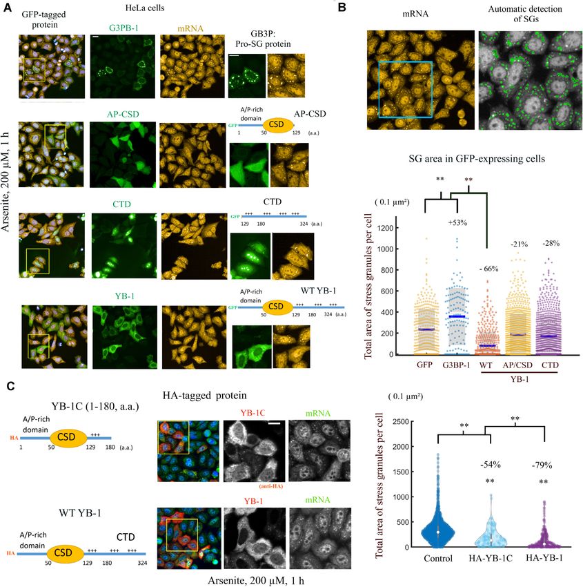
in HeLa cells to about 16–24% (Figure 8C–E and Supple- may negatively regulate SG assembly under physiological
mentary Figure S9a, short-term expression inhibition with conditions. However, in agreement with previous reports
siRNA has been preferred to reduce the risk of compensa- (39,40) including a recent large scale CRISPR screen (42),
tion mechanism). In support of this result, a positive regu- decreasing the expression level of YB-1 by using three differ-
lation of polysome assembly by YB-1 has already been re- ent siRNAs had no marked impact on SG assembly (Figure
ported by others in Caenorhabditis elegans and myeloma 9A, B). This result was expected as endogenous YB-1 lev-You can also read