The mechanism of HAMLET-induced cell death
←
→
Page content transcription
If your browser does not render page correctly, please read the page content below
Institutionen för Laboratoriemedicin, Lunds Universitet, Sverige

The mechanism of HAMLET-induced cell death
         -   cellular signalling, oncogenes and clinical perspectives

Akademisk avhandling som med vederbörligt tillstånd från Medicinska
Fakulteten vid Lunds Universitet för avläggande av doktorsexamen i
medicinsk vetenskap kommer att offentligt försvaras fredagen den 15e
juni 2012 kl 9.00 i GK-salen.

Handledare: Professor Catharina Svanborg
Fakultetsopponent: Professor Maria Masucci, Karolinska Institutet

                                                                        1
Till mina tjejer

                   2
LIST
OF
PAPERS
5
SUMMARY
6
SVENSK
SAMMANFATTNING
8
INTRODUCTION
10
The
Cancer
Cell
10
Oncogenes drive cancer progression
10
Tumour cells alter their metabolism
11
Ion channels as targets for anti-tumour therapy
12
The involvement of stress and MAPK signalling cancer
14
Exploiting the hallmarks of cancer for therapy
15
HAMLET
16
The discovery of HAMLET and molecular structure
16
The structure of the HAMLET complex
16
Molecular mechanism of HAMLETs effects
17
Mechanism of tumour cell death
18
Therapeutic aspects of HAMLET
18
PRESENT
INVESTIGATIONS
19
Aim
19
Paper
I
–
Conserved
features
of
cancer
cells
define
their
sensitivity
to
HAMLET-‐induced
death;
c-‐Myc
and
glycolysis
19
Aim
and
Background
19
Results
19
Conclusion
20
Paper
II
-‐
Selective
ion
channel
activation
by
HAMLET
explains
tumor
cell
death
20
Background
20
Results
20
Conclusion
21
Paper
III
-‐
Lipids
as
tumoricidal
components
of
HAMLET;
contributions
to
signalling
and
death
21
Background
21
Results
21
3Conclusion
22
Paper
IV-‐
HAMLET
treatment
of
colon
cancer
in
APCmin
mice;
peroral
application
and
disruption
of
the
Wnt/β-‐catenin
-‐signalling
pathway
22
Background
22
Results
22
Conclusion
23
DISCUSSION
23
HAMLET
acts
as
a
“magic
shotgun”
23
HAMLET
induces
a
transcriptional
response
characterized
by
p38
MAPK
signalling,
cell
death
genes
and
ER
stress
24
c-‐Myc
and
tumour
cell
metabolism
determine
HAMLET
sensitivity
25
The
HAMLET-‐activated
ion
channel
remains
elusive
26
HAMLET
triggers
ER
stress
in
carcinoma
cells
27
Glycolysis
inhibition
as
a
target
for
tumour
therapy
28
MAP
Kinases
are
critically
involved
in
the
tumouricidal
response
to
HAMLET
29
HAMLET
triggers
an
immunogenic
cell
death
which
should
be
optimal
for
in
vivo
anti-‐cancer
activity
30
HAMLET
shows
therapeutic
efficacy
in
a
model
of
colon
cancer
31
ACKNOWLEDGEMENT
32
REFERENCES
33
4LIST
OF
PAPERS
I
Conserved features of cancer cells define their sensitivity to HAMLET-
induced death; c-Myc and glycolysis
Petter Storm*, Sonja Aits*, Manoj K Puthia*, Alexander Urbano, Trent
Northen, Scott Powers, Ben Bowen, Yinxia Chao, Wolfgang Reindl, Do Y
Lee, Nancy L Sullivan, Jack Zhang, Maria Trulsson, Henry Yang, James D
Watson and Catharina Svanborg
Oncogene (2011) 30, 4765–4779, *Equal contribution
II
Selective ion channel activation by HAMLET explains tumor cell death
Petter Storm, Thomas Kjaer Klausen, Maria Trulsson, James Ho Chin
Shing, Marion Dosnon, Tomas Westergren, Yinxia Chao, Anna Rydström,
Priscilla Lay Keng Lim, Henry Yang, Stine Falsig Pedersen and Catharina
Svanborg
Submitted
III
Lipids as tumoricidal components of HAMLET;
Unique and shared effects on signalling and death
James Ho CS*, Petter Storm*, Anna Rydström, Ben Bowen, Fredrik Alsin,
Trent Northen and Catharina Svanborg
Manuscript, *Equal contribution
IV
Prevention of colon cancer development by peroral administration of
HAMLET (human !-lactalbumin made lethal to tumor cells) in
APCmin mice
Manoj K Puthia, Petter Storm, Aftab Nadeem, Godo Urbano, Catharina
Svanborg
Manuscript
5SUMMARY
Despite recent advances in cancer treatment, truly innovative approaches
are required to move beyond the modest benefits achieved to date.
HAMLET is a human protein-lipid complex originally discovered in
breast milk with properties making it a suitable candidate for cancer
therapy. Importantly, HAMLET has been shown to kill a wide range of
tumour cells while leaving healthy, differentiated cells unaffected.
HAMLET consists of α-lactalbumin, the most common protein in human
breast milk, which has adopted a molten globular structure that enables it
to bind oleic acid. The molecular mechanism of HAMLET induced cell
death has been extensively studied but a unifying mechanism for the
initiation and execution of cell death in response to HAMLET has not
been identified. The aim of this thesis was to identify components
dictating the HAMLET sensitivity of tumor cells and to define the events
that lead to cell death in response to HAMLET. Additionally, the relative
contribution of the fatty acid and the protein is discussed. Finally, the
therapeutic effects of HAMLET are extended to colon cancer, in a mouse
model with human relevance.
In Paper I we investigated the molecular basis of HAMLET sensitivity in
tumour cells. By systematic knockdown of ~1600 cancer-implicated genes
in an shRNA screen, c-Myc and glycolytic targets were identified as
important determinants of HAMLET sensitivity. Knockdown of c-Myc
with individual shRNAs in tumour cells rescued tumour cells from
HAMLET induced cell death. In contrast, glucose starvation of tumour
cells or the simultaneous application of glycolysis inhibitors significantly
enhances tumour cell death. Potential binding partners to HAMLET were
identified using a protein microarray approach. Hexokinase 1, the
enzyme responsible for trapping glucose in the cytosol, was identified as
a binding partner and HAMLET was shown to induce rapid metabolic
paralysis with a reduced abundance of metabolites in the glycolysis
pathways as well as accumulation of lipid metabolites. In conclusion,
these results show that the activity of genes responsible for hallmarks of
cancer, including oncogenes and the Warburg effect are important
determinants of HAMLET sensitivity.
In paper II the initiating events for tumour cell death are elucidated. By
rapid activation of a single ion channel the fluxes of cations were shown
to be disturbed in tumour cells. These fluxes could be amended by pre-
treatment with two broad ion channel inhibitors. These inhibitors also
abrogated the majority of HAMLET induced cellular changes including
uptake and cell shrinkage, transcriptional responses and ultimately, cell
death. The transcriptional response to HAMLET was further elucidated
and found to involve activation of the p38 MAPK signalling pathway.
Activation of p38 MAPK was found to be crucially involved in tumour
cell death as pre-treatment with p38-inhibitors or knockdown of p38
6reduced tumour cell death and morphological responses. In contrast to
tumour cells, healthy differentiated cells did not activate p38 but an
innate immune response. This provides a potential unifying mechanism
for HAMLET’s tumoricidal effect.
Paper III analyses the relative contribution of the protein and lipid
constituents to the tumoricidal effect of HAMLET. Oleate is the
deprotonated form of oleic acid and was identified as the functional lipid
in HAMLET formation and cell death, including ion channel activation,
gene expression effects, morphological changes and cell death. Oleate
alone did not account for the properties of the complex, as only minor
effects on ion fluxes and morphology were observed. By metabolomic
analysis, HAMLET-induced metabolic paralysis was not reproduced
when cells were treated with oleate or oleic acid alone. These results
show that the lipid acts on certain cellular targets in tumor cells, but that
the protein and lipid are both necessary for the tumoricidal effect of
HAMLET.
In Paper IV HAMLET was identified as a new colon cancer therapeutic,
which, if given early in life, also acts prophylactically. The risk to develop
colon cancer is increased in families carrying specific mutations and a
colon cancer model has been established in ApcMin/+ mice carrying the
same mutation. Heterozygous APC mutations cause the Wnt-signalling
pathway to be overactive and lead to adenoma formation in the mouse
intestine. Peroral HAMLET treatment of established tumours caused a
significant reduction in tumour numbers and mortality after 40 weeks,
accompanied by a reduction in nuclear β-catenin levels by an ion channel
dependent mechanism. Surviving tumours showed a reduction in a
number of onco-proteins and increased expression of glycolic enzymes,
indicating that active glycolysis might be essential to survive HAMLET
challenge. Finally, prophylactic use of HAMLET in the drinking water
was shown to prevent tumor formation in APC min mice.
In summary, this thesis defines the HAMLET-sensitive phenotype on
basis of an oncogenic program re-written by c-Myc and other oncogenes.
A unifying mechanism for HAMLET-induced cell death is also presented
and shown to involve rapid ion channel activation propagated through
p38 MAPK signalling. The charged form of oleic acid is shown to be the
important agonist in HAMLET and metabolic effects are discussed.
Finally, HAMLET is shown to be therapeutically and prophylactically
active in a mouse model of human colon cancer.
7SVENSK
SAMMANFATTNING
1971 förklarade president Richard Nixon, som
så många amerikanska presidenter före
honom, krig. Nixons fiende var dock något
oortodox; Nixon förklarade krig mot cancer. I
och med att senaten godkände ”National
Cancer Act”, av pressen snabbt döpt till ”War
Source: Wikipedia
on cancer”, hoppades Nixon att kunna
utradera cancer med hjälp av kraftigt ökade
anslag till cancerforskning kombinerat med
snabba framsteg inom molekylär- och
cellbiologi. Självklart har framsteg gjorts, men
40 år senare svårt att se Nixons krig som någon succé.
Behandlingsformerna är fortfarande trubbiga och biverkningarna ofta
svåra. Så varför är det så svårt att besegra cancer och vad gör en
cancercell speciell?
Varje cell har en uppsättning med ritningar i sig, cellens genom (som
består av DNA). Genomet innehåller instruktioner, gener, för hur
kroppens runt 20 000 olika proteiner ska byggas. I en cancercell har dessa
gener förändrats genom så kallade mutationer. En mutation kan ge
proteinet nya egenskaper eller göra det obrukbart. En tumörcell
karaktäriseras av att den har ackumulerat ett flertal felaktigheter i gener,
en process som tar många år, därav är cancer vanligare ju äldre man blir.
En cancercell har fått genetiska förändringar som ger den ’’överdrivna’’
egenskaper. Den delar sig ohämmat och har nästan oändligt liv då den är
okänslig för stoppsignaler och inte kan begå självmord. Dessutom har
cancerceller förmågan att undvika immunförsvaret och att sprida sig till
andra delar av kroppen. Vägen från en cell som får sin första mutation till
en fullt metastaserande tumör är dock lång och vinglig. Varje cancer i
varje patient tycks vara i det närmaste unik. Att prata om exempelvis
bröstcancer som en sjukdom blir därför felaktigt. Bröstcancer är
egentligen en uppsättning av olika cancerformer beroende på vilka gener
som förändrats, och varje unik genuppsättning ger en unik sjukdom, med
olika svar på behandling och chanser till överlevnad. Inte nog med att
samma tumörtyp skiljer mellan olika patienter, den senaste forskningen
visar att även tumörceller från samma patient tycks skilja sig mycket åt.
Därför det är så svårt att bota cancer. Vi har inte en enskild sjukdom att
besegra, utan ett spektrum av sjukdomar som kan ha mycket litet
gemensamt.
Forskningen skapar ständigt nya idéer och möjligheter för
cancerbehandling, och det gäller att utveckla dessa till nytta för patienten.
I den här avhandlingen presenterar jag vad som skulle kunna kallas “ett
magiskt hagelgevär” för cancerceller. HAMLET är ett humant protein-
derivat bestående av två i bröstmjölk rikligt förekommande
8komponenter; proteinet α-laktalbumin och fettsyran oleinsyra. I
HAMLET har dessa förenats och bildat en ny veckningsstruktur
stabiliserad av fettsyran. Denna nya veckningsstruktur ger i sin tur
HAMLET helt nya funktioner jämfört med α-laktalbumin; HAMLET har
förmågan att döda en bred grupp av tumörceller från olika organ, med
olika genetiska förändringar och från olika organismer. Till skillnad från
tumörceller tycks friska celler inte påverkas nämnvärt av HAMLET. Det
första arbetet i denna avhandling besvarar frågan – vad gör en tumörcell
känslig för HAMLET? Genom att systematiskt slå ut en rad gener i
tumörceller visar vi att ett protein kallat c-Myc är en drivande kraft. En av
c-Mycs funktioner är att optimera tumörcellens ämnesomsättning för
uppbyggnad av nya celler. Vi visar att denna förändrade
ämnesomsättning är en nyckel för HAMLET-känslighet samt att
HAMLET tycks stänga av tumörcellsmetabolism genom att direkta binda
till ett av nyckel-proteinerna för nedbrytande av socker.
HAMLET har tidigare visats påverka en rad viktiga funktioner i cellen
men det har tidigare saknats en enhetlig mekanism som förklarar varför
och hur HAMLET dödar så många olika typer av tumörceller. I
avhandlingens andra arbete studerar vi de första stegen som leder till
tumörcellernas död. Vi visar att HAMLET stör tumörcellernas
membraner och aktiverar jonkanaler, som ser till att balansen av joner
(kalcium, natrium och kalium) hålls på en för cellen hälsosam nivå.
HAMLET-behandling leder till att jonkanaler öppnas och obalansen i
jonnivåerna drar igång celldödsprogram med proteinet p38 som en viktig
komponent. Dessa resultat klarlägger HAMLETs initiala effekter på
tumörceller och föreslår också att jonkanaler kan vara attraktiva
målmolekyler för cancerterapi. I arbete tre visar vi att både fettsyran och
proteinet förklarar HAMLETs egenskaper och att båda är nödvändiga för
HAMLETs fulla effekt. Den laddade varianten av fettsyran är också den
biologiskt aktiva formen i t.ex. cellupptag, vilket är generellt intressant
information kring fettsyrors funktion i celler. Vi har också visat att
HAMLET fungerar som terapi mot cancer i tjocktarmen. Möss med en
mänsklig genetiska defekt utvecklar spontant tjocktarmscancer. Efter
peroral tillförsel av HAMLET kunde vi se en reduktion i antalet tumörer
och tumörstorlek. HAMLET var stabilt i tarmen och togs upp i
tumörvävnaden. Vi kunde också förebygga tumörutveckling genom att
tillföra HAMLET från tidig ålder. Betydelsen av dessa resultat kan inte
underskattas. Om resultaten bekräftas i kliniska studier skulle individer
genetisk benägenhet för koloncancer kunna erbjudas HAMLET i
förebyggande syfte.
Är HAMLET därmed det sista skottet i kriget mot cancern som Nixon
startade för 40 år sen? Det återstår att se men i denna avhandling har vi
lagt viktiga bitar i HAMLET-pusslet genom att visa på mekanismer för
tumörcellers död, egenskaper som gör dem känsliga för HAMLET samt
visar att HAMLET är effektivt i en tarmcancermodell.
9INTRODUCTION
The Cancer Cell
The cancer cell is characterized by a gain of genetic alterations that
enables the cancer cell to outgrow its non-transformed neighbours, evade
cell death, sustain proliferation in a hostile environment and invade and
metastasise into adjacent tissue. Hanahan and Weinberg proposed that
these biological capabilities constitute the “hallmarks of cancer” [1],
acquired by tumour cells during their multistep evolution from a single
transformed cell into a metastasising tumour. Importantly, tumours are
more than a mass of rapidly proliferating lump of cells but a complex
mixture of cell types participating in distinct biological processes. In
addition, tumours do not entail a single cell clone with identical genomic
alterations but rather a complex genomic mix up where different cells
within the tumour showing distinct advantages for proliferation,
resistance to apoptosis or chemotherapy. In addition to the heterogeneity
within tumours considerable heterogeneity exists between tumours [2].
Whereas two tumours originating from the same tissue or cell type,
through a process analogous to Darwinian evolution of species they
might end up with vastly different genotypes and phenotypes.
Collectively these traits make eradication of a tumour with a single
targeted agent a daunting task.
Oncogenes drive cancer progression
Arguably the most prominent fundamental characteristic of cancer cells is
their ability to sustain chronic proliferation. The life and death of non-
transformed cells is critically controlled by the production and release of
growth-promoting and anti-growth signals thereby maintaining tissue
homeostasis [3]. Cancer cells frequently abuse these signals to promote
indefinite proliferation thereby becoming masters of their own destiny
[4]. Whereas a normal cell is critically dependent on growth signals such
as EGF and NGF for proliferation, a tumour cell avoids this limitation by
deregulating pathways important for growth factor signalling.
Growth factor signalling starts by the engagement of a receptor on the
cell surface. Numerous examples of overexpression of these receptors
occur in tumour cells, for example HER2 in breast cancer [5] and EGFR in
brain cancers. By upregulation of these receptors, ambient levels of
growth factors might trigger cell proliferation or the upregulation itself
might induce ligand-independent signalling. More complex mechanism
of self-sustaining of growth signals might occur when downstream,
cytoplasmic signalling nexus proteins are mutated. In particular,
mutations in the RAS/RAF/MEK/ERK-pathway are found among a very
large fraction of tumours [6]. For example, a large proportion of
10melanomas carry a mutation in BRAF V599E, acting as phosphomimetic
turning the BRAF constantly on thereby constantly bombarding the cell
with mitogenic signals forcing the cell into a constant program of cell
cycling and cell division. It might be argued that activation of growth
signalling pathways might be present in all tumours although this is hard
to prove formally. In colon cancer, as an example, KRAS is found
mutated in around 50% of clinically assessed samples. The remaining
tumours could acquire growth signals by for example receptor over
expression, mutations in other downstream effectors or copy number
alterations of critical transcription factors.
One fundamental oncogene commonly altered to sustain proliferation is
c-Myc, a basic helix-loop-helix zipper (bHLHZ) motif–transcription
factor. Cellular levels of c-Myc are tightly regulated at multiple steps
including transcription, translation and protein. The activity is also
tightly regulated by its direct binding to another bHLHZ protein MAX
[7]. Cancers frequently shows overactive c-Myc due to gene
rearrangements and amplifications. Over activity can also stem from
mutations in the Ras/Raf/MEK/ERK pathway, causing increased c-Myc
mRNA transcription as well as stabilization of the c-Myc protein. c-Myc
activation can lead to transcriptional activation or repression of specific
genes. c-Myc plays a role in multiple signalling pathways including those
involved in cell growth, cell proliferation, metabolism, microRNA
regulation, cell death, and cell survival.
Tumour cells alter their metabolism
One fundamental consequence of oncogene activation seems to be a re-
wiring of tumour cell metabolism. Originally discovered in the 1920s and
extended in the 1950s [8], Otto Warburg made the striking observation
that, even in the presence of ample of oxygen, tumour cells favoured to
metabolize glucose by glycolysis rather than oxidative phosphorylation.
In the presence of oxygen, non-transformed cells metabolize glucose into
pyruvate and then completely oxidize pyruvate into CO2 during the
process of oxidative phosphorylation [9]. This is a highly efficient process
generating a net of up to 36 ATP molecules per molecule of glucose. In a
non-transformed cell low on oxygen, pyruvate is redirected from the
mitochondrial oxidative phosphorylation and instead converted into
lactate, a highly inefficient process generating a net output of only 2
molecules of ATP. What Warburg observed was that tumour cells,
regardless of oxygen availability, favoured glucose to lactate conversion.
This phenomenon, termed “Warburg effect”, was later shown to be
almost omnipresent in tumours and the concomitant increase in glucose
uptake has been exploited clinically for the detection of tumours by
flourodeoxyglucose positron emission tomography.
11Warburg was initially unable to explain the altered metabolism and early
research focused on defective mitochondria as the culprit for the Warburg
effect [10]. Recent research has however shown that most cancer cells
have fully functional mitochondria and that this is not the primary
explanation [11]. One alternative hypothesis would be that the hypoxic
environment within the tumour favours an oxygen-independent
metabolism. However, tumours cells with ample of oxygen, such as those
in blood and the lungs, also favours aerobic glycolysis [9]. Modern
research indicates that aerobic glycolysis is not an adaptation of tumour
cells to a harsh environment or an inborn error but that the altered
metabolism is beneficial for the tumour cell in that it allows the diversion
of glycolytic intermediates into various biosynthetic pathways [12]. In
essence, the Warburg metabolism is a metabolism fine-tuned for the
assembly of new cells, arguably the fundamental hallmark of cancer.
Importantly, signalling pathways activated by oncogenes have been
shown to directly control metabolic pathways. For example, activation of
the PI3K/Akt-pathway, a well characterised downstream effector of
growth factor receptors, cause upregulation of glucose transporters,
trapping of glucose intracellularly by hexokinase and commitment of
glucose to glycolysis by activation of phosphofructokinases [13,14].
Additionally, Ras, which is frequently mutated in a number of tumours,
increases glucose influx by upregulation of GLUT1 [15]. Also c-Myc has
prominent effects not only on the expression of glycolytic genes but has
also effect on mitochondrial biogenesis and glutamine utilization [16]. A
molecular explanation of the Warburg effect was recently provided by
Cantley and colleagues [17], where they provided evidence that an
alternative splice form of pyruvate kinase acts as gatekeeper for aerobic
glycolysis. This splice-form, termed PKM2, directs pyruvate towards
lactate and was found solely in tumour cells.
The identification of an altered cancer cell metabolism opens the avenue
for new, targeted therapies. Given that all cells rely on glycolysis for
survival it could be expected that a metabolism-targeting agent would
have precluding side effects and that the therapeutic window would be
narrow [18]. However, tumour cells have an unprecedented appetite for
glucose and molecules targeting either this weakness or drugs that forces
tumours into a normal-cell metabolism are plausible as effective anti-
cancer agents.
Ion channels as targets for anti-tumour therapy
Plasma membrane ion channels are involved in all basic cellular
processes important for tissue homeostasis, proliferation, differentiation
and cell death [19]. The major mechanisms by which ion channels
contribute to these effects include influx of essential signalling ions,
cellular volume regulation and maintenance of membrane potential. Ion
12channels can roughly be classified into three groups, according to the
mode of activation (gating). Voltage-gated channels are a group of ion
channels whose activity changes with the transmembrane voltage. The
binding of substances to the ion channel regulates the opening of ligand-
gated channels. Mechanosensitive channels comprise a group of channels
gated by mechanical force that is generally generated by membrane
stretch. Despite the variation in the stimulus type the outcome is the
same: channel opening and closing.
Ample of evidence for the involvement of ion channels in cancer
development are beginning to emerge [20]. In particular, K+ channels and
their regulation of membrane potential, which in turn regulates
transmembrane Ca2+ fluxes, have gained considerable interest [21]. For
example, members of the Transient Receptor Potential (TRP) channels
have been shown to be upregulated and involved in androgen
insensitivity and apoptosis resistance in prostate cancer cells [22]. K+ also
seems to be critically involved in regulation of metastasis and cell
movement, by regulation of downstream signalling pathways involving
tyrosine kinases and GTPases [23]. Ion channels also regulate intracellular
pH [24], which is fundamental to promote a proliferative phenotype, but
also to counteract negative feedback from the cancer metabolism depicted
above.
Ion fluxes are also involved in the regulation of cell death [25,26]. In
particular the cell shrinkage, an early event in apoptotic cell death, is
attributable to the efflux of K+ ions [27]. Concomitantly, activation of
caspases, mitochondrial depolarization and endonuclease activation is
dependent on K+ efflux. High concentrations of extracellular K+ have been
shown to abrogate both the extrinsic and intrinsic cell death programs by
inhibition of cytochrome c release [28]. In addition to K+, Ca2+ seems to
play an important role in apoptosis induction [29]. Cytosolic Ca2+ are
usually kept a low levels (~100 nM) through shuttling of intracellular
calcium into the ER and Ca2+ extrusion into extracellular space by the
plasma membrane Ca2+-ATPase. Ca2+ overload or perturbation of
intracellular calcium stores by for example ER stress or activation of
plasma membrane Ca2+ channels is able to initiate a number of apoptosis-
related events, including endonuclease activation and Ca2+ activated
cysteine proteases, such as calpains.
Ion channels are widely used as therapeutic targets for a wide range of
diseases, for example calcium blockers for myopathies and lidocain (a
sodium blocker) for local anaesthetics. However, the application of ion
channels for tumour therapy is still in its infancy [30]. In principle, an ion
channel drug could work by a multitude of mechanisms, for example, by
binding to the agonist site or blocking the pore. It could also act indirectly
by binding to allosteric sites or affect the binding of the ion channel to
downstream signalling partners. The K+ channel hERG1 has been under
13particular scrutiny as it is frequently overexpressed and known to control
a number of behaviours related to cancer cells [31]. hERG1 blockers have
been tested and shown to decrease proliferation in vitro and also decrease
the growth of tumour engraftments in mice [32]. However, the hERG1
blockers also induced life threatening cardiac repolarization side effects,
highlighting the pitfalls of targeting widely expressed channels for
tumour therapy. The identification of tumour specific ion channel
aberrations, for example upregulation, isoform assembly or mutations,
would be a great aid for the development of tumour therapy.
Concomitantly, agents acting specifically for the activation or inhibition
could have immense clinical benefit.
The involvement of stress and MAPK signalling cancer
The extensive rewiring of normal pathways for the benefit of the cancer
cell is stressful for the cellular machinery. Such stresses include DNA
damage/replication stress, proteotoxic stress, metabolic stress and
oxidative stress, collectively called the “stress phenotype of cancers” [33].
The DNA damage response originates from the highly unorganized state
of the tumour cell genome. Tumour cells frequently show alterations of
telomeres causing the formation of abnormal chromosomes and
numerous amplification and deletion events. In addition to this, DNA
damage repair pathways are usually inactivated or non-functional in
tumour cells. The highly glycolytic state of tumour cells will also put the
metabolic machinery under heavy stress, leading to a build up of
unwanted metabolites as well as reactive species. Finally, the constantly
dividing cancer cell shows a high protein production rate, exhausting the
protein synthesis machinery and leading to ER stress.
Mitogen activated proteins kinases (MAPKs) are crucial for the cellular
response to stress [34]. Three MAPK signalling units have been
characterized in detail: the extracellular signal-regulated kinases (ERKs),
the c-Jun amino-terminal kinases (JNK) or stress-activated protein kinases
(SAPK), and the p38 MAPKs (p38). Mitogens, inflammatory cytokines,
and growth factors are known to activate various MAPK signalling
pathways, whereas cellular stresses such as UV light, heat, or osmotic
shock selectively induce the JNK/SAPK and p38 MAPK pathways. A
shared feature among MAPKs is their activation by phosphorylation of
both threonine and tyrosine residues by a dual-specificity serine-
threonine MAPK. In turn, MAPKs frequently phosphorylate their
substrates at serine or threonine residues adjacent to prolines.
Fundamental outcomes of cellular stress include cell cycle arrest,
commitment to apoptosis, the activation of DNA-repair pathways,
regulation of protein translation, and the initiation of immune responses
[35]. p38 orchestrates these responses by direct phosphorylation of a
14number of substrates including the transcription factors ATF2 (a bZIP
family transcription factor with diverse roles in development, cell growth
and survival), MEF2 (important for cell differentiation and
organogenesis), DDIT3 (produced in response to DNA damage), and the
tumour suppressor p53.
Exploiting the hallmarks of cancer for therapy
The explosion of knowledge in cancer pathogenesis during the past
decades has evolved the paradigm of cancer therapy from relatively
nonspecific cytotoxic agents to selective, mechanism-based therapies [36].
Whereas traditional cancer chemotherapies were initially identified by
their ability to kill rapidly dividing cells, their efficacy is severely limited
by a narrow therapeutic index, significant toxicity due to their unspecific
mode of action and cancer cells ability to acquire resistance [37]. Targeted
therapies in contrast act by blocking essential pathways or mutant
proteins found exclusively in tumour cells. The first targeted agent, was
directed to a specific translocation (t(9;22)(q34;q11)) occurring in around
95% of patients with chronic myelogenous leukaemia and giving rise to
the fusion gene BCR-ABL1 [38]. Inhibiting the oncogenic kinase BCR–
ABL1 is a paradigm for clinically successful targeted therapy. A more
recent success is the treatment of melanoma, a cancer form with a dismal
prognosis. Overactivity of the RAS/RAF/MEK/ERK pathway feeds cells
with a constant bombardment of proliferative signals [39] and melanomas
frequently turn on this pathway by a specific mutation in BRAF.
Vemurafenib was developed to specifically inhibit BRAF bearing a
mutation at V599E present in around half of all melanomas [40]. The
observation that inhibition of a single mutant protein might eradicate an
entire tumour might seem counterintuitive, however. To explain this
phenomena the “oncogene addiction” hypothesis has been proposed.
This dogma entails that tumour cells for their well-being are highly
addicted to the activation of a single oncogene and that disturbance of
this oncogene alone is sufficient to kill a tumour cell.
Even though some of the targeted therapies have shown impressive
initial clinical response they usually fail to give long-term clinical
benefits. This probably reflects the high heterogeneity existing both
between different tumours originating from the same organ but also
intra-tumoural heterogeneity. For effective cancer therapy targeting a
single oncogenic aberration is unlikely to be enough, a more
comprehensive attack on the tumour is likely to be necessary.
15HAMLET
The discovery of HAMLET and molecular structure
HAMLET was discovered by serendipity while studying anti-adhesive
molecules in human milk [41]. Tumour cells were shown to undergo
morphological changes consistent with apoptosis when treated with the
casein-containing fraction. The protein component responsible for the
tumouricidal activity was identified as α-lactalbumin [42], the most
abundant protein in human breast milk with a well-known function as a
substrate specifier in the lactose synthase complex. However, a
tumouricidal function for α-lactalbumin had not been previously
described. Early studies of the α-lactalbumin complex suggested that a
mulitmeric form of α-lactalbumin was responsible for the activity and the
complex was hence named multimeric α-lactalbumin (MAL) [43].
However, MAL could not completely explain the effects seen and it was
soon realized that a cofactor was involved in the conversion of α-
lactalbumin into a cytotoxic entity. Mass spectrometry ruled out a
HAMLET-the first in a new family of
covalent modification whereas CD spectroscopy identified a stable,
tumoricidal molecules
partially unfolded form of α-lactalbumin. The factor responsible for the
stabilization was identified as oleic acid, the most common fatty acid in
human breast milk.
! = human alpha-lactalbumin
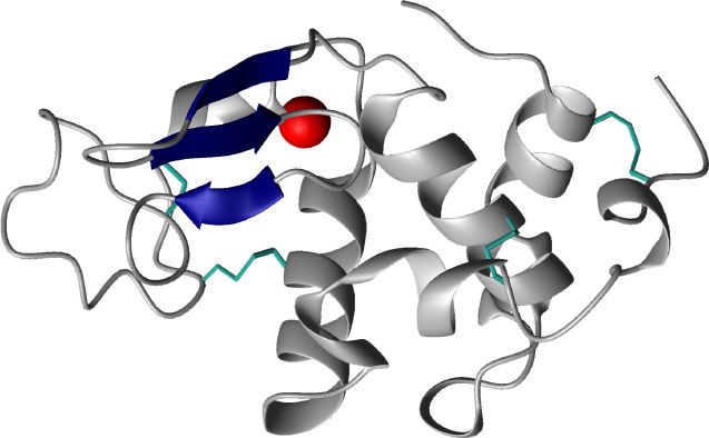
The structure made lethal to
of the HAMLET complex
tumour cells HAMLET consist of a single
molecule of α-lactalbumin protein
complexed with 4-7 oleic acid
residues [44]. α-lactalbumin is the
most abundant protein in human
breast milk and is made up of 123
amino acids [45]. In its native
conformation α-lactalbumin is a
tightly packed globular protein
that is stabilized by a calcium ion
+,-./0"/.1 and four disulphide bridges [46].
!"#$
Structurally, α-lactalbumin can be
divided into two α-helical
domains in the C- and N-terminal
%&'()* ends of the molecule and one
bridging β-sheet domain. When the calcium is removed from the protein,
using for example low pH or EDTA, the protein will adopt a molten
globule structure with retained secondary structure but loss of tertiary
structure.
16By unfolding of α-lactalbumin, new epitopes with the ability to bind oleic
acid is presented [47,48]. Structural analysis of the HAMLET complex by
ANS and CD spectroscopy, and more recently using SAXS (Ho et al.,
unpublished), has shown that the HAMLET complex retains a partially
unfolded state but with most of the secondary structure intact. SAXS
analysis identified a two-domain structure with the c-terminal end of the
polypeptide forming an extended leg, which might create a conformation
that is responsible for the cellular effects.
Molecular mechanism of HAMLETs effects
Unlike targeted therapies, by some designated as magical bullets,
HAMLET affects multiple cellular targets. HAMLET has been shown to
interact with artificial membranes suggesting that specific receptor
interactions are not required for these interactions to occur [49]. When
rounded, defined vesicles were challenged they changed morphology to
amorphous shapes reflecting the formation of long membrane distensions
and increased fluidity. A similar response to HAMLET was observed in
plasma membrane vesicles from carcinoma cells, suggesting that direct
membrane effects might contribute to the tumouricidal effect of
HAMLET. HAMLET has also been shown to rapidly enter tumour cells
but not non-transformed cells, through a mechanism that remains to be
explained [50]. Recent results also suggest that HAMLET interacts with
cellular proteins important for cell attachment [51]. Trulsson and co-
workers showed that HAMLET-binding to α-actinin disrupts focal
adhesions, causes detachment and effects signalling downstream of focal
adhesions.
Once intracellular, HAMLET reaches multiple targets. Early studies
identified histones as critical targets in tumour cells [52]. HAMLET was
found to strongly bind histone H3 and to lesser extent histones H4 and
H2B using immuno-precipitation and surface plasmon resonance
technology. In vitro, HAMLET formed insoluble precipitates with
histones probably defining the final act of the cell death program. In
congruence with these effects, pretreatment of tumour cells with histone
de-acetylase inhibitors (HDIs) was shown to enhance the lethal effect of
HAMLET [53] and the histone hyperacetylation response to HDIs
increased even further after HAMLET treatment. Internalized HAMLET
is also targeted to 20S proteasomes and inhibits proteasome activity and
perturbs proteasome structure [54]. In the same study, HAMLET was
also shown to be relatively resistant to degradation by proteasomes when
compared to α-lactalbumin.
17Mechanism of tumour cell death
Although HAMLET is able to activate an apoptotic response and caspases
in tumour cells, this activation is not critical for cell death as caspase
knock-out cells or cells pretreated with zVAD-fmk are still HAMLET
sensitive [55]. The notion that classical apoptosis is not critically involved
is further emphasized by the fact that neither p53 nor Bcl-2 affects
HAMLETs activity. Autophagy seems to play some role in the response
to HAMLET as HAMLET-treated tumour cells show definite signs of
autophagy, such as granular LC3-II staining and reduced levels of active
mTOR [56]. However, the contribution of autophagy to cell death is not
fully understood as opposing effects has been reported [57].
Jäättelä and colleagues identified the lysosome as one critical component
of the cell death pathway activated by HAMLET [57]. Using BAMLET,
the bovine counterpart of HAMLET, they were able to show that
stabilization of the lysosome by overexpression of HSP70 abrogated the
cytotoxic effect of HAMLET. Interestingly, HAMLET was recently shown
to kill bacteria with characteristics similar to that of apoptosis [58].
Streptococcus pneumoniae death was accompanied by apoptosis-like
morphology such as cell shrinkage, DNA condensation and degradation.
The effects on the bacteria were similar to those observed on eukaryotic
mitochondria, and both these effects were linked to calcium transport.
Therapeutic aspects of HAMLET
In addition to its efficiency as a cancer cell killer in vitro, HAMLET has
shown great promise as a therapeutic agent in vivo. In a placebo-
controlled clinical study, therapeutic efficacy against skin papillomas was
observed [59]. The lesion volume was reduced by 75% or more in all 20
patients treated with HAMLET compared to only 3 of 20 patients
receiving placebo. Rapid topical effects on human bladder cancers were
also seen in human patients receiving intra-uteral instillations of
HAMLET [60] with the long-term effects established in murine bladder
cancer model [61]. Whole body fluorescence imaging showed that Alexa-
labelled HAMLET was retained in the bladder of tumour bearing mice for
more than two days whereas tumour-free mice rapidly discarded the
HAMLET-solution. Local infusion of HAMLET into rat brains with
invasively growing human glioblastoma xenografts delayed tumour
development and prolonged survival [62]. Apoptosis was mainly
confined to the tumour area, although HAMLET diffused throughout the
infused hemisphere. In conclusion, clinical studies reported so far have
established HAMLET as a therapeutic agent inducing tumour specific cell
death and uptake and with no toxic effects on healthy tissues.
18PRESENT
INVESTIGATIONS
Aim
The overall aim of this investigation was to identify the mechanism
underlying HAMLET sensitivity as well as to identify critical components
explaining HAMLETs tumouricidal activity. The second objective was to
understand the mechanism of cell death, from the initial interaction of
HAMLET with the tumour cell to the final execution of death. Finally, the
translation of HAMLET into a therapy and the molecular mechanism
explaining HAMLETs in vivo effects were studied.
Paper I – Conserved features of cancer cells define their
sensitivity to HAMLET-induced death; c-Myc and glycolysis
Aim
and
Background
The genomes of all cancer cells carry somatic mutations. However,
tremendous heterogeneity exists both within tumours and between
tumours, an observation that has been unequivocally shown by recent
large scale sequencing efforts of wide ranges of tumours [2]. Interestingly,
HAMLET seems to identify characteristics shared by all cancer cells. In
this paper we investigated the basis for tumour cell sensitivity.
Results
Using a reverse-genetics approach we identified c-Myc as well as several
proteins related to the glycolytic flux as important modifiers of HAMLET
sensitivity. Using retroviral infection, an shRNA library was introduced
into A549 cells targeting 1600 cancer related genes. By treating these cells
with HAMLET and assessing the relative abundance of each shRNA
before and after HAMLET exposure we identified a candidate list of
genes important for HAMLET sensitivity. Among genes giving rise to
resistance to HAMLET, c-Myc was one of the most prominent hits. c-Myc
expression reflected the difference in HAMLET sensitivity, as lung and
kidney carcinoma cells show higher expression of c-Myc than healthy
cells. In addition, knockdown of c-Myc using individual shRNAs
conferred significant resistance to HAMLET. c-Myc is known to drive cell
cycle progression but this effect was not crucial for determining
HAMLET sensitivity. Instead, c-Mycs potential effect on the metabolic
state was investigated. Depletion of glucose or addition of the glycolysis
inhibitor 2-deoxyglucose significantly sensitized tumour cells to
HAMLET. Potential binding partners of HAMLET in the glycolytic
pathway were identified using a high-content functional protein array
containing 8000 human recombinant proteins. Hexokinase 1, with a
19pivotal role in trapping glucose within the cytosol, was identified as a
potential binding partner. The interaction between HAMLET and
Hexokinase 1 was confirmed using dot blots and confocal microscopy.
Finally, the overall impact of HAMLET on tumour cell metabolism was
investigated using a metabolomics approach. HAMLET-treated cells were
shown to contain high amount of oleic acid but also other fatty acid
metabolites, indicating a saturation of the lipolytic machinery. In
addition, a general shutdown of metabolism was evident 1 hour after
HAMLET treatment.
Conclusion
The sensitivity of tumour cells to HAMLET reflects the Hallmarks of
cancer, including oncogene expression and the Warburg effect.
Susceptibility was modified by c-Myc expression and by Hexokinase,
both affecting the metabolic state of tumour cells. HAMLET caused a
general shutdown of metabolism, as supported by the metabolomics
screen.
Paper II - Selective ion channel activation by HAMLET explains
tumor cell death
Background
HAMLET has been shown to enact a rapid, tumour cell-specific cell death
response. However, the identification of a ”HAMLET-receptor” has
remained elusive. In this study we aimed to reconcile recent observation
of a tumour membrane effect with early observation of an increase in
intracellular calcium [41]. We present a unifying mechanism for
HAMLET-induced cell death.
Results
Exposure of tumour cells to HAMLET was shown to induce a rapid efflux
of potassium and an influx of calcium and sodium. Broad inhibitors of
ion channels, including amiloride and barium chloride, were shown to
abrogate the fluxes, indicating the activation of an ion channel. Patch
clamping experiments confirmed these findings as evidence of activation
of a single ion channel with permeability for all three cations were
observed. Importantly, the ion channel activation could not be
reproduced using the free fatty acid or unfolded protein alone. Inhibitors
that prevented the current also prevented all aspects accompanying
20tumour cell death, including ATP drop, shrunken morphology and
signalling. Using transcriptomics we identified a transcriptional program
activated by HAMLET, characterised by activation of the p38 MAPK
pathway and upregulation of cell death and ER stress genes. The
activation of p38 was confirmed by phospho-specific antibodies and was
shown to be time and dose dependent. Activation of p38 is an integral
part of the cell death program as inhibition of p38, either by small
molecule inhibitors or siRNA, drastically reduced cell death. Healthy
cells responded to HAMLET challenge with innate immunity and
survival rather than a p38-dependent death response.
Conclusion
For the first time we provide a potentially unifying mechanism for
HAMLET induced cell death. A single ion channel is shown to be
activated by HAMLET and blocking ion fluxes abrogates most
downstream effects. These results also indicate that HAMLET may be
used to identify ion channels as attractive targets for cancer therapy.
Paper III - Lipids as tumoricidal components of HAMLET;
contributions to signalling and death
Background
The importance of the fatty acid for HAMLETs activity in relation to the
full complex remains controversial. Cis-monounsaturated fatty acid has
been identified as the optimal co-factor for HAMLET formation but the
relative contributions of the fatty acid, the unfolded protein and the
complex remains to be fully elucidated.
Results
HAMLET was formed with oleate or oleic acid and the resulting
complexes were compared in terms of their structure and ability to kill
tumour cells. Oleic acid or oleate formed HAMLET complexes with
similar efficiency and with structures that closely resembled each other.
Oleic acid was largely inert to tumour cells whereas oleate was able to
reproduce some of the effects of the HAMLET complex. Transcriptional
responses to HAMLET and high concentrations of oleate were largely
overlapping even though HAMLET caused upregulation of a distinct set
of DNA damage genes. By mass spectrometry analysis of the cellular
metabolome, both oleate and HAMLET triggered an increase in
intracellular lipid levels. However, only HAMLET triggered a general
21metabolic paralysis. In addition, HAMLET caused a depletion of cellular
carnitine levels and concomitant increase in oleoyl-carnitine levels.
Conclusion
Both oleic acid and oleate are efficient cofactors for formation of the
HAMLET complex. Cell appeared largely to be inert to oleic acid, but
oleate reproduced some of the complex responses to HAMLET. The full
complex consisting of both the fatty acid and the unfolded protein was
necessary for the full cell death response to occur.
Paper IV- HAMLET treatment of colon cancer in APCmin mice;
peroral application and disruption of the Wnt/"-catenin -
signalling pathway
Background
Previous studies have established HAMLET as an anti-cancer agent with
clinical potential. Colorectal tumours are frequently initiated by
inactivation of the APC tumour suppressor gene, which renders the Wnt-
signalling pathway overactive. Based on HAMLETs properties as a milk
protein and previously documented resistance to gastric enzymes, we
used APCMin/+ mice, which carry a germline mutation in APC, to study
the therapeutic potential of HAMLET in colon cancer.
Results
HAMLET was administered per-orally for ten days to tumour-bearing
mice and tumour development was assessed five weeks after the final
HAMLET administration. A significant reduction in tumour numbers,
tumour size, as well as in a number of key markers for proliferation and
oncogenesis were observed after HAMLET administration. HAMLET
was also shown to accumulate in tumour tissue but no gross side effects
were observed in adjacent healthy tissue. Transcriptomic analysis of
remaining tumour tissue identified a highly glycolytic phenotype in
surviving tumour cells, indicating that HAMLET might target glycolysis
for its effect. Untreated tumours showed higher expression of T-cell
marker genes, indicating that HAMLET might purge tumours of T-cells.
In vitro, HAMLET was shown to cause caspase, as well as ion channel,
dependent degradation of β-catenin and cause nuclear exit.
22Conclusion
These results indicate that HAMLET has potential as an anti-cancer agent
in colon cancer. It also reveals the Wnt pathway as one possible HAMLET
target. It also establishes the Warburg phenotype as important for
HAMLET sensitivity as well as confirms the importance of ion channels
for in vivo effects of HAMLET. The work confirms and extends previous
studies have established HAMLET as an anti-cancer agent with clinical
potential.
DISCUSSION
HAMLET acts as a “magic shotgun”
Contemporary targeted therapies have been described as magic bullets,
indicating that they targets tumour specific aberrations and thereby
specifically kills tumour cells. They are all designed to hit one single
target. HAMLET, in contrast appears to have multiple targets and this
feature is inherent to HAMLET’s tumoricidal effect. Since the discovery
of HAMLET, mitochondria, calcium signalling, histones, proteasomes, α-
actinins and the plasma membrane have been identified as HAMLET
targets. The cell death modality has been discussed as a function of
caspase activation, DNA damage, autophagy and lysosmal cell death
programs.
These mechanisms do not explain HAMLET-induced cell death, however.
Since the discovery of HAMLET we and others have searched for THE
events that initiate cellular attack by HAMLET, hoping for a unifying
mechanism of action. Knowing that the entire process of HAMLET
induced cell death is multifaceted and complex, is it still possible that
there are key molecular interactions that trigger the downstream events,
that converge on cell death? The ion fluxes induced by HAMLET suggest
that there are distinct early events, which, if inhibited, prevent
subsequent cellular responses. The molecular details of such interactions
need further study, however.
Acting on multiple targets and through multiple pathways is
traditionally not viewed as something positive. Concerns are associated
with unclear modes of action for drug development and litigation as well
as complex side effects. On the other hand, it is becoming increasingly
clear that targeting a single oncogenic aberration will rarely be curative,
as resistance due to acquired mutations develops rapidly.
23By the engagement of multiple pathways and cellular compartments,
HAMLET achieves several beneficial goals. Resistance does not develop
readily, as shown in vitro in cellular propagation studies. Loss of a single
target is unlikely to create resistance as many other interactions
potentially can take over and kill the cells. This spectrum of interactions
may also explain the broad effects of HAMLET against many different
tumours and the increased resistance of healthy, differentiated cells.
HAMLET induces a transcriptional response characterised by p38
MAPK signalling, cell death genes and ER stress
In the outset of this project we defined the transcriptional response to
HAMLET using microarray technology. By the simultaneous
quantification of all mRNA transcripts in one single experiment
microarray technology enables an unbiased approach to the study of cell
biology. By treatment of a carcinoma cell line with HAMLET, purification
of totalRNA and subsequent labelling and hybridisation to a whole-
genome human microarray we identified transcriptional regulation
falling in predominantly three categories; p38 MAPK signalling, cell
death genes and the ER stress pathway. HAMLET increased expression of
genes in the p38 MAPK signalling pathway. Cell survival and p38
signaling are transcriptionally and post-transcriptionally regulated,
through death receptors, survival pathways or pro- and anti-apoptotic
Bcl-2 proteins, which may activate p38 by secondary routes, e.g., by the
production of reactive oxygen species (ROS) [63]. The second category of
genes activated by HAMLET was involved in cell death. HAMLET has
been shown to increase the expression of for example KLF6, DDIT3,
GADD45 and ATF3, all key signaling components in cell death pathways.
Further analysis of the exact transcriptional regulation of cell death genes
will enable us to pinpoint more specifically the mechanism of cell death
execution by tumour cells after HAMLET treatment. Genes in the ER
stress pathway were also regulated by HAMLET, as discussed in detail
below. It can be debated if transcriptional regulation is crucial for
HAMLET induced cell death. However, when the p38 MAPK pathway
was blocked either using siRNA or pharmacological inhibitors, cells
failed to undergo cell death showing that microarray technology is
indeed useful to elucidate both cell death pathways but also bystander
phenomena.
Importantly, using transcriptomic technology, healthy cells were shown
to be much less responsive to HAMLET challenge. HAMLET was
identified as an immune activator, mostly targeting signaling pathways
involved in innate immunity. As a consequence, pro-inflammatory
cytokines were produced in healthy cells but this immune response was
lower or absent in tumor cells. The functional importance of these
pathways in healthy cells remains to be explored.
24c-Myc and tumour cell metabolism determine HAMLET
sensitivity
Deregulated expression of c-Myc has been shown to occur in a wide
range of human cancers and is often associated with poor prognosis,
indicating a key role for this oncogene in tumour progression [64].
Strikingly, it has recently been shown that the enhanced expression of c-
Myc contributes to almost all every aspect of cancer cell biology. The
ability of c-Myc to drive cellular proliferation and to inhibit cellular
differentiation has been known for a long time, but c-Mycs importance
for vasculogenesis, cell adhesion, cellular metabolism, metastasis and
genomic instability has recently gained considerable interest [65].
Consistent with this, disruption of c-Myc in c-Myc-induced transgenic
tumours triggers proliferative arrest and re-differentiation of tumour cells
usually resulting in rapid tumour regression [66]. Despite this, targeting
c-Myc for pharmacological therapy has proven challenging for a number
of reasons [67]; c-Myc exerts its effect through protein-protein and
protein-DNA interaction, which are usually not tractable with small
molecule inhibitors. Secondly, c-Myc is usually not mutated itself but its
effects are a consequence of induction by upstream oncogenic signals.
Finally, c-Myc is essential for proliferation of stem cells, indicating that
systemic blocking of c-Mycs function might trigger irreversible side
effects.
In paper I the activity of HAMLET was shown to be critically linked to
the expression of c-Myc. The dependence on c-Myc overexpression as a
determinant of HAMLET sensitivity was related to the metabolic
program employed by tumour cells. It should be noted that c-Myc has
widespread effects and is also known to induce expression of genes
driving cell proliferation. This effect is unlikely to explain the c-Myc-
dependence as cell cycle arrest of lung carcinoma cells (by serum
starvation or double-thymidine block) did not significantly alter
HAMLET sensitivity.
c-Myc does not only increase the expression of glycolysis related genes
but is also heavily involved in regulation of glutamine metabolism.
Glutamine is together with glucose the main substrate for catabolic
processes and tumour cells seems to have an extreme appetite for
glutamine, even so that some cancer patients shows depressed plasma
glutamine levels [68]. The knockdown of c-Myc is likely to force tumour
cells into a less “glutaminophilic” phenotype. Whether this effect of c-
Myc has any relation to c-Mycs effect on HAMLET sensitivity remains to
be elucidated.
Finally, c-Myc is also a critically regulated by β-catenin [65]. In paper IV,
intestinal tumours overexpressing β-catenin are shown to be sensitive to
25You can also read