Proteomics reveals synergy between biomass degrading enzymes and inorganic Fenton chemistry in leaf-cutting ant colonies - eLife
←
→
Page content transcription
If your browser does not render page correctly, please read the page content below
RESEARCH ARTICLE

                                   Proteomics reveals synergy between
                                   biomass degrading enzymes and
                                   inorganic Fenton chemistry in leaf-cutting
                                   ant colonies
                                   Morten Schiøtt*, Jacobus J Boomsma

                                   Centre for Social Evolution, Department of Biology, University of Copenhagen,
                                   Universitetsparken, Copenhagen, Denmark

                                   Abstract The symbiotic partnership between leaf-cutting ants and fungal cultivars processes
                                   plant biomass via ant fecal fluid mixed with chewed plant substrate before fungal degradation.
                                   Here we present a full proteome of the fecal fluid of Acromyrmex leaf-cutting ants, showing that
                                   most proteins function as biomass degrading enzymes and that ca. 85% are produced by the
                                   fungus and ingested, but not digested, by the ants. Hydrogen peroxide producing oxidoreductases
                                   were remarkably common in the proteome, inspiring us to test a scenario in which hydrogen
                                   peroxide reacts with iron to form reactive oxygen radicals after which oxidized iron is reduced by
                                   other fecal-fluid enzymes. Our biochemical assays confirmed that these so-called Fenton reactions
                                   do indeed take place in special substrate pellets, presumably to degrade plant cell wall polymers.
                                   This implies that the symbiotic partnership manages a combination of oxidative and enzymatic
                                   biomass degradation, an achievement that surpasses current human bioconversion technology.

                                   Introduction
*For correspondence:               Mutualistic mergers of simpler biological entities into organizationally complex symbioses have been
mosch@aqua.dtu.dk                  key steps in the evolution of advanced synergistic forms of life, but understanding the origins and
                                   secondary elaborations of such natural cooperative systems remains a major challenge for evolution-
Competing interests: The
                                   ary biology (Smith and Szathmáry, 1997; Bourke, 2011). Physiological complementarity may be a
authors declare that no
                                   main driver to maintain symbiotic associations, but new adaptations are also expected to jointly
competing interests exist.
                                   exploit the full potential of symbiosis (Herre et al., 1999; Foster and Wenseleers, 2006). The leaf-
Funding: See page 17               cutting ants belong to the genera Acromyrmex and Atta and form the phylogenetic crown group of
Received: 05 August 2020           the attine fungus-growing ants, which all live in mutualistic symbiosis with fungus-garden symbionts
Accepted: 09 January 2021          (Mueller et al., 2005). While the basal branches of the attine clade farm a considerable diversity of
Published: 12 January 2021         mostly leucocoprinaceous fungi, normally in underground chambers (Branstetter et al., 2017), the
                                   leaf-cutting ants almost exclusively farm what has been described as the highly variable cultivar spe-
Reviewing editor: Dieter Ebert,
University of Basel, Switzerland
                                   cies Leucocoprinus gongylophorus (Schultz and Brady, 2008). The ants cut leaf fragments from the
                                   canopy and understory and bring them back to their nest to be processed as growth substrate for
   Copyright Schiøtt and           the cultivar. This involves chewing forage material into smaller pieces, which are mixed with fecal
Boomsma. This article is
                                   fluid and deposited as new substrate on the actively growing parts of the garden (Weber, 1966).
distributed under the terms of
                                       The deep ancestors of the attine ants and their fungal cultivars were hunter-gatherers and
the Creative Commons
Attribution License, which         saprotrophs (Schultz and Brady, 2008), respectively, which implies that none of them were adapted
permits unrestricted use and       for efficiently degrading fresh plant material. Neither was there any need initially, because all phylo-
redistribution provided that the   genetically basal attine ants provision their gardens with dead plant debris. There is a striking differ-
original author and source are     ence between the small colonies of these basal attine ants and the large colonies of leaf-cutting ants
credited.                          which are conspicuous functional herbivores with a large ecological footprint (Mehdiabadi and

Schiøtt and Boomsma. eLife 2021;10:e61816. DOI: https://doi.org/10.7554/eLife.61816                                                  1 of 21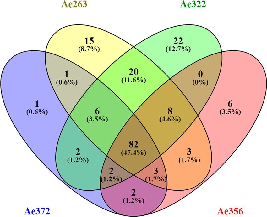
Research article                                                                                       Ecology Evolutionary Biology

                                  eLife digest Colonies of tropical leaf-cutting ants live in underground nests where a fungus
                                  grows that feeds them. The ants, in turn, provide the fungus with the freshly-cut leaf fragments it
                                  needs for nutrition. The relationship between the ants and the fungus, in which they live close
                                  together and help one another survive, is known as symbiosis. It is an ancient, extremely well
                                  integrated relationship, in which neither species can survive without the other. However, the details
                                  of how the ants and the fungus work together to break down the leaf fragments so they can be used
                                  for nutrition are not well understood.
                                     When the ants eat the fungus, they do not digest its enzymes (the proteins that accelerate
                                  chemical reactions in a cell). Instead, the fungal enzymes travel through the ants’ gut and into their
                                  fecal liquid, which gets deposited on the fresh-cut leaves when the ants collect them. The ants then
                                  make temporary pellets out of the new leaf fragments before providing them to the fungus. To
                                  better understand how each species contributes to the breakdown of the leaf fragments, Schiøtt
                                  and Boomsma identified all the proteins present in the fecal fluid of the ants. Once they had a
                                  complete list of about 100 proteins, they determined which of them were produced by the fungus
                                  and which by the ant. Schiøtt and Boomsma observed that certain combinations of fungal and ant
                                  enzymes could trigger a Fenton reaction – a chemical reaction that efficiently begins the breakdown
                                  of the tough walls around plant cells. This reaction is so aggressive that it is rarely found in nature,
                                  but it could help explain the high efficiency of the fungus and the ants symbiotically processing leaf
                                  fragments.
                                     But could a Fenton reaction actually proceed in the ants’ nest without hurting the ants or
                                  affecting the rest of the fungal garden? The evidence obtained suggested that the temporary pellets
                                  made by the ants serve to isolate the reaction, so the aggressive chemistry takes place away from
                                  the ants and detached from the fungal gardens.
                                     Schiøtt and Boomsma showed that the symbiotic relationship between the ants and the fungus
                                  has led to a sustainable and efficient way of breaking down plant materials to use them for nutrition.
                                  The Fenton reaction is economically important in many industries, including bioethanol production,
                                  the detergent industry, and food production. Emulating the methods used by leaf-cutting ants,
                                  which have been fine-tuned by millions of years of natural selection, may allow humans to develop
                                  more efficient technologies for breaking down organic compounds.

                                  Schultz, 2010; Mueller, 2002; Shik et al., 2021). However, most of the leaves that the ants harvest
                                  are heavily protected against herbivory by recalcitrant cell walls and toxic substances (Chen, 2008),
                                  defenses that needed to be overcome for obtaining net nutrition. In recent decades, a substantial
                                  amount of research has elucidated the selection forces that shaped the functional herbivory niche of
                                  the leaf-cutting ants. It was shown that: 1. the ‘higher’ attine ants to which the leaf-cutting ants
                                  belong rear specialized, gongylidia-producing cultivars that lost their free-living relatives and could
                                  thus begin to co-evolve with the ants (Schultz and Brady, 2008); 2. the leaf-cutting ant cultivar Leu-
                                  cocoprinus gongylophorus evolved to be obligatorily multinucleate and polyploid, which may well
                                  have allowed higher crop productivity (Kooij et al., 2015a); 3. leaf-cutting ant queens evolved to be
                                  multiply inseminated, in contrast to all phylogenetically more basal attines studied; this meant that
                                  not only the garden symbiont but also the farming ant families became genetically ‘chimeric’, albeit
                                  not as cell-lines but as lines of fungal nuclei and individual patrilines of ants (Villesen et al., 2002); 4.
                                  the leaf-cutting ants evolved distinct division of labor within the worker caste, with small, medium,
                                  and large workers and specialized soldiers (the latter only in Atta) (Weber, 1966); and 5. the crown
                                  group of the attine ants underwent significant changes in their gut microbiota that likely facilitated
                                  the conversion efficiency of fungal diet substrate into ant biomass (Sapountzis et al., 2015;
                                  Sapountzis et al., 2019).
                                     While these advances have significantly enriched our understanding of the spectacular biology of
                                  the attine ants in general and the leaf-cutting ants in particular, very few studies have addressed the
                                  molecular synergy mechanisms that were decisive for integrating the complementary innovations of
                                  the ants and their cultivars. Recent genome sequencing showed that cultivars likely changed their
                                  chitinase processing abilities in co-evolution with the ants’ labial glands (Nygaard et al., 2016), but

Schiøtt and Boomsma. eLife 2021;10:e61816. DOI: https://doi.org/10.7554/eLife.61816                                                     2 of 21
Research article Ecology Evolutionary Biology
the clearest and most detailed evidence of functional physiological complementarity has been found
for the digestive enzymes of the mutualistic farming symbiosis. These insights were initiated by pio-
neering work by Michael M. Martin (Boyd and Martin, 1975b; Boyd and Martin, 1975a;
Martin and Martin, 1971; Martin and Martin, 1971), which suggested that proteases in the fecal
fluid of Atta leaf-cutting ants had actually been produced by the fungal symbiont and passed
unharmed through the ant guts to mediate protein degradation of the plant forage material when it
was mixed with fecal fluid. Our previous work not only confirmed this hypothesis for Acromyrmex
leaf-cutting ants (Kooij et al., 2014b), but also expanded the list of cultivar-derived fecal fluid pro-
teins to include pectinases, hemicellulases, and laccases (Rønhede et al., 2004; Schiøtt et al., 2010;
De Fine Licht et al., 2013; Kooij et al., 2016). These studies also showed that all these enzymes are
produced in the specialized gongylidia of the fungal cultivar that the ants ingest as their almost
exclusive food source (Schiøtt et al., 2010; De Fine Licht et al., 2013; Kooij et al., 2014b;
Kooij et al., 2016).
While these recent studies generalized Martin’s findings across the two leaf-cutting ant genera
and for multiple groups of enzymes, they left a number of deeper questions regarding the symbiotic
decomposition process unanswered. First, the degradation of leaf fragments in Acromyrmex fungus
gardens proceeds very efficiently, as ant-produced pellets of chewed leaf material deposited on top
of the garden turn black within a few hours to then be spread out over the garden by the ants. Sec-
ond, the fungal cultivar has lost a major ligninase domain (Nygaard et al., 2016), raising the ques-
tion how these recalcitrant (difficult-to-degrade) cell wall components are breached to allow fungal
hyphae access to the nutritious interior of live plant cells. To address what processes we might have
missed, the present study focused on obtaining a full proteome of the fecal fluid of the leaf-cutting
ant Acromyrmex echinatior, extending the earlier partial proteome from 33 to 101 identified pro-
teins (Schiøtt et al., 2010). This revealed a surprisingly high proportion of oxidoreductases, which
made us conjecture that the mutualistic ant and fungal partners might jointly realize aggressive bio-
mass conversion via the inorganic Fenton reaction, which produces hydroxyl radicals that can break
down lignocellulose. We thus set up a series of experiments to investigate whether Fenton chemistry
indeed takes place when fecal fluid interacts with chewed leaf fragments concentrated in the ca. 3
mm diameter pellets that the ants construct. We confirmed our basic hypothesis and also found
other enzymatic and iron components of the fecal fluid participating in the recycling of Fenton chem-
istry reactants. The synergistic process of enzymatic and inorganic chemistry appears to sustain a
continuous production of hydroxyl radicals until the ants terminate the process. They do that by dis-
mantling their Fenton bioconversion pellets to offer the pre-digested plant substrate to the fungal
hyphae as small fragments and seemingly without any collateral damage to ant or fungal tissues due
to uncontrolled hydroxyl radicals.
Results
Characterization of the fecal fluid proteome
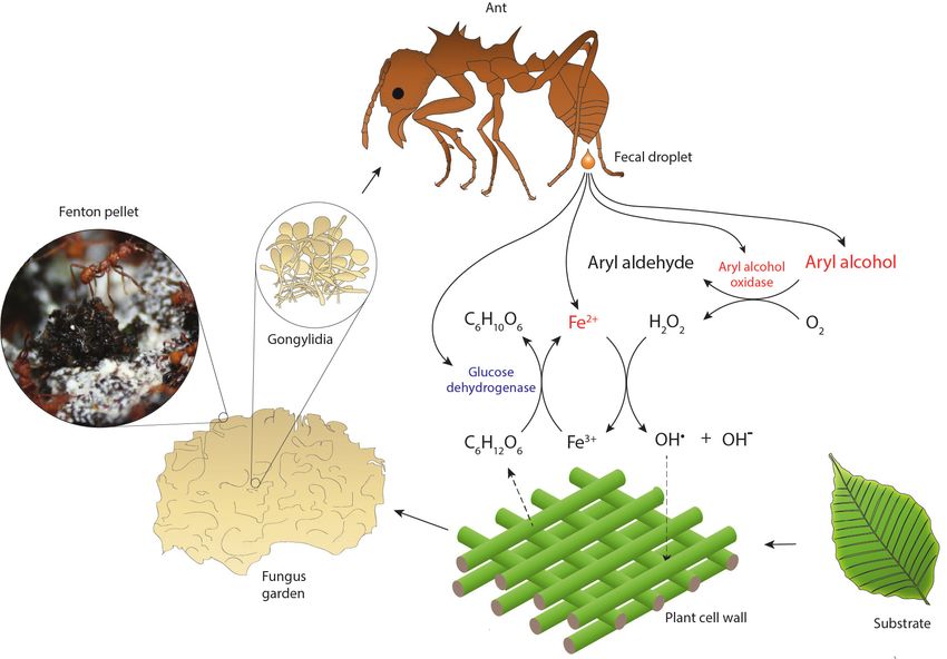
Our proteomic analysis of worker fecal fluid from four different A. echinatior colonies identified 174
proteins (Supplementary file 1), which showed substantial overlap among the pooled colony-spe-
cific samples (Figure 1A). To avoid including spurious proteins, we restricted our analyses to a short-
list of 101 proteins present in at least three of the four colony samples, and we were able to
annotate 91 (90%) of these proteins (Table 1). This core proteome consisted mainly of degradation
enzymes belonging to five major functional categories: oxidoreductases, proteolytic enzymes, carbo-
hydrate active enzymes (CAZymes), phosphate-liberating enzymes, and lipid degrading enzymes (lip-
idolytic enzymes) (Figure 1B). Of the 10 remaining proteins, only three could not be functionally
annotated. The vast majority (86) of the core fecal proteome originated from the fungal symbiont,
with only 15 being encoded by the ant genome. Based on the number of spectra obtained for each
identified protein, MaxQuant produced an approximate measure of relative abundance of each pro-
tein called label-free quantification intensity (LFQ intensity) (Table 1). This revealed that lipidolytic
enzymes constitute only a very small fraction and that the proteins in the ‘miscellaneous’ category
collectively had a moderate representation although some of these proteins had high abundance.
The four remaining categories had summed LFQ intensities of the same order of magnitude
(Figure 1B) and two specific proteins stood out as being exceptionally abundant
Schiøtt and Boomsma. eLife 2021;10:e61816. DOI: https://doi.org/10.7554/eLife.61816 3 of 21Research article Ecology Evolutionary Biology
A B
Proteolytic enzymes
Oxidoreductases
Miscellaneous
CAZymes Lipidolytic enzymes
Phosphate−liberating enzymes
Figure 1. Statistics of the fecal fluid proteome of Acromyrmex echinatior. (A) Venn diagrams, constructed using the web application Venny 2.1 (http://
bioinfogp.cnb.csic.es/tools/venny/index.html) showing the overlap of protein profiles identified in the four fecal fluid samples obtained from colonies
Ae263, Ae322, Ae356, and Ae372. (B) Pie chart showing the abundances of proteins across colonies assigned to six functional categories based on the
label-free quantification (LFQ) values provided by MaxQuant.
(Supplementary file 1), the Alkaline phosphatase (Protein ID 75 in Table 1) and the previously
described Laccase LgLcc1 (Protein ID 97 in Table 1; De Fine Licht et al., 2013), which jointly made
up almost a quarter of the total fecal protein mass. Three other proteins also showed remarkably
high LFQ-abundance levels: a 1,3-beta-glucanase (Protein ID 88 in Table 1), a Metallopeptidase M35
(Protein ID 206 in Table 1), and a Serine protease S53 (Protein ID 64 in Table 1), which implies that
the five most abundant proteins contributed more than 40% of the total fecal protein mass. These
results confirmed our previous findings that a large proportion of the fecal fluid proteins consists of
proteases (Kooij et al., 2014b), pectinases (Schiøtt et al., 2010), hemicellulases (Kooij et al., 2016),
and laccases (De Fine Licht et al., 2013), but we now also identified several phosphate-liberating
and lipidolytic enzymes.
The complementary components needed for Fenton chemistry
Five of the identified oxidoreductases were annotated as fungal glucose-methanol-choline (GMC)
oxidoreductases. A subgroup of these enzymes are the aryl alcohol oxidases that are believed to
play an important role in lignin degradation by brown-rot fungi (Guillén et al., 2000). This category
of wood degrading fungi is distinct from the white-rot fungi, which degrade lignin using ligninolytic
peroxidases (Arantes et al., 2012; Arantes and Goodell, 2014). The latter mechanism has most
likely evolved only once in the Agaricomycotina lineage (to which Leucocoprinus belongs), but has
subsequently been lost many times giving a complex phylogenetic pattern (Floudas et al., 2012).
We showed previously (Nygaard et al., 2016) that the leaf-cutting ant cultivar has lost its ligninolytic
peroxidases, so their decomposition spectrum would functionally be more reminiscent of brown-rot
fungi than white-rot fungi although they are not wood degrading fungi and are thus not formally
covered by this terminology. Assuming that convergent adaptation could have taken place in the
Leucocoprinus symbiont, we thus pursued the pathways that could enable the Fenton reaction in
more detail.
In order to break down lignocellulose, brown-rot fungi produce aromatic alcohols, which are sub-
sequently oxidized by aryl alcohol oxidases to produce hydrogen peroxide (H2O2). The hydrogen
peroxide is subsequently used in a Fenton reaction in which reduced iron (Fe2+) reacts with hydro-
gen peroxide to produce hydroxyl radicals that can diffuse into the lignocellulose substrate and
break the chemical bonds responsible for the rigid structure of lignocellulose. This in turn will loosen
the cell walls and allow access for more bulky degradation enzymes to target specific components of
the plant cell walls. Unless the oxidized iron (Fe3+) is recycled by reduction to Fe2+, the production
Schiøtt and Boomsma. eLife 2021;10:e61816. DOI: https://doi.org/10.7554/eLife.61816 4 of 21Research article Ecology Evolutionary Biology
Table 1. Fecal fluid proteins that were found in three or four of the examined colony samples (Figure 1), and thus likely to belong to
the core fecal fluid proteome.
For each protein the predicted function, its fungal or ant origin, the number of samples containing the protein, the relative abundance
of the protein, and an identifier number are listed. Proteins were assigned to one of six functional categories (Figure 1). Although a
number of oxidoreductases are listed in the CAZy database in the subcategory of auxiliary activities we have for the present study
kept them separate. This is because it remains ambiguous whether they are true CAZymes if they are defined as redox enzymes that
‘act in conjunction with CAZymes’. This separation also resolved the problem that some of the oxidoreductases listed in Table 1 are
not covered by the CAZy database. Question marks indicate that annotations were inconclusive.
Category Protein ID Predicted function Source Samples with protein Relative abundance
Oxidoreductases 15 Laccase Fungus 4 106.0
(including CAZymes with
97 Laccase Fungus 4 431.8
auxilliary activity)
180 Laccase Fungus 4 14.7
184 Laccase Fungus 4 21.4
35 4-carboxymuconolactone decarboxylase, alpha-beta hydrolase Fungus 4 4.3
81 Copper radical oxidase, glyoxal oxidase, galactose oxidase Fungus 4 8.9
186 Copper radical oxidase, glyoxal oxidase, galactose oxidase Fungus 4 8.3
98 FAD/FMN-containing isoamyl alcohol oxidase Fungus 4 41.1
9 GMC oxidoreductase, aryl-alcohol oxidase Fungus 4 5.9
86 GMC oxidoreductase, aryl-alcohol oxidase Fungus 4 10.3
178 GMC oxidoreductase, aryl-alcohol oxidase Fungus 4 6.9
191 GMC oxidoreductase, aryl-alcohol oxidase Fungus 4 18.3
23 GMC oxidoreductase, aryl-alcohol oxidase Fungus 3 1.9
145 GMC oxidoreductase, glucose dehydrogenase Ant 4 82.2
149 Succinate dehydrogenase (ubiquinone) flavoprotein subunit Ant 3 4.8
Proteolytic enzymes 6 Aspartic peptidase A1A, polyporopepsin Fungus 4 12.0
111 Aspartic peptidase A1A, saccharopepsin Fungus 4 74.3
125 Metallopeptidase M1, ERAP2 aminopeptidase Ant 4 11.3
112 Metallopeptidase M14A, zinc carboxypeptidase Ant 4 27.4
144 Metallopeptidase M14A, zinc carboxypeptidase Ant 4 7.3
141 Metallopeptidase M28D, carboxypeptidase Q Ant 4 15.9
188 Metallopeptidase M35, deuterolysin Fungus 4 23.9
206 Metallopeptidase M35, peptidyl-lys metallopeptidase Fungus 4 218.6
207 Metallopeptidase M35, peptidyl-lys metallopeptidase Fungus 4 11.4
56 Metallopeptidase M36 Fungus 4 4.3
133 Serine protease S1A, chymotrypsin Ant 4 56.0
134 Serine protease S1A, chymotrypsin Ant 4 3.8
135 Serine peptidase S1A, chymotrypsin-2 Ant 4 8.8
36 Serine peptidase S8A, cuticle degrading peptidase Fungus 4 7.2
103 Serine peptidase S8A, cerevisin Fungus 4 51.6
16 Serine peptidase S10, carboxypeptidase Fungus 3 2.0
14 Serine peptidase S10, carboxypeptidase Fungus 4 1.3
24 Serine peptidase S10, carboxypeptidase OcpA Fungus 4 31.5
95 Serine peptidase S10, serine-type carboxypeptidase F Fungus 4 11.7
199 Serine peptidase S28, carboxypeptidase Fungus 4 6.3
33 Serine peptidase S53, grifolisin Fungus 4 20.5
64 Serine protease S53, grifolisin Fungus 4 192.4
Table 1 continued on next page
Schiøtt and Boomsma. eLife 2021;10:e61816. DOI: https://doi.org/10.7554/eLife.61816 5 of 21Research article Ecology Evolutionary Biology
Table 1 continued
Category Protein ID Predicted function Source Samples with protein Relative abundance
Phosphate- 74 Acid phosphatase, 3-phytase A Fungus 3 7.2
liberating enzymes
0 Acid phosphatase, nucleotidase Fungus 4 16.9
61 Acid phosphatase, nucleotidase Fungus 4 24.8
75 Alkaline phosphatase Fungus 4 432.8
100 Alkaline phosphatase Fungus 4 14.8
109 Phytase esterase-like Fungus 4 105.1
59 Phytase esterase-like Fungus 3 19.2
193 Phytase, histidine acid phosphatase domain Fungus 4 6.5
187 Phytase, phosphoglycerate mutase, histidine acid phosphatase Fungus 4 25.2
72 PLC-like phosphodiesterase Fungus 3 3.5
37 Ribonuclease T1 Fungus 4 5.6
34 Ribonuclease T2 Fungus 4 11.5
73 GH3, beta-glucosidase Fungus 4 52.0
99 GH3, beta-xylosidase Fungus 4 76.0
55 GH5, endocellulase Fungus 3 3.7
179 GH5, exo-1,3-beta-glucanase Fungus 4 9.8
183 GH5, mannan endo-1,4-beta-mannosidase F Fungus 4 7.6
13 GH5, mannan endo-1,4-beta-mannosidase Fungus 3 11.7
38 GH10, endo-1,4-beta-xylanase Fungus 3 3.9
Carbohydrate 121 GH12, xyloglucanase Fungus 3 4.8
active enzymes
132 GH13, alpha-glucosidase, maltase Ant 3 2.9
123 GH15, glucoamylase Fungus 4 66.5
88 GH17, 1,3-beta-glucanase Fungus 4 290.8
8 GH18, chitinase Fungus 3 16.8
66 GH20, betahexosaminidase Fungus 3 1.3
214 GH25?, lysozyme Fungus 4 102.4
25 GH27, alpha-galactosidase Fungus 4 27.9
83 GH28, endo-polygalacturonase Fungus 4 83.3
80 GH29, alpha-L-fucosidase Fungus 3 1.8
104 GH31, alpha-glucosidase Fungus 4 39.1
157 GH31, alpha-glucosidase Ant 4 7.9
108 GH35, beta-galactosidase Fungus 4 13.6
154 GH37, trehalase Ant 4 19.8
90 GH43, arabinan-endo-1,5-alpha-L-arabinosidase Fungus 4 21.8
11 GH51, arabinofuranosidase Fungus 4 85.7
46 GH53, arabinogalactanase Fungus 4 36.2
77 GH55, exo-1,3-beta-glucanase Fungus 4 34.5
7 GH78, a-L-rhamnosidase Fungus 4 14.0
63 GH79, betaglucoronidase Fungus 4 35.0
62 GH88, glucuronyl hydrolase Fungus 4 3.0
76 GH92, exo-alpha-mannosidase? Fungus 4 16.6
68 GH92, exo-alpha-mannosidase? Fungus 3 3.8
57 CE8, pectinesterase Fungus 4 69.0
40 CE12, rhamnogalacturonan acetylesterase Fungus 4 5.8
Table 1 continued on next page
Schiøtt and Boomsma. eLife 2021;10:e61816. DOI: https://doi.org/10.7554/eLife.61816 6 of 21Research article Ecology Evolutionary Biology
Table 1 continued
Category Protein ID Predicted function Source Samples with protein Relative abundance
67 CE12, Rhamnogalacturonan acetylesterase Fungus 4 27.6
208 PL1, pectate lyase Fungus 4 34.6
122 PL4, rhamnogalacturonan lyase Fungus 4 9.6
Lipidolytic enzymes 124 Lipase, triacylglycerol lipase Fungus 4 2.5
65 Neutral/alkaline nonlysosomal ceramidase Fungus 4 6.2
138 Pancreatic lipase-related protein Ant 4 6.5
44 Phosphatidylglycerol/phosphatidylinositol transfer protein Fungus 3 2.8
32 Alpha/beta hydrolase, cephalosporin esterase Fungus 4 10.1
18 Alpha/beta hydrolase, triacylglycerol lipase, carotenoid ester lipase Fungus 4 5.5
27 Carboxylesterase; alpha-beta hydrolase; lipase Fungus 4 1.5
Miscellaneous proteins 212 Fruit-body specific protein D Fungus 4 44.0
31 Plant expansin, papain inhibitor Fungus 3 1.7
129 Regucalcin Ant 4 5.8
113 SnodProt1, cerato platanin Fungus 4 114.7
10 Ubiquitin Fungus 4 1.7
136 Epididymal secretory protein E1 Ant 4 6.4
158 CEN-like protein 2, OV-16 antigen Fungus 4 4.8
106 Hypothetical protein Fungus 4 39.2
195 Hypothetical protein Fungus 3 4.5
119 Hypothetical protein, symbiosis related protein, MAPK? Fungus 4 52.8
of reactive oxygen species will end when all Fe2+ has been used. It has therefore been suggested
that fungi secrete secondary metabolites such as hydroquinones to reduce Fe3+ to Fe2+
(Suzuki et al., 2006). In spite of the decomposition potential of the Fenton reaction, it remains a
major challenge for any living tissues to be exposed to concentrated doses of free radicals. Numer-
ous adaptations have in fact evolved to remove low concentrations of free radicals from living tissues
(Matés et al., 1999), so we conjectured that attine ants should have found ways to avoid such dam-
age in case they would use Fenton chemistry.
Against this background knowledge from brown-rot fungi, we drew up a hypothetical scenario
(Figure 2) that might allow the leaf-cutting ant symbiotic partnership to use Fenton chemistry, to
sustain and control that reaction according to need, and to have it take place in a spatial setting
where collateral damage to living tissues would be avoided. As it appears, the spatial issue appears
to have been resolved because Acromyrmex colonies concentrate their chewed leaf fragments in
pellets of ca. 3 mm diameter at the top of their fungus gardens for some hours before the material
is dispersed across the actively growing ridges of the fungal mycelium mass. However, if these pel-
lets would be the optimal location for a sustained Fenton reaction, the symbiotic partnership would
have been required to also evolve a fecal fluid enzyme that reduces Fe3+ while decomposing degra-
dation products from the plant substrate. We hypothesized that glucose dehydrogenase (Protein ID
145 in Table 1), the most abundant ant-encoded fecal fluid protein (Table 1, Supplementary file 1),
could have this function using glucose as electron donor to reduce Fe3+, either directly or indirectly
via an intermediate redox compound (Figure 2). We therefore set up a number of biochemical
assays to test the proposed scenario and report the results below.
Hydrogen peroxide production by fungal GMC oxidoreductases in ant
fecal fluid
A phylogenetic tree of all five fungal GMC oxidoreductases isolated from fecal fluid, together with
representative GMC oxidoreductases from various basidiomycete fungi, showed that these five
enzymes clustered within the aryl alcohol oxidases (Figure 3), while being distinct from the four
Schiøtt and Boomsma. eLife 2021;10:e61816. DOI: https://doi.org/10.7554/eLife.61816 7 of 21Research article Ecology Evolutionary Biology
Figure 2. Hypothesized reaction scheme for the generation of hydroxyl radicals when ant fecal fluid interacts with chewed leaf fragments in temporary
substrate pellets, based on the presence of enzymes in the total fecal fluid proteome (Supplementary file 1). Fungal enzymes produced in gongylidia
of the symbiotic garden-cultivar that the ants ingest pass unharmed through the gut to end up in the fecal fluid (Boyd and Martin, 1975b; Kooij et al.,
2014b; Schiøtt et al., 2010; De Fine Licht et al., 2013; Kooij et al., 2016). After droplets of fecal fluid are deposited and become exposed to
oxygen, the fungal oxidoreductases produce hydrogen peroxide while aryl alcohols are converted to aryl aldehydes. The hydrogen peroxide then
reacts with reduced iron (Fe2+) to produce hydroxyl radicals (OH ) in a Fenton reaction, which aggressively breaks down cell walls of the plant substrate.
.
3+
Oxidized iron (Fe ) can then be reduced again by ant-encoded glucose dehydrogenase, using glucose released via plant cell wall decomposition as
electron donor. The leaf substrate is initially concentrated in green pellets of ca. 3 mm diameter distributed across the top of fungus gardens, which
turn black in a few hours when subjected to Fenton-mediated degradation (inset image). Compounds ultimately derived from the fungal symbiont are
in red text and compounds directly produced by the ants in blue.
other known functional groups (glucose oxidases, methanol oxidases, pyranose 2-oxidases, and cel-
lulose dehydrogenases) (Ferreira et al., 2015). We subsequently measured the amount of hydrogen
peroxide that these GMC oxidoreductases produce with and without substrates, which showed that
fecal fluid contained a substantial amount of hydrogen peroxide. Addition of aromatic veratryl alco-
hol further increased this amount, whereas glucose and methanol did not have any effect
(Figure 4A), confirming the presence of aryl alcohol oxidases in the fecal fluid. As an extra control
assay to check that the increase in absorbance was directly caused by hydrogen peroxide, we added
the enzyme catalase, which is a very effective and specific hydrogen peroxide degrading enzyme. As
expected, catalase completely removed the signal, confirming that hydrogen peroxide is indeed nat-
urally produced in fecal fluid (Figure 4A). Two other fecal fluid oxidoreductases (Protein IDs 81 and
186 in Table 1) were annotated as copper radical oxidases with close similarity to glyoxal oxidases.
Schiøtt and Boomsma. eLife 2021;10:e61816. DOI: https://doi.org/10.7554/eLife.61816 8 of 21Research article Ecology Evolutionary Biology
These enzymes may also produce hydrogen peroxide, using the small organic compound glyoxal
and other aldehydes as substrate (Daou and Faulds, 2017). Experimental addition of glyoxal to fecal
fluid of Acromyrmex worker did indeed increase the amount of hydrogen peroxide, confirming that
glyoxal oxidases are also present in the fecal fluid (Figure 4A).
Does ant fecal fluid contain sufficient iron to maintain Fenton
chemistry?
For the Fenton reaction to take place, the produced hydrogen peroxide must react with Fe2+ atoms.
Brown-rot fungi are believed to be able to actively increase the concentration of iron atoms in their
wood substrate for this purpose (Kirker et al., 2017), but such a mechanism would not be applica-
ble to the fecal fluid Fenton reaction since it is separated from the fungal hyphae. To be viable, this
reaction must therefore rely on iron present in the leaf material collected by the ant workers and in
the fecal fluid itself. Making fungal iron available would be adaptive for the ant farming symbiosis as
a whole because direct use of iron from live plant tissues might well be constrained because plants
also produce iron chelator compounds to inhibit the growth of plant pathogens (Dellagi et al.,
2005). We measured the iron levels in fecal fluid and found an average (± SD) level of 370 ± 110 mM
iron across six ant colonies. This concentration of iron should be sufficient to allow the Fenton reac-
tion to occur (Hegnar et al., 2019; Elias and Waterhouse, 2010). It corresponds to the levels rele-
vant for natural wood degrading systems (Hegnar et al., 2019) although it is one to two orders of
magnitude lower than the levels typically used for pretreatment of plant biomass in vitro
(Jung et al., 2015). However, in such human bioconversion experiments, oxidized iron is not
recycled into reduced iron, which means that the pool of useable iron is continuously decreasing and
therefore needs a high starting concentration. In living systems there are limits to how much iron can
be accumulated without posing a risk for unintended oxidation of organic compounds. Natural selec-
tion producing a system for iron recycling could thus be highly adaptive.
Are fecal fluid hydroxyl radicals produced by Fenton chemistry?
The hydroxyl radicals that Fenton chemistry should produce can be measured via their ability to
break down 2-deoxy-D-ribose into thiobarbituric acid-reactive substances (TBARS) (Halliwell et al.,
1988). To quantify this effect, we adapted the standard 2-deoxy-D-ribose assay to accommodate
the small volumes of fecal fluid that could practically be collected directly from the ants and found
pronounced amounts of hydroxyl radicals to be present in the ant fecal fluid (Figure 4B). Adding a
strong metal chelator 1,10-phenanthroline caused a significant reduction in absorbance. This is con-
sistent with the hydroxyl radicals being produced by Fenton chemistry because the Fenton reaction
is known to be inhibited by 1,10-phenanthroline’s strong iron sequestering activity (Winter-
bourn, 1995). This effect was not observed when only the solvent of the 1,10-phenanthroline chemi-
cal (methanol) was added.
Is iron reduction controlled by the ant encoded glucose hydrogenase?
The Fenton reaction can only proceed as long as there are Fe2+ atoms present to be converted to
Fe3+ atoms, and brown-rot fungi are believed to maintain their Fenton reactions via enzyme systems
that convert Fe3+ back to Fe2+ (Guillén et al., 2000; Arantes et al., 2012). A similar mechanism
would not work for the initial stages of leaf degradation in the fungus garden, as the mycelium is
separated from the Fenton pellets. We therefore conjectured that the ant-encoded glucose dehy-
drogenase identified in the fecal fluid proteome could mediate the reduction of Fe3+ to Fe2+ using
glucose as electron donor. We indeed found a measurable capacity for Fe3+ reduction in fecal fluid,
which was further increased by the addition of glucose. This was consistent with Fe3+ being actively
recycled into Fe2+ via fecal fluid compounds and with glucose dehydrogenase most likely being
responsible for at least part of this activity (Figure 4C). An alternative pathway for Fe3+ reduction is
mediated by laccase (Guillén et al., 2000), which was also found in high quantities in the fecal fluid.
Preliminary experiments using laccase substrates in these assays did, however, not change the rate
of Fe3+ reduction. This makes it less likely that laccase plays a role in this process, although more
elaborate experimentation will be needed in order to rule out this possibility.
Schiøtt and Boomsma. eLife 2021;10:e61816. DOI: https://doi.org/10.7554/eLife.61816 9 of 21Research article Ecology Evolutionary Biology
15 PHACH_135972
FDP-191
96 Aryl-alcohol
RHOPL_54008
oxidases
TRAVE_176148
95 31
89 FDP-23
FDP-9
99 FDP-178
97
92 FDP-86
RHOPL_108489
93
PHACH_131961
Glucose
61 RHOPL_128830
oxidases
23
PHACH_126879
Methanol
100 PHLBR_157963
98 TRAVE_43286
oxidases
99 PHLBR_123747
PHACH_137275
Pyranose-2
100
TRAVE_174721
oxidases
48 TRAVE_73596
PHLBR_160653
Cellulose
100
PHACH_11098
Dehydrogenases
0.5
Figure 3. Gene tree of the five fungal glucose-methanol-choline (GMC) oxidoreductases identified in the ant fecal
fluid (present study) and in representative other basidomycete fungi, all assigned to functional groups based on a
previous study (Ferreira et al., 2015). All fungal GMC oxidoreductases from the fecal fluid (Protein IDs 9, 23, 86,
178, and 191 in Table 1; red text) clustered among the known aryl-alcohol oxidases. Other closely related
functional groups are glucose oxidases, methanol oxidases, pyranose-2 oxidases, and cellulose dehydrogenases,
which were retrieved in Phanerochaete chrysosporium (PHACH), Rhodonia placenta (RHOPL), Trametes versicolor
(TRAVE), and Phlebia brevispora (PHLBR), but not in fecal fluid of the leaf-cutting ant A. echinatior. Note that the
ant-encoded glucose dehydrogenase is also a GMC oxidoreductase, but would sequence-wise not fit into this
phylogeny of fungal proteins. Numbers are aLRT SH-like support values for nodes. The scale bar represents 0.5
substitutions per site.
The online version of this article includes the following source data for figure 3:
Source data 1. Amino acid sequences used for the gene tree shown in Figure 3.
Discussion
Acromyrmex fecal fluid has a highly specialized proteome
The presence of fungal enzymes in the fecal fluid of leaf-cutting ants was suggested already in the
1970s based on the biochemical similarity of proteases in fecal fluid and the fungal cultivar
(Boyd and Martin, 1975b; Boyd and Martin, 1975a). We achieved increasing confirmation of this
idea in a series of previous studies (Schiøtt et al., 2010; Kooij et al., 2014b; De Fine Licht et al.,
2013; Kooij et al., 2016), but our present study presents the first overall proteome that is complete
enough to allow inferences of hitherto unknown chemical processes. The comprehensive list of A.
echinatior fecal fluid proteins that we obtained (Supplementary file 1) was now also sufficiently rep-
licated to confirm that the large majority of the identified proteins are fungal in origin and serve the
farming symbiosis after being deposited on new plant substrate via the ant fecal fluid. When run on
SDS-Page gels (Schiøtt et al., 2010), the fecal fluid proteins always gave the same banding pattern,
rejecting the null hypothesis that they are a random collection of proteins that happen to escape
digestion. Overall, 101 of the 174 identified proteins were found in at least three fecal fluid samples
Schiøtt and Boomsma. eLife 2021;10:e61816. DOI: https://doi.org/10.7554/eLife.61816 10 of 21Research article Ecology Evolutionary Biology
A B C 0.30
8 p = 0.00058 0.6 p = 0.0013
Nano moles of H2O2 per µl of fecal fluid
p = 0.00047
Amount of Fe3+ reduced to Fe2+ (µM min-1)
p = 0.000081
7 p = 0.0022
p = 0.00083
Change in absorbance at 532 nm
0.5 0.25
p = 0.00073 NS
6 NS
0.4 0.20
NS
5
4 0.3 0.15
3 0.10
0.2
2
0.1 0.05
1
0 0.0 0.00
e
e
nt
e
l
l
se
l
e
ro
os
ro
l
in
Ca l
Gl l
try oxa
as
no
ho
ro
os
lve
la
ol
nt
nt
uc
al
nt
ha
uc
lco
ta
hr
y
Co
Co
So
t
Gl
Co
Gl
ca
et
nt
la
M
l+
na
ho
e
ra
Ph
lco
Ve
la
try
ra
Ve
Figure 4. Bioassays to demonstrate that inorganic. Fenton chemistry must be taking place when ant fecal fluid is
exposed to oxygen while being deposited on leaf pulp pellets chewed by the ants, testing the key interactions
hypothesized in Figure 2. (A) Bar plot showing the concentrations (means ± SE across six colonies) of hydrogen
peroxide in fecal fluid after adding potential substrates (glucose, methanol, glyoxal, or veratryl alcohol) of glucose-
methanol-choline (GMC) oxidoreductases with or without the hydrogen peroxide degrading enzyme catalase.
One-way ANOVA showed a highly significant overall effect of treatments: F6,36 = 14.597, p=2.863e-08, and
pairwise post hoc t-tests on matching samples from the same ant colonies (corrected for multiple testing with the
Holm–Bonferroni method) confirmed both the enhancing effects of veratryl alcohol and glyoxal and the inhibiting
effect of catalase (p-values in plot unless non-significant) (NS). (B) Deoxyribose assays showing that ant fecal fluid
has the capacity to produce hydroxyl radicals (means ± SE across six colonies). Phenanthroline is known to work as
an iron chelator and significantly reduced degradation of 2-deoxy-D-ribose while the solvent (methanol) of
phenanthroline did not. Paired t-tests followed the same protocol as in the A-panel except that the overall
ANOVA was omitted because there were only three means to compare. (C) A Ferrozine assay (means ± SE across
six colonies) showing the capacity of ant fecal fluid to reduce Fe3+ to Fe2+, confirming that addition of glucose
increases the rate of iron reduction. Statistics as in the B-panel.
The online version of this article includes the following source data for figure 4:
Source data 1. Data used for Figure 4A, B, and C.
from different ant colonies, and for many of them we were able to measure the corresponding enzy-
matic activity using biochemical assays (this study and Schiøtt et al., 2010; De Fine Licht et al.,
2013; Kooij et al., 2014b; Kooij et al., 2016). These results strongly suggest that most if not all of
the fecal proteins have condition-dependent adaptive significance, have been selected to avoid deg-
radation in the digestive system of the ants, and serve the efficiency of the entire symbiosis between
farming ants and fungal cultivars.
While many of the enzyme activities found in the ant fecal fluid have been discussed in previous
publications, the roles of phosphate-liberating enzymes and lipidolytic enzymes have not. Just like
nitrogen and carbon, phosphorus is a major macronutrient required for many biological processes.
Enzymatic degradation of organic matter to liberate C, N, and P is known to take place in a stoichio-
metrically balanced way (Sinsabaugh et al., 2009), so it is not surprising that the fecal fluid also con-
tains a range of enzymes able to release phosphorus from various substrates. That alkaline
phosphatase (Protein ID 75 in Table 1) had the highest abundance of all fecal proteins underlines
the importance of phosphorus mineralization. Several enzymes able to release phosphorus from phy-
tin were also present. Phytin is a phosphorus-storage compound in plant seeds, but also occurs in
pollen and vegetative tissues (Brinch-Pedersen et al., 2002). Laboratory colonies of A. echinatior
were occasionally fed with dry rice grains, but in nature leaf-cutting ants are not known to forage on
Schiøtt and Boomsma. eLife 2021;10:e61816. DOI: https://doi.org/10.7554/eLife.61816 11 of 21Research article Ecology Evolutionary Biology
seeds to any significant degree (Kooij et al., 2014a). However, they do harvest flowers, whose pol-
len may be a source of phytin. Finally, the fecal fluid contained ribonucleases that will degrade RNA,
another major source of phosphorus. The fecal lipidolytic enzymes had low abundances overall.
Apart from digesting lipids into assimilatory products, these enzymes may also hplay a function in
degrading cell membranes to liberate cytosolic compounds as has been shown for biotrophic fungi
(Ghannoum, 2000). They may also interfere with signal transduction pathways involved in plant resis-
tance toward pathogens (Wu and Li, 2015), but all these putative functions require that the plant
substrate is alive for a while after having been fragmented and placed in the fungus garden, which
may well be the case.
Multiple lines of evidence for fecal fluid Fenton chemistry after mixing
with fresh leaf substrate
The key compounds hypothesized to be involved in inorganic Fenton chemistry (GMC oxidoreduc-
tases, hydrogen peroxide, high concentrations of iron, and glucose dehydrogenase) were always
found in all four colonies for which we investigated fecal fluid samples. Compared to white-rot fungi,
brown-rot fungi are believed to have a reduced array of enzymes targeting lignocellulose, and to
specifically lack lignin peroxidases, which is also the case for L. gongylophorus (Nygaard et al.,
2016). Brown-rot fungi compensate this lack of enzymatic potential by using Fenton chemistry to
degrade lignocellulose while producing extracellular hydroxyl radicals (Arantes and Goodell, 2014).
In the Fenton reaction, hydrogen peroxide reacts with ferrous iron (Fe2+) and hydrogen ions to pro-
duce ferric iron (Fe3+), water, and hydroxyl radicals (Guillén et al., 2000; Arantes et al., 2012). The
very reactive hydroxyl radicals produced in these free-living fungi are known to damage a wide
range of biomolecules (Halliwell et al., 1988). Their putative functions are: 1. to release soluble car-
bohydrate fragments from plant cell walls (Schweikert et al., 2002) and 2. to reduce the viscosity of
solutions containing plant cell wall components such as xyloglucan, pectin, and cellulose (Fry, 1998),
as expected when degradation of these polysaccharides has taken place. The small size of hydroxyl
radical molecules allows them to initiate penetration and cleavage of lignin, cellulose, and hemicellu-
lose polymers in preparation for further degradation into assimilatory monomers by specific enzymes
(Arantes et al., 2012). Brown-rot fungi are believed to use GMC oxidoreductases or copper radical
oxidases to aerobically oxidize fungal alcohols or aldehydes in order to produce hydrogen peroxide.
In this process they then obtain the required iron from the wood substrate (Arantes et al., 2012) or
via actively mycelial transport into the degrading wood (Kirker et al., 2017). It has been suggested
that fungal laccases are then oxidizing hydroquinones into the semiquinones that react with ferric
iron (Fe3+) to produce quinones and ferrous iron (Fe2+), which would produce the recycling process
needed to maintain the Fenton reaction over time (Guillén et al., 2000).
It is intriguing that the details of Fenton chemistry in brown-rot fungi have remained rather little
known in spite of the economic importance of these fungi (Arantes and Goodell, 2014), and that
the leaf-cutting ants offered an unexpected window for understanding this elusive process. The lac-
cases, GMC oxidoreductases, and copper radical oxidases were similarly found in high quantities in
the fecal fluid of Acromyrmex leaf-cutting ants, consistent with Fenton chemistry being a standard
procedure in this fungus-farming symbiosis to degrade plant cell walls and/or toxic secondary plant
polymers meant to deter herbivores (Figure 2). We could verify that the identified GMC oxidoreduc-
tases were aryl alcohol oxidases (Figure 3), which are known to produce hydrogen peroxide from
aromatic alcohols. We also verified that fecal fluid contains concentrated hydrogen peroxide and
that the concentration increased further after adding veratryl alcohol or glyoxal, which are substrates
for GMC oxidoreductases and copper radical oxidases, respectively (Guillén et al., 1992; Daou and
Faulds, 2017). We further documented that the iron content of the fecal fluid is maintained at levels
that are amply permissive for Fenton chemistry to occur. Also the acid pH of ca. 4 in the fecal fluid
should be ideal for Fenton chemistry (Bishop et al., 1968; Erthal et al., 2004) although we cannot
be sure this is the ambient pH because the plant material might also influence the final pH in the
substrate pellet. Finally, a separate assay showed that fecal fluid components degrade deoxyribose
into thiobarbituric acid-active products, which can only be explained by the presence of hydroxyl
radicals (Halliwell et al., 1988). To obtain more direct evidence for the produced hydroxyl radicals
contributing to plant substrate breakdown, future research should focus on using Fourier transform
infrared spectroscopy to analyze plant cell wall preparations subjected to fecal fluid degradation, a
type of analytical chemistry that was beyond the scope of the present study. Without such validation,
Schiøtt and Boomsma. eLife 2021;10:e61816. DOI: https://doi.org/10.7554/eLife.61816 12 of 21Research article Ecology Evolutionary Biology
we cannot rule out other possible functions of these radicals such as general sanitation of the plant
substrate. However, such alternative function has never been conjectured or shown for the ant fecal
fluid, whereas brown-rot fungi are widely believed to use hydroxyl radicals for substrate degradation
(Arantes and Goodell, 2014). We also note that specialized functions of the ant fecal fluid aimed at
plant substrate degradation are uncontroversial (Boyd and Martin, 1975b; Boyd and Martin,
1975a; Martin and Martin, 1971; Kooij et al., 2014b; Rønhede et al., 2004; Schiøtt et al., 2010;
De Fine Licht et al., 2013; Kooij et al., 2016). Our Fenton chemistry scenario is therefore supported
by consistent inferential evidence, but in need of further experimental validation.
The high concentrations of laccase LgLcc1 (Protein ID 97 in Table 1) in the ant fecal fluid suggest
a similar mechanism for maintaining reliable conversion of Fe3+ back to Fe2+ (using hydroquinone as
an electron donor) as the Fenton chemistry known from brown-rot fungi (Guillén et al., 2000). How-
ever, the glucose dehydrogenase (Protein ID 145 in Table 1) encoded by the ant genome was also
abundant in the fecal proteome and could convert Fe3+ back to Fe2+ via simultaneous oxidization of
glucose. We verified this putative function as likely with a Ferrozine-based assay (Goodell et al.,
2006), including its dependence on glucose availability. Interestingly, however, preliminary experi-
ments indicated that adding laccase substrates did not influence the velocity of this reaction, which
suggests that glucose liberated from the plant polysaccharides is primarily used to keep the Fenton
reaction going. This substrate dependence may provide an additional feedback loop for the farming
symbiotic partnership to control the inorganic Fenton chemistry process. The ant-derived glucose
dehydrogenase (Protein ID 145 in Table 1) alternative might allow the ants to also use the fungal lac-
cases specifically for the decomposition of secondary plant compounds as found in one of our previ-
ous studies (De Fine Licht et al., 2013). That study also suggested that LgLcc1 (Protein ID 97 in
Table 1) came under positive selection in the common ancestor of the leaf-cutting ants, which would
be consistent with laccase becoming redundant for iron reduction and therefore undergoing direc-
tional selection for other functions.
Symbiotic complementarity is necessary for making fast Fenton
chemistry work
The assembly of complementary components from different sources implies that the fungal cultivar
and the farming ants synergistically cooperate to accomplish the continued production of hydroxyl
radicals, but only as long as Fenton chemistry is needed. The ants do not appear to keep adding
fecal fluid to existing Fenton pellets and they take them apart when they have turned black after
some hours to distribute the Fenton exposed leaf fragments over the actively growing ridges of the
fungus garden where fungal decomposition takes over. Both the processing efficiency and the ulti-
mate behavioral control by the ants strongly suggest refined co-adaptation at the molecular level to
enable colonies to operate as integrated functional herbivores. An earlier study found a high expres-
sion of an NADH-quinone oxidoreductase in fungus gardens of A. echinatior (Grell et al., 2013), an
enzyme converting quinones to hydroquinones that can react with Fe3+ to form Fe2+ after conver-
sion to semiquinones by laccase. This is consistent with the ant encoded glucose dehydrogenase
being relevant only for the degradation process in the Fenton pellets, and with NADH-quinone oxi-
doreductases taking over after the leaf fragments have been dispersed over the garden and the fun-
gal hyphae decompose what remains of the substrate.
Aryl alcohol oxidases use aromatic alcohols as a substrate to produce hydrogen peroxide. White-
rot fungi produce aromatic veratryl alcohol as a secondary metabolite, which plays several important
roles for ligninolysis (Jensen et al., 1994). Veratryl alcohol is derived from phenylalanine via the phe-
nylalanine/tyrosine pathway that has previously been shown to be upregulated in the gongylidia of
the A. echinatior cultivar L. gongylophorus (De Fine Licht et al., 2014). This might suggest that
veratryl alcohol (or a derivative thereof) is produced in the gongylidia of the fungal cultivar to be
ingested but not digested by the ants so it can function in the fecal fluid to facilitate the initial stages
of substrate degradation. The hydrogen peroxide needed in the Fenton reaction may also be
derived from the activity of the glyoxal oxidases that we identified, which use various aldehydes as
substrate for hydrogen peroxide production (Daou and Faulds, 2017). Such aldehydes could then
either originate from the oxidized aryl alcohols produced by the aryl alcohol oxidases, or could be
direct degradation products from plant cell wall components (Daou and Faulds, 2017). These multi-
ple parallel pathways suggest that the co-evolved interactions between inorganic Fenton chemistry
Schiøtt and Boomsma. eLife 2021;10:e61816. DOI: https://doi.org/10.7554/eLife.61816 13 of 21Research article Ecology Evolutionary Biology
and organic enzyme functions are likely to have robust operational functionality under a variety of
environmental (i.e. substrate) conditions.
Finally, as we explained in the introduction, it is important to note that hydroxyl radicals are
highly toxic also for the organisms producing them. This probably constrains the use of naturally pro-
duced hydroxyl radicals for biomass degradation unless the Fenton chemistry producing these radi-
cals remains distal to hyphal growth and is terminated by the time hyphae grow into the substrate
patches pre-treated in this manner. This complication will slow down the rate of decomposition in
free-living fungi, but the leaf-cutting ant symbiotic partnership appears to have resolved this con-
straint by compartmentalizing the complementary elements of initial aggressive break down of plant
substrate. Although the necessary compounds for the Fenton reaction are all present in the guts of
the ants, the low oxygen pressure there would presumably prevent the production of hydrogen per-
oxide inside the ants and thereby any collateral damage to ant tissues. Defecation onto concen-
trated pellets of chewed leaf fragments thus ensures that the reaction does not start until
atmospheric oxygen is available. Consequently, the Fenton pellets appear to function as small bio-
conversion reactors that are spatially isolated from both the fungal and the ant tissues so that poten-
tially harmful side-effects are avoided. Also in this respect robustness against potential malfunction
appears to have evolved. Our recent genome sequencing study of Acromyrmex ants and their culti-
vars revealed that A. echinatior has more glutathione S-transferase genes than other (non-attine)
ants, despite having fewer detoxification genes in general (Nygaard et al., 2011). Glutathione
S-transferase is known to be involved in the breakdown of oxidized lipids, which would likely be pro-
duced if reactive oxygen species would be formed in the gut and react with the lipid cell membranes
of the gut tissues. This suggests that physiological mechanisms to ameliorate inadvertent damage
caused by hydroxyl radicals may be in place, should they appear in the ant gut before the excretion
of fecal fluid.
Implications for our general understanding of the leaf-cutting ant
symbiosis and its bioconversion efficiency
The extent to which cellulose is degraded in the fungus garden has been widely disputed (see
Grell et al., 2013 and references therein). It is noteworthy that the fecal fluid proteome only con-
tained a single 1,4-beta-glucanase (Protein ID 55 in Table 1) to target the cellulose backbone, and
that this protein was found in low quantities and in only three of the four examined colonies. We
also found a single 1,4-beta-glucosidase (Protein ID 73 in Table 1) that releases glucose moieties
from cellobiose and oligosaccharides, but this enzyme is also targeting hemicellulose in addition to
cellulose. Finally, effective degradation of cellulose normally requires the enzyme cellobiohydrolase,
which was only found in one colony in very low quantities (Supplementary file 1). These findings are
consistent with our earlier inference (Grell et al., 2013; Moller et al., 2011) that cellulose degrada-
tion is not a primary function of the fecal fluid, supporting earlier findings that recalcitrant plant cell
wall cellulose is not degraded to a significant degree in Acromyrmex fungus gardens, but discarded
from the colony as waste (Moller et al., 2011). Our finding that Fenton chemistry may be used for
breaking chemical bonds in cell wall lignocellulose and is likely sufficient to give hyphal degradation
enzymes access to their target substrates in the interior of plant cells, appears to corroborate this
notion. More complete cellulose degradation would release an excess of assimilatory sugars for the
fungal symbiont but without simultaneously providing nitrogen or phosphorus, which are the limiting
factors for growth in any functional herbivore (Elser et al., 2007). Such sugars could well be a bur-
den for the symbiosis if they would allow inadvertent microorganisms to thrive in fungus gardens
(Grell et al., 2013).
While the documentation of Fenton chemistry substantially increases our understanding of the
way in which Acromyrmex leaf-cutting ants could become crop pests by robust functional herbivory,
it is intriguing that we have never observed Fenton pellets in any of the Atta colonies that we have
maintained in the lab for ca. 25 years. This suggests that the colonies of this other genus of leaf-cut-
ting ants, which are approximately two orders of magnitude larger in size and considerably more
damaging as agricultural pests have evolved alternative mechanisms to boost the efficiency and
robustness of their functional herbivory. Given that also Atta colonies produce enormous amounts of
cellulose rich waste, we do not expect that the explanation for Atta’s success as functional herbi-
vores will be fundamentally different. This is because both genera rear very closely related lineages
of the L. gongylophorus cultivars in sympatry at our Panamanian sampling site (Kooij et al., 2015b)
Schiøtt and Boomsma. eLife 2021;10:e61816. DOI: https://doi.org/10.7554/eLife.61816 14 of 21You can also read