Adipose Tissue Macrophage Polarization in Healthy and Unhealthy Obesity
←
→
Page content transcription
If your browser does not render page correctly, please read the page content below
REVIEW
                                                                                                                                           published: 17 February 2021
                                                                                                                                         doi: 10.3389/fnut.2021.625331

                                               Adipose Tissue Macrophage
                                               Polarization in Healthy and Unhealthy
                                               Obesity
                                               Alistaire D. Ruggiero 1 , Chia-Chi Chuang Key 2 and Kylie Kavanagh 1,3*
                                               1
                                                Section on Comparative Medicine, Department of Pathology, Wake Forest University School of Medicine, Winston-Salem,
                                               NC, United States, 2 Section on Molecular Medicine, Department of Internal Medicine, Wake Forest University School of
                                               Medicine, Winston-Salem, NC, United States, 3 Department of Biomedicine, University of Tasmania, Hobart, TAS, Australia

                                               Over 650 million adults are obese (body mass index ≥ 30 kg/m2 ) worldwide. Obesity
                                               is commonly associated with several comorbidities, including cardiovascular disease
                                               and type II diabetes. However, compiled estimates suggest that from 5 to 40% of
                                               obese individuals do not experience metabolic or cardiovascular complications. The
                                               existence of the metabolically unhealthy obese (MUO) and the metabolically healthy
                                               obese (MHO) phenotypes suggests that underlying differences exist in both tissues
                                               and overall systemic function. Macrophage accumulation in white adipose tissue (AT)
                                               in obesity is typically associated with insulin resistance. However, as plastic cells,
                           Edited by:          macrophages respond to stimuli in their microenvironments, altering their polarization
                        Norbert Stefan,
                                               between pro- and anti-inflammatory phenotypes, depending on the state of their
      University of Tübingen, Germany
                                               surroundings. The dichotomous nature of MHO and MUO clinical phenotypes suggests
                       Reviewed by:
             Vibeke H. Telle-Hansen,           that differences in white AT function dictate local inflammatory responses by driving
 Oslo Metropolitan University, Norway          changes in macrophage subtypes. As obesity requires extensive AT expansion, we posit
                 Marijana Todorcevic,
         Oxford Centre for Diabetes,           that remodeling capacity with adipose expansion potentiates favorable macrophage
      Endocrinology and Metabolism             profiles in MHO as compared with MUO individuals. In this review, we discuss how
          (OCDEM), United Kingdom
                                               differences in adipogenesis, AT extracellular matrix deposition and breakdown, and AT
                 *Correspondence:
                                               angiogenesis perpetuate altered AT macrophage profiles in MUO compared with MHO.
                      Kylie Kavanagh
           kkavanag@wakehealth.edu             We discuss how non-autonomous effects of remote organ systems, including the liver,
     orcid.org/0000-0001-8772-6186             gastrointestinal tract, and cardiovascular system, interact with white adipose favorably in
                                               MHO. Preferential AT macrophage profiles in MHO stem from sustained AT function and
                    Specialty section:
          This article was submitted to        improved overall fitness and systemic health.
             Nutrition and Metabolism,
                                               Keywords: adipose, macrophage, metabolically healthy, metabolically unhealthy, obesity
                a section of the journal
                    Frontiers in Nutrition

       Received: 02 November 2020
        Accepted: 05 January 2021
                                               INTRODUCTION
       Published: 17 February 2021
                                               As of February 2020, more than 1.9 billion adults worldwide were overweight [body mass index
                              Citation:        (BMI): 25–29.9 kg/m2 ], and over 650 million were obese (BMI ≥ 30 kg/m2 ) (1). Obesity decreases
         Ruggiero AD, Key C-CC and
                                               lifespan and increases the risk of developing hypertension, dyslipidemia, and type II diabetes
   Kavanagh K (2021) Adipose Tissue
   Macrophage Polarization in Healthy
                                               (T2D) (2–4). Despite the number and variety of deployed weight-loss interventions, very few
               and Unhealthy Obesity.          overweight or obese patients maintain weight loss over time, and globally, the number of obese
                Front. Nutr. 8:625331.         individuals continues to increase (5). Across the BMI spectrum, not all obese individuals suffer
      doi: 10.3389/fnut.2021.625331            the same comorbidities. Roughly 60% of obese individuals present with dysglycemia, hypertension,

Frontiers in Nutrition | www.frontiersin.org                                         1                                        February 2021 | Volume 8 | Article 625331
Ruggiero et al. Adipose Macrophages in Obesity Phenotypes and/or dyslipidemia, and cutoff criteria associated with each of physiological processes, including tissue remodeling and insulin these maladies define obesity as either healthy [metabolically sensitivity. Macrophage accumulation was originally thought healthy obese (MHO)] or unhealthy [metabolically unhealthy to be universally pro-inflammatory and contributory to insulin obese (MUO)] (6, 7). The majority of individuals are classified as resistance. However, macrophage subtypes stimulate different MUO; however, between 5 and 40% of obese individuals do not responses within AT. Macrophage subtypes exist along a present with metabolic abnormalities and are defined as MHO continuum, as they demonstrate variable metabolic activation (6–8). Definitions of MHO vary, as some studies identify only and ranges in inflammatory signaling (22, 23). As such, they are insulin-sensitive individuals as MHO, whereas others identify often classified by whether they are more pro-inflammatory or individuals with two or fewer metabolic abnormalities as MHO more anti-inflammatory. M1 macrophages are thought to be (7, 9, 10). A recently proposed definition of MHO identifies more pro-inflammatory and secrete pro-inflammatory cytokines individuals based on the diagnosis of obesity and the following that ultimately inhibit proper insulin signaling in adipocytes criteria: serum triglycerides ≤150 mg/dl, HDL-cholesterol (23, 24). Contrarily, M2 macrophages are thought to be more concentrations >40 mg/dl in men or >50 mg/dl in women, anti-inflammatory and secrete anti-inflammatory cytokines that systolic blood pressure ≤130 mmHg, diastolic blood pressure maintain functional insulin signaling (23, 24). ≤85 mmHg, no antihypertensive treatment as an alternative In obese states, macrophages play crucial roles in damage indicator, fasting blood glucose ≤100 mg/dl, and no treatment response. Macrophage polarization patterns are influenced by with glucose lowering agents (11). Significant controversy exists environmental cues and inflammatory signaling (23), and over the definitions and stability of MHO classifications. There macrophages accumulate when danger signals propagate and is no universally accepted definition of MHO; many MHO incite more inflammation to manage their resolution. In individuals progress to MUO over time, and, although MHO obese states, macrophages clear dead adipocytes and other individuals do have higher all-cause mortality and an increased cell debris, exocytose excess lipid, secrete both pro- and anti- risk of cardiovascular events compared with healthy lean inflammatory cytokines, and contribute to adipose remodeling individuals (12, 13), they are at a decreased risk of cardiovascular (23). Macrophage responses are interrupted by insulin resistance, complications and all-cause mortality compared with the MUO hypoxia, and reactive oxygen species generation, metabolic individual. Despite this controversy, understanding the biological endotoxemia or cell senescence, and death. While macrophage mechanisms that maintain metabolic health with overt obesity accumulation in obese individuals is typically affiliated with would aid the development of therapeutics to convert MUO inflammation and these downstream consequences, healthy individuals to MHO and ultimately reduce the financial burden obese individuals do not demonstrate the same levels of of obesity-related comorbidities. inflammation as their MUO counterparts. In this review, we Although obesity results in white adipose tissue (AT) discuss how differences in white AT components give rise to expansion, maintenance of metabolic function may underlie contrasting macrophage M-phenotypes in MHO compared with MHO individuals’ superior metabolic homeostasis. AT functions MUO persons. We also discuss how diet and aspects of systemic as an endocrine organ that maintains energy equilibrium, metabolic health impact AT macrophages. but function can differ by location. White AT accumulates throughout the body, including in the epicardial, mesenteric, omental, retroperitoneal, gonadal, subcutaneous abdominal, ADIPOSE MACROPHAGE SUBTYPES gluteal, and femoral regions (14). Intra-abdominal, or visceral, and subcutaneous white AT depots perform different functions Obesity alone incites the recruitment and proliferation of AT and thus differentially impact metabolic health. Visceral AT macrophages, the predominant adipose leukocyte population accumulation is positively associated with cardiometabolic (25, 26). As plastic cells that respond to their microenvironments, risk factors (15) and correlation with decreased insulin macrophages range from highly pro-inflammatory, or M1-like, to sensitivity (16). On the contrary, subcutaneous white AT highly anti-inflammatory, or M2-like (27). M1 macrophages fight accumulation protects against cardiometabolic risk factors (15) against intracellular pathogens, are induced by pro-inflammatory and corresponds with maintained insulin sensitivity (17), as factors including lipopolysaccharide and interferon-γ, and evidenced by subcutaneous adipose transplantation into visceral secrete inflammatory cytokines including interleukin (IL)-6, IL- depots alleviating metabolic dysregulation (18). Both adipocytes 1β, and monocyte chemoattractant protein-1 (MCP-1) (28). and immune cells in AT express and secrete bioactive hormones Hematopoietic-derived M1 macrophages utilize glycolysis and and signaling proteins that regulate metabolism (19–21). The are recruited into AT and where they can proliferate (29–31). M2 goal of this review is to summarize macrophages as key AT macrophages, on the other hand, contribute to tissue repair and immune cells and their influences, which drive tissue function produce anti-inflammatory cytokines, including IL-4 and IL-13. (Figure 1). Elucidating changes in white AT composition is Contrary to M1 macrophages, yolk sac-derived M2 macrophages crucial to unraveling the mechanisms behind the observed utilize oxidative phosphorylation (31). White AT homeostasis metabolic differences between MHO and MUO groups. requires a balance of both these pro- and anti-inflammatory Obesity-associated AT expansion often results in the macrophage subtypes. Excess M1 macrophage infiltration results accumulation of immune cells, including macrophages, in increased tissue inflammation, whereas an overabundance contributing to low-grade chronic inflammation. Macrophages of M2 macrophages can lead to aberrant fibrogenesis, limiting are the most abundant leukocytes in AT and assist in regulating the remodeling required for AT to respond to changing lipid Frontiers in Nutrition | www.frontiersin.org 2 February 2021 | Volume 8 | Article 625331
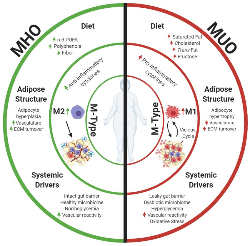
Ruggiero et al.                                                                                                        Adipose Macrophages in Obesity Phenotypes

 FIGURE 1 | Metabolically healthy obesity (MHO) and metabolically unhealthy obesity (MUO) may be defined by differences in AT. This review focuses on macrophage
 phenotypes (M-Types) as a key element driving adipose health, as these immune cells have potent effects on the local AT niche and are influenced by diet and other
 systemic health characteristics. Differences in dietary components and aspects of adipose expansion perpetuate MHO and MUO adipose macrophage phenotypes.
 Increased consumption of omega 3 polyunsaturated fatty acids (n-3 PUFAs), polyphenols, and fiber results in anti-inflammatory adipose macrophage (M2)
 programming in MHO. These dietary components combined with improved white adipose adipogenesis and corresponding adipocyte hyperplasia increase tissue
 vascularization and extracellular matrix (ECM) turnover, and downstream anti-inflammatory signaling propagates anti-inflammatory M2 macrophage proliferation while
 abating harmful pro-inflammatory M1 macrophage recruitment into tissue. Parallel to effects of diet and adipose structure, systemic drivers, including functional
 liver-adipose cross talk and improved vasoreactivity, also decrease MHO AT pro-inflammatory signaling and maintain insulin sensitivity. Alternatively, increased
 consumption of saturated fat, cholesterols, trans fat, and fructose incites pro-inflammatory macrophage recruitment in MUO adipose. Consumption of these dietary
 components in conjunction with dysfunctional adipogenesis results in adipocyte hypertrophy. This combined with decreased angiogenic signals, disrupted ECM
 turnover, and downstream pro-inflammatory cytokine secretion stimulates pro-inflammatory M1 macrophage recruitment. Systemic dysfunction in the form of a
 decreased vasoreactivity and oxidative stress concurrently fosters insulin resistance while promoting pro-inflammatory M1 macrophage recruitment into MUO adipose.
 Once M1 macrophages enter the tissue, they secrete additional pro-inflammatory cytokines that recruit more M1 macrophages. This vicious cycle of inflammation,
 perpetuation of unhealthy AT, and greater multisystemic dysfunction characterize MUO individuals who are defined by increased expression of metabolic syndrome
 components. Created with Biorender.com.

storage needs (32, 33). In diet-induced obese states, macrophage                     remainder of this text, metabolically active macrophages are
polarization shifts from a 4:1 M2-to-M1 ratio in lean animals to                     associated with decreased nicotinamide adenine dinucleotide
a 1.2:1 ratio in obese animals, as M1 macrophages are recruited                      phosphate oxidase 2, which reduces the inflammation associated
into white AT (29).                                                                  with obesity and downstream insulin resistance (30, 35, 36).
   MHO individuals display an anti-inflammatory adipose                              These macrophages are thought to better regulate lipid,
macrophage profile that more closely resembles that of                               catecholamine, and iron availability and perform other
metabolically healthy lean individuals, including an increased                       functions, including modulating local inflammation and
M2:M1 ratio (34). Of note, MHO individuals likely accumulate                         clearing dead adipocytes during prolonged obesity (30, 35, 36).
metabolically active adipose macrophages, which is a subtype                         This suggests that, in contrast to MUO, MHO individuals’ anti-
that may be distinct from coarsely defined M1 or M2. Although                        inflammatory AT macrophage profile helps maintain insulin and
we will focus primarily on M1 and M2 macrophages in the                              glucose regulation and deters pro-inflammatory macrophage

Frontiers in Nutrition | www.frontiersin.org                                     3                                         February 2021 | Volume 8 | Article 625331
Ruggiero et al.                                                                                        Adipose Macrophages in Obesity Phenotypes

recruitment. In the following sections, we will discuss the               either adipocyte hypertrophy or preadipocyte differentiation
relationships between aspects of AT function, macrophage                  and adipocyte hyperplasia. In MHO, de novo adipogenesis
polarization, and metabolic regulation in MHO compared                    driven by improved glucose uptake and anti-inflammatory
with MUO.                                                                 signaling—including increased PPARγ and adiponectin—results
                                                                          in smaller, more numerous adipocytes (54); the resulting
                                                                          adipocyte hyperplasia maintains a more anti-inflammatory AT
ADIPOCYTE FUNCTION AND                                                    macrophage profile (54). In MUO, hypertrophic adipocytes
ADIPOGENESIS                                                              communicate with recruited M1 macrophages and secrete pro-
                                                                          inflammatory leukotrienes, such as LTB4, which inhibit insulin
MHO adipocyte function mediates anti-inflammatory AT                      signaling in metabolic tissues and thus further recruit more pro-
macrophage activation and polarization (37). Adipocytes secrete           inflammatory macrophages (55). Proteomic analyses of visceral
cytokines, depending on their inflammatory state, that influence          AT from T2D MUO obese individuals reveal mitochondrial
immune cells. Secretion of Th2 cytokines, including IL-                   dysfunction and reduced adipocyte differentiation, which
4 and IL-13, induces macrophage peroxisome proliferator-                  additionally incites M1 macrophage recruitment (56). This
activated receptor (PPAR)δ activation, a regulator of fatty               vicious cycle continues with tumor necrosis factor-alpha and IL-
acid metabolism (38), which also improves whole-body insulin              1β secreted by M1 macrophages, further impairing adipogenic
sensitivity (39). Insulin-sensitive obese individuals have higher         differentiation, with reduced adipogenic gene expression in
PPARγ (i.e., PPARγ2) messenger RNA (mRNA) expression levels               subcutaneous AT and, in some cases, increased numbers of
in peripheral blood mononuclear cell and their visceral adipose           small adipocytes (48, 57, 58). In contrast, normoglycemic
than do insulin-resistant obese individuals (40, 41). Ablation of         obese individuals show an increase in the percent of adipose
both PPARγ, a regulator of adipogenesis and lipogenesis, and              progenitors within their tissue compared with both pre-diabetic
PPARδ renders macrophages unable to transition to the M2                  and T2D obese subjects (59). MHO individuals benefit from
subtype (38, 42, 43). Therefore, in MHO individuals, adipocyte            a positive insulin sensitizing cycle, where anti-inflammatory
cytokine secretion and its downstream effects on fatty acid               signals from adipocytes result in M2 macrophage polarization,
metabolism and adipogenesis regulators incite M2 macrophage               which promotes healthy adipocyte hyperplasia and further anti-
polarization and whole-body insulin sensitivity while promoting           inflammatory signaling from adipocytes.
sufficient adipogenesis (i.e., proliferation and differentiation of
preadipocytes) to manage the concurring nutrient overload.
    Fluctuations in AT macrophage ratios correspond to changes            EXTRACELLULAR MATRIX REMODELING
in preadipocyte differentiation and adipogenic signaling. The
factors secreted by pro-inflammatory M1 macrophages possess               Healthy AT expansion requires extracellular matrix (ECM)
anti-adipogenic properties (44). Unlike M1 macrophages,                   remodeling. Adipocytes are surrounded by a network of ECM
both M2 macrophages and inactive macrophages promote                      proteins that serve as a mechanical support and respond
preadipocyte survival by releasing a platelet-derived growth              to different signaling events (60). AT expansion relies on
factor (PDGF) (45). However, excess M2 macrophages have                   adaptive cellular and extracellular responses to prevent ectopic
been shown to impair preadipocyte differentiation through the             lipid deposition and lipotoxicity (61–63). Within AT, collagens
transforming growth factor-beta (TGF-β) pathway (46). The                 produced primarily by adipocytes and endothelial cells comprise
effects of AT macrophage balance on preadipocyte differentiation          most non-cell tissue mass, whereas integrins are the major tissue
and adipose expansion require further investigation (47);                 receptors for cell adhesion to ECM proteins (64, 65). Increased
however, differences in the AT macrophage profiles of MHO                 interstitial fibrosis due to excess collagen deposition likely
and MUO individuals likely drive the rate and format of                   decreases ECM flexibility and reduces tissue plasticity, which
adipogenesis, as in vitro differentiation protocols illustrate that       leads to adipocyte dysfunction and immune cell infiltration
adipogenesis is greater in MHO than MUO people (48–51). The               (66). Insulin-resistant individuals demonstrate aberrant ECM
nuclear hormone receptor PPARγ acts as a master transcription             deposition and insufficient ECM breakdown (59, 67).
factor of adipocyte differentiation by inducing and maintaining               Important protein families that comprise the ECM include
the expression of key adipogenic genes, such as GLUT4 and                 matrix metalloproteinases (MMPs), enzymes that process and
adiponectin, which are necessary for normal adipocyte function            degrade pericellular substrates and play a vital role in regulating
and downstream insulin sensitivity (52). Diabetic patients treated        ECM remodeling in normal and disease states (68). Tissue
with PPARγ-activating thiazolidinediones, a class of antidiabetic         inhibitors of metalloproteinases (TIMPs), which comprise a
drugs, often experience weight gain in the form of subcutaneous           family of four protease inhibitors, inhibit MMPs to achieve a
AT expansion. Accordingly, the subcutaneous AT expansion that             balance in production and breakdown (69). Adipose expression
occurs through adipocyte hyperplasia (increasing number of cells          of MMP-9, increased in MUO compared with MHO (38, 70),
through differentiation of new adipocytes) appears metabolically          positively correlates with insulin resistance and cardiovascular
favorable and contributes to systemic insulin sensitization (53),         risk in obese persons. Similarly, visceral AT MMP-14 expression
and the same PPARγ signaling promotes an M2-positive AT                   correlates with adipose accumulation and insulin resistance in
balance. In obese states, AT macrophages provide signals that             women (71). MMP-11 is also increased in the white adipose
communicate with mature adipocytes and preadipocytes to incite            of obese insulin-resistant mice (72). TIMP-3 regulates white

Frontiers in Nutrition | www.frontiersin.org                          4                                   February 2021 | Volume 8 | Article 625331
Ruggiero et al. Adipose Macrophages in Obesity Phenotypes
adipose inflammation and insulin sensitivity, and its deletion reduces macrophage expression of pro-inflammatory cytokines
in mice increases M1 macrophage accumulation in white AT through adenosine diphosphate ribosylation factor 6 (88).
(73), whereas its overexpression in macrophages resulted in MiRNA-145 also promotes preadipocyte differentiation and
improved glucose tolerance and insulin sensitivity and decreased angiogenesis, leading to healthier AT (89). Adipocyte-derived
inflammation in high-fat diet-fed mice (74). exosomes contain many miRNAs still being characterized,
Individuals with worsening glycemic control exhibit excess such as miRNA-23b, miRNA-148b, and miRNA-4429, which
AT deposition of collagens I, III, IV, and VI (59). Collagen VI are expected to alter TGF-β signaling and potentially mitigate
gene expression coincides with more visceral adipose mass and downstream fibrosis (90). For example, upregulation of miRNA-
pro-inflammatory macrophage accumulation (75, 76). Excess 23b mitigated kidney fibrosis in leptin-deficient mice (91),
collagen VI deposition imparts stress by inhibiting adipocyte suggesting that exosomal miRNAs secreted from MHO adipose
expansion (60). AT fibrosis creates rigidity, restrains adipocyte likely promote both anti-inflammatory macrophage polarization
expansion, and, ultimately, triggers adipocyte inflammation and preadipocyte differentiation while mitigating fibrosis.
in response to the increased mechanical stress. Collagen IV, Current data indicate that MHO adipose corresponds
which accounts for up to 50% of the basement membrane, with sufficient ECM deposition and breakdown coupled with
also increases with TGFβ-1 and TGFβ-3 gene expression in balanced AT macrophage populations and anti-fibrotic/pro-
human subcutaneous adipose, resulting in pro-inflammatory and adipogenic miRNA secretion. Of note, miRNA promotion
pro-fibrotic phenotypes (77). Increases in collagens, including of preadipocyte differentiation co-occurring with new blood
Col24α1, are associated with insulin resistance in AT and vessel formation (89) highlights that adipose expansion
skeletal muscle (78, 79). Also, gene expression of CD44, which necessitates vascularization.
regulates cell–cell and cell–matrix interactions, is 3-fold higher
in subcutaneous AT of MUO individuals, and CD44 density on
macrophages is associated with the M1 phenotype (80). These ANGIOGENESIS
data indicate that a vicious cycle of aberrant ECM turnover
and increased inflammatory signaling, including M1 macrophage Adipose vascularity dictates tissue metabolism and
recruitment, results in insulin resistance in obese AT. insulin sensitivity. In obese states, AT expansion requires
MHO individuals possess improved ECM turnover rates, as neovascularization that allows for sufficient oxygenation,
their more flexible ECM constitution allows for increased lipid nutrient delivery, and adipocyte differentiation. Adipose
storage. As previously mentioned, the expandability of MHO microvasculature plays a primary role in glucose homeostasis, as
individuals’ subcutaneous AT is thought to contribute to their impaired tissue perfusion results in decreased glucose uptake and
decreased visceral AT accumulation and healthier metabolic is a hallmark of T2D (92). Adipocyte hypertrophy that can occur
profile (60), resulting in less cell death and decreased M1 within just three days of high-fat diet consumption incites signals
macrophage recruitment (23). Decreased amounts of MMPs and to increase angiogenesis and alleviate hypoxia (93). Successful
TIMPs, including TIMP-1, allow MHO adipocytes to differentiate neovascularization and its concomitant ECM remodeling reduce
more readily (81), as insulin-sensitive obese patients demonstrate hypoxia to maintain AT health (94).
less fibrosis than diabetic obese patients before and after bariatric MHO individuals typically accumulate subcutaneous white
surgery (82). Improving ECM turnover in MUO individuals AT, which possesses improved angiogenic capabilities as
could permit increased subcutaneous fat mass and ameliorate compared with visceral adipose (95). MHO individuals maintain
metabolic dysfunction and shifts in macrophage balance (83). peripheral capillary density similar to metabolically healthy
M2 secretion of TGF-β provides essential structural support lean individuals, which allows for enhanced nutrient flow, and
and necessary remodeling. However, in pathological instances, demonstrate improved fitness compared with MUO (96–98).
this secretion results in aberrant fibrosis development. Secretion Vascular endothelial growth factor (VEGF) contributes to new
of TGF-β by M2 macrophages is intended to promote anti- blood vessel formation, as it induces the growth of both
inflammatory tissue remodeling, though if aberrant, results in preexisting and new vessels (65). Adequate AT VEGF signaling
increased collagen deposition, downstream fibrosis, and insulin in high-fat diet-fed mice protected the animals against insulin
resistance (84). MHO individuals’ M2/M1 AT macrophage ratio, resistance by reducing hypoxia and, in turn, increasing their
corresponding TGF-β secretion, and adequate ECM turnover M2/M1 tissue macrophage profile (99–101). Increases in capillary
allow for decreased adipocyte mechanical stress. density coincided with improvements in metabolic function
Genes and gene product regulation in different obesity in obese rats with metabolic syndrome (98). Improved fitness
subtypes also determine ECM turnover by controlling achieved through aerobic training of high-fat diet-fed rats
macrophage polarization. For example, microRNAs (miRNAs) increased AT capillary density and increased the number of M2
alter gene expression and modulate downstream glucose tissue macrophages (102).
metabolism and insulin sensitivity in obesity (85), and exosomes Improved vascularity in MHO AT also is likely to increase
from AT-derived stem cells control M2 macrophage polarization numbers of adipose progenitor cells, as such cells reside
(86). Specifically, exosomal miRNA-34a secreted by adipocytes within adipose vasculature (103). Increased numbers of adipose
suppresses M2 macrophage polarization and promotes obesity- progenitors allow for hyperplastic expansion, which perpetuates
induced adipose inflammation and metabolic dysfunction (87), an anti-inflammatory immune profile, including an increased
whereas increased expression of miRNA-145 in visceral adipose M2/M1 macrophage ratio, as demonstrated by increased
Frontiers in Nutrition | www.frontiersin.org 5 February 2021 | Volume 8 | Article 625331Ruggiero et al. Adipose Macrophages in Obesity Phenotypes
adipocyte hyperplasia in the subcutaneous AT of obese women patterns (DAMPs), which share common effector pathways.
(104). These data suggest that hyperplastic subcutaneous depot These patterns have been best explored with “microbial”
expansion that co-occurs with increased AT vascularity facilitates inputs where these pathogen signals (endotoxin, lipoproteins,
an anti-inflammatory AT milieu in the MHO. membrane proteins, peptidoglycans, DNA fragments, and
Prolonged hyperglycemia makes the cells within AT vessels lipoteichoic acid are examples) bind to receptors and initiate
susceptible to injury and promotes microvascular dysfunction. both phagocytosis/destruction and activation of inflammatory
A vicious cycle occurs in MUO patients, where impaired outcomes crucial to effective innate immunity (111). Toll-like
glycemic control worsens vascular reactivity, which then receptors (TLRs), scavenger receptors, and mannose receptors
exacerbates AT hypoxia, inflammation, and tissue insulin are pattern recognition receptors present in all macrophages,
resistance. MUO patients have a 44% decrease in capillary and binding and activation of the inflammasome and subsequent
density and 58% lower VEGF signaling in the subcutaneous release of cytokines and interferons lead to polarization toward
adipose, highlighting the occurrence of vascular rarefaction the M1 type of local resident macrophages, recruitment of
with hyperglycemia (105). Likewise, VEGF expression in both circulating monocytes for activation, and proliferation of
subcutaneous and visceral adipose decreases in a stepwise fashion macrophages in situ (112).
with worsening insulin resistance (106). ECM dysregulation Diet has profound effects on the magnitude of DAMPs
contributes to insufficient vascularization, as obese patients with and PAMPs to which adipose macrophages are exposed. One
T2D demonstrate increased basement membrane thickness (59) of the important observations made in people was coined
and higher collagen VI synthesis, which correlated inversely with “metabolic endotoxemia,” whereby caloric excess was related
AT oxygenation in MUO patients (105). The two-hit process of to increased biomarkers of microbial translocation, such as
impaired ECM remodeling and poor vascularization stimulates lipopolysaccharide-binding protein 1 (LBP1) (113). LBP1 is
a pro-inflammatory immune cell response in MUO adipose, released from the liver into the circulation in response to
exemplified by CD68 mRNA and macrophage inflammatory PAMPs and functions as a co-receptor for TLRs present on
protein 1α expression inversely correlating with AT oxygenation the macrophage cell membrane, thus initiating inflammatory
in MUO subjects (105). Accordingly, contrary to MHO adipose, responses in peripheral tissues, such as adipose (114, 115).
decreased angiogenic capacity and increased vessel injury In addition to just caloric excess, specific dietary components
in MUO adipose result in increased pro-inflammatory M1 are known to induce LBP1 and PAMP signaling through
macrophage recruitment and pro-inflammatory signaling. modulation of the microbial interactions at the intestinal
As angiogenic capacity influences adipose macrophage mucosal barrier. Fructose is the best described dietary factor
subtypes, existing macrophages in adipose impact angiogenesis. proven to increase intestinal barrier dysfunction in rodents,
Macrophage deletion results in a reduction of vascular density humans, and non-human primates (116–120). Fructose is
in AT (107). Macrophages are a significant source of AT additionally associated with excess caloric consumption, obesity,
PDGF, which assists with blood vessel growth and repair and metabolic diseases, such as diabetes and metabolic-associated
of damaged vessels; their deletion also leads to a significant fatty liver, and these disease states further augment macrophage
reduction in PDGF mRNA (108). M2 macrophages promote accumulation and the inflammatory cycle of insulin resistance
angiogenic signaling, as evidenced by increased endothelial in AT (27). Lipoproteins are additionally recognized as PAMPs,
cells and tubular structures in subcutaneous adipose post-M2 and low-density lipoproteins reliably increase in concentration
macrophage injection (109). M2 macrophage polarization, but in response to caloric excess and fructose exposure, as the liver
not the M1 phenotype, caused a substantial downregulation of packages and processes TGs for export (121). This lipoprotein-
TIMP-1 expression, resulting in the production of the angiogenic delivered TG is the substrate for AT to uptake and store
activated zymogen, proMMP-9 (110). These data suggest that the peripherally, which, in unhealthy obesity, may not be an efficient
anti-inflammatory MHO adipose macrophage profile stimulates process. Impaired insulin sensitivity and excess adipocyte
angiogenesis, which not only ensures sufficient AT nutrient hypertrophy lead to hypoxia, further inflammatory signaling
supply but also contributes to MHO individuals’ improved fitness and macrophage recruitment, and even adipocyte apoptosis or
and systemic health. necrosis—the sequelae being more local DAMPs to drive local
macrophages to respond, recruit, and additionally augment the
M1 response.
NUTRITION AND ADIPOSE MACROPHAGE The endotoxemia resulting from caloric excess or dietary
PROGRAMMING fructose has been described as sterile; however, more recently,
antibiotic and probiotic therapy deployed to modify the pathogen
Nutrient overload caused by excess food consumption is the response has shown effectiveness in improving inflammatory and
most common initial trigger for obesity, and diet represents the metabolic outcomes (117). More evidence to suggest that local
paramount environmental health factor that is both modifiable adipose PAMP responses are to actual pathogens includes the
and variable across populations. High caloric intake per se recent demonstration of an adipose microbiome in obese people
can effect macrophage polarization (27); however, dietary (122–124). These bacteria are confirmed to include whole live
factors can influence either augmentation of M1 subtype organisms and be present in the circulation and visceral and
abundance or support M2 programming. Macrophages have subcutaneous AT depots (124). From the data suggesting that
evolved to be effective responders to pathogen-associated dietary calories and ingredients increase microbial translocation
molecular patterns (PAMPs) and damage-associated molecular and shape the microbiome, it is likely that the number and
Frontiers in Nutrition | www.frontiersin.org 6 February 2021 | Volume 8 | Article 625331Ruggiero et al. Adipose Macrophages in Obesity Phenotypes
type of microbes filtered out into adipose also are diet driven Omega-3 polyunsaturated fatty acids (n-3 PUFA) cannot
(125) and will influence the abundance and polarization of be synthesized de novo by humans due to the lack of delta-
adipose macrophages. 12 and delta-15 desaturase enzymes and must, therefore,
Saturated fat is another dietary component with the ability be acquired from the diet (144). The major n-3 fatty acid
to function as a PAMP/DAMP. Structurally, a longer chain of in the diet, α-linolenic acid (18:3n-3), can be converted to
single carbon–carbon bonds may mimic the saturated fatty acids other more anti-inflammatory lipids, such as eicosapentaenoic
in phospholipids of most microbial membranes and the long fatty acid (20:5n-3), docosahexaenoic acid (22:6n-3), and the less
acid chains incorporated in the structure of endotoxins, which recognized docosapentaenoic acid (22:5n-3), which can be
in intact gram-negative microbes reside in the outer membrane directly sourced through consumption of fish and derived
(126). Saturated fat intake has been related to endotoxemia, fish oils. The utilization of dietary n-3 fatty acids in the
but studies that include calorie control are not available, and synthesis of complex PUFAs, such as docosahexaenoic acid,
caloric excess alone is sufficient to elevate LBP1 and induce eicosapentaenoic acid, and anti-inflammatory prostaglandins
peripheral inflammation (127–129). Similarly, trans-fatty acids is well-noted and thought to contribute to the reduction of
structurally resemble saturated fatty acids and are presumed to pathologies associated with chronic disease, including metabolic
act as pro-inflammatory danger signals and a potent dietary syndrome. The challenge is that the conversion of α-linolenic
ingredients famous for induction of metabolically unhealthy acid into these anti-inflammatory lipids is very limited in people;
obesity (130, 131). A diet rich in unhealthy attributes, such thus, increasing dietary intake, coupled with counseling to reduce
as excess caloric amounts, high fructose or added sugars, the negative dietary features described earlier, is a popular
cholesterol (132), saturated and/or trans-fatty acids, all drive strategy to improve metabolic health in obesity. N-3 PUFAs
macrophage activation through highly conserved pathways directly interact with G-protein coupled receptor (GPR) 120 to
evolved to detect pathogens and resolve tissue damage (133). generate an intracellular signaling complex that inhibits multiple
The result perpetuates an inflammatory state and M1 phenotypic inflammatory pathways, such as NF-κB and activated c-Jun N-
predominance in AT in response to signals that indicate the terminal kinase, which are downstream of TLR and cytokine
need for active phagocytic and antigen presentation functions. receptors (145). This effect is not limited to macrophage signaling
Depot differences are not well-described; however, some evidence and shifting the profile toward a resolving M2 phenotype;
for dietary factors inducing intra-abdominal fat shifts do exist. these healthy long-chain fatty acids also signal through GPR120
Examples include trans-fat consumption being linked to visceral on adipocytes to reduce inflammation and improve insulin
fat accumulation, and in an obese patient cohort, the abundance sensitivity, leading to less DAMP signaling from hypoxic,
of ectopic bacteria in omental fat tissue was slightly higher than stressed adipocytes and decreased paracrine inflammatory effects
that in subcutaneous fat, both of which are consistent with on tissue-resident macrophages. In summary, healthy dietary
the body of knowledge that indicates intra-abdominal AT is features, such as fiber, n-3 PUFAs, and bioactive flavonoids can
more contributory to unhealthy obesity than is subcutaneous fat directly and indirectly drive the macrophage profile toward an
expansion (122, 124). M2 anti-inflammatory profile and a healthier state, even if the
Few dietary factors directly influence macrophages positively subject is obese (27). Diet can influence the balance of M1 and
to effect an M2 inflammation-resolving state. Polyunsaturated M2 macrophages in adipose both directly, by modifying the
fatty acids (PUFAs) do have a direct role on macrophage burden of DAMP/PAMP signaling and indirectly by influencing
function (134), whereas most dietary components have indirect insulin sensitivity and tissue function of adipocytes and vascular
contributions to adipose health and consequential reductions cells in adipose. Therapeutic strategies that capitalize on dietary
in DAMP/PAMP sensing by local macrophage populations mechanisms are in development, including synthetic GPR120
(135). These indirect effects will not be discussed, but examples ligands, probiotics, and synbiotics to improve intestinal barrier
include dietary fiber, which shifts the microbiome and improves function, as methods to improve health in obese persons
mucosal barrier function, thus decreasing LBP1 and endotoxemia (70, 138).
(136–138), dietary components, such as isoflavones, which are
rich in fermented foods, and polyphenols, which are rich in
fruits and vegetables. Isoflavones can have a lipid-lowering IMPACTS OF NON-ALCOHOLIC FATTY
effect (139), thus decreasing lipoprotein sensing by scavenger LIVER DISEASE ON ADIPOSE
receptors, and can have estrogen receptor (ER) activity, which MACROPHAGE TYPES
indirectly can reduce inflammation and promote vascular
reactivity. Macrophages express ER (predominantly α-isoforms MHO individuals’ liver composition and inflammatory signaling
and G-protein coupled ER1), and dietary isoflavones can bind moderate the anti-inflammatory profile of their peripheral
and decrease Nuclear factor-kappa B (NF-κB) activation and tissues. Obesity-related nutrient overload incites spillover of
cholesterol oxidation in the context of lipid and cholesterol free fatty acids from AT that are taken up by the liver
exposure (140), thus facilitating or maintaining M2 polarization through the portal vein. Accordingly, increased visceral adipose
in culture and vascular tissue, an effect likely to be also seen accumulation corresponds with liver triglyceride accumulation.
in adipose macrophages (141). Polyphenols can be effective free The severity of non-alcoholic fatty liver disease and non-
radical scavengers, thus reducing local inflammation and tissue alcoholic steatohepatitis has been shown to correspond with
damage signaling (142, 143). an expression of inflammatory genes in AT (146). Increased
Frontiers in Nutrition | www.frontiersin.org 7 February 2021 | Volume 8 | Article 625331Ruggiero et al. Adipose Macrophages in Obesity Phenotypes
pro- and anti-inflammatory macrophage infiltration in visceral hypoxia, oxidative stress, and pro-inflammatory macrophage
adipose was observed in obese patients with non-alcoholic infiltration (158, 159). These data suggest that MHO perivascular
steatohepatitis (146). MHO individuals demonstrate less liver AT successfully facilitates glucose uptake while promoting anti-
triglyceride accumulation and liver fibrosis and overall improved inflammatory macrophage accumulation.
liver function compared with MUO individuals (147, 148). Like perivascular AT, interactions between healthy epicardial
Decreased liver fibrosis corresponded with fewer omental AT AT and the myocardium mitigate pro-inflammatory signaling
macrophages in obese humans, as macrophage accumulation in MHO individuals. Epicardial AT, located between the
decreased with decreasing fibro-inflammation indexes (149). myocardium and visceral pericardium, acts as an energy source
Adiponectin, an adipokine that promotes AT lipid storage, for the myocardium, as epicardial adipose has a higher capacity
lipid oxidation, and downstream anti-inflammatory signaling, for uptake and release of free fatty acids and a lower rate of
is increased in MHO compared with MUO, providing another glucose utilization than other visceral depots (160). Given its
physiologic mechanism for MHO individuals’ decreased liver ability to take up free fatty acids, epicardial adipose is thought
triglyceride accumulation (150–152). Adiponectin has also been to act as a buffer for the myocardium against lipotoxicity
shown to correlate with insulin resistance in obese female (160, 161). However, in pathological settings, the epicardial
people (153). Decreased liver triglyceride accumulation and adipose may provide excess free fatty acids associated with
fibrosis, along with increased effectiveness of anti-inflammatory myocardial steatosis and systemic insulin resistance (162).
signaling from the liver, correspond with reduced macrophage Insulin resistance and T2D are associated with increased
accumulation in MHO and allow for improved adipose storage MCP-1 expression in epicardial adipose, and peri-coronary
and sustained AT glucose uptake. adipose displays increased M1 macrophage infiltration compared
with other regions distal to the coronaries (163, 164). The
importance of whole-body health highlights that peri-coronary
PARACRINE ADIPOSE EFFECTS ON THE epicardial adipose inflammation may influence vascular function
CARDIOVASCULAR SYSTEM negatively as well as positively (163). In unhealthy obese
states, hypoxic perivascular adipose transports macrophages that
In addition to their decreased risk of all-cause mortality, may carry oxidized cholesterol from systemic circulation to
MHO individuals experience a decreased risk of heart epicardial adipose through the neovascularized vasa vasorum
failure even compared with metabolically unhealthy lean (165, 166). Local epicardial AT inflammation also stems from
individuals (154). The interactions between MHO individuals’ dysregulated miRNA expression. Patients with coronary artery
AT and their cardiovascular system explain the observed disease demonstrate increased miR-103-3b upregulation, which
cardiometabolic outcomes. is a potential modulator of the pro-inflammatory cytokine CCL13
The AT perivascular and epicardial fat depots are in direct (167). Insulin resistance and T2D are characterized by changes
proximity to cardiovascular tissue and interact positively in in miRNAs, including miR-29a and miR-143, which regulate AT
a paracrine fashion with the myocardium and vasculature browning and inflammation (168). Importantly, miR-29a has
in MHO persons. Perivascular AT, located around the large been associated with myocardial fibrosis, whereas miR-143 is a
arteries, produces nitric oxide and secretes adipocyte-derived biomarker of vascular smooth muscle cell activation that is linked
relaxing factors and other adipokines that relax vascular smooth to atherosclerosis and hypertension (169, 170). Adipose acts as a
muscle cells and are able to go into microcirculation (155). local renin–angiotensin system by producing angiotensinogen, a
As perivascular adipose maintains vascular bed homeostasis, it precursor to angiotensin II (171). The hypertension medication
controls the effects of insulin on microcirculatory systems in telmisartan—an angiotensin II type 1 receptor blocker and
metabolic tissues. For instance, perivascular adipose successfully PPARγ agonist—improved insulin resistance while decreasing
facilitates insulin-mediated vasoreactivity and glucose uptake in M1 and increasing M2 macrophage gene expressions in visceral
skeletal muscle (156). Interestingly, loss of perivascular AT in adipose from high-fat diet-fed mice (171). These data suggest
lipoatrophic mice (A-ZIP/F1) enhances the contractile responses that a hypertensive MUO person who has more M1 macrophages
of blood vessels, which results in hypertension (157). In instances recruited into their adipose, when treated with telmisartan, may
of pathological perivascular dysfunction, the perivascular AT experience a shift in their adipose macrophage profile and a
release of adipocyte-derived relaxing factors diminishes, whereas reduction in local inflammation. While epicardial AT function
its release of pro-inflammatory cytokines, including IL-6, protects MHO individuals from lipotoxicity and maintains the
tumor necrosis factor-alpha, and MCP-1, increases, and a anti-inflammatory immune cell profile, more work is needed to
negative cycle of perfusion and AT dysfunction perpetuates understand the crosstalk between epicardial adipose miRNAs,
as described earlier. This directly impacts endothelial and the cardiovascular system, and their relationship to health
vascular smooth muscle cells and incites vascular inflammation and disease.
(155). Pro-inflammatory perivascular AT signaling is initiated
by decreased nitric oxide production, increased reactive oxygen
species, and pro-inflammatory cytokines released by the DISCUSSION
dysfunctional endothelium, vascular smooth muscle cells, or
vascular macrophages (155). In MUO individuals, dysfunctional In this review, we discuss how macrophage phenotypes drive
perivascular AT alterations stem from adipocyte hypertrophy, adipose health in MHO and MUO persons, as these immune
Frontiers in Nutrition | www.frontiersin.org 8 February 2021 | Volume 8 | Article 625331Ruggiero et al. Adipose Macrophages in Obesity Phenotypes
cells affect the local AT niche and are heavily influenced by diet reduced macrophage accumulation and pro-inflammatory
and systemic health characteristics (Figure 1). Improved white signaling while restoring metabolic function in obese mice
adipose function in conjunction with the consumption of n-3 and people (172–174). Calcium/calmodulin-dependent protein
PUFAs, polyphenols, and fiber results in anti-inflammatory M2 kinases (CaMKs) play roles in myocardial ischemia/reperfusion
macrophage programming in MHO. Functional white adipose injury, regulating food intake and energy expenditure. Activation
adipogenesis, increased tissue vascularization, ECM turnover, of CaMK II δ in cardiomyocytes prompted pro-inflammatory
and downstream anti-inflammatory signaling in combination macrophage recruitment and associated NF-κB signaling that
with consumption of the dietary components mentioned results in fibrosis and myocardial dysfunction (175). Loss of
earlier propagate M2 maintenance and proliferation while CaMK kinase II (CaMKK2) reduced AT M1 macrophage-
abating harmful pro-inflammatory M1 macrophage recruitment. derived NF-κB signaling caused by a high-fat diet, highlighting
Gut mucosal barrier integrity, functional liver-adipose, and an important function for CaMKK2 in controlling diet-induced
cardiovascular system-adipose cross talk parallel the effects of adipose M1 macrophage inflammation (176). For example,
diet by decreasing MHO AT pro-inflammatory signaling and treatment with a CAMKK2 inhibitor, tilianin, decreased pro-
maintaining insulin sensitivity. Alternatively, increased dietary inflammatory signaling in cardiomyocytes (177). The use of
consumption of saturated fat, cholesterols, trans fat, and fructose CaMKK2 inhibitors in MUO may reduce AT inflammation,
incites pro-inflammatory adipose macrophage recruitment in although more research is needed to determine how CaMK
MUO adipose, which inhibits adipogenesis. Consumption of inhibition impacts AT immune cell populations over time.
these dietary components, in conjunction with dysfunctional As obesity rates continue to rise and weight-loss interventions
adipogenesis, results in augmented adipocyte hypertrophy. prove largely unsuccessful, understanding how to mediate
This, combined with decreased angiogenic signals, disrupted the vicious AT macrophage cycle in MUO individuals is
ECM turnover, and downstream pro-inflammatory cytokine imperative. Although long-term obesity ultimately increases
secretion, stimulates further pro-inflammatory M1 macrophage the risks of multisystem adverse events, breaking the pro-
recruitment. Impaired gut mucosal barrier integrity in the MUO inflammatory macrophage cycle will potentially shift MUO
drives multi-organ inflammation. This results in dysfunctional individuals to MHO and reduce current health burdens.
adipose-liver and adipose-cardiovascular system cross talk,
which concurrently promote pro-inflammatory M1 macrophage
recruitment in MUO adipose. Once M1 macrophages enter AUTHOR CONTRIBUTIONS
the tissue, they secrete additional pro-inflammatory cytokines
that recruit more M1 macrophages. This vicious cycle of All authors listed have made a substantial, direct and intellectual
inflammation and perpetuation of unhealthy AT, further contribution to the work, and approved it for publication.
expansion, and greater multisystemic dysfunction characterizes
MUO individuals. FUNDING
Clearing M1 macrophages from unhealthy adipose may
reestablish metabolic health. Obesity incites senescent cell The funding source for this work was the National Institutes of
accumulation in AT and M1 macrophage recruitment, and Health, National Heart, Lung, and Blood Institute. The grant
pharmacological senescent cell clearing agents have effectively number to be listed as R01HL142930.
REFERENCES 8. Phillips CM. Metabolically healthy obesity: definitions,
determinants and clinical implications. Rev Endocr Metab
1. World Health Organization. Obesity and Overweight. (2003). Available Disord. (2013) 14:219–27. doi: 10.1007/s11154-013-9
online at: https://www.who.int/news-room/fact-sheets/detail/obesity-and- 252-x
overweight 9. Durward C, Hartman TJ, Nickols-Richardson SM. All-cause mortality risk
2. Engin A. The definition and prevalence of obesity and metabolic syndrome. of metabolically healthy obese individuals in NHANES III. J Obes. (2012)
Adv Exp Med Biol. (2017) 960:1–17. doi: 10.1007/978-3-319-48382-5_1 2012:460321. doi: 10.1155/2012/460321
3. Renehan AG, Tyson M, Egger M, Heller RF, Zwahlen M. Body- 10. National Cholesterol Education Program (NCEP) Expert Panel on Detection,
mass index and incidence of cancer: a systematic review and meta- Evaluation, and Treatment of High Blood Cholesterol in Adults (Adult
analysis of prospective observational studies. Lancet. (2008) 371:569–78. Treatment Panel III). Third report of the National Cholesterol Education
doi: 10.1016/S0140-6736(08)60269-X Program (NCEP) expert panel on detection, evaluation, and treatment of
4. Saklayen MG. The global epidemic of the metabolic syndrome. Curr high blood cholesterol in adults (Adult Treatment Panel III) final report.
Hypertension Rep. (2018) 20:12. doi: 10.1007/s11906-018-0812-z Circulation. (2002) 106:3143–421.
5. Mann T, Tomiyama AJ, Westling E, Lew AM, Samuels B, Chatman J. 11. Lavie CJ, Laddu D, Arena R, Ortega FB, Alpert MA, Kushner RF. Healthy
Medicare’s search for effective obesity treatments: diets are not the answer. weight and obesity prevention: JACC health promotion series. J Am Coll
Am Psychol. (2007) 62:220. doi: 10.1037/0003-066X.62.3.220 Cardiol. (2018) 72:1506–31. doi: 10.1016/j.jacc.2018.08.1037
6. Primeau V, Coderre L, Karelis A, Brochu M, Lavoie M, Messier 12. Caleyachetty R, Thomas GN, Toulis KA, Mohammed N, Gokhale
V, et al. Characterizing the profile of obese patients who are KM, Balachandran K, et al. Metabolically healthy obese and incident
metabolically healthy. Int J Obes. (2011) 35:971–81. doi: 10.1038/ijo.201 cardiovascular disease events among 3.5 million men and women. J Am Coll
0.216 Cardiol. (2017) 70:1429–37. doi: 10.1016/j.jacc.2017.07.763
7. Blüher M. Metabolically healthy obesity. Endocr Rev. (2020) 41:bnaa004. 13. Zheng R, Zhou D, Zhu Y. The long-term prognosis of cardiovascular disease
doi: 10.1210/endrev/bnaa004 and all-cause mortality for metabolically healthy obesity: a systematic review
Frontiers in Nutrition | www.frontiersin.org 9 February 2021 | Volume 8 | Article 625331Ruggiero et al. Adipose Macrophages in Obesity Phenotypes
and meta-analysis. J Epidemiol Community Health. (2016) 70:1024–31. of metabolically healthy and unhealthy obese monkeys. Obesity. (2017)
doi: 10.1136/jech-2015-206948 25:689–96. doi: 10.1002/oby.21762
14. Wronska A, Kmiec Z. Structural and biochemical characteristics of 35. Ni Y, Ni L, Zhuge F, Xu L, Fu Z, Ota T. Adipose tissue macrophage
various white adipose tissue depots. Acta Physiol. (2012) 205:194–208. phenotypes and characteristics: the key to insulin resistance in obesity and
doi: 10.1111/j.1748-1716.2012.02409.x metabolic disorders. Obesity. (2020) 28:225–34. doi: 10.1002/oby.22674
15. Elffers TW, de Mutsert R, Lamb HJ, de Roos A, Willems van Dijk K, 36. Coats BR, Schoenfelt KQ, Barbosa-Lorenzi VC, Peris E, Cui C, Hoffman
Rosendaal FR, et al. Body fat distribution, in particular visceral fat, is A, et al. Metabolically activated adipose tissue macrophages perform
associated with cardiometabolic risk factors in obese women. PLoS ONE. detrimental and beneficial functions during diet-induced obesity. Cell Rep.
(2017) 12:e0185403. doi: 10.1371/journal.pone.0185403 (2017) 20:3149–61. doi: 10.1016/j.celrep.2017.08.096
16. Park K, Rhee B, Lee K, Kim S, Lee H, Koh C-S, et al. Intra-abdominal 37. Miyachi Y, Tsuchiya K, Shiba K, Mori K, Komiya C, Ogasawara N,
fat is associated with decreased insulin sensitivity in healthy young men. et al. A reduced M1-like/M2-like ratio of macrophages in healthy
Metabolism. (1991) 40:600–3. doi: 10.1016/0026-0495(91)90050-7 adipose tissue expansion during SGLT2 inhibition. Sci Rep. (2018) 8:1–13.
17. McLaughlin T, Lamendola C, Liu A, Abbasi F. Preferential fat deposition in doi: 10.1038/s41598-018-34305-x
subcutaneous vs. visceral depots is associated with insulin sensitivity. J Clin 38. Kang K, Reilly SM, Karabacak V, Gangl MR, Fitzgerald K, Hatano B,
Endocrinol Metab. (2011) 96:E1756–60. doi: 10.1210/jc.2011-0615 et al. Adipocyte-derived Th2 cytokines and myeloid PPARδ regulate
18. Foster MT, Softic S, Caldwell J, Kohli R, Annette D, Seeley RJ. Subcutaneous macrophage polarization and insulin sensitivity. Cell Metab. (2008) 7:485–95.
adipose tissue transplantation in diet-induced obese mice attenuates doi: 10.1016/j.cmet.2008.04.002
metabolic dysregulation while removal exacerbates it. Physiol Rep. (2013) 39. Sugii S, Olson P, Sears DD, Saberi M, Atkins AR, Barish GD, et al. PPARγ
1:e00015. doi: 10.1002/phy2.15 activation in adipocytes is sufficient for systemic insulin sensitization. Proc
19. Kahn CR, Wang G, Lee KY. Altered adipose tissue and adipocyte function in Natl Acad Sci USA. (2009) 106:22504–9. doi: 10.1073/pnas.0912487106
the pathogenesis of metabolic syndrome. J Clin Invest. (2019) 129:3990–4000. 40. Macias-Gonzalez M, Moreno-Santos I, García-Almeida J, Tinahones F,
doi: 10.1172/JCI129187 Garcia-Fuentes E. PPARγ2 protects against obesity by means of a mechanism
20. Gustafson B, Gogg S, Hedjazifar S, Jenndahl L, Hammarstedt A, that mediates insulin resistance. Eur J Clin Invest. (2009) 39:972–9.
Smith U. Inflammation and impaired adipogenesis in hypertrophic doi: 10.1111/j.1365-2362.2009.02198.x
obesity in man. Am J Physiol Endocrinol Metab. (2009) 297:E999–1003. 41. Garcia-Fuentes E, Murri M, Garrido-Sanchez L, Garcia-Serrano S, García-
doi: 10.1152/ajpendo.00377.2009 Almeida JM, Moreno-Santos I, et al. PPARγ expression after a high-fat meal
21. Adamczak M, Wiecek A. The adipose tissue as an endocrine organ. Semin is associated with plasma superoxide dismutase activity in morbidly obese
Nephrol. (2013) 33:2–13. doi: 10.1016/j.semnephrol.2012.12.008 persons. Obesity. (2010) 18:952–8. doi: 10.1038/oby.2009.314
22. Atri C, Guerfali FZ, Laouini D. Role of human macrophage polarization 42. Odegaard JI, Ricardo-Gonzalez RR, Goforth MH, Morel CR, Subramanian
in inflammation during infectious diseases. Int J Mol Sci. (2018) 19:1801. V, Mukundan L, et al. Macrophage-specific PPARγ controls alternative
doi: 10.3390/ijms19061801 activation and improves insulin resistance. Nature. (2007) 447:1116–20.
23. Russo L, Lumeng CN. Properties and functions of adipose doi: 10.1038/nature05894
tissue macrophages in obesity. Immunology. (2018) 155:407–17. 43. Moreno-Indias I, Tinahones FJ. Impaired adipose tissue expandability and
doi: 10.1111/imm.13002 lipogenic capacities as ones of the main causes of metabolic disorders. J
24. Thomas D, Apovian C. Macrophage functions in lean and obese adipose Diabetes Res. (2015) 2015:970375. doi: 10.1155/2015/970375
tissue. Metabolism. (2017) 72:120–43. doi: 10.1016/j.metabol.2017.04.005 44. Lacasa D, Taleb S, Keophiphath M, Miranville A, Clement K.
25. Weisberg SP, McCann D, Desai M, Rosenbaum M, Leibel RL, Ferrante AW. Macrophage-secreted factors impair human adipogenesis: involvement of
Obesity is associated with macrophage accumulation in adipose tissue. J Clin proinflammatory state in preadipocytes. Endocrinology. (2007) 148:868–77.
Invest. (2003) 112:1796–808. doi: 10.1172/JCI200319246 doi: 10.1210/en.2006-0687
26. Xu H, Barnes GT, Yang Q, Tan G, Yang D, Chou CJ, et al. Chronic 45. Sorisky A, Molgat AS, Gagnon A. Macrophage-induced adipose tissue
inflammation in fat plays a crucial role in the development of dysfunction and the preadipocyte: should I stay (and differentiate) or should
obesity-related insulin resistance. J Clin Invest. (2003) 112:1821–30. I go? Adv Nutr. (2013) 4:67–75. doi: 10.3945/an.112.003020
doi: 10.1172/JCI200319451 46. Nawaz A, Aminuddin A, Kado T, Takikawa A, Yamamoto S, Tsuneyama K,
27. Lumeng CN, Bodzin JL, Saltiel AR. Obesity induces a phenotypic switch in et al. CD206+ M2-like macrophages regulate systemic glucose metabolism
adipose tissue macrophage polarization. J Clin Invest. (2007) 117:175–84. by inhibiting proliferation of adipocyte progenitors. Nat Commun. (2017)
doi: 10.1172/JCI29881 8:1–16. doi: 10.1038/s41467-017-00231-1
28. McLaughlin T, Ackerman SE, Shen L, Engleman E. Role of innate and 47. Martinez-Santibañez G, Nien-Kai Lumeng C. Macrophages and the
adaptive immunity in obesity-associated metabolic disease. J Clin Invest. regulation of adipose tissue remodeling. Annu Rev Nutr. (2014) 34:57–76.
(2017) 127:5–13. doi: 10.1172/JCI88876 doi: 10.1146/annurev-nutr-071812-161113
29. Lumeng CN, DelProposto JB, Westcott DJ, Saltiel AR. Phenotypic 48. McLaughlin T, Sherman A, Tsao P, Gonzalez O, Yee G, Lamendola C, et al.
switching of adipose tissue macrophages with obesity is generated by Enhanced proportion of small adipose cells in insulin-resistant vs insulin-
spatiotemporal differences in macrophage subtypes. Diabetes. (2008) sensitive obese individuals implicates impaired adipogenesis. Diabetologia.
57:3239–46. doi: 10.2337/db08-0872 (2007) 50:1707–15. doi: 10.1007/s00125-007-0708-y
30. Caslin HL, Bhanot M, Bolus WR, Hasty AH. Adipose tissue macrophages: 49. Almuraikhy S, Kafienah W, Bashah M, Diboun I, Jaganjac M, Al-
Unique polarization and bioenergetics in obesity. Immunol Rev. (2020) Khelaifi F, et al. Interleukin-6 induces impairment in human subcutaneous
295:101–13. doi: 10.1111/imr.12853 adipogenesis in obesity-associated insulin resistance. Diabetologia. (2016)
31. O’Neill LA, Kishton RJ, Rathmell J. A guide to immunometabolism for 59:2406–16. doi: 10.1007/s00125-016-4031-3
immunologists. Nat Rev Immunol. (2016) 16:553. doi: 10.1038/nri.2016.70 50. Yang X, Jansson P-A, Nagaev I, Jack MM, Carvalho E, Sunnerhagen KS, et al.
32. Catrysse L, van Loo G. Adipose tissue macrophages and their Evidence of impaired adipogenesis in insulin resistance. Biochem Biophys Res
polarization in health and obesity. Cell Immunol. (2018) 330:114–9. Commun. (2004) 317:1045–51. doi: 10.1016/j.bbrc.2004.03.152
doi: 10.1016/j.cellimm.2018.03.001 51. Park HT, Lee ES, Cheon YP, Lee DR, Yang KS, Kim YT, et al. The
33. Hou J, Shi J, Chen L, Lv Z, Chen X, Cao H, et al. M2 macrophages relationship between fat depot-specific preadipocyte differentiation and
promote myofibroblast differentiation of LR-MSCs and are associated metabolic syndrome in obese women. Clin Endocrinol. (2012) 76:59–66.
with pulmonary fibrogenesis. Cell Commun Signal. (2018) 16:1–14. doi: 10.1111/j.1365-2265.2011.04141.x
doi: 10.1186/s12964-018-0300-8 52. Maryam A, Jae M, Nasun H, Christopher L, Annette R, Michael D. PPARγ
34. Kavanagh K, Davis AT, Peters DE, LeGrand AC, Bharadwaj MS, Molina signaling and metabolism: the good, the bad and the future. Nat Med. (2013)
AJ. Regulators of mitochondrial quality control differ in subcutaneous fat 19:557–66. doi: 10.1038/nm.3159
Frontiers in Nutrition | www.frontiersin.org 10 February 2021 | Volume 8 | Article 625331You can also read