Building an Asymmetrical Brain: The Molecular Perspective
←
→
Page content transcription
If your browser does not render page correctly, please read the page content below
REVIEW
                                                                                                                                               published: 30 April 2019
                                                                                                                                       doi: 10.3389/fpsyg.2019.00982

                                                Building an Asymmetrical Brain: The
                                                Molecular Perspective
                                                Judith Schmitz*, Onur Güntürkün and Sebastian Ocklenburg

                                                Biopsychology, Department of Psychology, Institute of Cognitive Neuroscience, Ruhr University Bochum, Bochum, Germany

                                                The brain is one of the most prominent examples for structural and functional differences
                                                between the left and right half of the body. For handedness and language lateralization,
                                                the most widely investigated behavioral phenotypes, only a small fraction of phenotypic
                                                variance has been explained by molecular genetic studies. Due to environmental factors
                                                presumably also playing a role in their ontogenesis and based on first molecular evidence,
                                                it has been suggested that functional hemispheric asymmetries are partly under epigenetic
                                                control. This review article aims to elucidate the molecular factors underlying hemispheric
                                                asymmetries and their association with inner organ asymmetries. While we previously
                                                suggested that epigenetic mechanisms might partly account for the missing heritability
                                                of handedness, this article extends this idea by suggesting possible alternatives for
                                                transgenerational transmission of epigenetic states that do not require germ line epigenetic
                                                transmission. This is in line with a multifactorial model of hemispheric asymmetries,
                           Edited by:           integrating genetic, environmental, and epigenetic influencing factors in their ontogenesis.
                          Mattie Tops,
            VU University Amsterdam,            Keywords: laterality, handedness, language lateralization, epigenetics, Nodal pathway
                          Netherlands

                      Reviewed by:
                       Rachel Tomer,
            University of Haifa, Israel
                                                INTRODUCTION
                     Bernard Crespi,
     Simon Fraser University, Canada            In 1866, German zoologist Ernst Haeckel introduced promorphology – the science of an
                                                organism’s external form – and proposed symmetry as a fundamental criterion for classifying
                 *Correspondence:
                       Judith Schmitz
                                                organisms (Haeckel, 1866). The clade of bilateria (animals displaying mirror-inverted body
               Judith.Schmitz@rub.de            halves), including (but not restricted to) all vertebrates, was created in 1888 (Hatschek, 1888).
                                                Besides asymmetry (organisms without any axis or plane of symmetry, e.g., the majority of
                   Specialty section:           sponges) and radial symmetry (organisms with one axis, but several planes of symmetry, e.g.,
         This article was submitted to          starfish), bilateral symmetry is considered one of the three major types of body plans (Manuel,
                             Cognition,         2009). However, bilateral symmetry is frequently broken by either the position of non-paired
               a section of the journal         internal organs in one body half (e.g., the left-sided stomach and the right-sided liver) or by
               Frontiers in Psychology
                                                anatomical differences between the left and right half of paired internal organs. For example,
         Received: 24 October 2018              the human lungs are constituted of two lobes on the left and three lobes on the right side.
           Accepted: 15 April 2019              Based on these observations, humans and other vertebrates have also been described as “pseudo-
           Published: 30 April 2019
                                                bilateral” (see Figure 1; Levin, 2005).
                           Citation:               The brain is one of the most striking examples for structural and functional differences
         Schmitz J, Güntürkün O and
                                                between the left and right half of the body. Functional hemispheric asymmetries are found
     Ocklenburg S (2019) Building an
             Asymmetrical Brain: The
                                                in several aspects of cognition such as memory, emotion, attention, language, and executive
               Molecular Perspective.           functions (Ocklenburg et al., 2014b). This review article is aimed at elucidating the molecular
              Front. Psychol. 10:982.           factors underlying the asymmetrical development of the human brain. We recently reviewed
     doi: 10.3389/fpsyg.2019.00982              the evidence for environmental factors in handedness ontogenesis and suggested that functional

Frontiers in Psychology | www.frontiersin.org                                        1                                               April 2019 | Volume 10 | Article 982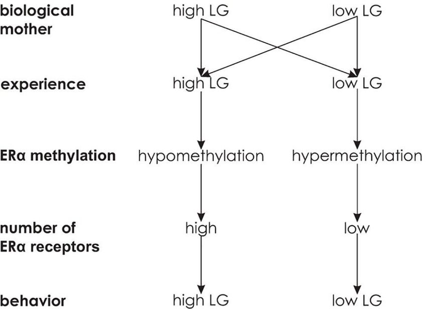
Schmitz et al.                                                                                                                    Building an Asymmetrical Brain

                                                                                  evidence suggests a molecular genetic association of handedness
  A                                        B                                      with the ontogenesis of visceral asymmetries (Brandler et al.,
                                                                                  2013; Brandler and Paracchini, 2014). As handedness and
                                                                                  language lateralization are not completely independent from
                                                                                  each other (Knecht et al., 2000; Jansen et al., 2007; Somers
                                                                                  et al., 2015a), the same could hold true for language lateralization.
                                                                                  Thus, to investigate the molecular factors underlying the
                                                                                  ontogenesis of functional hemispheric asymmetries and a possible
                                                                                  relationship with visceral asymmetries, it is important to consider
                                                                                  the emergence of visceral as well as structural and functional
  C
                                                                                  hemispheric asymmetries in human development.
                                           D

                                                                                  The Emergence of Visceral Asymmetries
                                                                                  The human body is symmetric at the beginning of embryonic
                                                                                  development. The first visceral asymmetry is detected when the
                                                                                  heart, initially a straight tube, starts to loop at the end of 5 gestational
                                                                                  weeks and occupies its typical left-sided position (Steding, 2009).
                                                                                  During an embryonic twist along the rostrocaudal axis, asymmetries
                                                                                  of other organs emerge (Kathiriya and Srivastava, 2000). The
                                                                                  liver is larger on the right than the left side at 6 gestational
                                                                                  weeks (Hutchins and Moore, 1988). The lung divides into the
  FIGURE 1 | The major types of body plans. (A) Asymmetry. (B) Radial             left and right lung buds differing in length at 7 gestational weeks.
  symmetry. (C) Bilateral symmetry. (D) Due to the asymmetrical position of       One week later, the lung buds develop three lobes in the right
  internal organs, humans and other vertebrates have been described as            and two lobes in the left body half (Steding, 2009). The development
  pseudo-bilateral (Levin, 2005).
                                                                                  of the stomach is characterized by left-convex bending starting
                                                                                  at 7 gestational weeks. While the liver extends massively, the
                                                                                  stomach grows into its characteristic left-sided position (Steding,
hemispheric asymmetries such as handedness are partly under                       2009). Overall, the establishment of visceral asymmetries is
epigenetic control (Schmitz et al., 2017b). However, in a fast-                   completed at the end of the embryonic period [end of 10 gestational
moving field like genetics, significant progress has been made                    weeks (see Figure 2A; O’Rahilly, 1979)].
in the meantime. First empirical studies reported the effects
of DNA methylation on the strength of functional hemispheric
asymmetries in the individual (Leach et al., 2014; Schmitz                        The Emergence of Structural
et al., 2018a,b). Here, we aim to focus on the relationship of                    Hemispheric Asymmetries
functional hemispheric asymmetries with inner organ                               Like the visceral organs, the brain develops from an unpaired
asymmetries. Moreover, we previously (Schmitz et al., 2017b)                      structure, the neural tube. Initially, it mostly grows ventrally,
suggested that epigenetic mechanisms might partly account                         resulting in the emergence of its main subregions prosencephalon,
for the large gap between heritability estimates for handedness                   mesencephalon, and rhombencephalon at the age of 6 gestational
from twin and adoption studies of up to 0.66 (Risch and                           weeks. The cerebral hemispheres differentiate toward the end
Pringle, 1985) and the small variance explained by molecular                      of 8 gestational weeks (Chi et al., 1977a; Steding, 2009). The
genetic studies (Eriksson et al., 2010; Armour et al., 2014),                     earliest structural hemispheric asymmetry that has been found
also known as the missing heritability problem (Maher, 2008).                     to date is an enlargement of the left compared to the right
However, in order to account for this gap, this mechanism                         choroid plexus in terms of length, area, and circumference at
requires transgenerational transmission of epigenetic states in                   11–13 gestational weeks (Abu-Rustum et al., 2013). Thus, in
families. As germ line epigenetic inheritance is highly                           contrast to visceral asymmetries which are established in the
controversial in humans (Ambeskovic et al., 2017b), we suggest                    embryonic period, structural hemispheric asymmetries do not
an alternative mechanism by which epigenetic mechanisms                           seem to emerge before the fetal period starting at the 11th
might account for the missing heritability of functional                          gestational week (see Figure 2B; O’Rahilly, 1979). As suggested
hemispheric asymmetries.                                                          by Corballis (2013), the early structural asymmetry in the choroid
                                                                                  plexus might be a precursor for other hemispheric asymmetries.
                                                                                  Specifically, interhemispheric differences in the production of
THE DEVELOPMENT OF ASYMMETRY                                                      cerebrospinal fluid, which synthesizes peptides, growth factors,
                                                                                  and cytokines, might underlie the pronounced leftward planum
The consistency of population-level lateralization in handedness                  temporale asymmetry evident at the 31st gestational week
across centuries (Faurie and Raymond, 2004) and continents                        (Chi et al., 1977b; Kasprian et al., 2011). Starting at the 20th
(Raymond and Pontier, 2004) is likely the result of asymmetrical                  gestational week, a leftward asymmetry in cortical volume has
prenatal CNS development (Willems et al., 2014). Recent                           been reported for the occipital lobes (Weinberger et al., 1982)

Frontiers in Psychology | www.frontiersin.org                                 2                                               April 2019 | Volume 10 | Article 982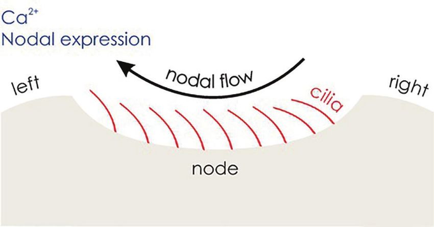
Schmitz et al.                                                                                                                    Building an Asymmetrical Brain

as well as for the entire left hemisphere (Hering-Hanit et al., 2001;             turning preference after birth (Hepper et al., 1990, 1991) and
Kivilevitch et al., 2010; Andescavage et al., 2017).                              handedness at school age (Hepper et al., 2005). The finding of
                                                                                  a prenatal hand preference that is consistent until school age
The Emergence of Motor Asymmetries                                                was lately confirmed by kinematic analysis of fetal arm movements:
In line with structural hemispheric asymmetries already being                     In fetal development, individuals acted faster and more precisely
apparent in human fetal development, prenatal ultrasound                          using the hand that they would report as their dominant hand
studies indicated early signs of motor asymmetries in terms                       at age 9. By analyzing movement or deceleration time for touching
of head turning and arm movements. The majority of human                          the eye or mouth, subsequent handedness could be classified
fetuses display a strong preference for turning the head toward                   with an accuracy between 89 and 100% (Parma et al., 2017).
the right side. Ultrasound observations of fetuses positioned
in the cephalic (head-first) presentation revealed a right side                   The Emergence of Language Lateralization
preference between 30 and 38 gestational weeks (Ververs et al.,                   Language lateralization in newborns and infants has mainly
1994). While this pattern was confirmed in a subsequent study,                    been studied in terms of perceptional asymmetries. At the
a midline preference was observed in breech-positioned (feet-                     behavioral level, a right ear advantage for dichotically presented
first) fetuses (Fong et al., 2005). Newborns also show a                          syllables has been found in infants (Entus, 1980) and 4-day-old
population-level asymmetry toward turning the head to the                         neonates (Bertoncini et al., 1989). Moreover, greater left-
right side (Dunsirn et al., 2016), and the preferred direction                    hemispheric temporal activation in response to language
of head turning predicts hand use in reaching tasks in infants                    stimuli has been reported in neuroimaging studies within
(Coryell and Michel, 1978; Michel, 1981; Konishi et al., 1986).                   the first postnatal week (Peña et al., 2003; Gervain et al.,
Congenital muscular torticollis involves unilaterally shortened                   2008). In order to investigate language lateralization as early
neck muscles, which leads to a permanent head tilt sustaining                     as possible, preterm infants between 28 and 30 gestational
visual experience toward either the left or right hand. Ocklenburg                weeks were tested with a linguistic (typically left hemisphere
et al. (2010) found a strong impact of sustained visual experience                dominant) and a non-linguistic (typically right hemisphere
toward one hand on the probability of being right- or left-handed                 dominant) discrimination task. For linguistic discrimination,
in children affected by this disorder. In adults, 64.5% of kissing                functional optical imaging revealed that posterior temporal
couples turn their head toward the right side, indicating a                       areas showed faster and more sustained activation in the
persistence of head turning bias into adulthood (Güntürkün,                       left hemisphere. The left frontal region (Broca’s area) was
2003). Moreover, the preferred side of head turning is correlated                 responsive to linguistic, but not to non-linguistic discrimination
with handedness LQ (Ocklenburg and Güntürkün, 2009). Overall,                     (see Figure 2D; Mahmoudzadeh et al., 2013). In contrast,
the tendency to turn the head toward one side is likely to                        pitch processing has been shown to be right-lateralized in
be associated with motor preferences in later life.                               fetuses and preterm infants (Schleussner et al., 2004; Mento
    Using ultrasound, individual arm movements are detectable                     et al., 2010). However, Perani et al. (2011) found rather
at 9 gestational weeks (de Vries et al., 2001). Starting at the                   bilateral processing of language perception in neonates. For
10th gestational week, a strong preference of right arm movements                 word production as assessed by fTCD, it has been shown
is apparent in three quarters of fetuses (Hepper et al., 1998),                   that only 60% of 6- to 11-year-old children, but 95% of
which is persistent throughout fetal development (see Figure 2C;                  adults, showed left-hemispheric language lateralization (Haag
McCartney and Hepper, 1999). Starting at the 15th gestational                     et al., 2010). Thus, it has been assumed that language
week, 90% of fetuses prefer right-sided thumb sucking. This                       lateralization develops in the course of language acquisition
early preference is highly persistent as it is correlated with head               (Minagawa-Kawai et al., 2011; Bishop, 2013).

                              A

                              B

                              C

                              D

  FIGURE 2 | The time course of asymmetry development. (A) Visceral asymmetries. (B) Structural hemispheric asymmetries. (C) Motor asymmetries. (D) Language
  lateralization.

Frontiers in Psychology | www.frontiersin.org                                 3                                               April 2019 | Volume 10 | Article 982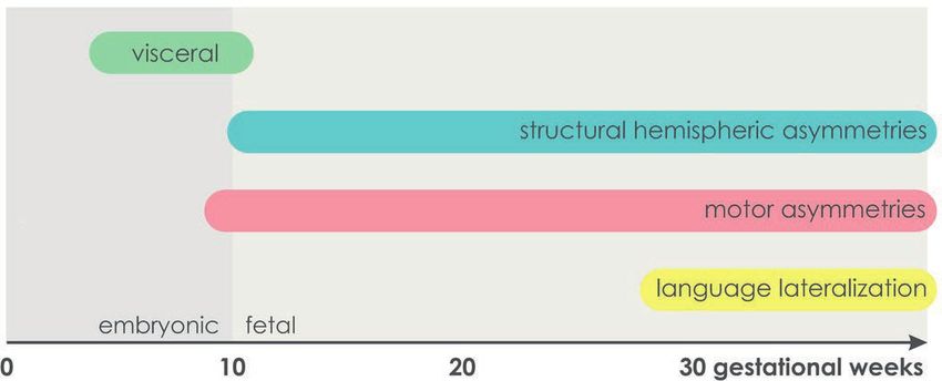
Schmitz et al.                                                                                                        Building an Asymmetrical Brain

THE ROLE OF GENETICS AND                                                   determined by the dichotic listening task. In contrast, there
GENE EXPRESSION                                                            was significant heritability for attentional modulation of language
                                                                           lateralization (Ocklenburg et al., 2016c). In contrast, a genetic
The fact that functional hemispheric asymmetries are already               linkage study estimated the heritability for language lateralization
established in the human fetus is in line with a genetic influence         based on fTCD to be 0.31 (Somers et al., 2015b). Based on
(Hepper, 2013). The observation that handedness direction is               an elevated incidence of atypical language lateralization in
more similar within than between families has inspired genetic             schizophrenia, Crow (2008) proposed shared genetic mechanisms
theories of handedness since the early twentieth century (Ramaley,         between schizophrenia and functional hemispheric asymmetries.
1913). Early genetic models assumed one gene with two alleles              The “Big Bang” theory suggests that a genetic speciation event
to establish handedness and language lateralization (Annett,               involving Protocadherin11X and Y gave rise to the development
1964; McManus, 1985). However, after sequencing of the human               of hemispheric asymmetries and human language, while an
genome, molecular genetic studies on functional hemispheric                absence of hemispheric asymmetries is reflected in schizophrenic
asymmetries revealed a far more complex picture of the                     symptoms. However, a large-scale GWAS did not confirm a
underlying molecular factors.                                              role of this gene pair in schizophrenia (Schizophrenia Working
                                                                           Group of the Psychiatric Genomics Consortium, 2014).
The Genetics of Hemispheric Asymmetries                                    Molecular genetic evidence regarding candidate gene for language
The neurogenetics of handedness has been reviewed in detail                lateralization suggest a role of the Forkhead box P2 gene
elsewhere (Ocklenburg et al., 2013c). Shortly, two genome-wide             (FOXP2) (Pinel et al., 2012; Ocklenburg et al., 2013b), the
association studies (GWAS) did not reveal any single nucleotide            KIAA0319/TTRAP/THEM2 locus (Pinel et al., 2012), the NMDA
polymorphism (SNP) associated with handedness direction                    receptor 2B subunit gene (GRIN2B) (Ocklenburg et al., 2011)
(Eriksson et al., 2010; Armour et al., 2014). A linkage study              and the Cholecystokinin A receptor gene (CCKAR) (Ocklenburg
in a Dutch population isolate confirmed this result but found              et al., 2013a). The association of the Proteolipid Protein 1
suggestive linkage for handedness in in chromosomal region                 gene (PLP1) with language lateralization suggests modulation
22q13 (Somers et al., 2015b). Candidate genes for handedness               via the corpus callosum (Ocklenburg et al., 2018). Overall,
include the androgen receptor gene (AR) (Medland et al., 2005;             although to date no specific environmental factors have been
Hampson and Sankar, 2012; Arning et al., 2015), the catechol-              associated with language lateralization, the fact that several
O-methyltransferase gene (COMT) (Savitz et al., 2007), the                 studies in newborns and infants find rather bilateral processing
leucine-rich repeat transmembrane neuronal 1 gene (LRRTM1)                 of language (Bishop, 2013) and the moderate heritability estimates
(Francks et al., 2007), and the PCSK6 gene (proprotein convertase          from twin and family studies suggest that environmental factors
subtilisin/kexin type 6) (Scerri et al., 2011; Arning et al., 2013;        also contribute to the development of language lateralization.
Brandler et al., 2013). Two recent studies found an association
of handedness with the SETDB2 gene (SET domain, bifurcated 2)              The Molecular Link Between Visceral and
encoding for a methyltransferase regulating hemispheric                    Hemispheric Asymmetries
asymmetries in the zebrafish model (Ocklenburg et al., 2016a;              Over the past years, there has been evidence for a molecular
Crespi et al., 2018). Overall, handedness is likely to be a complex,       genetic link of handedness with the development of body
polygenic trait. However, several large-scale twin studies estimated       asymmetries (Brandler et al., 2013; Brandler and Paracchini,
24–26% of phenotypic variance to be explained by genetic factors           2014; Schmitz et al., 2017a). Visceral asymmetries at the structural
with the remainder being influenced by shared and unique                   level are preceded by a cascade of molecular events leading to
environment (Medland et al., 2006, 2009; Vuoksimaa et al.,                 an asymmetric body plan in the embryo. A universal model
2009). Thus, as pointed out by Hepper (2013), the early emergence          of visceral asymmetry development in vertebrates has been
of handedness in fetal development is in line with a genetic               established over the course of the recent years. An initial symmetry
effect on the initial appearance but does not exclude an effect            break starts around 8 days embryonic age in mice. At this early
of perinatal and postnatal environmental factors on handedness.            stage of development, the vertebrate embryo has an elongated
    To date, no GWAS for language lateralization has been                  body form with the head and heart at the anterior and a cavity
performed yet. Early twin studies found no correlation between             known as the node at the posterior end. Within the node, the
dichotic listening task performances of monozygotic twins and              clockwise rotational movement of motile cilia (hair-like cell
concluded an absence of genetic effects on language lateralization         organelles with the ability to beat) induces a leftward flow of
(Springer and Searleman, 1978; Jäncke and Steinmetz, 1994).                extracellular fluid (Nonaka et al., 1998). In a second step, Pkd2
In twin pairs concordant for handedness, the correlation of                (polycystic kidney disease 2) transduces this leftward nodal flow
language lateralization quotients was 0.74, suggesting a genetic           into stronger left-sided Ca2+ signaling on the edge of the node
component. However, in twin pairs discordant for handedness,               (McGrath et al., 2003; Takao et al., 2013) as well as stronger
the correlation was only 0.18 (Sommer et al., 2002). Bryden                left-sided expression of Nodal (see Figure 3). Nodal is an
(1975) reported a positive correlation between maternal and                intercellular signaling protein of the transforming growth factor
offspring lateralization, but no such correlation between paternal         beta (TGF-beta) family (Zhou et al., 1993).
and offspring lateralization. In a more recent study, Ocklenburg               With the help of the growth/differentiation factor 1 (Gdf1),
et al. (2016c) found no heritability for language lateralization           another member of the TGF-beta family, left-sided Nodal

Frontiers in Psychology | www.frontiersin.org                          4                                           April 2019 | Volume 10 | Article 982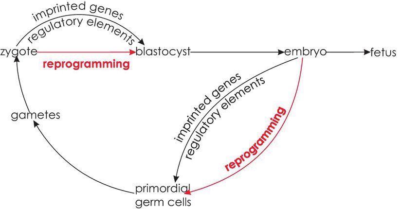
Schmitz et al.                                                                                                                   Building an Asymmetrical Brain

is transmitted to the lateral plate mesoderm (LPM), an                                 nucleus (Concha et al., 2000). Thus, Nodal signaling is responsible
embryonic structure anterior to the node (Rankin et al.,                               for the determination of epithalamic asymmetry direction rather
2000). The Nodal signaling pathway (see Figure 4) is only                              than for establishing asymmetry per se (Roussigne et al., 2012).
activated in the left LPM, expressing Nodal, as well as Lefty2,                        In wild-type zebrafish, the initial symmetry break is induced
and Pitx2. Lefty2 encodes a protein that suppresses the Nodal                          by the fibroblast growth factor (FGF) pathway, which causes
pathway in the right LPM and thereby maintains asymmetry                               parapineal precursor cells to migrate toward the left hemisphere.
(Meno et al., 2001; Sakuma et al., 2002). Finally, Pitx2 encodes                       Interestingly, the SETDB2 gene, a candidate gene for handedness,
a transcription factor that remains asymmetrically expressed                           encodes for a methyltransferase that suppresses fgf8 expression
during the development of the heart and other organs and                               (Ocklenburg et al., 2016a). In zebrafish, no epithalamic asymmetry
plays a direct role in their asymmetric morphology (Piedra                             develops when Fgf8 function is inactivated (Regan et al., 2009),
et al., 1998). Research in zebrafish suggests that prior to                            indicating an overlap of molecular mechanisms involved in
cilia movement, Atp6ap1b, a protein involved in ATPase                                 the ontogenesis of hemispheric asymmetries in zebrafish and
proton pumps, regulates the establishment of the ciliated                              humans in the FGF pathway.
organ (Kupffer’s vesicle in zebrafish) (Gokey et al., 2015),                              Another line of evidence suggests an involvement of the
while the Wnt (wingless-related integration)/beta-catenin                              Nodal pathway in human handedness ontogenesis. PCSK6,
pathway regulates cilia length and number (Zhu et al., 2015)                           mentioned above as one of the key candidate genes for
and functioning of the Nodal signaling pathway (Hüsken                                 handedness (Scerri et al., 2011; Arning et al., 2013), has
and Carl, 2013). Evidence that the Nodal signaling pathway                             been shown to play a role in left-right determination and
also corresponds to visceral asymmetry development in                                  the Nodal pathway in mice. At the structural level, a loss-
humans is given by a strong overlap of genes involved in                               of-function mutation of pcsk6 leads to defects in the
the Nodal signaling cascade and genetic variants involved                              development of the left-right axis such as right-sided stomach,
in disorders characterized by abnormal asymmetry of human                              spleen, or pancreas. Moreover, nodal, pitx2, and lefty are
internal organs. In line with findings in mice and                                     expressed in both the left and the right LPM, suggesting
chicks, among the genes identified to be involved in                                   that their usual asymmetric expression patterns are regulated
laterality defects in humans are NODAL, LEFTY2, and GDF1                               by Pcsk6 (Constam and Robertson, 2000). In humans, PCSK6
(Shiraishi and Ichikawa, 2012; Deng et al., 2015).                                     is most strongly expressed in the liver and spinal cord as
    Besides its involvement in visceral asymmetry development,                         well as the corpus callosum, the largest commissure connecting
the Nodal signaling cascade affects structural hemispheric                             the hemispheres (Johnson et al., 2003). Based on the findings
asymmetries in zebrafish. The zebrafish epithalamus consists                           of PCSK6 being involved in visceral asymmetry development,
of bilateral habenular nuclei and the pineal complex containing                        other genes causing asymmetry defects when knocked out
the medial epiphysis and the left-hemispheric parapineal nucleus                       in mice have been examined with respect to hand performance
(Roussigne et al., 2012). About 20 h after fertilization, genes                        (Brandler et al., 2013). The most common task used for
involved in the Nodal pathway, such as lefty1 and pitx2c, are                          the determination of hand performance is the Pegboard
expressed in the structure later developing into the left                              task. Participants are instructed to move pegs from one
epithalamus. Experimentally manipulated symmetrical expression                         row of holes to another with either the left or the right
of Nodal genes as well as the absence of Nodal gene expression                         hand. A quantitative measure of fine motor skill is obtained
does not prevent parapineal migration. However, left- and                              by relating the times required to complete left- and right-
right-sided parapineal nuclei are equally distributed, and the                         hand trials resulting in the so-called PegQ measure
habenular nucleus is larger on the side containing the parapineal                      (Ocklenburg et al., 2014a). While PCSK6 again showed the
                                                                                       strongest association with PegQ in a cohort selected for
                                                                                       reading disability, PKD2, meiosis-specific structural protein
                                                                                       (MNS1), regulatory factor X 3 (RFX3), and GLI family zinc
                                                                                       finger 3 (GLI3) were also among the top hits. The strongest
                                                                                       association in a general population cohort was found for
                                                                                       a SNP upstream of the Glypican 3 gene (GPC3), whose
                                                                                       disruption in mice causes lung and heart asymmetry defects.
                                                                                       Moreover, genes involved in double outlet right ventricle,
                                                                                       heterotaxia, and situs inversus (mirror reversal of viscera)
                                                                                       were significantly overrepresented in genes associated with
                                                                                       hand skill in both cohorts (Brandler et al., 2013). Overall,
                                                                                       there is evidence that the molecular pathways controlling
                                                                                       visceral laterality may be partly contributing to handedness.
 FIGURE 3 | The development of visceral asymmetries. Motile cilia (red)                As genes involved in ciliopathies are associated with the
 induce a leftward nodal flow, which is transduced into stronger left-sided Ca2+       early development of brain midline structures such as the
 signaling and Nodal expression, triggering the Nodal signaling pathway in the
                                                                                       corpus callosum and the cerebellar vermis, molecular
 left lateral plate mesoderm (LPM).
                                                                                       mechanisms determining visceral asymmetries might

Frontiers in Psychology | www.frontiersin.org                                      5                                          April 2019 | Volume 10 | Article 982
Schmitz et al. Building an Asymmetrical Brain
FIGURE 4 | The Nodal signaling cascade. Facilitated by Gdf1, Nodal is transmitted to the left LPM, expressing Nodal, Lefty2, and Pitx2. The protein encoded by
Lefty2 suppresses the Nodal pathway in the right LPM. Pitx2 encodes a transcription factor that is involved in asymmetric morphogenesis.
be reused for brain midline structures and therefore affect behavioral asymmetries already being apparent in the human
behavioral laterality (Brandler and Paracchini, 2014). fetus set the starting point for the examination of lateralized
A recent study investigated structural and functional gene expression in the human fetal brain. Sun et al. (2005)
asymmetry in situs inversus subjects with and without primary found consistent asymmetrical gene expression between the
ciliary dyskinesia (PCD), a recessive disorder resulting in left and right fetal perisylvian cortex. One of the consistently
disruption of motile cilia. The authors found an elevated rate asymmetrically expressed genes was Lim Domain Only 4
of left-handedness in situs inversus subjects without PCD, but (LMO4). Knockdown of Lmo4 expression in the right anterior
not in those with PCD. Moreover, the typical counter-clockwise cortex in mice resulted in reduced right-hemispheric neuron
bending of the brain (Yakovlevian torque) was reversed in number, thinner right- than left-hemispheric axonal
situs inversus subjects, while structural gray and white matter projections, and a rightward shift of paw preference (Li
asymmetries and functional language lateralization were not et al., 2013). As, in humans, the corticospinal tract connecting
(Vingerhoets et al., 2018). Subsequent whole genome sequencing precentral gyrus and spinal cord does not reach the spinal
of the same sample found a probable monogenetic cause for cord at this early fetal developmental stage (ten Donkelaar
situs inversus in those subjects with PCD, while no candidate et al., 2004), gene expression asymmetries in the developing
mutations were identified for most of the situs inversus subjects motor cortex are unlikely the underlying factor for early
without PCD (Postema et al., 2018). These findings are in line indications of motor asymmetries. Ocklenburg et al. (2017)
with a link between handedness and visceral asymmetry that found pronounced gene expression asymmetries in the fetal
is, however, independent of genes involved in cilia function, spinal cord. In line with the finding that most fetuses show
indicating different mechanisms for different asymmetry more right- than left-sided arm movements, the majority
phenotypes (Vingerhoets et al., 2018). of asymmetrically expressed transcripts was expressed more
strongly in the right spinal cord. These findings suggest that
asymmetrical gene expression in the spinal cord induces
Hemispheric Asymmetries in asymmetries in motor output that lead to use-dependent
Gene Expression plasticity processes in the brain (Ocklenburg et al., 2017).
The comprehensive literature on visceral asymmetry A major limitation of studies on gene expression in human
development in mice and chicken as well as on the ontogenesis tissue is the fact that the impact of gene expression on behavioral
of hemispheric asymmetries in zebrafish suggests that besides outcomes is impossible to determine. However, lateralized gene
genetic variants associated with hemispheric asymmetries in expression has successfully been associated with lateralized
humans, gene expression patterns might also play a decisive behavior in animal models. Grabrucker et al. (2017) reported
role. Thus, the reports of structural hemispheric as well as asymmetrical expression of the candidate genes found by Sun
Frontiers in Psychology | www.frontiersin.org 6 April 2019 | Volume 10 | Article 982Schmitz et al. Building an Asymmetrical Brain
et al. (2005) in the left and right hemispheres of wild-type that subtle expression asymmetries of individual genes might
mice. In the T-maze test, these animals displayed a strong translate to strong expression asymmetries at the level of
rightward bias. In contrast, prenatal zinc-deficient mice, a functional gene groups. Lateralized gene expression was found
mouse model of autism spectrum disorder (ASD), displayed for several functional gene groups involved in signal transmission
an absence of lateralized gene expression as well as an absence in the nervous system (Karlebach and Francks, 2015). In a
of side preference in the T-maze test (Grabrucker et al., 2017). comparative study, Muntané et al. (2017) investigated gene
More direct evidence for an impact of gene expression expression in the left- and right-hemispheric ventrolateral
asymmetries on behavior comes from a study investigating prefrontal cortex, superior temporal cortex, and primary motor
predation behavior in cichlid fish (Lee et al., 2017). While cortex in humans and macaques. No gene was asymmetrically
some individuals preferentially attack the left side of their expressed between the hemispheres after correction for multiple
prey (associated with a right-turn, therefore called right-handed), testing. However, weighted gene co-expression network analysis,
others show the opposite left-handed pattern and only few which clusters genes into modules based on correlations of
are non-lateralized. The authors determined individual laterality expression patterns, revealed modules that showed different
indices based on the number of left- and right-sided attacks. expression levels between hemispheres in all three cortical areas
Gene expression was determined for the left and right tectum in humans. These asymmetric modules contained several
opticum, telencephalon, and hypothalamus. Although, in each candidate genes involved in brain asymmetry (e.g., AR, LEFTY1,
of the three brain structures, more than 300 genes showed LMO4, and PCSK6). Moreover, these modules were enriched
nominally significant left-right differences in their expression for functional gene groups such as receptor activity in the
patterns, none survived correction for multiple testing. For superior temporal cortex and locomotion in the primary motor
each gene and brain structure, the fold change of expression cortex. Interestingly, no module showed differential expression
between the left and right hemisphere was linked to the levels between the left and right macaque cortex (Muntané
behavioral laterality index. In the tectum opticum, 140 genes et al., 2017). Thus, the lack of expression asymmetries at the
showed a linear relationship between gene expression asymmetry level of individual genes does not necessarily indicate the
and behavioral asymmetry during predation. Most of these absence of gene expression asymmetries at the level of functional
genes were upregulated in the hemisphere facing toward gene groups.
(ipsilateral to) the prey. In contrast, the 173 genes showing a To determine whether early structural and behavioral
linear relationship between gene expression asymmetry in the asymmetries in human prenatal development are preceded by
telencephalon and behavioral asymmetry were mostly upregulated early asymmetrical gene expression in the CNS, de Kovel et al.
in the hemisphere not facing (contralateral to) the prey. (2017) compared gene expression in the left and right human
Interestingly, one of the genes displaying this pattern was fetal spinal cord and hindbrain at 6–10 gestational weeks. In
lrrtm1, a candidate gene for handedness (Francks et al., 2007). contrast to the results reported by Ocklenburg et al. (2017),
In the hypothalamus, the 79 genes showing a linear relationship no gene showed individual expression asymmetry after controlling
between gene expression asymmetry and behavioral asymmetry for multiple comparisons. However, functional analysis revealed
were also mostly upregulated in the hemisphere contralateral leftward lateralization of gene groups involved in glutamate
to the prey (Lee et al., 2017). Although this study does not receptor signaling and neurotransmitter transport in the human
allow for causal conclusions, it is a hint toward an impact of fetal spinal cord. As the expression of both gene groups increases
lateralized gene expression on lateralized behavior. Importantly, during fetal development, the leftward lateralization was
this relationship was found, although no gene displayed gene interpreted as the left spinal cord maturing faster than the
expression asymmetry strong enough to survive correction for right. In contrast, the functional gene groups ‘mRNA metabolism’,
multiple testing. ‘DNA strand elongation’, ‘chromosome segregation’, and ‘protein
translation’ showed rightward lateralization. The expression of
Gene Ontology: Considering these functional gene groups decreases in the course of fetal
Gene Functions development, which is also consistent with the assumption of
Similar to the findings by Lee et al. (2017) and in contrast the left spinal cord outpacing the right spinal cord. This pattern
to the studies in human CNS tissue (Sun et al., 2005; Ocklenburg was found to be reversed in the hindbrain, consistent with
et al., 2017), there are a number of studies not replicating the cross-over of nerve tracts in the inferior hindbrain (de
expression asymmetries at the level of individual genes in the Kovel et al., 2017). In a subsequent study, de Kovel et al.
human fetal (Johnson et al., 2009; Lambert et al., 2011) or (2018) complemented this dataset with left and right midbrains
adult brain (Hawrylycz et al., 2012; Pletikos et al., 2014; Muntané and forebrains at 7 gestational weeks. At this stage of development,
et al., 2017). These results might imply that asymmetries in the midbrain showed a similar pattern to the hindbrain with
gene expression are strongly development-dependent and can an advanced maturation rate in the right hemisphere. In contrast,
only be detected during critical time frames. Another, yet not the forebrain showed no differences in maturation rates; however,
contradicting, possibility is that gene expression asymmetries genes expressed in the left forebrain were enriched in the
are mostly too subtle to detect at individual gene level, especially functional gene group ‘cerebral cortex neuron differentiation’,
when correcting for multiple comparisons. Thus, Karlebach while ‘extracellular structure organization’ was enriched in the
and Francks (2015) performed a reanalysis at the level of right forebrain. The authors performed the same analyses on
functional gene groups instead of individual genes, assuming brain samples obtained at 9–15 gestational weeks with finer
Frontiers in Psychology | www.frontiersin.org 7 April 2019 | Volume 10 | Article 982Schmitz et al. Building an Asymmetrical Brain
subdivisions. Similar to hindbrain and midbrain at earlier that approximately one third of variance in asymmetrical gene
developmental stages, they confirmed faster right-hemispheric expression could be explained by epigenetic regulation.
maturation for the right cerebral cortex (excluding the temporal Interestingly, these epigenetic factors were enriched in the
lobe). In contrast, the left diencephalon, temporal lobe, basal TGF-beta signaling pathway, adding additional evidence for an
ganglia, and choroid plexus of the lateral ventricle showed overlap between molecular mechanisms involved in the
faster maturation than their right-hemispheric counterparts. development of visceral asymmetry and hemispheric asymmetries.
This finding is especially interesting for the choroid plexus as This is also in line with the association of SETDB2 with handedness
the left-faster-than-right maturation rates at 9–16 gestational in adults, as the protein encoded by this gene not only regulates
weeks are in line with the left-larger-than-right structural left-right asymmetry in zebrafish but also regulates gene expression
asymmetry at 11–13 gestational weeks (Abu-Rustum et al., epigenetically (Ocklenburg et al., 2016a). As access to DNA in
2013). This structural asymmetry has been suggested to underlie healthy humans is limited to peripheral tissues, the influence
structural asymmetries in the temporal lobe (Corballis, 2013), of epigenetic modulation on hemispheric asymmetries in adults
a brain region that also shows left-faster-than-right maturation has so far only been investigated in buccal samples. Although
rates (de Kovel et al., 2018). DNA methylation is tissue-specific, it has been proposed that
its degree in peripheral tissue can be interpreted as a biomarker
for CNS-related phenotypes (Klengel et al., 2014; Freytag et al.,
THE ROLE OF EPIGENETIC 2017). Leach et al. (2014) investigated DNA methylation in 19
REGULATION CpG sites in the LRRTM1 promoter region. Principal component
analysis revealed a block of CpG sites that was negatively
The described gene expression asymmetries in early human correlated with strength of hand preference in the overall sample
CNS development raise the question of the underlying molecular as well as in female participants. Thus, stronger DNA methylation
factors. Gene expression includes the transcription and translation in these CpG sites was associated with a tendency toward
of a gene, affecting the encoded protein products. Epigenetic ambidexterity. The authors conclude that this finding suggests
mechanisms summarize several chemical modifications to the an effect of environmental factors on handedness via epigenetic
DNA or proteins involved in DNA packaging that regulate modulation (Leach et al., 2014). We recently found that DNA
the accessibility of so-called transcription factors to the DNA, methylation in promoter regions of genes asymmetrically expressed
which results in activation or repression of transcription (Zhang in the human fetal CNS predicts handedness direction (Schmitz
and Meaney, 2010). Only about 1.2% of the human genome et al., 2018a). Moreover, the amount of experienced birth stress
is translated into proteins (Mattick and Makunin, 2006), whereas in pre- and perinatal development was correlated with DNA
non-coding DNA has historically been considered as “junk methylation in NEUROD6, whose expression is tripled in the
DNA.” However, starting with the Nobel Prize-awarded work left compared to the right perisylvian cortex at 12 gestational
of Jacob and Monod (1961), research has shown that an weeks (Sun et al., 2005). Moreover, we found an association
important function of non-coding DNA, such as promoter between language lateralization in the dichotic listening task
regions of genes, is the regulation of transcription and translation. and DNA methylation in the KIAA0319 promoter region in
The most investigated and best understood form of epigenetic buccal cells. The effect was not found for language lateralization
mechanisms is DNA methylation, the addition of a methyl per se, but for the conditions reflecting attentional modulation,
group directly to the DNA, more specifically to the 5-position indicating KIAA0319 as an epigenetic marker for cognitive
of cytosine guanine (CpG) dinucleotides. Within promoter control processes. As KIAA0319 is a major candidate gene for
regions, stronger DNA methylation typically represses dyslexia and presumably involved in the ontogenesis of visceral
transcription of that gene (Jaenisch and Bird, 2003). The complex asymmetries, this finding is partly in line with an overlap of
interplay of epigenetic mechanisms has been shown to play a genes involved in visceral asymmetries and the ontogenesis of
key role in pre- and postnatal brain development (Bale, 2015). hemispheric asymmetries (Schmitz et al., 2018b).
While an important function of epigenetic regulation is tissue-
specific differentiation of cells and therefore a mechanism by Transgenerational Epigenetic Inheritance:
which cells with an identical genotype are able to take different Is That Possible?
forms, epigenetic mechanisms can also change as a function We recently argued that heritable epigenetic mechanisms might
of environmental factors (Tammen et al., 2013). partly account for the missing heritability of handedness (Schmitz
et al., 2017b). However, since the existence of transgenerational
Epigenetics in the Development of epigenetic inheritance in humans is highly controversial (Babenko
et al., 2015), how can epigenetic mechanisms contribute to
Hemispheric Asymmetries
lateralization at the population level?
A number of environmental factors have more or less consistently
been shown to affect handedness (de Kovel et al., 2019).
We previously reviewed the empirical evidence and suggested Germ Line Epigenetic Inheritance
that the epigenetic regulation might be an underlying factor Transgenerational epigenetic inheritance typically refers to direct
connecting environment and phenotype (Schmitz et al., 2017b). transmission via the germ line, meaning that an environmental
In the human spinal cord, Ocklenburg et al. (2017) could show factor influences the epigenetic state in the F0 generation, which
Frontiers in Psychology | www.frontiersin.org 8 April 2019 | Volume 10 | Article 982Schmitz et al. Building an Asymmetrical Brain
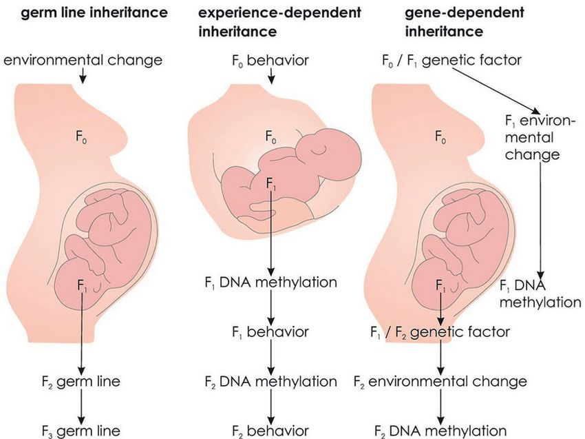
is transmitted to subsequent generations via the germ line region in the hypothalamus, resulting in more pronounced
(Danchin et al., 2011). There are two criteria to the demonstration production of ERα receptors. In contrast, low LG experience
of germ line epigenetic inheritance. First, the environmental leads to relative ERα promoter hypermethylation and less ERα
factor has only affected the F0 generation, but not subsequent receptors. The increase or decrease in ERα receptors causes
generations. Second, because germ cells might be directly the offspring (F1) to show more or less LG behavior toward
influenced by the environmental factor, germ line epigenetic their own offspring (F2), respectively (see Figure 7).
inheritance requires transmission over at least three generations Importantly, cross fostering confirmed the experience of LG
in the female or at least two generations in the male germ behavior being causative for ERα expression. Biological offspring
line (see Figure 5A; Crews, 2008). of high LG mothers that was cross fostered to low LG mothers
However, due to the phenomenon of reprogramming, it had a similarly small amount of ERα receptors in the hypothalamus
has long been assumed that germ line epigenetic inheritance as regular offspring of low LG mothers and thus showed low
is not possible. Reprogramming refers to the erasure of LG behavior themselves. In contrast, biological offspring of low
epigenetic signatures between generations at two times during LG mothers cross fostered to high LG mothers showed a high
development. First, reprogramming takes place shortly after amount of ERα receptors in the hypothalamus and high LG
fertilization in the zygote. Second, epigenetic signatures are behavior as adults (Champagne et al., 2006). Overall, this indicates
removed in primordial germ cells of the developing embryo that an epigenetic state influences an organism’s behavior such
that later develop into gametes. These reprogramming events that it induces the same epigenetic state and behavior in its
ensure restoring of pluripotency of germ cells and zygotes offspring. Thus, in contrast to germ line epigenetic transmission,
(Babenko et al., 2015). The fact that epigenetic states are experience-dependent epigenetic transmission induces recreation
erased during embryonic development conflicts with the idea of epigenetic states in every new generation (see Figure 5B).
that epigenetic states can be inherited. However, it has been While parental behavior and epigenetics interactively influence
shown that some epigenetic states escape both reprogramming inheritance, changes in the environment can interrupt the
processes, such as imprinted genes (Bartolomei, 2009) and transmission across generations (Danchin et al., 2011).
regulatory elements (see Figure 6; Hackett et al., 2013), Interestingly, paw preference and hemispheric dominance
potentially leading to transgenerational epigenetic inheritance in terms of dendritic complexity and spine density have been
over the germ line. compared in F4 generations of rats with ancestors being
In mammals, transgenerational epigenetic inheritance has exposed to stress multigenerationally (female rats in the F0–F3
mainly been reported in mice and rats, while it remains generation had been exposed to stress) or transgenerationally
controversial in humans (Ambeskovic et al., 2017a) due to (only female rats in the F0 generation had been stressed).
the correlational nature of studies reporting these effects. A While both transgenerational and multigenerational stress led
group of epidemiological studies suggest that in utero exposure to an increase in left paw preference in the F4 male offspring,
to the Dutch famine in 1944 and 1945 is associated with less this shift was more pronounced and significantly different
DNA methylation of the insulin-like growth factor 2 (IGF2) from non-stressed controls in the multigenerationally stressed
gene (Heijmans et al., 2008) and affects birthweight and height individuals. Moreover, a preference for the left paw was
of grandchildren (Painter et al., 2008; Veenendaal et al., 2013). associated with increased dendritic complexity and spine
However, no data are currently available for the critical F3 density in the right parietal cortex (Ambeskovic et al., 2017b).
generation. Another group of studies examined a cohort from As stress during pregnancy has been shown to affect maternal
Överkalix, a small isolated village in Sweden with detailed behavior (Yao et al., 2014), maternal behavior might have
records on food availability (Bygren et al., 2001). A surfeit of induced experience-dependent epigenetic inheritance of
food during paternal grandfathers’ childhood was associated hemispheric asymmetries.
with a fourfold increase of diabetes in grandchildren (Kaati Experience-dependent epigenetic inheritance is also conceivable
et al., 2002, 2007). As this effect was transmitted through in humans. For example, an individual’s attachment to their mother
grandfathers and fathers, these studies indicate sperm-mediated is predictive of their infant’s attachment to them, and the experience
transgenerational epigenetic transmission over the germ line of less parental care is reflected in lower sensitivity toward the
(Pembrey, 2002). Based on these epidemiological studies and own children (Champagne, 2008). Applied to handedness, parental
research in rodents, transgenerational epigenetic effects on brain (F0) behavior might lead to specific DNA methylation patterns
functions transmitted over the germ line (see Figure 5A) have in the offspring (F1), initiating the same behavior toward their
been postulated to exist in humans as well (Bohacek et al., children (F2). This in term might result in transmission of DNA
2013). However, there is currently no indication of a unique methylation patterns to the F2 generation (see Figure 5B).
environmental factor in preceding generations to affect an
individual’s handedness. Gene-Dependent Epigenetic Inheritance
The findings of environmental factors involved in handedness
Experience-Dependent Epigenetic Inheritance (Schmitz et al., 2017b, 2018a) open up another possibility of
There are alternative forms of transgenerational epigenetic transgenerational epigenetic inheritance. Coren (1995) suggested
inheritance. In female rats, the experience of high maternal that the familial transmission of left-handedness might indicate
licking and grooming (LG) behavior leads to relative a genetic origin but could also be explained by the possibility
hypomethylation of the estrogen receptor alpha (ERα) promoter that left-handed mothers have experienced more birth
Frontiers in Psychology | www.frontiersin.org 9 April 2019 | Volume 10 | Article 982Schmitz et al. Building an Asymmetrical Brain
A B C
FIGURE 5 | Alternative forms of transgenerational epigenetic inheritance. (A) Germ line epigenetic inheritance: An environmental factor acts on the F0 generation
and induces an epigenetic state that is transmitted to subsequent, unaffected individuals via the germ line. (B) Experience-dependent epigenetic inheritance:
Maternal behavior induces an epigenetic state in the offspring (F1) that in turn influences F1 behavior toward its offspring (F2) transmitting behavior and epigenetic
states across generations. (C) Gene-dependent epigenetic inheritance: A genetic factor modulates the probability of an environmental factor that influences F1
epigenetic states. As F1 likely transmits the genetic factor to F2, epigenetic states are transmitted across generations.
complications than right-handed mothers. The author suggests This third possibility of transgenerational epigenetic inheritance
that the experience of a complicated birth might lead to (see Figure 5C) requires three criteria.
non-specified “internal conditions,” which elevate the probability
(1) There is a genetic risk for an environmental factor
of complications during delivery and left-handed offspring. This
hypothetical scenario was partly confirmed as left-handed Individuals who were delivered in breech position are more
mothers reported more birth complications (especially breech likely to deliver in breech position themselves in their first
birth and prolonged labor) and more left-handed offspring pregnancy than individuals who were delivered in cephalic position
than right-handed mothers, while left-handed fathers did not (odds ratio = 2.3) (Nordtveit et al., 2008). Moreover, the odds
play a role in the number of birth complications or left-handed ratio of breech delivery after one previous breech delivery is
offspring. However, the author assumed that left-handed mothers elevated to 4.3 (Albrechtsen et al., 1998). Interestingly, the usual
had experienced more complications during their own birth rightward head turning preference of fetuses in a cephalic position
without empirically testing this assumption (Coren, 1995). It is not present in fetuses in breech position that rather show a
might be possible that there is interindividual variability in head midline preference (Fong et al., 2005). As direction of head
proneness to the risk of a certain environmental event (e.g., turning is strongly associated with later handedness (Coryell and
birth complications) due to the genetic setup. The environmental Michel, 1978; Michel, 1981; Konishi et al., 1986; Ocklenburg
factor might be associated with epigenetic modifications (for et al., 2010), a genetic influence on fetal position might be associated
example, in the NEUROD6 promoter region, Schmitz et al., with ontogenesis of hemispheric asymmetries. Mothers who were
2018a) eventually leading to changes in behavior (handedness). born prematurely have an enlarged risk of preterm delivery
Frontiers in Psychology | www.frontiersin.org 10 April 2019 | Volume 10 | Article 982Schmitz et al. Building an Asymmetrical Brain
FIGURE 6 | Epigenetic reprogramming. Reprogramming occurs shortly after fertilization in the zygote and in the primordial germ cells of the developing embryo.
At both stages, some epigenetic states are able to escape reprogramming.
FIGURE 7 | Experience-dependent epigenetic inheritance. Independent of licking and grooming behavior (LG) of the biological mother, the experience of high LG is
associated with a large number of ERα receptors and high LG behavior, while the experience of low LG behavior is associated with a low number of ERα receptors
and low LG behavior.
compared to women born at term (Porter et al., 1997) with an (Lunde et al., 2007). Overall, pronounced genetic factors seem
odds ratio of 1.5 (Wilcox et al., 2008). Preterm birth seems to to influence different types of birth complications.
be a heritable trait with a polygenic cause (Chaudhari et al.,
(2) This environmental factor affects DNA methylation
2008) with twin studies estimating genetic factors to account for
30% of the phenotypic variance in gestational age at delivery Prenatal stress has been shown to exert long-term effects
(Kistka et al., 2008). Similar results have been reported for on the hypothalamic-pituitary-adrenocortical (HPA) axis. For
birthweight (Clausson et al., 2000) where genetic factors of the example, the experience of severe stress in utero leads to a
fetus accounted for 31% of phenotypic variance and maternal stronger cortisol response to the Trier Social Stress Test (TSST)
genetic factors accounted for 22% of the variation in birth weight in young adults as compared to control subjects without prenatal
Frontiers in Psychology | www.frontiersin.org 11 April 2019 | Volume 10 | Article 982Schmitz et al. Building an Asymmetrical Brain
stress experience (Entringer et al., 2009). Similar effects have Moreover, both MZ and DZ twins are more likely to
been reported for prenatal stress and cortisol reactivity toward experience a complicated birth and to display lower weight
temporary stressors such as vaccination or maternal separation and shorter gestational age at birth. This is in line with a
(Tollenaar et al., 2011). Several studies have also reported an Finnish large-scale study reporting that the often described
effect of prenatal stress on basal cortisol levels (Glover et al., increased probability of left-handedness in twins is absent after
2010). Recent research suggests that prenatal stress induces its controlling for these two factors, showing that birth weight
associated outcomes via epigenetic modification (Glover, 2015). and gestational age might rather contribute to handedness
For example, DNA methylation in genes associated with HPA development than twinning per se (Heikkilä et al., 2015). As
axis regulation is altered in cord blood and placenta in individuals the probability to conceive twins is partly genetically determined
with chronic stress experience during pregnancy compared to (Machin, 2009; Painter et al., 2010; Mbarek et al., 2016), a
controls (Kertes et al., 2016). The association between prenatal genetic factor might modulate the probability of certain prenatal
stress and DNA methylation in the glucocorticoid receptor environmental factors such as reduced placental blood supply
(GR) gene (NR3C1) (Perroud et al., 2014) has been confirmed and thus access to important nutrients (Nugent and Bale,
by meta-analysis (Palma-Gudiel et al., 2015). Birth stress might 2015) or perinatal complications in general. As mentioned
also modify fetal DNA methylation via its associated enhanced above, differences in prenatal environment between MZ twins
levels of oxidative stress (Saphier et al., 2013; Ávila et al., likely induce the observed differences in DNA methylation at
2015). Interestingly, cortisol and other glucocorticoids released birth (Gordon et al., 2012), which might have effects on the
by acute or chronic stress might also play a role in modulating ontogenesis of hemispheric asymmetries. Thus, an identical
hemispheric asymmetries, which often results in stronger right- genetic background does not contradict the idea of gene-
hemispheric involvement (Ocklenburg et al., 2016b). dependent epigenetic inheritance, as genetic factors do not
determine prenatal environment, but modulate it in a
(3) DNA methylation affects the phenotype
probabilistic way.
As mentioned above, the study by Leach et al. (2014), as well Overall, a complex interplay of genetic, environmental, and
as our own work (Schmitz et al., 2018a,b), provides first hints epigenetic factors might contribute to handedness development.
that epigenetic markers for handedness and language lateralization A similar mechanism has been shown to lead to the ontogenesis
can be found in buccal cells. Moreover, findings from spinal of visual processing asymmetries in birds. In pigeons, the left
cord tissue in the human fetus suggest an involvement of epigenetic hemisphere outperforms the right hemisphere in categorization
mechanisms in asymmetrical gene expression, potentially leading and discrimination tasks. This functional hemispheric asymmetry
to motor asymmetries (Ocklenburg et al., 2017). can be prevented by incubating pigeons in darkness thereby
Overall, gene-dependent epigenetic inheritance (see Figure 5C) withdrawing the environmental factor light. This is possible
is an alternative mechanism for epigenetic transmission that because birds occupy an asymmetrical position within the egg.
is worth being empirically tested. Although this proposed While the right eye faces toward the eggshell, the left eye is
mechanism is based on an initial genetic factor, it would not covered by the own body. Thus, a genetically determined body
necessarily be captured by classic molecular genetic studies. position results in an environmental factor (light exposure)
For example, genetic polymorphisms involved in certain birth stimulating the right, but not the left eye, through the semi-
complications would not be captured by GWAS on the handedness translucent eggshell initiating a cascade of molecular events
phenotype, as they might only exert their influence on handedness eventually leading to asymmetries at the behavioral level. While
depending on the interplay with environmental factors. This epigenetic mechanisms are thought to play a role in this cascade
is also illustrated by the special case of twins. Sharing the of molecular events, this possibility has not yet been confirmed
same genetic background, MZ twins have the same genetic by empirical testing (Güntürkün and Ocklenburg, 2017;
risk of birth complications. However, this does not necessarily Ocklenburg and Güntürkün, 2018). In humans, a genetic
mean that both co-twins are equally affected by birth predisposition might lead to an enhanced probability of birth
complications. For example, 75% of MZ twins are monochorionic, complications that then induces epigenetic modification of the
i.e., share a placenta (Cordero et al., 2005). Unequal placenta NEUROD6 promoter region or other genes. As mentioned
sharing, i.e., one co-twin receives blood from more than 60% above, this mechanism is most likely to affect DNA methylation
of the placenta, has repeatedly been shown to affect discordance in brain cells, while our results in buccal cells are better
of birth weight in MZ twins (Fick et al., 2006; Zhang et al., described as an epigenetic signature for handedness in
2013). As the placenta is crucial for providing the embryo or non-neuronal tissue.
fetus with nutrients (Nugent and Bale, 2015), these findings The potential mechanism of gene-dependent epigenetic
argue for different intrauterine environments even in MZ twin inheritance for familial transmission of handedness raises
pairs. These environmental differences might underlie the reported the question if a similar mechanism is conceivable for language
differences in DNA methylation patterns in DZ and MZ twin lateralization. However, environmental factors involved in
pairs immediately after birth (Ollikainen et al., 2010; Gordon the development of language lateralization have been
et al., 2012). Interestingly, preeclampsia, one of the most fatal investigated sparsely, and we found an effect of DNA
birth complications, is associated with reduced levels of DNA methylation on cognitive modulation of language lateralization,
methylation and increased expression of genes involved in the but there was no effect on language lateralization per se
TGF-beta signaling pathway such as PITX2 (Martin et al., 2015). (Schmitz et al., 2018b). Thus, further research is needed in
Frontiers in Psychology | www.frontiersin.org 12 April 2019 | Volume 10 | Article 982You can also read