Extracellular Vesicles in Oral Squamous Cell Carcinoma and Oral Potentially Malignant Disorders: A Systematic Review - MDPI
←
→
Page content transcription
If your browser does not render page correctly, please read the page content below
International Journal of
            Molecular Sciences

Review
Extracellular Vesicles in Oral Squamous Cell
Carcinoma and Oral Potentially Malignant Disorders:
A Systematic Review
Tami Yap * , Neha Pruthi, Christine Seers , Simone Belobrov, Michael McCullough and
Antonio Celentano
 Melbourne Dental School, The University of Melbourne, 720 Swanston Street, Carlton, VIC 3053, Australia;
 npruthi@student.unimelb.edu.au (N.P.); caseers@unimelb.edu.au (C.S.);
 belobrov@student.unimelb.edu.au (S.B.); m.mccullough@unimelb.edu.au (M.M.);
 antonio.celentano@unimelb.edu.au (A.C.)
 * Correspondence: tspyap@unimelb.edu.au

 Received: 29 January 2020; Accepted: 10 February 2020; Published: 11 February 2020

 Abstract: Extracellular vesicles (EVs) are secreted from most cell types and utilized in a complex
 network of near and distant cell-to-cell communication. Insight into this complex nanoscopic
 interaction in the development, progression and treatment of oral squamous cell carcinoma (OSCC)
 and precancerous oral mucosal disorders, termed oral potentially malignant disorders (OPMDs),
 remains of interest. In this review, we comprehensively present the current state of knowledge of
 EVs in OSCC and OPMDs. A systematic literature search strategy was developed and updated to
 December 17, 2019. Fifty-five articles were identified addressing EVs in OSCC and OPMDs with all
 but two articles published from 2015, highlighting the novelty of this research area. Themes included
 the impact of OSCC-derived EVs on phenotypic changes, lymph-angiogenesis, stromal immune
 response, mechanisms of therapeutic resistance as well as utility of EVs for drug delivery in OSCC
 and OPMD. Interest and progress of knowledge of EVs in OSCC and OPMD has been expanding on
 several fronts. The oral cavity presents a unique and accessible microenvironment for nanoparticle
 study that could present important models for other solid tumours.

 Keywords: oral squamous cell carcinoma; extracellular vesicles; exosomes; oral premalignant lesions;
 mouth neoplasms; microvesicles; oral potentially malignant disorders

1. Introduction
      Oral squamous cell carcinoma (OSCC) is a highly debilitating disease that is often fatal. Early stage
OSCC has the most favourable prognosis and requires less aggressive treatment; however, more than
half of patients present with an advanced disease [1]. Advances in the early identification and improved
targeted treatment to reduce morbidity continue to be sought. An area that presents an opportunity
for the early identification of OSCC is the study of oral potentially malignant disorders (OPMD),
considered a group of disorders that precede OSCC development, which can be visualized and assessed
clinically. The oral cavity presents a unique and accessible environment to source nano-sized particles
for study of the presence of OSCC and OPMD.
      Extracellular vesicles (EVs), composed of an outer lipid bilayer containing proteins and nucleic
acids, have been identified as an important means of cell-to-cell communication, both near and
distant, in the body [2]. Tumour derived EVs can impact cellular processes of effector cells within
the tumour environment as well as at distant sites, creating favourable environments for tumour
growth and spread [3]. A specific type of EV are exosomes containing nucleic acids, proteins and

Int. J. Mol. Sci. 2020, 21, 1197; doi:10.3390/ijms21041197                         www.mdpi.com/journal/ijms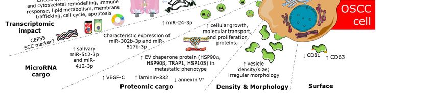
Int. J. Mol. Sci. 2020, 21, 1197 2 of 29
lipids that appear to be purposefully packaged. This is exemplified by the observable differences
between exosome-contained cargo, such as non-coding microRNA with microRNA from donor cells,
suggesting a key way that tumour cells can influence their surrounding microenvironment [4,5].
Interest is expanding in the use of biofluid EVs as a potential non-invasive source of prognostic and
diagnostic biomarkers. Furthermore, fully understanding the role that EVs have in influencing the
tumour phenotype, immune modulation and preparation of metastatic bed has the potential for the
development of more sophisticated and targeted treatments.
EVs are a complex group of vesicles that originate from distinct subcellular compartments.
Cells release many types of vesicle subpopulations such as exosomes, microvesicles, apoptotic vesicles,
lipoproteins and chylomicrons. The two most characterised types of EVs have differing physiological
and pathological functions: exosomes [6] derived from multi-vesicular bodies (MVBs), a specialised
subset of endosomes that contain intra-luminal membrane-bound vesicles; and microvesicles or
ectosomes [7] that are derived from the cytoplasmic membrane [8,9]. In addition to different sites of
origins, the two types of EVs have differing intracellular life before discharge. It has now been well
established that although the term “exosomes” has often been used in research articles for isolated
EVs, most historical and novel purification protocols such as differential ultracentrifugation, filtration
and precipitation kits, co-isolate different types of EVs [10]. Therefore, the 2018 minimal information
for studies of extracellular vesicles (MISEV) guidelines recommend that unless specific markers of
subcellular origin can be established, future authors are encouraged to use terms for EV subtypes
that refer to physical characteristic, biochemical composition or description of conditions or cell of
origin [11]. This will enable a more accurate determination of the contribution of cargoes of each EV
subtype to a physiologic state.
In this systematic review, we present the current state of knowledge with respect to EV studies
conforming with the MISEV 2018 guidelines that involve clinically defined OSCC and OPMDs and
OSCC-derived cell lines.
2. Results
2.1. Study Selection and Characteristics
The primary literature search, as of 12 October 2018, identified 1247 papers, of which 801 were
unique. Screening of these by title resulted in retention of 280 records and following abstract screening,
108 reports were retained for full text review. Finally, of the 108 articles screened, 32 articles were
accepted by the reviewers for final inclusion. A second search performed on 17 December 2019,
identified a further 23 articles eligible for inclusion and review. This indicates a rapid rise in interest in
the field of EV analysis in relation to OSCC and OPMDs and/or a greater propensity for investigators
to adhere to the MISEV guidelines.
Of the 55 retained studies, 43 included in vitro components, 12 included in vivo components,
and 16 studies included clinical sample derived EV components in their design. There were 13 studies
which compared EVs from clinical cohorts (Table 1) and 29 studies presenting novel findings of EVs
derived from and on primary or established cell lines (Table 2).
Various EV isolation and purification techniques were reported with ultracentrifugation being the
most common technique for EV isolation and immunoblotting for characterization and classification
of EVs. The majority of the studies were conducted in China, Japan and the USA. The oldest study
included in this review was from 2005 [12], with 53 of 55 articles published from 2015 onwards.
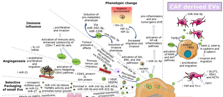
The articles variously describe the EV morphologic characters and cargo and the role of specific
molecules in EV-mediated and EV-employed influence from and on OSCC, OPMD and stromal cells
types. The main features of OSCC- EVs have been graphically summarized in Figure 1.Int. J. Mol. Sci. 2020, 21, 1197 3 of 29
Table 1. Findings from eligible studies assessing extracellular vesicles (EVs) in oral squamous cell carcinoma (OSCC) and oral potentially malignant disorders (OPMD)
clinical cohort samples.
Study Specimen Type Markers Assay Findings
quantification - Increased vesicle size, irregular morphology, increased
AFM, SMFS-CD63 mapping, intervesicular aggregation observed in OSCC
[13] Salivary EVs morphology
Western blot analysis - Higher CD63 single vesicle density (by AFM) in OSCC
CD63 surface density
- Proteome of OSCC EVs enriched in proteins involved in
[14] Salivary EVs shotgun protein analysis MS molecular transport and cellular growth and proliferation
- Significantly higher concentration (p = 0.01) and size (p = 0.002)
quantification NTA, AFM, TEM, ELISA, of nanoparticles in OSCC
[15] Salivary exosomes
CD81, CD9, CD63 Western blotting - lower expression of CD 81 (p = 0.032) in OSCC
- miR-302b-3p and miR-517b-3p expressed only OSCC-EVs
vs. controls
[16] Salivary EVs microRNA qPCR array; qPCR - miR-512-3p and miR-412-3p were up-regulated in OSCC-EVs
vs. controls
- Increased (I1,404/I2,924) (p = 0.005), (I1,033/I1,072) (p = 0.024)
Fourier-transform IR and (I2,924/I2,854) (p = 0.026) in OSCC with sensitivity 100%,
[17] Salivary exosomes spectroscopy intensity ratios
spectroscopy specificity 89%
- 109 miRNA exhibited changes in their expression levels in
OSCC EVs compared to normal controls
[18] Salivary exosomes microRNA microarray; qPCR - miR-24-3p was significantly higher in OSCC EVs in comparison
to healthy controls (p < 0.05)Int. J. Mol. Sci. 2020, 21, 1197 4 of 29
Table 1. Cont.
Study Specimen Type Markers Assay Findings
- Higher quantitative levels in OSCC (p < 0.05) vs. normal and
benign ulceration
TEM; dynamic light - Annexin V+ decreased in high OSCC pathological grade
Salivary MVs and
[19] Quantification; Annexin V scattering; CFSE labelling; (p < 0.01) and poorer survival (p < 0.05)
circulating MVs
flow cytometry - Higher quantitative levels of circulating MVs in OSCC
(p < 0.001)
- Exosomal fraction compared to free plasma shared all 9
[20] Plasma EVs microRNA microarray upregulated and 6 of 7 downregulated microRNAs
- Increased EV number (p < 0.001) and EV size (p < 0.05) in OSCC
vs. controls
[21] Plasma EVs Quantification; microRNA NTA; qPCR - Increased miR-21, miR-27b and miR-27a increased in EV
fraction vs. non-EV fraction in OSCC
- Non-significant decrease in CD63 post OSCC resection
(p = 0.091)
[22] Plasma EVs CD63, Cav-1 immunocapture - non-significant increase in Cav-1 post OSCC resection
(p = 0.237)
- 37 differential proteins in OSCC with lymph node metastasis vs.
LC-MS; healthy controls
[23] Serum exosomes protein MS; - 28 differentially expressed proteins in OSCC with lymph node
qPCR metastasis vs. no lymph node metastasis esp. PF4V1, XCXL7,
F13A1, ApoA1Int. J. Mol. Sci. 2020, 21, 1197 5 of 29
Table 1. Cont.
Oral Lichen Planus
Sample Type Markers Assay Findings
[24] Salivary exosomes microRNA microarray; qPCR - miR-4484 significantly upregulated in OLP
- Increased expression of miR-34a-5p, miR-130b-3p,
and miR-29c-3p
[25] Plasma exosomes microRNA microarray - Decreased miR-301b- 3p and miR-144-3p
- miR-34a-5p correlated with increasing disease severity
Abbreviation List: AFM: Atomic force microscopy; CFSE: carboxyfluorescein succinimidyl ester; ELISA: enzyme linked immunosorbent assay; LC-MS: Liquid chromatography-mass
spectrometry; MV: microvesicle; MS: mass spectrometry; NTA: nanoparticle tracking analysis; OSCC: oral squamous cell carcinoma; qPCR: quantitative polymerase chain reaction; TEM:
transmission electron microscopy; SMFS: single molecular force spectroscopy.Int. J. Mol. Sci. 2020, 21, 1197 6 of 29
Table 2. Findings from eligible studies assessing EVs in cell-based samples.
OSCC
Study Cell Type Main Findings
EVs Derived from EVs Studied on
OSCC and Keratinocytes
OSCC cell-derived exosomes promote source cell line proliferation, migration,
[26] OSCC lines (OSC-3, OSC-4) OSCC lines (OSC-3, OSC-4)
and invasion in a dose dependent manner
OSCC—EVs produced under hypoxic conditions increased the migration and
[27] OSCC lines (Cal-27, SCC9) OSCC lines (Cal-27, SCC9) invasion of normoxic OSCC cells in a hypoxia-inducible factors—HIF1α and
HIF-2α–dependent manner which was abrogated by miR-21 depletion
SQUU-B- exosomes s conferred metastatic ability to non-metastatic SQUU-A
[28] OSCC line (SQUU-B (metastatic)) OSCC line (SQUU-A (non-metastatic)
cells and reduced mRNA expression of cytokeratin 13
OSCC line (HOC313-LM (highly metastatic sub OSCC line (HOC313-P (parent cell HOC313-LM exosomes transferred oncogenic miR-343-3p and miR-1246 to
[29]
line)) line)) HOC313-P cells and resulted in increase in cell motility and invasive ability
EVs released from cisplatin-resistant OSCC cells transmit miR-21 to induce
[30] Cisplatin resistant OSCC cell lines (HSC3, SCC9) Parental OSCC (HSC3 and SCC9)
cisplatin resistance of OSCC cells
OSCC derived EGFR-containing EVs were able to transform RT7 cells, effects
[31] OSCC line (HSC3) Oral keratinocytes (RT7)
of which were largely blocked by cetumixab
OSCC lines (Ca1 CALH2, SQCC/Y1)
Premalignant buccal oral keratinocyte (SVpgC2a) Exposure to OSCC-derived exosomes specifically modulated mRNA
[32] Transformed malignant (SVFN9) Primary normal oral keratinocytes transcripts associated with matrix remodeling, cell cycle, differentiation,
normal oral keratinocyte lines (OH113, NK4, apoptosis, transcription and translation
NOK368)
OSCC & Fibroblasts
HSC3-exosomes lead to uptake by NOFs with resulting upregulation of
[33] OSCC line (HSC3) NOF
expression of non-coding RNA Lnc-CAF
Cal-27 MVs were internalized increased levels of CAF markers (FAP and Tn-c)
were isolated from HCFs, Cal-27 MV treated HGFs also showed increased
[23] OSCC line (Cal-27) HGFs (human gingival fibroblasts)
glucose and lactate production with an increased expression of GLUT1, PDK1
and MCT4 but a decrease in CaV1Int. J. Mol. Sci. 2020, 21, 1197 7 of 29
Table 2. Cont.
OSCC
Study Cell Type Main Findings
EVs Derived from EVs Studied on
OSCC and Keratinocytes
Exosome transfer from TGFβ signalling-competent fibroblasts increased
transforming growth factor-beta receptor II (TBRII) levels and the TBRII signal
[34] Primary OSCC CAFs Primary OSCC keratinocytes
transducing protein SMAD2 (but not SMAD3) phosphorylation in OSCC
keratinocytes
Primary OSCC CAFs and adjacent tissue fibroblasts CAF derived EVs increased C25(OTSCC) cell proliferation and migration
[35] OSCC lines (SCC25, SCC4)
(AF) compared with exosome depleted media or controls
CAF derived exosomes containing low or lentiviral plasmid restored
[36] Primary OSCC CAFs and matched NOFs OSCC lines (Cal-27, SCC15)
expression of miR-34a-5p are transferred to OSCC cells
CAF-EVS significantly increased the invasive, migration and apoptosis rate of
OSCC lines (HSC3, SAS, SCC15,
[37] Primary CAFs and NOFs, HSC-3, SAS and SCC-15 but not SCC-25 with HSC-3 the most was most
SCC25)
responsive to pooled CAF-EVs with deeper invasion, small tumour islands
Primary cancer associated fibroblasts; normal
[38] OSCC line (Cal-27) Significantly increased Cal-27 migration and invasion
fibroblasts
Normal fibroblast and keratinocyte derived EVs suppressed OSCC
[39] Human dermal fibroblasts; normal keratinocytes OSCC line (TR146)
proliferation but only at particular doses
OSCC & Endothelial Cells
Both OSCC-derived exosomes increased VEGFR2 expression in HUVECS;
SQUU-B exosomes increased tube formation in HDLECs, both OSCC cell line
OSCC cell lines (SQUU-A (non-metastatic), SQUU-B
[40] endothelial cells (HUVECs, HDLECs) derived exosomes stimulate expression of HDLEC mRNA expression of
(metastatic))
VEGFRs1-3 but only SQUU-B exosomes increased expression of VEFG-A,-C
and -D
Cal-27-MVs carrying Sonic hedgehog (Shh) protein significantly induce tube
[41] OSCC lines (Cal-27) Endothelial cells (HUVECs)
formation in HUVECS
SSC15-EVs showed significant HUVEC tube formation, migration and
increased apoptotic bodies vs. HSC3—EVs which significantly inhibited tube
[42] OSCC lines (SCC15 AND HSC3) Endothelial cells (HUVECs)
formation and proliferation; EVs derived from different OSCC cell lines are
either pro-or anti angiogenicInt. J. Mol. Sci. 2020, 21, 1197 8 of 29
Table 2. Cont.
OSCC
Study Cell Type Main Findings
EVs Derived from EVs Studied on
OSCC and Keratinocytes
PCI-13– exosomes caused significant increase in VEGF mRNA levels and
[43] OSCC lines (PCI-13, UMSCC47) Endothelial cells (HUVECs) IGFBP-3 mRNA expression levels in the recipient cells; no significant changes
after co-incubation of HUVECs with UMSCC47-derived exosomes
Metastatic OSCC subline (LN1-1) and parent line Human dermal lymphatic LN1-1 derived EVs significantly increased migration and tube formation
[44]
(OEC-M1) endothelial cells (LECs) compared to incubation with parent cell
OSCC & Immune Cells
OSCC serum MV fractions were FasL positive and induced DNA
OSCC patient sera; T cells (Jurkat) and OSCC line
[12] T-blast cells, T cells (Jurkat) fragmentation, decreased the MMP potential or induced apoptosis of Jurkat
(PCI-13)
cells, T blast cells or activated T lymphocytes
Increase in miR-21-5p and activation of NF- κB suggesting pro-inflammatory,
[21] OSCC line (Cal-27) derived EVs THP1 monocytes
pro-tumorigenic shift
OSCC exosomes enhanced cytotoxicity of NK cells via the interferon
[45] OSCC cell lines (SCC-25, Cal27) NK cells regulatory factor 3 (IRF-3) pathway by delivery of that NF-κB-activating
kinase-associated protein 1 (NAP1)
immortalized keratinocytes (HIOEC) leukoplakia Macrophages (THP-1 derived); OSCC—exosomes but not HIOEC- or Leuk1- exosomes THP-1 and PBMCs
[46]
cell line (Leuk1) OSCC cell lines (SCC25, Cal27) healthy donor PBMCs derived macrophages into a M1 phenotype associated with tumor suppression
OSCC derived exosomes produced under normoxic conditions activated
[47] OSCC lines (Cal-27; SCC-29) Primary γδ T cells
cytotoxicity of γδ T cells against these same oral cancer cell lines
OSCC- exosome co-cultured macrophages showed higher expression levels of
OSCC line (SCC9, Cal-27), immortalized Macrophages (THP-1 derived), protein markers of M2 macrophage subtype: CD163, CD206, Arg-1, and IL-10;
[48]
keratinocytes (HIOEC) HBMCs media of above cultured macrophages increased proliferation and invasive
ability of OSCC cell lines with this effect abrogated by inhibition of miR-29a-3pInt. J. Mol. Sci. 2020, 21, 1197 9 of 29
Table 2. Cont.
OSCC and Mesenchymal Stem Cells
Primary mesenchymal stem cell (MSCs) from
OSCC line (SCC-15); oral dysplasia LK and OSCC mesenchymal stem cell derived exosomes both accelerated
[49] normal oral mucosa, dysplastic leukoplakia (LK)
line (DOK) proliferation, invasion and migration of both SCC-15 and DOK cells
and OSCC
hBMSCs transfected with miR-101-3p-Cy3-derived exosomes donated
Primary human bone marrow mesenchymal stem
[50] OSCC line (TCA 8113) miR-101-3p to OSCC cells repressing invasion and migration and reducing
cells
colony forming ability
OPMD
Study Cell Type Main Findings
EVS Derived from EVs Studied on
T-cell proliferation and migration significantly increased with erosive
[51] OLPPlasma-derived exosome from OLP patients T lymphocytes (Jurkat)
LP-derived exosomes but not non-erosive LP exosomes
Abbreviation list: CAFs: cancer associated fibroblasts; HUVECs: human umbilical vein endothelial cells; HDLECs: human dermal lymphatic endothelial cells; NOFs: normal oral
fibroblasts; OLP: oral lichen planus; OPMD: oral potentially malignant disorder; OSCC: oral squamous cell carcinoma; PBMC: peripheral blood mononuclear cells.Int. J. Mol. Sci. 2020, 21, 1197 10 of 29
Int. J. Mol. Sci. 2020, 21, x FOR PEER REVIEW 8 of 27
Figure
Figure 1. 1.Known
Knownroles
roles and
and interactions
interactionsofofEVs
EVsin in
OSCC.
OSCC.
2.2. Increased Abundance
2.2. Increased andand
Abundance Altered
AlteredMorphology
Morphology of OSCC-Derived
OSCC-DerivedEVs
EVs
Several
Several EV EV studies
studies suggestmorphological
suggest morphological and andor orvolume
volumedifferences
differences between
betweenEV isolated from from
EV isolated
healthy individuals and individuals with OSCC and following OSCC
healthy individuals and individuals with OSCC and following OSCC resection. These observations resection. These observations
correlate with observations from other body fluids [52]. Using atomic force microscopy (AFM),
correlate with observations from other body fluids [52]. Using atomic force microscopy (AFM), Sharma
Sharma et al. (2011) [13] showed that salivary exosomes from healthy patients (n = 5) had
et al. (2011) [13] showed that salivary exosomes from healthy patients (n = 5) had homogeneous and
homogeneous and circular morphology, and had a significantly smaller size range than salivary
circular morphology,
exosomes from oraland had
cancer (OC)a significantly
patients (n = 5)smaller
(40–80 nmsizevs.range
20–400thannm, salivary exosomes from
p < 0.05). Furthermore, the oral
cancer = (40–80 nm vs. 20–400 nm, p <
exosomes in saliva of OC patients were 2- to 4-fold more abundant and had greater morphologicalsaliva
(OC) patients (n 5) 0.05). Furthermore, the exosomes in
of OC patients
variation in were
shape 2- to aggregation
with 4-fold more andabundant and had
multivesicular greater
bodies present morphological variationetinal.shape
[13]. Zlotogorski-Hurvitz
with (2016)
aggregation
[15] also and multivesicular
examined OC bodies derived present
exosomes[13].inZlotogorski-Hurvitz
unstimulated salivaetisolatedal. (2016) [15] also
using
ultracentrifugation.
examined Initially they
OC derived exosomes utilised nano-tracking
in unstimulated analysisusing
saliva isolated (NTA)ultracentrifugation.
to show that exosomes were they
Initially
of significantly
utilised nano-tracking higher concentration
analysis (NTA) to show in thethat
(pooled)
exosomes oralwere
fluidsof of OC patients
significantly (36.0concentration
higher ± 7.5E8
particles/mL) than in the pooled oral fluid healthy individuals (17.9 ± 12.45E8
in the (pooled) oral fluids of OC patients (36.0 ± 7.5E8 particles/mL) than in the pooled oral fluid particles/mL) (p = 0.01).
healthy
The NTA analysis also indicated that exosomes were significantly larger in size in the OC patient oral
individuals (17.9 ± 12.45E8 particles/mL) (p = 0.01). The NTA analysis also indicated that exosomes
fluid pool samples (95.36 ± 36.76 nm, n = 11) than the healthy individual sample pool (49.05 ± 32.87
were significantly larger in size in the OC patient oral fluid pool samples (95.36 ± 36.76 nm, n = 11) than
nm, n = 17) (p = 0.002) [15]. Pooling of the samples from the cancer and healthy controls unfortunately
the healthy
precludedindividual
them from sample
being poolable (49.05 ± 32.87the
to determine nm, n = 17)
median (p = 0.002)
exosome [15]. Pooling
size distribution of the samples
between the
fromclinical
the cancer and healthy controls unfortunately precluded them from
types. NTA analysis was not performed on isolated exosomes due to difficulty in separating being able to determine
the median exosome
the tightly packed size distribution
vesicles resulting frombetween the clinical types.This
the ultracentrifugation. NTA analysis was
observation would notindicate
performed
on isolated
that someexosomescautiondue to difficulty
should be shown in separating the tightly packed
towards “individual” exosome vesicles
data resulting
derived from from the
ultracentrifuged exosome
ultracentrifugation. pellets. In would
This observation an alternative
indicateapproach,
that some bio-structural
caution should differences
be shown between
towards
salivary exosomes from OC patients and healthy individuals were assessed
“individual” exosome data derived from ultracentrifuged exosome pellets. In an alternative approach, using Fourier-transform
infrared (FTIR)
bio-structural spectroscopy,
differences between revealing
salivary 3 signal
exosomesintensity
fromratios that wereand
OC patients significantly increased in were
healthy individuals
OSCC exosomes versus controls (I1,404/I2,924, p = 0.005), (I1,033/I1,072, (p = 0.024) and (I2,924/I2,854, p = 0.026) [17].
assessed using Fourier-transform infrared (FTIR) spectroscopy, revealing 3 signal intensity ratios that
A developed discrimination function model enabled differentiation of salivary exosomes with a
were significantly increased in OSCC exosomes versus controls (I1,404 /I2,924 , p = 0.005), (I1,033 /I1,072 ,
sensitivity of 100% and specificity of 89%, finding that early- and late-stage OSCC clustered together [17].
(p = 0.024) and (I2,924
In contrast, /I2,854et, pal.=(2019)
Zhong 0.026) [17].
[19] whoA developed
reported discrimination
a rigorous saliva sample function model
collection enabled
protocol,
differentiation
did not find ofthat
salivary exosomes
salivary with a(MV)
microvesicles sensitivity of 100%
in samples sourcedandfrom
specificity
65 OSCC ofpatients,
89%, finding that early-
21 patients
and late-stage OSCC clustered together [17].
In contrast, Zhong et al. (2019) [19] who reported a rigorous saliva sample collection protocol,
did not find that salivary microvesicles (MV) in samples sourced from 65 OSCC patients, 21 patientsInt. J. Mol. Sci. 2020, 21, 1197 11 of 29
with an oral ulcer or 42 healthy donors differed in physical morphology, size distribution and zeta
potential, as assessed by TEM, flow cytometry and dynamic light scattering. However, there were
significantly higher levels of salivary MVs in the total cohort of OSCC patients compared to healthy
individuals (p < 0.01) and patients with oral ulcers (p < 0.05). Furthermore, increased salivary MV
abundances were correlated with metastasis and tumour stage, but discrimination between healthy
donors and the early stage OSCC subgroup alone (Stage I + II) did not reach significance with this
cohort. Circulating MVs were also found to be significantly higher in OSCC patients compared to
healthy donors (p < 0.001), although no correlation in quantification between salivary and circulating
MVs was identified [19]. However, no significant relationship was found between salivary MV levels
and overall survival [19], and so although salivary MV abundance might serve as an indicator of
disease presence and progression, further investigation is to understand its potential as needed to
assess prognosis.
2.3. Differential Expression of OSCC-Derived EV Surface and Cargo Proteins
It has been reported that exosomes from OSCC patients showed an increase in CD63 surface
density assessed by force spectroscopy compared to those from healthy controls, although this was not
described statistically [13]. Increased EV surface expression, of either CD63 or CD9, was not found
using ELISA, although CD81 expression was significantly lower in OC derived fluids (p = 0.032) [15].
Zorilla et al. (2019) [22] collected plasma samples from 10 OSCC patients with large tumours
(T4) with no distant metastasis both pre- and shortly post-tumour and lymph node resection. Isolated
plasma EVs were characterized for CD63 and CAV-1 using immunocapture-based assay. Overall,
this pilot study did not report findings that reached statistical significance, but found trends that CD63
positive plasmatic EV levels decreased in the 7 days following surgery (p = 0.091), whereas CAV-1
levels increased (p = 0.237), most likely due to post-surgery inflammatory response. Further trends
suggested poor survival associated with continuing high plasmatic CD63 EV levels following resection
(p = 0.808) [22]. Expansion of a study of this type using a larger cohort may indicate if this approach
has some promise as a prognostic survival indicator.
Using a shotgun proteomic approach with salivary EVs of healthy (n = 10) and OC (n = 7)
subjects, Winck et al. (2015) identified 381 proteins by mass spectrometry with 18 proteins detected
exclusively in the healthy group EVs, 4 proteins exclusively identified in the OC group EVs and 8
proteins differentially expressed (p < 0.05) between the groups [14]. Gene ontology analysis of these
proteins revealed over-representation of antigen binding and enzyme inhibitory functions, proteins
related to transport, particularly of metals, as well as cellular growth and proliferation [14].
Questioning evolution of EV proteomic cargo in OSCC metastasis, Ono et al. (2018) [53] have
characterized the EV proteomes of an OSCC line (HSC-3) and a metastatic sub-line (HSC-3-M3) with
particular interest in heat shock proteins as chaperone proteins. The metastatic OSCC cells secreted
larger EVs than those derived from the parental OSCC cells, as measured by TEM (p = 0.045) and 32%
of the proteome was different between the two cell lines. Of 192 EV proteins identified, 9 chaperones
were at higher levels in HSC-3-M3 EVs as compared to HSC-3 EVs, including HSP90α, HSP90β,
TRAP1 and HSP105, suggesting that these EV proteins may potentially be biomarkers of a metastatic
phenotype [53].
Zhong et al. (2019) found that the abundance of vascular endothelial growth factor C (VEGF-C) in
the tumour samples of OSCC patients correlated with patient levels of salivary MVs [19]. VEGF-C is
one of the major pro-lymphangiogenic growth factors, thus correlating metastasis to lymph nodes
with salivary MV levels. Interestingly, the percentage of annexin V+ , as a marker of apoptotic cell
MV origin, was approximately 50% in salivary MVs of both OSCC patients and healthy donors,
whereas few circulating MV of OSCC patients were annexin V+ . Furthermore, within the OSCC group,
the percentage of annexin V+ MV was significantly decreased in salivary MVs of patients with a higher
pathological grade (III) when compared to those with lower pathological grades (I and II) (p < 0.01),
which was associated with poorer survival rates (p < 0.05) [19].Int. J. Mol. Sci. 2020, 21, 1197 12 of 29
Serum exosomal protein content has been measured and explored as an indicator of locoregional
spread. Li et al. (2019) [54] isolated exosomes from serum of subjects, 10 each with OSCC with lymph
node metastasis (LNM), OSCC with no LNM (NLNM) and healthy controls (HC). Proteomic comparison
using a combination of liquid chromatography-mass spectrometry and mass spectrometry followed by
clustering into gene ontology categories. Of 415 proteins identified, 37 and 28 differentially expressed
proteins were identified in OSCC-LNM compared to HC and OSCC-NLNM, respectively. Combined
with qPCR-based validation findings from tissue, serum and whole blood samples, they highlighted
that serum exosomal proteins, PF4V1, CXCL7, F13A1 and ApoA1 may be related to the OSCC lymph
node metastasis [54]. ROC analysis using relative abundances of ApoA1, CXCL7, PF4V1 and F13A1 in
serum, serum exosomes and whole blood indicated some potential application as novel predictive
circulating biomarkers for OSCC with LNM but are not useful in prognosis. Expansion of the cohort
size would test the validity of these conclusions.
Laminin-332 in cancer cells has been shown to promote cell growth, invasion and metastasis [55].
In accord with this, Wang et al. (2019) [44] found that Laminin-332 proteins (laminin α3, β3 and γ2)
were upregulated in LN1-1 cell line EVs versus the levels found in the EV of the OEC-M1 cell line,
from which LN1-1 cells were derived (>1.5 fold) and the number of laminin γ2-positive gold particles
per EV was greater in LN1-1 EVs than in OEC-M1 EVs (p < 0.01) [44]. Comparing 10 healthy controls
and 20 OSCC, ELISA detected a significantly higher level of plasma EV-borne laminin-332 in lymph
node positive OSCC patients than in lymph node negative OSCC patients, which were both higher
than healthy controls (p < 0.01). The protein load ex vivo fluorescent signals from the cervical lymph
nodes of mice orthotopically implanted with PKH-26-labeled laminin γ2-deficient EVs (from LN1-1
LAMC2-knockdown cells) demonstrated a significant decrease in uptake relative to the corresponding
controls (p < 0.01), suggesting a reduced ability to drain into lymph nodes in comparison with the
control EVs [44].
Overall, these data indicate that in combination it may be that various salivary MV biomarkers
including HSP90α, HSP90β, TRAP1, HSP105, VEGF-C, annexin V and laminin-332 could be of use in
tumour detection, staging and prognosis.
2.4. Directed microRNA Cargo of OSCC-Derived EVs
Differential microRNA expression profiles of OSCC-EVs have been reported from both in-vitro and
clinical studies. Rabinowits et al. (2017) compared the microRNA expression of tongue OSCC-derived
EVs, matched benign tissue as well as plasma from one single patient [20]. They found significant
differences in the microRNA expression profiles of tumour and matched benign tissue, with nine
differentially upregulated and seven downregulated in tumour tissue out of 359 microRNAs found [20].
Comparison of the microRNA present in exosomes isolated from the conditioned media from 4 HNSCC
cell lines: H413 (from buccal mucosa OSCC), Detroit 562 (pharyngeal cancer metastatic to pleura),
FaDu (hypopharyngeal cancer) and Cal 27 (tongue OSCC), with similarly isolated EVs from normal
gingival keratinocytes identified 32 common differentially abundant microRNA in the 4 HNSCC cell
lines [56]. Further, comparison of the microRNA present in EVs isolated from the saliva of OSCC
patients (n = 5) and 5 cancer-free controls found several of the microRNA that were upregulated
in cancer culture cells were increased in salivary exosomes of HNSCC patients when compared to
controls [56].
In a similar study comparing the concentration, size and microRNA content of EVs from the
saliva of 5 OSCC patients and 5 healthy controls, Gai et al. (2018) showed that two microRNAs,
miR-302b-3p and miR-517b-3p, were expressed only in OSCC patients while a further two, miR-512-3p
and miR-412-3p, were present in greater abundance in comparison to EVs from the saliva from healthy
controls [16].
Salivary exosomal miRNA microarray profiling by He et al. (2020) [18] of 4 OSCC patients and
4 healthy controls found 50 miRNA were significantly increased (fold change 3.60 to 345.62) and 59
miRNA were decreased (fold change 0.49 to 0.02). Particularly, miR-24-3p was significantly increasedInt. J. Mol. Sci. 2020, 21, 1197 13 of 29
(fold change 121.54). The level of miR-24-3p was 5.73-fold higher in cancer patients than in healthy
controls (p < 0.01) in a validation cohort of 45 OSCC vs. 10 healthy controls with an AUC of 0.738
(95% CI: 0.589–0.886, p = 0.02). The molecular mechanism of miR-24-3p in tumorigenesis was also
investigated using plasmid transfection of HSC6 and SCC25 OSCC cell lines with the overexpression
of miR-24-3p significantly increasing the colony formation and growth rate of OSCC cells. PER1 was
recognized as a predicted target of miR-24-3p with its levels significantly decreased (p < 0.01) in OSCC
patients compared to normal controls [18]. MicroRNA cargo of OSCC-derived EVs continue to be of
interest as biomarkers and therapeutic targets.
2.5. Selective microRNA Cargo Loading into EVs in OC and Oral Dysplasia
Quantitative PCR profiling of EVs isolated from plasma of patients with OSCC showed that miR-21,
miR-27b and miR-27a were specifically enriched in EVs compared to the non-EV fraction (p < 0.05) [21].
Comparing microRNA content of CAL27 cells vs. CAL27-derived EVs, demonstrated selected
packaging with several microRNA differentially expressed between cells and EVs including miR-7d-5p,
miR-130a-3p, miR-181a-5p, miR-107, miR-21-5p, miR-103a-3p, miR-22-3p and miR-20a-5p [21].
Dickman et al. (2017) [57] also demonstrated selective packaging of microRNAs into EV of OSCC and
oral dysplasia cell lines, showing miR-142-3p, miR-451a, miR-150-5p and miR-223-3p were consistently
dysregulated compared to their donor cells. Furthermore, miR-142-3p was found to be consistently
increased in concentration in donor cells upon shRNA-mediated inhibition of Rab27A, which functions
in a pathway responsible for selective packaging of microRNAs into small EVs [57]. Further evaluation
of miR-142-3p, for its role in promoting cancer growth, found it to target TGFBR1, reducing its
expression in donor cells leading to suppressed tumour growth and colony formation. Conversely,
release of miR-142-3p in the small EVs was found to reduce TGFBR1 activity and promote tumour
growth both in vitro and in vivo [57].
2.6. OSCC-Derived EVs Influence Phenotypic Change and Confer Tumorigenicity
The EV cargoes can influence expression and behaviour of target cells. Qadir et al. (2018) [32]
measured mRNA transcripts following exposure of normal oral keratinocytes to exosomes isolated from
8 cells lines: 3 OSCC (Ca1, CaLH2, SQCC/Y1), one transformed malignant (SVFN8), one premalignant
buccal oral keratinocyte (SVpgC2a) and 3 normal oral keratinocytes (OH113, NK4, NOK368). Exposure
to exosomes from all cell lines impacted expression of mRNA transcripts involved in matrix modulation,
cytoskeletal remodelling, immune response, lipid metabolism and membrane trafficking; whereas
OSCC cell line-derived exosomes specifically modulated matrix and membrane remodelling, cell cycle,
differentiation, apoptosis and transcription and translation [32].
Kawakubo et al. (2016) [28] found that purified exosome samples derived from cell culture
supernatant of the metastatic cell line SQUU-B induced the invasive properties of non-metastatic cells
(SQUU-A). This was associated with reduction in mRNA expression of KRT13 [28] coding cytokeratin
13, the down-regulation of which is strongly linked to malignant transformation [58]. Sento et al.
(2016) revealed that OSCC cell-derived exosomes promote tumour cell proliferation, migration and
invasion of OSCC cells, potentially through the activation of Akt, ERK and JNK signalling pathways.
Furthermore, OSCC-4 exosomes induced tumours of significantly larger size than parental OSCC-4
cells in a murine in vivo model [26]. Similarly, injection of EVs from the metastatic OSCC subline
LN1-1 into mice induced tumours of increased volume than in mice injected the parental cell line
OEC-M1 EVs or PBS (p < 0.01) [26].
Heparin can inhibit cancer cell exosomes internalization by blocking cell-surface heparin sulfate
proteoglycan (HSPG) receptors [59,60]. Sento et al. 2016 [26], found that heparin treatment abrogated
the uptake of OSCC-4 cell-derived exosomes by OSC-4 cells and subsequent effects such as promotion
of cell proliferation, migration and invasion. However, these effects were transient in vitro and in vivo
effects and required repeated heparin dosing [26]. Together, this data indicates that uptake of exosomesInt. J. Mol. Sci. 2020, 21, 1197 14 of 29
by OSCC cells uses similar mechanism(s) to that used by other cancer cell types and that exosomes can
influence cancer cells in an autocrine and paracrine fashion.
The phenotypic changes induced by EVs are thought to be influenced by EV microRNA content.
Sakha et al. (2016) [29] demonstrated that exosomes mediated transfer of microRNA to adjacent or
distant cells for intercellular communication in OSCC cell lines when oncogenic microRNAs were
delivered from a highly metastatic OC cell line HOC313-LM to the poorly metastatic cell line HOC313-P
through exosomes. It was revealed that exosomal miR-342-3p and miR-1246 induced a pro-metastatic
phenotype [29]. Involvement of exosomal delivered microRNA in metastasis was also demonstrated
by Kawakubo-Yasukochi et al. (2018) [61]. The mRNA transcriptome of SQUU-A cells treated with
exosomes from SQUU-A and SQUU-B had differential expression of 22 mRNA. Further analysis of 6
selected microRNAs revealed miR-200c-3p as a key pro-invasion factor that silenced the targets CHD9
and WRN, significantly accelerating the invasive potential of SQUU-A cells [61].
Exosomes isolated from the murine HNSCC cell line SCCVII plus the human cell lines SCC90
(oropharyngeal SCC) and PCI-13 were injected into immunocompetent C57bL/6 mice with premalignant
tumours induced using the 4NQO. The numbers of developing tumours per mouse increased in
the SCCVII exosome treated group relative to the controls (6.2 vs. 3.2, p < 0.001) and per mouse
tumour burden increased (p < 0.037) [62]. Importantly, the tumours had reduced inflammatory
infiltrates. However, human cell line exosome injection did not result in significant tumour burden
changes. Nonetheless, there was a significant reduction of immunofluorescence using both anti-CD4
and anti-CD8 antibodies in the murine tumours exposed to exosomes indicating there is a reduced
inflammatory phenotypic effect induced by each EV type [62].
Further evidence of microRNA delivery by exosomes is shown by uptake of CAL 27-derived EVs
by THP1 monocytes, with a corresponding increase in oncogenic miR-21-5p presence and associated
activation of the NF-κB inflammatory pathway [21]. This was accompanied by an increase in measured
levels of IL-6, CCL2, PEG2 and MMP9, suggesting a pro-inflammatory and pro-tumorigenic shift [21].
It was proposed that in particular, the transmission of miR-21 promotes monocyte migration and
infiltration, which in turn promotes angiogenesis [21].
Further evidence of the importance of miR-21 in oral cancer was demonstrated by Li et al.
(2019 [47], who using C3H mice showed that integrated anti-PD-L1 and miR-21 knockdown improved
the antitumor effect of tumour-derived exosomes [47].
Collectively, these studies demonstrate that EVs derived from OSCC directly influence cellular
phenotype by promoting cell proliferation, invasion and metastasis.
2.7. EV Mediated Fibroblast Interaction in OSCC
Exosomes derived from cultured human dermal fibroblasts (NHOK, NHDF) and oral keratinocytes
(HaCaT) were shown to be produced at similar amounts but to possess different exosomal markers [39].
NHOK and NHDF exosomes were CD9, annexin V and Flotillin-1 positive, whereas HaCaT exosomes
were only CD9 positive, pointing to altered protein packaging in these exosomes [39]. When co-cultured
with healthy keratinocyte and fibroblasts, proliferation was suppressed, similar to the effect of the
corticosteroid dexamethasone. Fibroblast exosomes had the more suppressive effect. Co-culture of these
exosomes with OSCC cell line TR146 also suppressed proliferation at particular doses, but keratinocyte
exosomes had the greater anti-proliferative effect [39]. Thus, non-malignant cells produce exosomes
with differential effects on non-malignant and malignant cells.
Languino et al. (2016) [34] investigated whether exosomes purified from stromal fibroblasts are
able to confer TGFβ signalling to SCC keratinocytes deficient in TGFβ ligand response. TGFβ signalling
in cancer is complex and can have proliferative and anti-proliferative effects, dependent on the context.
Languino et al. demonstrated that exosome transfer from TGFβ signalling-competent fibroblasts
increased transforming growth factor-beta receptor II (TBRII) levels and the TBRII signal transducing
protein SMAD2 phosphorylation in OSCC keratinocytes, while SMAD3 phosphorylation did not
consistently change. Furthermore, stimulation of OSCC keratinocytes with recombinant TGFβ1 afterInt. J. Mol. Sci. 2020, 21, 1197 15 of 29
exosome transfer resulted in a small, but further increase of phosphorylated SMAD2 [34]. Thus,
exosome delivery and uptake influence TGFβ signalling.
Principe et al. (2018) [35] had a main focus to identify cancer-associated fibroblast (CAF)-enriched
secreted molecules that are shuttled between fibroblasts and cancer cells and that can affect cancer
cell progression. To this end, they have determined a comprehensive CAF proteome. They found
that CAF secreted exosomes significantly increased SCC25 cell proliferation and migration compared
with exosome-depleted media or controls. In addition, when OTSCC cells were treated with human
recombinant microfibrillar-associated protein 5 (MFAP5), which they identified as enriched in the CAF
secretome, cell proliferation and migration via activation of MAPK and AKT oncogenic pathways
were observed [35]. Investigation of MFAP5 levels in patient tumours showed elevated levels in
cancer-associated stroma [35].
In their analysis, Li et al. (2018) [36] observed that the expression of miR-34a-5p was significantly
reduced in CAF- and SCC cell line (SCC25 and Cal-27)– derived exosomes. They demonstrated transfer
of cy3-tagged miR-34a-5p to the OSCC cells. In xenograft experiments, miR-34a-5p overexpression
in CAFs could inhibit the tumorigenesis of OSCC cells [36]. Sun et al. (2019) [38] observed that
exosomes derived from CAFs and normal fibroblast (NF) medium when added to a medium of
OSCC Cal-27 cells, exerted strong effects on upregulating MMP-3, MMP-9, N-cadherin and β-catenin.
Furthermore, CAL-27 migration and invasion assays significantly increased when exposed to CAF
exosomes (p < 0.05) [38]. The onco-microRNA miR-382-5p is associated with several cancers and
has been shown to be more abundant in CAFs than in NFs [63]. Direct transfection of CAL-27 with
miR-382-5p mimics promoted migration and invasion capacity [63].
OSCC-derived MVs may influence normal fibroblasts that in turn can influence tumour progression.
The glycometabolic influence of MVs derived from CAL-27 OSCC cells was studied by Jiang et al.
(2019) in HGFs (human gingival fibroblasts) from healthy individuals, PNFs (paracancerous normal
fibroblasts) and CAFs 3 OSCC patients [23]. HGFs incubated with the OSCC-derived MVs internalized
these tumour MVs (TMVs) and subsequently increased levels of CAF markers (FAP and Tn-c).
TMV-treated HGFs also showed increased glucose and lactate production with an increased expression
of GLUT1, PDK1 and MCT4, but a decrease in CaV1. Knockdown experiments on HGFs showed
CAV1 knockdown caused increased expression of PDK1 and MCT4, but not GLUT1, indicating Cav 1
is related to aerobic glycolysis of HGFs through ERK1/2 activation pathway. Injection of a combination
of untreated and TMV-treated HCFs with CAL-27 xenograft into nude mice found that treating with
TMVs was associated with a significant increase in tumour weight (p < 0.05). The invasive potential of
OSCC cells was demonstrated to be increased after being co-cultured with TMV-treated HCGs and
partially reversed by knockdown of MCT1 in CaL-27 cells [23].
Effects of EVs derived from 5 primary CAF lines and 5 primary NF lines were studied on 4
TOSCC cell lines: HSC3 and SAS (higher invasive and metastatic potential) and the less aggressive
SSC-15 and SSC-25 cell lines [37]. The invasive potential of HSC-3, SAS and SCC-15, but not SCC-25,
was significantly increased (p < 0.05) by co-culture with individual CAF-EVs compared to NF-EVs and
controls. HSC-3 was the most responsive to pooled CAF-EVs and invaded deeper (p < 0.01) and with
smaller tumour islands (p < 0.05) when compared to NF-EVs and controls. Further, migration (p < 0.05)
and apoptosis rate (p < 0.05) were increased. Microarray was performed on total RNA isolated from
HSC3 cell line treated with CAF-EV or NF-EV, identifying thirty-two differentially expressed (FC ≥ 1.3)
genes. These findings support CAF-EV mediated influence on OSCC behaviour and gene expression
which may be those OSCCs that already possess higher invasive and metastatic potential [37].
Ding et al. (2018) [33] studied long non-coding RNA profiles in the transformation of NFs
into CAFs within the OSCC microenvironment, particularly identifying upregulation of lncRNA
LOC400211, also known as Lnc-CAF. Lnc-CAF is located in 14q23.1-q23.2, identified as a H&N SCC
cancer risk locus [64]. Expression of Lnc-CAF in OSCC cell line HSC3-derived exosomes was found
to be high. Incubation with HSC3 exosomes lead to uptake by NFs with resulting upregulation ofInt. J. Mol. Sci. 2020, 21, 1197 16 of 29
expression of Lnc-CAF (p < 0.05) [33]. This is the only study to date reporting expression of lncRNA in
exosomes in OSCC [33].
Together, these studies demonstrate a bi-direction EV mediated CAF influence in
OSCC progression.
2.8. OSCC-Derived EVs Stimulate Changes in Endothelial Cells Consistent with Angiogenesis
and Lymphangiogenesis
Several studies demonstrated effects from OSCC-derived EVs on vascular and lymphatic
endothelial cell lines with changes in gene expression and protein abundances consistent with
angiogenic and lymphangiogenic processes. Morioka et al. (2016) exposed HUVECs (human umbilical
vein endothelial cells) and HDLECs (human dermal lymphatic endothelial cells) to exosomes isolated
from 2 OSCC cells lines developed from tumours isolated from the same patient, one non-metastatic
(SQUU-A) and one metastatic (SQUU-B). The HDLEC response to SQUU-A exosome exposure was an
increase in VEGFR1, VEGFR2 and VEGFR3, whereas the response to exosomes from SQUU-B cells
led to an increase in all VEGFRs, as well as VEGF-A, VEGF-C and VEGF-D. HUVEC response to
exposure to exosomes from each OSCC cell lines was an increased level of mRNA and protein of only
VEFGR2 [40]. Exposure to exosomes from SQUU-B cell lines lead to increased branching of HDLEC,
indicating stronger tube formation, but neither exosome population affected HUVEC branching [40].
In contrast, de Andrade et al. (2017) [42] have shown OSCC cell line SSC15 derived EVs induced
significant HUVEC tube formation, migration and increased apoptotic bodies in comparison to HSC3
cell line EV that significantly inhibited tube formation and proliferation [42]. SCC15 and HSC3 release
EV similar in size and concentration, but HSC3 cells exhibit higher CD63 and annexin II levels. Thus,
pro and anti-angiogenic effects can be exerted on HUVEC by EVs derived from different cell lines.
Whether CD63 and annexin II exposure influences HUVECs remains to be determined.
Aberrant activation of the sonic hedgehog (Shh) pathway in OSCC specimens was shown to be
closely related to angiogenesis via the RhoA/ROCK signalling pathway [41]. Using immunohistochemistry
to detect sonic hedgehog (SHH) in primary and metastatic FFPE specimens of 80 OSCC, Huaitong
et al. (2017) [41] found that SHH was positively associated with micro-vessel density, TNM stage,
tumour recurrence and lymph node metastasis. SHH was assessed by Western blot and shown to be
abundant in the OSCC cell line CAL 27; however, the amount of SHH in CAL 27-derived microvesicles
was up to 5-fold that of the cell line itself. HUVECS treated with CAL 27-derived microvesicles
followed by Matrigel culturing were found to increase tube formation. Furthermore, this effect was
blocked utilizing exoenzyme C3 transferase and shRNA targeting RhoA. This together supported that
OSCC-derived SHH in microvesicles may modulate angiogenesis and vascular bed preparation of
primary tumours and future metastatic sites [41]. Therefore, it appears that cytoskeletal system can
enhance cancer cell invasiveness and the metastatic potential via exosomes.
Ludwig et al. (2018) [43] aimed to analyse to what extent exosomes produced by OSCC cell
lines, the human papilloma virus-(HPV)-negative line PCI-13 and HPV-positive lateral tongue SCC
cell line UMSCC47 play in blood vessel formation in vitro using HUVEC and an immunocompetent
orthotopic mouse model. Culturing the tumour cells under hypoxic conditions significantly increased
the yield of total exosome proteins for both cell lines. Co-incubation of HUVECs for 24 h with
20 mg of PCI-13–derived exosomes caused an increase in VEGF mRNA levels and IGFBP-3 mRNA
expression levels in the recipient cells (p < 0.05), suggesting growth promotion. However, no significant
changes were seen after co-incubation of HUVECs with UMSCC47-derived exosomes. Nor were
significant differences observed in internalization of PCI-13 and UMSCC47-derived exosomes by
HUVECs. Results using the in vivo 4-NQO oral carcinogenesis model [65] showed tongue base cell
line UMSCC90, PCI-13 or murine cell line SCCVII-derived exosome-treated groups have an increased
amount of vascular structures within the tumour tissue. Overall, the tumour-derived exosomes
promoted angiogenesis [43]. Plasma analysis showed that relative to healthy controls, patients with
HNSCC with active disease have elevated levels of exosomes that interact with HUVECs and stimulateInt. J. Mol. Sci. 2020, 21, 1197 17 of 29
their proliferation [43]. Wang et al. (2019) [44] found that incubation of human dermal lymphatic
endothelial cells (LEC) with metastatic subline LN1-1- EVs did not affect growth but significantly
increased migration and tube formation of LEC when compared to LEC incubation with non-metastatic
parent cell line OEC-M1-derived EVs [44].
Thus, pro- and anti-angiogenic effects can be exerted on HUVEC and other non-malignant cell
lines by EVs derived from different cell lines. In addition, specific tropism shown by the HUVEC and
HDLEC cells [40] points to avenues of investigation regarding the metastatic process.
2.9. Immune Influence of OSCC Derived EVs
Correlation has been found between Fas ligand (FasL) expression on tumour cells and T cells
undergoing apoptosis within the tumour microenvironment in subsets of OSCC patients [66,67]. Kim et
al. (2005) were able to isolate MVs from the sera of 27 OSCC patients, whereas they were unable to
isolate MVs from healthy controls. They found that 21 patients had Fas ligand positive MVs (FasL+)
and were able to induce apoptosis of Jurkat and T-cell blasts. The biological activity of the FasL+ MVs
was partially blocked by ZB4 anti-Fas monoclonal antibody. In addition, FasL levels were found to be
correlated with the patient tumour stage, highlighting that FasL+ MVs may play a role in tumour bed
immune response [12].
The effects of EVs from tongue OSCC cells on the phenotype and cytotoxic activity of immune
cells in vitro and on the innate immune system in vivo using a zebrafish model was assessed by Al
-Samadi et al. (2017) [68]. They demonstrated that activated immune cells significantly decreased the
proliferation and invasive abilities of tongue OSCC cell lines. Furthermore, EVs from SCC-25 increased
the cytotoxic activity of CD8+T and NK cells more than those from HSC-3 cells. In zebrafish, the amount
of IL-13 mRNA was decreased following exposure to SCC-25 derived EVs [68], suggestive of reduced
B-cell and macrophage activation, amongst other possible effects. Wang et al. (2018) [69] showed that
OC-derived exosomes directly enhanced the cytotoxicity of NK cells. In addition, NF-κB-activating
kinase-associated protein 1 (NAP1) was significantly enriched in the corresponding donor cells.
Following internalization of NAP1 derived from OSCC cells, there was an augmentation of levels of
downstream molecules associated with NK cell activation and tumour-suppressive function [69]. Xiao et
al. (2018) [46] demonstrated that conditioned media from OSCC-exosome-activated macrophages
significantly promoted the migration of OSCC cells. However, this was not found with exosomes
derived from normal keratinocytes and leukoplakia cell lines [46].
OSCC-stromal mononuclear cells or macrophages can differentiate into subtypes, mainly M1 with
antitumor effects, M2 with tumour-promoting effects and with M2 subtype macrophage infiltration
levels negatively correlated with prognosis in OSCC [70]. Exosomes from OSCC cell lines SSC-9 and
CAL-27 were cultured by Cai et al. (2019) [48] with monocytic cell line THP-1- derived macrophages to
study exosome effect on polarization of macrophages to M2 subtype. These co-cultured macrophages
showed higher expression levels of protein markers of M2 macrophage subtype: CD163, CD206,
Arg-1 and IL-10. Additionally, the medium of the coculture promoted the proliferation and invasion
of the source OSCC cell lines. OSCC tissues were found to have high expression miR-29a-3p, low
suppressor of cytokine signaling 1 (SOCS1) and highly expressed phosphorylated signal transduction
and transcriptional activator 6 (p-STAT6) compared to adjacent tissue [48]. Inhibition of miR-29a-3p in
the OSCC cell lines found that co-culture with subsequent derived exosomes led to inhibition of M2
phenotype conversion, higher levels of SOCS1 and lower levels of STAT6 expression. Accompanying
the invasive and proliferative ability following placement of the culture medium with the sources OSCC
cell line was also found to be decreased [48]. This demonstrated an EV-mediated interaction of tumour
promotive effects through promotion of M2 macrophage differentiation, mediated by EV-transferred
miR-20a-3p via the SOCS1/STAT6 signalling pathways.
Together the data indicate that EVs carry chemical signals that direct the immune system to reduce
function and also increase pathogenic potential of malignant cells.Int. J. Mol. Sci. 2020, 21, 1197 18 of 29
2.10. Modulating OSCC-EV Production and Packaging
There has been some evidence presented that extracellular factors impact EV production and
contents. Annexin A1 (ANXA1) has been implicated as an important factor in HNSCC cellular
proliferation via EGFR signalling pathway [71]. ANXA1 has been negatively correlated with increasing
tumour grade in OSCC [72,73]. Raulf et al. (2018) [71] observed an inverse relationship between
ANXA1 levels and EGFR, with dysplastic tissue rich in EGFR and poor in ANXA1. Down-regulation
of ANXA1 resulted in increased EGFR activity and downstream PI3K-AKT signalling that may be
via exosomal EGFR. It was also shown that shRNA knockdown of ANXA1 in HN5 tongue OSCC
cell line resulted in reduced production levels of EVs and concurrent reduced levels of exosomal
phosphorylated-EGFR [71]. Furthermore, functional studies using shRNA to knockdown ANXA1
resulted in a clear increase in cellular proliferation in 4 different HNSCC cell lines.
When Momen-Heravi et al. (2019) challenged cultured CAL-27 OSCC cells with different doses
of LPS and ethanol, it resulted in increased EV production with particular increase in larger subset
of microvesicles compared to the smaller exosomes, although this did not appear to markedly alter
microRNA cargo of EVs [21].
Normoxic and hypoxic conditions were shown to affect packaging of tumour-derived exosomes
and downstream T cell activity. Li et al. (2016) [27] found that exosomes derived from hypoxic
OSCC cells increased the migration and invasion of OSCC cells in a HIF1α and HIF-2α–dependent
manner. Out of 108 microRNAs differentially expressed under hypoxic conditions, miR-21 was the
most significantly upregulated. Notably, miR-21 depletion in hypoxic OSCC cells led to decreased
miR-21 levels in exosomes and significantly reduced cell migration and invasion. This same group later
demonstrated that tumour derived exosomes from Cal-27 and SCC-9 cells produced under normoxic
conditions activate cytotoxicity of γδ T cells against these same OC cell lines, an effect attenuated
under hypoxic conditions. Similarly, C3H mice injected with SCCVII and previously immunized
with normoxic exosomes, had significantly decreased xenograft tumour growth, whereas this was
significantly increased by hypoxic exosomes. It was found that myeloid-derived suppressor cells
(MDSCs) played an important role in hypoxic exosomes EV-induced suppression of γδ T-cell function,
purported to be mediated by a miR-21/PTEN/PD-L1 regulation axis [27].
OSCC-EV production appears to be influenced by tumour bed environment and may in turn have
downstream influence on tumour behaviour.
2.11. Therapeutic Influence on OSCC-EVs
Melatonin has been demonstrated to reduce OSCC tumorigenesis in vivo [74]. Hunsaker et al.
(2019) [75] studied the effect of the addition of melatonin to culture on exosomal expression of the
OSCC-associated microRNAs, miR-21 miR-133a and miR-155, in the OSCC cell lines SSC25, CAL27
and SSC9. Increased expression of both miR-21 [76] and miR-155 [76] have been associated with poor
prognosis, whereas miR-133-3p has been reported to inhibit OSCC in vitro [77]. Exosomal miRNA
expression assessed by qPCR following addition of melatonin found significantly increased expression
of miR-21 from all three cell lines (p < 0.05), significant reduction of miR-155 expression (p < 0.05),
but no impact on miR-133a expression [75]. This exosomal microRNA modulation did not necessarily
clarify the role of melatonin’s possible affects against oral cancer but indicates the potential that
therapeutic regimes could have on OSCC-EVs.
2.12. OSCC-EV Modulated Influence on Therapeutic Response
Selective EV packaging and production by OSCC cells appears to influence resistance to
chemotherapeutic agents, such as the nucleotide binding, cisplatin, and human-mouse chimeric
monoclonal antibody, cetuximab, which binds to EGFR. Liu et al. (2017) [78] found that miR-21 was
highly expressed in exosomes derived from two cisplatin-resistant OSCC cell lines, SCC-9-R and
HSC-3-R, which were developed from SCC-9 and HSC-3 cells, respectively. Treatment with EVsYou can also read