Histone methyltransferase MLL4 controls myofiber identity and muscle performance through MEF2 interaction - JCI
←
→
Page content transcription
If your browser does not render page correctly, please read the page content below
RESEARCH ARTICLE                                                                                                                               The Journal of Clinical Investigation

Histone methyltransferase MLL4 controls myofiber
identity and muscle performance through MEF2
interaction
Lin Liu,1,2 Chenyun Ding,1,2 Tingting Fu,1,2 Zhenhua Feng,3 Ji-Eun Lee,4 Liwei Xiao,1,2 Zhisheng Xu,1,2 Yujing Yin,1,2 Qiqi Guo,1,2
Zongchao Sun,1,2 Wanping Sun,1,2 Yan Mao,1,2 Likun Yang,1,2 Zheng Zhou,1,2 Danxia Zhou,1,2 Leilei Xu,3 Zezhang Zhu,3
Yong Qiu,3 Kai Ge,4 and Zhenji Gan1,2
State Key Laboratory of Pharmaceutical Biotechnology and MOE Key Laboratory of Model Animals for Disease Study, Department of Spine Surgery, Nanjing Drum Tower Hospital, The Affiliated Hospital
1

of Nanjing University Medical School, Model Animal Research Center, Nanjing, China. 2Chemistry and Biomedicine Innovation Center (ChemBIC), Nanjing University, Nanjing, China. 3Department of Spine
Surgery, Nanjing Drum Tower Hospital, Affiliated Hospital of Nanjing University Medical School, Nanjing, China. 4Adipocyte Biology and Gene Regulation Section, National Institute of Diabetes and Digestive
and Kidney Diseases, NIH, Bethesda, Maryland, USA.

            Skeletal muscle depends on the precise orchestration of contractile and metabolic gene expression programs to direct fiber-
            type specification and to ensure muscle performance. Exactly how such fiber type–specific patterns of gene expression are
            established and maintained remains unclear, however. Here, we demonstrate that histone monomethyl transferase MLL4
            (KMT2D), an enhancer regulator enriched in slow myofibers, plays a critical role in controlling muscle fiber identity as well
            as muscle performance. Skeletal muscle–specific ablation of MLL4 in mice resulted in downregulation of the slow oxidative
            myofiber gene program, decreased numbers of type I myofibers, and diminished mitochondrial respiration, which caused
            reductions in muscle fatty acid utilization and endurance capacity during exercise. Genome-wide ChIP-Seq and mRNA-Seq
            analyses revealed that MLL4 directly binds to enhancers and functions as a coactivator of the myocyte enhancer factor 2
            (MEF2) to activate transcription of slow oxidative myofiber genes. Importantly, we also found that the MLL4 regulatory circuit
            is associated with muscle fiber–type remodeling in humans. Thus, our results uncover a pivotal role for MLL4 in specifying
            structural and metabolic identities of myofibers that govern muscle performance. These findings provide therapeutic
            opportunities for enhancing muscle fitness to combat a variety of metabolic and muscular diseases.

Introduction                                                                                             ing the capacity of mitochondria to burn fatty acids and glucose
Muscle fitness is a key determinant of human health and disease.                                         as their chief fuels (6–10). Conversely, reduced muscle fitness,
Muscle performance and fatigability are determined by the struc-                                         including a shift away from slow oxidative fibers and decreased
tural and metabolic properties of the specialized myofibers (1–3).                                       mitochondrial oxidative capacity, is a common consequence of
Myofibers differ markedly in their contractile and energy metabo-                                        a variety of human illnesses, including metabolic disorders and
lism functions and can generally be classified as slow-twitch (type                                      muscular diseases (3, 7, 11, 12).
I) and fast-twitch (type II). Type I myofibers are rich in mitochon-                                          Myofibers depend on the precise orchestration of contrac-
dria, rely largely on mitochondrial oxidative metabolism, and                                            tile and metabolic gene expression programs to direct fiber-type
are resistant to fatigue (4, 5), whereas type II myofibers generally                                     specification and ensure muscle performance. Exactly how such
contain fewer mitochondria, have lower oxidative capacity, are                                           fiber-type–specific patterns of gene expression are established
fatigue sensitive, and can be subclassified as type IIa, IIx, or IIb                                     and maintained remains unclear, however. Previous studies have
in rodents based on the type of myosin heavy chain (MHC) iso-                                            identified multiple transcriptional factors, such as nuclear recep-
form expressed (4, 5). Myofibers maintain remarkable plasticity                                          tor PPARs, ERRs, and myocyte enhancer factor 2 (MEF2), along
for undergoing metabolic and structural remodeling during devel-                                         with coregulators PGC-1s, NCoR1, and HDACs in the regulation
opment and in response to physiological stimuli and systemic dis-                                        of diverse metabolic and structural gene expression in muscle
eases. Exercise enhances muscle performance and endurance by                                             (13–21). As a critical step for gene activation and the primary
increasing the proportion of slow oxidative fibers and by promot-                                        site of the gene-environment interaction, enhancer activation is
                                                                                                         increasingly recognized as a key layer of regulation for adapting
                                                                                                         gene transcription to environmental cues. Active enhancers are
Authorship note: LL and CD are co–first authors and contributed equally to this work.                    marked by the presence of monomethylated histone H3 lysine 4
Conflict of interest: The authors have declared that no conflict of interest exists.
                                                                                                         (H3K4me1) and acetylated histone H3 lysine 27 (H3K27ac) (22,
Copyright: © 2020, American Society for Clinical Investigation.
Submitted: January 8, 2020; Accepted: May 29, 2020; Published: August 4, 2020.
                                                                                                         23). Whether and how the epigenetic regulation of enhancer acti-
Reference information: J Clin Invest. 2020;130(9):4710–4725.                                             vation orchestrates muscle fiber–type–specific patterns of gene
https://doi.org/10.1172/JCI136155.                                                                       expression are unclear.

    4710              jci.org   Volume 130   Number 9   September 2020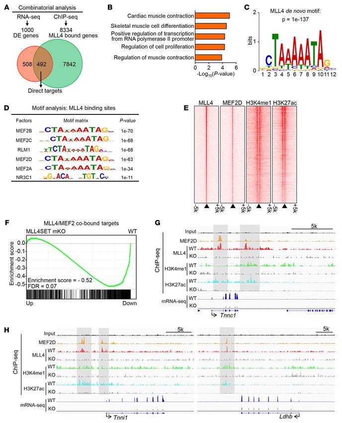
The Journal of Clinical Investigation                                                                             RESEARCH ARTICLE

     Mixed-lineage leukemia 4 (MLL4/KMT2D), a major his-                   ly published data (37, 38), efficient ablation of Mll4 mediated by
tone H3 lysine 4 (H3K4) monomethyl transferase, is an essential            Mck-Cre did not occur until approximately 7 days after birth (Sup-
histone writer for enhancer activation (24–28). MLL4 has been              plemental Figure 1A; supplemental material available online with
shown to colocalize with cell type–specific transcription factors to       this article; https://doi.org/10.1172/JCI136155DS1). As expected,
establish active enhancers during cell differentiation in multiple         the expression of Mll4 mRNA and protein levels were markedly
cell types (26–29). Loss of MLL4 prevents the establishment of             reduced in fast and slow muscles from both muscle-specific Mll4-
H3K4me1 and H3K27ac signatures on enhancer regions, leading                KO mouse lines (Figure 1B and Supplemental Figure 1, B–E). Nota-
to significant defects in enhancer activation and, consequently,           bly, deleting the enzymatic SET domain destabilized endogenous
gene transcription (26–28, 30). Studies in mouse models further            MLL4 protein in muscle (Figure 1B and Supplemental Figure 1D),
highlighted the importance of MLL4 in regulating a wide range              which is consistent with recently published data (29). Both Mll4-
of biological processes, including embryonic development, meta-            mKO and Mll4SET-mKO mice were born at normal Mendelian
bolic homeostasis, and cancer (27, 29, 31–33). Moreover, frequent          ratios and did not exhibit an overt metabolic phenotype compared
mutations in MLL4 have been implicated in several human genet-             with WT littermates on standard chow. These phenotypes includ-
ic diseases, including Kabuki syndrome, congenital heart disease,          ed similar body weight, food intake, locomotor activity, energy
and various types of cancer (33–35).                                       expenditure, and muscle weight (Supplemental Figure 2 and Sup-
     As a critical regulator of cell differentiation and cell-fate tran-   plemental Figure 3). This indicates that postnatal ablation of Mll4
sition, MLL4 has been documented as directing embryonic mus-               neither affects muscle differentiation nor development. Examina-
cle and fat tissue development (27, 29). However, it has yet to be         tion of myofiber cross-sectional area by histochemical staining
explored whether MLL4 has a role in adult skeletal muscle. In this         revealed that fiber-size distribution was altered in gastrocnemius
study, we generated 3 independent skeletal muscle–specific Mll4-           (GC) muscle from both muscle-specific Mll4-KO mouse lines, with
KO mouse lines and found that slow myofiber–enriched MLL4 is               a shift toward an increased cross-sectional area relative to that of
required for slow oxidative type I fiber formation to ensure muscle        WT controls (Figure 1C and Supplemental Figure 3M). Muscle
endurance. Mice with muscle-specific ablation of MLL4 exhibited            phenotyping and transcriptional profiling revealed that fiber-
marked downregulation of the slow oxidative myofiber gene pro-             type programs were substantially regulated in the muscle-specific
gram and had a decreased number of type I myofibers and amount             Mll4-KO mouse lines. Expression of the gene encoding the major
of mitochondrial activity, resulting in reduced muscle fatty acid          slow-twitch type I myosin isoform MHC1 (Myh7 gene) and slow-
utilization and endurance capacity during exercise. Mechanisti-            twitch troponin genes (Tnni1, Tnnc1, and Tnnt1) was reduced in
cally, we demonstrated that MLL4 drives the slow oxidative type I          both Mll4-mKO and Mll4SET-mKO GC muscle (Figure 1D). In
fiber program in cooperation with MEF2. Our studies also strong-           contrast, expression of the fast-twitch troponin gene (Tnnt3) was
ly suggest that the MLL4-dependent regulation of the type I fiber          increased in muscle from the 2 Mll4-KO mouse lines (Figure 1D).
program is operational in human muscle.                                    Similar observations were made in soleus and WV muscles from
                                                                           the Mll4SET-mKO mice (Supplemental Figure 4). Consistent with
Results                                                                    the gene expression results, MHC1 immunofluorescence staining
Slow myofiber–enriched MLL4 is required for type I fiber formation.        revealed a marked reduction in the number of type I fibers in sev-
As an initial step for exploring the potential function of MLL4 in         eral muscle groups from both Mll4-mKO and Mll4SET-mKO mice
adult muscle, we examined the expression patterns of MLL4 in               (Figure 1, E and F). Notably, the Mll4SET-mKO mouse line did
different muscle types from adult WT mice. Comparison of slow              not exhibit overt muscle phenotypes in young mice at P10 (Sup-
fiber–dominant soleus muscle to fast fiber–enriched white vas-             plemental Figure 5), suggesting that the observed results were not
tus lateralis (WV) by Western blot revealed that MLL4 protein is           a consequence of changes in muscle development or early fiber-
expressed preferentially (~2-fold) in soleus muscle (Figure 1A). In        type patterning in Mll4SET-mKO mice.
contrast, the protein levels of EZH2 and SUZ12, key components                  An in vitro primary skeletal myocyte culture system further
of the polycomb repressive complex 2, were markedly decreased              demonstrated that the MLL4 protein levels were induced during
in soleus muscle compared with those in WV (Figure 1A). These              differentiation of myoblasts into mature myotubes (Supplemen-
results suggest that MLL4 may activate the expression of slow              tal Figure 6A). Consistent with the established role of MLL4 in
fiber genes in slow myofibers.                                             maintaining H3K4me1 levels on active enhancers (27, 28, 30), the
    To directly examine the role of MLL4 in skeletal muscle, we            induced expression of slow myosin paralleled the elevated levels
generated 2 independent skeletal muscle–specific Mll4-KO mouse             of H3K4me1 on the Myh7 enhancer during muscle cell differentia-
lines using Mll4fl/fl and Mll4SETfl/fl mice. In the Mll4fl/fl strain,      tion (Supplemental Figure 6, A and B). An MLL4 loss-of-function
the exons 16–19 were flanked by 2 loxP sites (27), while in the            study was also conducted in primary skeletal myocyte. We used
Mll4SETfl/fl strain, the exons 50 and 51 encoding the enzymatic SET        adenoviral vectors to express Cre or control viruses in Mll4SETfl/fl
domain of MLL4 were floxed (29). Mll4fl/fl and Mll4SETfl/fl mice           myoblasts. Cells were then induced to differentiate into myo-
were bred with mice expressing Cre in postnatal skeletal muscle,           tubes. Adenoviral Cre-mediated KO of Mll4 in myoblasts result-
in which Cre expression is under the control of the muscle creatine        ed in diminished expression of Myh7 and decreased proportion of
kinase (Mck) promoter (36), to achieve deletion of Mll4 in both            slow myosin–positive myotubes (Figure 1, G and H). In contrast,
fast- and slow-twitch muscle fiber types (referred to as Mll4-mKO          fast myosin gene Myh4 expression was not decreased by Mll4 dele-
and Mll4SET-mKO, respectively). This resulted in efficient post-           tion (Figure 1, G and H). Consistent with reduction of Myh7 mRNA
natal deletion of Mll4 in skeletal muscle. Consistent with previous-       in Mll4-KO myotubes, MLL4 deficiency reduced the amounts of

                                                                           jci.org   Volume 130   Number 9   September 2020           4711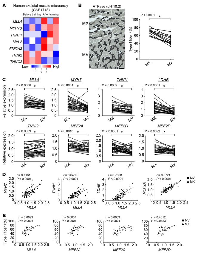
RESEARCH ARTICLE                                           The Journal of Clinical Investigation

 4712   jci.org   Volume 130   Number 9   September 2020

The Journal of Clinical Investigation                                                                                RESEARCH ARTICLE

Figure 1. Slow myofiber–enriched MLL4 is required for type I muscle fiber     exercise in both Mll4SETfl/flHSA-Cre mice and the WT control
formation. (A) Representative Western blot analysis of protein extracts       group, indicative of a switch to carbohydrates as the chief fuel
prepared from WV and soleus muscles of WT mice using indicated anti-
                                                                              (Supplemental Figure 8, A–D). Interestingly, in this high-intensity
bodies. Quantification of the MLL4/tubulin, EZH2/tubulin, and SUZ12/
tubulin signal ratios normalized (=1.0) to the WV and presented below         exercise challenge, both genotypes demonstrated similar ener-
the corresponding bands. n = 5–6 mice per group. (B) Representative           gy substrate utilization (RER) during the course of exercise, and
Western blot analysis of MLL4 expression in GC muscles of indicated mice.     Mll4SETfl/flHSA-Cre mice ran distances and achieved maximal
n.s., nonspecific band. n = 3 mice per group. (C) Top: representative WGA     speed similarly to their control littermates (Supplemental Figure
staining of GC muscle from 8-week-old male Mll4-mKO and Mll4SET-mKO
                                                                              8, C–F). Mice that performed the VO2max test were then subjected
mice. Scale bar: 50 μm. Bottom: cross-sectional areas of GC myofibers
were measured by ImageJ. n = 4–5 mice per group. (D) Expression of slow-      to a low-intensity (endurance type) protocol, during which ani-
twitch myosin gene (Myh7) and representative slow/fast-twitch troponin        mals ran at a constant speed of 60% of their maximal running
genes (qRT-PCR) in GC muscle from indicated genotypes. n = 5–8 mice           speed (Supplemental Figure 8G). While RER values increased
per group. (E) Cross section of (top) soleus and (bottom) GC muscle from      during the beginning of the exercise test, both WT and Mll4SETfl/fl
8-week-old male Mll4-mKO and Mll4SET-mKO mice stained for MHC1
                                                                              HSA-Cre mice displayed a drop in RER after approximately 14
(green) and MHC2b (red). Scale bars: 250 μm. (F) Quantification of IF data
shown in E. n = 3–5 mice per group. (G and H) Primary myoblasts isolated      minutes of endurance exercise (Supplemental Figure 8, G–I). This
from Mll4SETfl/fl mice were infected with an adenovirus overexpressing        decrease indicates a shift in substrate usage from glucose to fat
Cre or control virus (Ctrl), followed by differentiation into myotubes. (G)   metabolism. Despite no difference in RER during the exercise
Results of qRT-PCR and Western blot analysis in skeletal myotubes. n = 3      period, the Mll4SETfl/flHSA-Cre mice ran shorter distances com-
independent experiments. (H) Left: IF staining of skeletal myotubes was
                                                                              pared with the WT control group (Supplemental Figure 8, H–K).
performed using antibodies directed against myosin-slow or myosin-fast.
Scale bars: 100 μm. Right: quantification of the myosin-slow IF data          These results suggest that muscle MLL4 deficiency resulted in
expressed as mean percentage of total myotubes. n = 3 independent             reduced capacity for persistent use of fat as a fuel during endur-
experiments. Values are represented as mean ± SEM. *P < 0.05 vs. corre-       ance exercise. Furthermore, we also measured glucose and fatty
sponding controls, 2-tailed unpaired Student’s t test.                        acid levels in blood from WT and Mll4SETfl/flHSA-Cre mice before
                                                                              and after endurance exercise. Mll4SETfl/flHSA-Cre mice showed
                                                                              significantly lower blood glucose levels compared with WT con-
                                                                              trols at baseline and after 80 minutes of endurance exercise (Fig-
H3K4me1 on the Myh7 enhancer (Supplemental Figure 6C). Thus,                  ure 2D). Conversely, blood triglyceride (TG) and fatty acid levels
in vitro manipulation of Mll4 in myocytes provided further evi-               were significantly higher after exercise in Mll4SETfl/flHSA-Cre
dence that MLL4 exerts control upon slow fiber gene expression.               mice (Figure 2E). Notably, blood ketone body β-hydroxybutyrate
Taken together, these data demonstrate an essential role of MLL4              levels mirrored the changes of blood fatty acids, as they were also
in the regulation of type I muscle fiber type.                                increased in Mll4SETfl/flHSA-Cre mice at basal conditions or after
     Loss of muscle MLL4 causes reduced running endurance. Muscle             exercise (Figure 2E). Together, these results demonstrate a change
fiber-type composition and the capacity to burn fatty acids and               in slow type I fibers and muscle metabolism in the absence of mus-
glucose are important determinants of muscle performance and                  cle MLL4 that compromises running endurance.
endurance, and fast-twitch muscle fibers exhibit a glycolytic burst                MLL4 coordinates gene programs controlling muscle contraction
metabolism and are more susceptible to endurance exercise–                    and energy metabolism. Type I myofibers depend on the precise
induced fatigue (5). We next sought to determine the physiologi-              orchestration of slow contractile and oxidative metabolic gene
cal impact of muscle MLL4 deficiency. To rule out possible effects            expression to ensure muscle endurance. To more thoroughly ana-
mediated by MLL4 in the heart on exercise phenotypes, Mll4SETfl/fl            lyze the fiber-type–specific patterns of gene expression changes
mice were bred with human skeletal actin Cre (HSA-Cre) mice                   that result from loss of muscle MLL4, we performed RNA-Seq
to establish another skeletal muscle–specific deletion of the Mll4            analysis on mRNA isolated from GC muscles of Mll4SET-mKO
alleles. As expected, the protein expression of Mll4 was dramat-              mice and littermate controls. We found that MLL4 regulated
ically decreased in multiple muscle types, but not in the heart,              a total of 1000 genes in skeletal muscles, with 447 up- and 553
in Mll4SETfl/flHSA-Cre mice (Supplemental Figure 7A). In addi-                downregulated, respectively (Figure 3A). In addition to the key
tion, MHC1-positive fibers were markedly reduced in Mll4SETfl/fl              slow-twitch MHC gene Myh7 and troponin complex described
HSA-Cre GC muscle (Figure 2, A and B, and Supplemental Figure                 above (Figure 1D), gene ontology (GO) analysis of MLL4-regu-
7B). We next assessed acute running endurance performance in                  lated genes also revealed significant enrichment in fiber-type–
the Mll4SETfl/flHSA-Cre mouse line using a run-to-exhaustion pro-             specific isoforms of sarcomeric components as well as ion chan-
tocol on a motorized treadmill. Consistent with the observed alter-           nels involved in excitation-contraction coupling (Figure 3B). The
ations in muscle fiber–type proportion, Mll4SETfl/flHSA-Cre mice              comparative RNA-profiling strategy revealed extensive fiber-type
could run for significantly shorter times and distances (~30%)                switching and novel myofiber-specific gene expression. As shown
compared with WT littermates (Figure 2C).                                     in Figure 3C, a broad array of contraction-related genes that differ
     To further evaluate muscle fuel utilization during exercise,             between muscle fiber type were altered in Mll4SET-mKO muscle
WT or Mll4SETfl/flHSA-Cre mice were first subjected to a forced               (Figure 3C). Real-time PCR confirmed that the expression of many
maximal exercise capacity test (VO2max test) consisting of increas-           genes encoding slow contractile proteins (Myl2, Myl3, Tpm3, and
ing speed every 2 minutes at 10° inclination until exhaustion (Sup-           Myom3) was reduced in the GC muscles of Mll4SET-mKO mice
plemental Figure 8A). Consistent with a shift to muscle glucose               compared with those of WT controls (Figure 3D). The expression
utilization, the respiratory exchange ratio (RER) increased with              of slow fiber calcium-handling genes (Atp2a2, Casq2, Smtnl1, and

                                                                              jci.org   Volume 130   Number 9   September 2020          4713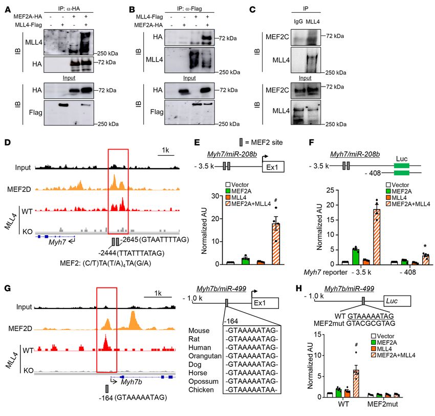
RESEARCH ARTICLE                                                                                The Journal of Clinical Investigation

                                                                                            Figure 2. Loss of muscle MLL4 causes reduced
                                                                                            running endurance. (A) MHC fiber typing by IF of
                                                                                            GC muscle of indicated genotypes. MHC1 (green);
                                                                                            MHC2b (red). Representative images are shown.
                                                                                            Scale bar: 250 μm. (B) Quantification of IF data
                                                                                            shown in A expressed as type I fibers per section.
                                                                                            n = 5–6 mice per group. (C) Left: schematic
                                                                                            depicts increments of speed over time. Right:
                                                                                            bars represent mean running time and distance
                                                                                            for 10-week-old male Mll4SETfl/flHSA-Cre mice
                                                                                            and WT on a motorized treadmill. n = 11–13 mice
                                                                                            per group. (D and E) Blood glucose, TG, NEFA,
                                                                                            and β-hydroxybutyrate levels in mice of indicated
                                                                                            genotypes at rest or after 80 minutes of exercise.
                                                                                            n = 8–9 mice per group. Values are represented
                                                                                            as mean ± SEM. *P < 0.05 vs. corresponding WT
                                                                                            controls, 2-tailed unpaired Student’s t test.

Myoz2) was also reduced in the Mll4SET-mKO muscles compared            vation of slow oxidative muscle fiber gene programs by MLL4,
with those in WT controls (Figure 3D). Moreover, we also found         we examined the possibility that MLL4 regulates factors known
a decreased expression of genes associated with fatty acid and         to regulate slow myofiber gene expression. Esrrg mRNA levels
glucose metabolism (Cpt1b, Slc27a1, Fabp3, Dgat2, Fads6, Phyhd1,       were modestly increased and Ppard mRNA levels were lower in
Ldhb, and Ppp1r1a) in Mll4SET-mKO muscle (Figure 3D). The              Mll4SET-mKO muscles (Supplemental Figure 9), whereas Fnip1,
expected lactate dehydrogenase (LDH) isoenzyme activity shifts         Ppargc1a, Ppargc1b, Ppara, Esrra, and Esrrb gene expression were
due to changes in Ldhb expression were confirmed by gel-activity       not changed in Mll4SET-mKO muscles (Supplemental Figure 9).
studies (Figure 3E). To further assess the metabolic effects of mus-   These results led us to the hypothesis that MLL4 may act as a
cle MLL4, mitochondrial respiration rates were determined in the       direct activator of slow-myofiber gene expression.
extensor digital longus (EDL) muscle of Mll4SET-mKO mice and               We next sought to examine the genome-wide MLL4 occu-
WT controls using pyruvate or palmitoylcarnitine as substrates.        pancy on WT mouse muscle chromatin by ChIP coupled with
State 3 (maximal ADP stimulated) respiration rates were signifi-       high-throughput DNA sequencing (ChIP-Seq). As a control
cantly lower in Mll4SET-mKO muscle compared with that of WT            for specificity, ChIP-Seq experiments were also performed in
controls (Figure 3F). Moreover, measurement of oxygen consump-         Mll4-mKO muscle; 9403 high-confidence MLL4 genomic bind-
tion rate (OCR) using an extracellular flux analyzer also revealed     ing regions were obtained by filtering out nonspecific signals
that Mll4SET-KO myotubes had a reduced OCR in the presence of          observed in MLL4-deficient muscle (Supplemental Figure 10,
uncoupler FCCP, a sign of reduced maximal mitochondrial respi-         A–C). MLL4-binding sites in skeletal muscle were predominantly
ratory capacity (Figure 3G), whereas no difference in the extra-       located in the intergenic, intronic, and promoter regions (Supple-
cellular acidification rate (ECAR) was observed in Mll4SET-KO          mental Figure 10D), which is consistent with our previous MLL4
myotubes compared with Mll4SETfl/fl controls (data not shown).         ChIP-Seq results in myocytes (27). Correlating the MLL4 cistrome
Together, these results demonstrate that MLL4 programs type            in muscle and myocytes with the global mRNA changes upon mus-
I muscle metabolism by coordinate regulation of gene programs          cle Mll4 deletion revealed that approximately 49% of MLL4-reg-
controlling muscle contraction and energy metabolism.                  ulated genes were directly bound by MLL4 (based on the genes
    MLL4 drives the slow oxidative muscle fiber program in cooper-     nearest to the MLL4-binding peaks) (Figure 4A). Furthermore,
ation with MEF2. To define the mechanism involved in the acti-         GO analysis revealed that the 492 directly regulated genes were

   4714        jci.org   Volume 130   Number 9   September 2020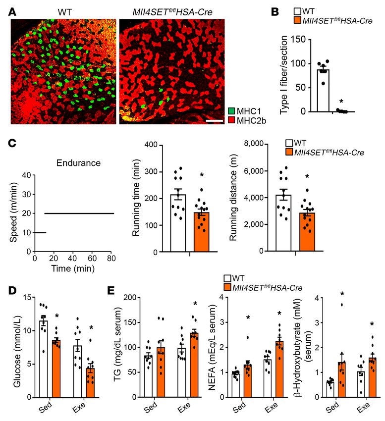
The Journal of Clinical Investigation                                                                                     RESEARCH ARTICLE

Figure 3. MLL4 coordinately regulates gene programs controlling muscle contraction and energy metabolism. (A) Volcano plot showing fold changes ver-
sus P values for analyzed RNA-Seq data generated from the GC muscle of 8-week-old male Mll4SET-mKO mice compared with littermate controls (WT).
Significantly upregulated genes are represented by red dots, whereas downregulated genes are represented by blue dots. (B) GO enrichment analysis of
gene transcripts regulated in Mll4SET-mKO muscle. (C) Heatmap analysis of contraction-related genes regulated in Mll4SET-mKO muscle compared with
WT controls. n = 3 independent samples per group. Color scheme for fold change is provided. (D) Expression of genes (qRT-PCR) involved in muscle con-
traction, calcium handling, and metabolism in GC muscle from the indicated genotypes. n = 5–6 mice per group. (E) Left: LDH isoenzymes were separated
by polyacrylamide gel electrophoresis using whole-cell extracts from heart (Ht, control) and GC muscle from indicated mice. A representative gel showing 4
independent mice per group is shown. Right: quantification of LDH isoenzyme activity gel electrophoresis shown on the left. (F) Mitochondrial respiration
rates were determined from the EDL muscle of the indicated genotypes using pyruvate or palmitoylcarnitine as a substrate. Pyruvate/malate (Py/M) or
palmitoylcarnitine/malate (PC/M) stimulated, ADP-dependent respiration, and oligomycin induced (oligo) are shown. n = 6–7 mice per group. (G) OCRs in
skeletal myotubes harvested from Mll4SETfl/fl mice subjected to adenovirus-based overexpression of Cre compared with control virus. Basal OCR was first
measured, followed by administration of 10 mM sodium pyruvate, and 2 μM oligomycin (to inhibit ATP synthase), uncoupler FCCP (2 μM), or rotenone/
antimycin (Rot/A; 1 μM), as indicated. n = 6 separate experiments done with 5 biological replicates. Values are represented as mean ± SEM. *P < 0.05 vs.
corresponding controls, 2-tailed unpaired Student’s t test.

                                                                               jci.org   Volume 130   Number 9   September 2020                 4715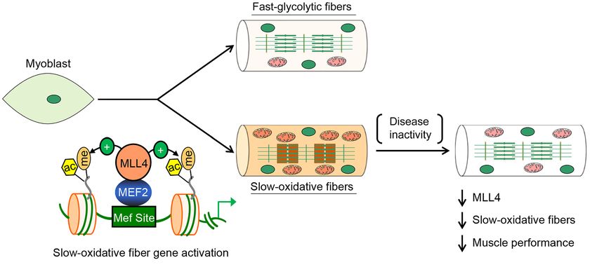
RESEARCH ARTICLE                                           The Journal of Clinical Investigation

 4716   jci.org   Volume 130   Number 9   September 2020
The Journal of Clinical Investigation                                                                                  RESEARCH ARTICLE

Figure 4. MLL4 colocalizes with MEF2 on enhancers of slow oxidative            MLL4 and MEF2 cobound target genes in muscle (Figure 4F).
muscle genes. (A) Analysis of the MLL4 ChIP-Seq data in muscle and             These data suggest that MLL4 acts coordinately with MEF2 in
myocytes combined with mRNA-Seq data set upon muscle Mll4 deletion
                                                                               regulating muscle gene expression. Genomic colocalization of
defines a set of genes directly regulated by MLL4. (B) GO enrichment anal-
ysis of MLL4 direct targets, with the top 5 terms shown. (C) De novo motif     MLL4 and MEF2 was confirmed on many slow oxidative mus-
analysis of MLL4-binding regions in muscle using HOMER. Shown is the           cle fiber gene loci in myocytes. For example, slow fiber–related
top-scoring motif present in the top 5000 emergent MLL4-binding sites.         gene enhancers (e.g., Myh7, Tnnc1, Tnni1, Tnnt1, Ldhb, Casq2,
(D) Known TF motifs with the highest relative enrichment in MLL4-binding       and Mybph) showed strong MLL4 and MEF2 binding as well as
regions in muscle. (E) MLL4 colocalizes with MEF2 on active enhancers in
                                                                               the presence of H3K4me1 and H3K27ac signals (Figure 4, G and
myocyte. Heatmap shows the ChIP-Seq binding signal intensity for MLL4,
MEF2D, H3K4me1, and H3K27ac. Binding is ranked from the strongest              H, and Supplemental Figure 11). Importantly, deletion of Mll4
to the weakest MLL4-binding sites. Active enhancers are defined with           markedly decreased levels of H3K4me1 and H3K27ac, which was
enhancer markers H3K4me1 and H3K27ac. (F) GSEA showing that expres-            consistent with pronounced reduction of slow oxidative muscle
sion of MLL4/MEF2 cobound target genes is significantly downregulated          fiber gene mRNA in Mll4SET-mKO muscle (Figure 4, G and H,
in Mll4SET-mKO muscle compared with that of WT controls. All genes
                                                                               and Supplemental Figure 11). Together, these results suggest that
in mRNA-Seq profiling from Mll4SET-mKO muscle were ranked by fold
difference compared with WT controls and expressed on the x axis. This         MLL4 occupies the enhancers in cooperation with MEF2 to acti-
data set was compared with the gene list of the nearest genes identified in    vate transcription of the slow oxidative muscle genes.
MLL4/MEF2 cobound regions in muscle. (G and H) MLL4-dependent active                A series of co-immunoprecipitation (co-IP) studies were next
enhancers on slow oxidative gene loci are shown. Top: ChIP-Seq binding         conducted to determine whether MLL4 interacts with MEF2.
profiles for MLL4, MEF2D, and histone modifications in WT or Mll4-KO
                                                                               HEK293 cells were cotransfected with expression vectors for
myocytes. mRNA-Seq data from WT and Mll4SET-mKO muscle is shown at
the bottom, indicating a high correlation of the 2 data sets. Input, genomic   HA-MEF2A and Flag-MLL4. Anti-HA was found to co-IP MLL4
DNA from myocytes. Gray boxes indicate the high-confidence MLL4-               and MEF2A (Figure 5A). Using MLL4 as the IP target, MEF2A
binding regions corresponding to Tnnc1, Tnni1, and Ldhb genes. Data were       was pulled down (Figure 5B). Furthermore, IP of MLL4 in C2C12
obtained from published data sets GSE50466 and GSE43223. The current           myotubes with an anti-MLL4 antibody confirmed that endog-
data sets GSE138994 and GSE137368 were also analyzed.
                                                                               enous MLL4 and MEF2 actually interact in muscle cells (Figure
                                                                               5C). To explore functional correlates of the MLL4/MEF2 inter-
                                                                               action, we focused on the Myh7 and Myh7b loci. These 2 MLL4-
                                                                               regulated genes encode the major slow myosin and drive expres-
enriched with muscle contraction-related terms similarly to those              sion of miR-208b and miR-499, which further promote muscle
seen in the Mll4SET-mKO transcriptome (Figure 4B). In addition,                fiber remodeling (41, 42). Upon examination of the MLL4 and
we also found that MLL4 could occupy many fatty acid utilization               MEF2 ChIP-Seq data, we uncovered a MLL4/MEF2 cobound
genes (e.g., Cpt1b and Fabp3) (Supplemental Figure 10E). Togeth-               region located approximately 3 kb upstream of the Myh7 tran-
er, the strong correlation between the cistromic and transcriptom-             scription start site (TSS). We identified 2 highly conserved DNA
ic findings supports a direct role for MLL4 in regulating muscle               sequences conforming to the consensus binding sites for MEF2
fiber–type gene programs.                                                      ([C/T]TA[T/A]4TA[G/A]) in the Myh7/miR-208b cis-proximal
     Interestingly, de novo motif analysis of the top 5000 emer-               enhancer region (Figure 5D). Cell cotransfection studies were
gent MLL4-binding regions in muscle using HOMER revealed                       next conducted using a rat 3.5 kb Myh7 promoter reporter contain-
a sequence element, 5′-CTAAAAATAG-3′, as the highest score                     ing the enhancer element (Figure 5E). The Myh7 luciferase report-
motif, with a P value of 1 × 10 –137 (Figure 4C). This motif closely           er was not activated by MLL4 alone, but when it was expressed
corresponded to a consensus site that has been previously known                together with MEF2A, synergistic activation was observed (Figure
as the MEF2-binding site. Indeed, the search for known motifs                  5E), suggesting a cooperative transcriptional activation by the
confirmed that the most enriched motif within MLL4-binding                     2 factors. To further assess the activation of the Myh7/miR-208b
regions was the binding site for MEF2 family members, key reg-                 enhancer by MLL4/MEF2, deletion mapping studies were used to
ulators of the type I fiber-type program (refs. 18, 39, and Figure             demonstrate that this MLL4/MEF2 synergistic effect was mark-
4D). Moreover, we found that MLL4 could occupy both Mef2a                      edly diminished by deleting the upstream enhancer sequences
and Mef2d genes (Supplemental Figure 10F), indicating that                     (Figure 5F). We also detected MLL4 and MEF2 colocalization
Mef2a and Mef2d are also direct targets of MLL4. These results                 on the Myh7b promoter region (Figure 5G). Cell cotransfection
suggest that MEF2s may be coregulatory transcription factors                   studies were further conducted using a mouse Myh7b promoter-
that are involved in the MLL4-mediated regulation of the type I                reporter containing a highly conserved MEF2-binding site (Figure
fiber-type program. We next interrogated a published myocyte                   5H). As expected, the combination of MLL4 and MEF2 resulted
MEF2 ChIP-Seq data set (40) in conjunction with our previously                 in synergistic activation of the Myh7b reporter (Figure 5H). This
published myocyte ChIP-Seq data for MLL4 and histone modi-                     synergistic effect was completely abolished upon mutation of the
fications (H3K4me1 and H3K27ac) associated with enhancer                       MEF2 site (Figure 5H), providing further evidence that the MEF2
activities (27). As shown in Figure 4E, heatmap visualization                  site cooperates with MLL4 in the activation of Myh7b/miR-499
of the MEF2 binding and H3K4me1 and H3K27ac signal at                          gene transcription. Taken together, these results demonstrate that
the MLL4-binding events demonstrated a substantial degree                      MLL4 cooperates with MEF2 to activate slow muscle gene tran-
of MLL4 and MEF2 cooccupancy on active enhancers (Figure                       scription, thereby driving fiber-type remodeling.
4E). Gene set enrichment analysis (GSEA) further demonstrat-                        MLL4 levels are regulated in response to physiological and patho-
ed that Mll4 deletion significantly downregulated expression of                physiological stimuli in human muscle. To determine the relevance

                                                                               jci.org   Volume 130   Number 9   September 2020            4717
RESEARCH ARTICLE The Journal of Clinical Investigation Figure 5. MLL4 cooperates with MEF2 to activate slow myofiber gene transcription. (A and B) Co-IP experiments were performed by cotransfecting HA-MEF2A and Flag-MLL4 in HEK293 cells, as indicated at the top. Antibodies against the HA or Flag epitope were used for co-IP. Extracts (Input) from the HEK293 cells and proteins from the IP were analyzed by immunoblotting. Representative results for co-IP are shown. n = 3 independent experi- ments. (C) Co-IP results with extracts prepared from C2C12 myotubes using anti-MLL4 antibody or control IgG. Representative results are shown. n = 3 independent experiments. (D) MLL4 and MEF2D ChIP-Seq tracks from myocytes at the Myh7 locus. Two putative conserved MEF2-binding sites within the cis-proximal enhancer region of the Myh7 gene are shown. (E) MLL4 and MEF2 synergistically activate Myh7 gene promoter. The rat Myh7.Luc.3.5k promoter reporter was used in cotransfection studies in HEK293 cells in the presence of expression vectors indicated. Values represent mean (± SEM) firefly/renilla luciferase activity shown as arbitrary units (AU) normalized (=1.0) to vector control. n = 4 independent experiments. (F) Results of transient transfection performed with rMyh7.Luc.3.5k and truncation mutant of rMyh7.Luc.408 in HEK293 cells in the presence of expression vectors indicated. n = 4–5 independent experiments. (G) Left: MLL4 and MEF2D ChIP-Seq tracks from myocyte at the Myh7b locus. Right: putative conserved MEF2-binding site within the mouse Myh7b promoter regions. (H) Top: site-directed mutagenesis was used to abolish the MEF2 response element. Bottom: mMyh7b.Luc.1k (WT) or MEF2mut.mMyh7b.Luc.1k promoter reporters were used in cotransfection studies in HEK293 cells in the presence of expression vectors indicated. n = 5 independent experiments. Values are represented as mean ± SEM. *P < 0.05 vs. corresponding controls; #P < 0.05 compared with MEF2A alone. P values were determined using 1-way ANOVA coupled to a Fisher’s LSD post hoc test. of the MLL4 regulatory circuit in humans, we investigated whether database [GEO] GSE1718) from a group of sedentary subjects who MLL4 function could be altered by physiological or pathophysio- underwent 20 weeks of an endurance exercise training program logical stimuli in human muscle. We first analyzed the gene expres- (43). This analysis revealed that the MLL4 regulatory circuit was sion profiles in human muscle (NCBI’s Gene Expression Omnibus induced in human muscle by exercise training. Increased mRNA 4718 jci.org Volume 130 Number 9 September 2020
The Journal of Clinical Investigation                                                                          RESEARCH ARTICLE

levels of MLL4 associated with elevated expression of its targets     over, our data also established that the MLL4 regulatory circuit
(MYH7B, TNNT1, MYL2, and ATP2A2) were observed in the muscle          is associated with muscle fiber–type switching in humans. There-
tissue from the “posttraining” group compared with the “pretrain-     fore, MLL4 likely represents a previously unrecognized molecular
ing” group (Figure 6A). In contrast, the levels of TNNI2 and TNNC2    switch that specifies myofiber structural and metabolic identities
mRNA showed a decrease in posttraining muscle (Figure 6A).            that govern muscle performance.
     Skeletal muscle dysfunction, including fiber-type switch-             Enhancer activation is a critical step for gene activation. While
ing and reduced oxidative capacity, has been associated with a        transcriptional regulation of myofiber gene expression through
variety of human diseases. Patients with adolescent idiopathic        multiple transcription factors, such as nuclear receptor PPARs,
scoliosis (AIS), the most common type of scoliosis, which affects     ERRs, MEF2, SOX6, and Tbx15, along with coregulators have
1%–4% adolescents, have generalized muscle dysfunction (44,           been established (13–21, 49, 50), it remains unclear how fiber type–
45). Although causal mechanisms of AIS remain unclear, paraspi-       specific patterns of gene expression are controlled at the enhancer
nal muscle imbalance due to muscle fiber–type switching has long      activation layer. In this study, we showed that the enhancer regu-
been recognized as a possible factor underlying the pathology of      lator MLL4 drives the contractile and metabolic specification of
AIS (46–48). Therefore, we examined the regulation of MLL4 sig-       type I muscle fibers in both primary skeletal myotubes and skeletal
naling in paraspinal muscle samples from 40 patients with AIS.        muscle. We also confirmed a marked reduction in active enhancer
Consistent with previous reports (47, 48), metachromatic ATPase       hallmarks on slow muscle gene enhancers in Mll4-KO myocytes.
staining revealed that muscle fiber–type programs were altered in     Notably, deletion of the enzymatic SET domain destabilized the
paraspinal muscle from AIS patients. The biopsy specimens from        MLL4 protein, thus limiting the study of the role of its enzymatic
the concave side (MV) of the curvature contained a lower percent-     activity in muscle. It is also worth noting that we used Mck-Cre and
age of type I muscle fibers compared with the convex side (MX)        HSA-Cre mice for creating mice with the Mll4 deletion specifical-
(Figure 6B). As expected, muscle biopsies from the MV group           ly in skeletal muscle in this study and that we did not observe a
exhibited lower slow fiber gene (MYH7, TNNI1, TNNC1, TNNT1,           change in muscle differentiation or development upon muscle Mll4
and LDHB) expression, but higher fast fiber gene (TNNI2 and           deletion. In the Mll4SET-mKO model using Mck-Cre, which is not
TNNC2) expression compared with those from the MX group (Fig-         fully active until after birth (37, 38), we did not observe significant
ure 6C and Supplemental Figure 12A). Expression levels of MLL4        changes in muscle fiber type in Mll4SET-mKO muscle compared
and MEF2s were also lower in the MV group compared with the           with that of WT controls at stage P10. This could reflect muscle
MX group (Figure 6C). In addition, the levels of MLL4 mRNA            fiber–type remodeling during postnatal development. It is possi-
exhibited a significant positive correlation with slow fiber gene     ble that other postnatal programs, such as neuronal or hormone
(MYH7, TNNI1, TNNT1, and LDHB) and MEF2s mRNA levels, but             signals, are more dominant during the first weeks after birth (5).
not with a marker of the fast fiber program (TNNI2) (Figure 6D        Remarkably, we found that MLL4 is required for the maintenance
and Supplemental Figure 12B). The relationship between MLL4           of type I fibers in adult muscle. As such, ablation of muscle MLL4
expression and levels of the MEF2s with type I fiber percentage       profoundly affects muscle fatigability and exercise performance.
was also assessed. As shown in Figure 6E, both MLL4 and MEF2s         Thus, our study has expanded the role of the enhancer regulator
exhibited a significant positive correlation with type I fiber per-   MLL4 to include pivotal muscle fiber specification and exercise
centage (Figure 6E). Together, these results strongly suggest that    physiological functions other than development and cell differen-
the MLL4 regulatory circuit is operative in the regulation of the     tiation. Conceivably, this muscle-specific regulatory action may
type I muscle program in response to physiological and pathophys-     reflect the multifunctions of MLL4 in using its enhancer remodel-
iological stimuli in humans.                                          ing activity to direct context-specific biological processes.
                                                                           Type I muscle fibers are equipped with slow-twitch contrac-
Discussion                                                            tile machinery and a high-capacity fuel-burning system poised
The mechanisms underlying the precise orchestration of con-           for endurance exercise. Our data suggest that MLL4 orchestrates
tractile and metabolic gene expression that specify muscle fiber      the structural and metabolic programs controlling type I muscle
identity and function remain unclear. Herein, using loss-of-func-     metabolism. Genome-wide transcriptional analysis revealed that
tion strategies in mice and primary muscle cells together with        MLL4 acts by coordinately regulating genes controlling slow-
assessment of muscle biopsies from humans, we have uncovered          twitch myofibers, calcium handling, and oxidative metabolism. At
an essential role for the enhancer regulator MLL4 in controlling      a functional level, these changes resulted in substantial impaired
the structural and metabolic programs that govern myofiber iden-      muscle endurance during exercise in muscle-specific Mll4-KO
tity and muscle performance (Figure 7). MLL4 is highly expressed      mice. Interestingly, we found that muscle MLL4 deficiency did not
in type I muscle fibers, and deletion of Mll4 specifically in skel-   compromise muscle performance during high-intensity (sprint)
etal muscle resulted in decreased numbers of type I myofibers         exercise, perhaps reflecting that fast type II fibers are involved
and diminished mitochondrial respiration, leading to substan-         in rapid bursts of contraction. Our data suggest that MLL4 is a
tial defects in muscle endurance during exercise. These changes       critical upstream epigenetic switch that specifies slow oxidative
resulted from marked downregulation of the slow oxidative mus-        myofiber identity by coactivating MEF2 transcriptional regula-
cle gene programs in skeletal muscle lacking MLL4. We found that      tors. Since the expression of slow oxidative muscle genes is also
MLL4 directly binds to enhancers and functions as a coactivator       regulated by nuclear receptors, such as PPARβ/δ and ERRs (14, 15,
of MEF2 to activate the transcription of slow oxidative myofiber      17, 19), it remains to be tested whether MLL4 may also contribute
genes, thereby driving the muscle fibe–type remodeling. More-         to the activation of PPARβ/δ and ERRs in muscle. Additionally, it

                                                                      jci.org   Volume 130   Number 9   September 2020             4719
RESEARCH ARTICLE The Journal of Clinical Investigation Figure 6. MLL4 regulatory circuit is associated with muscle fiber–type remodeling in humans. (A) Relative expression of MLL4 and representative fiber type–specific genes in human vastus lateralis muscles before or after endurance training. Data were extracted from GEO GSE1718. (B–E) Paraspinal muscle samples from the convex (MX) and concave (MV) sides of the curvature from 40 patients with AIS were used for this analysis. (B) Left: representative sec- tions of paraspinal muscle from AIS patients stained for myosin ATPase activity (pH = 10.2, type II fibers dark, type I fibers light). Scale bar: 100 μm. Right: quantification of ATPase-staining data shown on the left expressed as mean percentage of total muscle fibers. n = 15 patients. *P < 0.05, paired Student’s t test. (C) mRNA expression levels of MLL4, MEF2s, and slow oxidative muscle fiber genes were determined by qRT-PCR. n = 40 patients. *P < 0.05, paired Student’s t test. (D) Correlation between MLL4 gene expression and that of slow oxidative myofiber genes and MEF2s. n = 40 patients. Pearson’s correla- tion analysis was used to determine the correlation. (E) Correlation between MLL4 and MEF2s expression and the type I fiber percentage. n = 15 patients. Spearman’s correlation analysis was used. 4720 jci.org Volume 130 Number 9 September 2020
The Journal of Clinical Investigation                                                                                   RESEARCH ARTICLE

Figure 7. Model of MLL4 in the control of muscle fiber–type specification and function. The schematic depicts a proposed model for the MLL4 regulatory
mechanism that specifies muscle fiber identity and muscle performance.

will be of interest to determine whether MLL4 plays an opposing              for MEF2 leads to the recruitment of alternate coregulatory com-
role as a direct transcriptional repressor of fast myofiber–specific         plexes (40, 52). It is also possible that MEF2 exon switching could
gene expression. Coordinate control of muscle fiber type specifi-            alter its association with MLL4.
cation and fuel-burning capacity occurs during exercise training.                 Myofiber shift from slow oxidative toward fast glycolytic has
Interestingly, we found that exercise induces an increase in mRNA            been associated with a variety of chronic illnesses, including met-
abundance of MLL4 and its targets in human muscle, suggesting                abolic disorders and muscular diseases (3, 7, 12). In this study, our
the MLL4 regulatory circuit unveiled here could be involved in the           survey of the human muscle samples demonstrated that the MLL4
response to exercise training.                                               regulatory circuit is associated with muscle fiber–type switching in
     Our results from global MLL4-directed transcriptional anal-             AIS patients. Specifically, MLL4 and MEF2s levels were reduced in
ysis indicate that the MLL4-driven slow oxidative muscle gene                muscle samples from the MV group compared with those in the MX
program acts in cooperation with MEF2. The observed role of                  group. We also found that the expression of MLL4 and MEF2s was
MEF2 is of interest, given its known role in the regulation of               strongly correlated with the expression of slow fiber genes and type
muscle development and muscle fiber–type remodeling in adult-                I fiber-type proportion. Based on our data, it is possible that down-
hood (18, 39). We found that MLL4 interacts and cooperates with              regulated MLL4 signaling leads to reduced type I muscle fibers,
MEF2 to activate slow oxidative muscle gene transcription. This              contributing to the paraspinal muscle imbalance in AIS patients.
is consistent with the observation that elevated expression of               Notably, MLL4 has also been identified as a major causative muta-
MLL4 parallels high MEF2 activity in slow fiber–dominant sole-               tion gene in Kabuki syndrome, a human genetic disease that caus-
us muscle. Notably, MLL4 also directly binds to Mef2a and Mef2d              es multiple malformations, including muscle hypotonia (34, 53). It
gene loci, which likely adds another regulation layer downstream             is tempting to speculate that loss-of-function mutations in MLL4
of MLL4 to enhance the mechanisms described here and boosts                  contribute to the muscle dysfunction in Kabuki syndrome.
the highest level of MLL4/MEF2 cooperation. MEF2D has been                        In summary, we demonstrate an essential role for enhancer
shown to recruit the Ash2L methyltransferase complex to MyoD                 regulator MLL4 in specifying myofiber structural and metabolic
target genes during myogenesis (51). The interaction between                 identities that govern muscle performance. Given that many dis-
MLL4 and MEF2 and the genomic colocalization of MLL4 with                    ease states are associated with reduced muscle endurance, these
MEF2 suggest that MEF2 plays critical roles in recruiting MLL4               findings provide the possibility of therapeutic opportunities for
to establish active enhancers on slow oxidative muscle genes.                enhancing muscle fitness to combat a variety of metabolic and
The precise mechanism whereby MLL4 transcriptional activa-                   muscular diseases.
tion complex is tethered to MEF2-binding sites to establish active
enhancers, however, remains to be determined. The class II                   Methods
HDACs and NCoR1 are known to corepress MEF2 (18, 21); there-                 Animal studies. Male C57BL/6J WT mice were from MARC of Nan-
fore, it is likely that MEF2-mediated regulation of the slow fiber           jing University. Generation of Mll4fl/fl and Mll4SETfl/fl mice has been
phenotype is controlled by the balance between MLL4 and class                described elsewhere (27, 29). Mice were backcrossed to the C57BL/6J
II HDACs/NCoR1 signaling. MLL4 and MEF2 cooperation allows                   background for more than 6 generations. To generate mice with a
enhancer activation and the establishment of slow fiber–specific             muscle-specific disruption of the Mll4 allele, Mll4fl/fl and Mll4SETfl/fl
gene expression patterns, while the class II HDACs and NCoR1                 mice were crossed with mice expressing Cre recombinase under the
put brakes on the activation of MEF2. Interestingly, previous                control of an Mck promoter (Jackson Laboratory, stock no. 006475)
studies also suggest that the muscle-specific switch in exon usage           or an HSA promoter (Jackson Laboratory, stock no. 006139) to

                                                                             jci.org   Volume 130   Number 9   September 2020                4721
RESEARCH ARTICLE The Journal of Clinical Investigation
achieve muscle-specific deletion of Mll4. Male offspring were geno- F3, catalog AB 2266724; Developmental Studies Hybridoma Bank).
typed, and mice at the age of 1 day to 20 weeks were used. Mice were Tissue sections were stained with H&E (MilliporeSigma) according to
randomly assigned to various analyses. Littermate controls were standard protocols. Wheat germ agglutinin (WGA) staining was per-
used in all cases. Investigators involved in immunofluorescence (IF) formed using FITC-conjugated WGA (MilliporeSigma, L4859). Quan-
imaging, RNA-Seq/ChIP-Seq, and histological analysis were blind- tification of cross-sectional area of the myofibers was performed with
ed. Investigators performing animal handling, sampling, and raw ImageJ software (NIH).
data collection were not blinded. RNA-Seq studies. Transcriptomics analyses were performed using
Human studies. Details on subject characteristics are provided in RNA-Seq as described previously (56). Total RNA was isolated from the
Supplemental Table 1. AIS patients who underwent posterior instru- GC muscle of 8-week-old male Mll4SET-mKO and WT control mice
mentation and spinal fusion surgery were recruited from the Drum using RNAiso Plus (Takara Bio). RNA-Seq using Illumina HiSeq 4000
Tower Hospital. Patients with scoliosis secondary to known etiology was performed by the Beijing Novogene Bioinformatics Technology
were excluded from the present study. Deep paraspinal muscle biop- Co. Three independent samples per group were analyzed. Paired-end,
sies were taken at both the concave and convex sides of the curve’s 150 nt reads were obtained from the same sequencing lane. Tran-
apex during spinal fusion surgery, cleaned, and mounted for fiber typ- scriptome sequencing libraries averaged 33 million paired reads per
ing or flash frozen in liquid nitrogen for RNA isolation. sample, with 81.8% alignment to the mouse genome (UCSC mm10).
Exercise stress test. Mice were acclimated (run for 9 minutes at 10 The sequencing reads were then aligned to the UCSC mm10 genome
m/min followed by 1 minute at 20 m/min) to the treadmill for 2 con- assembly using TopHat, version 2.0.14, with the default parameters.
secutive days before the experimental protocol. Low -intensity (endur- Fragments per Kb of exon per million mapped reads (FPKM) were cal-
ance) exercise studies were conducted as described previously (41, culated using Cufflinks, version 2.2.1. The criteria for a regulated gene
54). In brief, fed mice were run for 10 minutes at 10 m/min, followed were a fold change greater than 1.5 (either direction) and a significant
by a constant speed of 20 m/min until exhaustion. P value (< 0.05) versus WT. For pathway analysis, the filtered data sets
RER during exercise were determined as described previously (41, were uploaded into DAVID Bioinformatics Resources 6.8 to review the
54, 55). Briefly, mice were placed in an enclosed treadmill attached to biopathways using the Functional Categories database.
the Comprehensive Laboratory Animal Monitoring System (CLAMS) The GO analysis was used to interpret data, and the regulated terms
(Columbus Instruments) for 10 minutes at a 10° incline and at 0 m/ ranked by P value are shown in Figure 3B and Figure 4B. The volcano
min. To determine maximal exercise capacity, mice were subjected to plot and heatmap analysis of regulated genes were generated by using
a high-intensity exercise (wind sprints) test consisting of an increasing R software, version 3.3.2, and the ggplot2/gplots package. The RNA-Seq
speed every 2 minutes at 10° inclination until exhaustion. The increas- data were deposited in the NCBI’s GEO database (GSE137368).
ing speeds used in the protocol were 10, 14, 18, 22, 26, 28, 30, 32, 34, RNA analyses. Quantitative reverse-transcriptase PCR (qRT-PCR)
36, 38, 40, 42, 44, 46, and 48 m/min. One week later, the same group was performed as described previously (56). Briefly, total RNA was
of mice performed a low-intensity exercise (endurance) challenge. extracted from mouse muscle or primary myotubes using RNAiso Plus
After a brief warm-up, the mice were challenged with a constant speed (Takara Bio). Isolated total RNA integrity was electrophoretically veri-
of 60% of their maximal running speed at 10° inclination until exhaus- fied by ethidium bromide staining. Then, 1 μg total RNA samples were
tion. Measurements were collected before the exercise challenge and reverse transcribed with the PrimeScript RT Reagent Kit with gDNA
throughout the challenge. Eraser (Takara Bio) using random hexamer primers according to the
Metabolic measurements in vivo. Mice were housed individually manufacturer’s instructions. Real-time qPCR was performed using
in metabolic cages under a 12-hour light/12-hour dark cycle with free the ABI Prism Step-One System with SYBR Premix Ex Taq (Takara
access to food and water using the CLAMS (Columbus Instruments). Bio). Specific oligonucleotide primers for target gene sequences are
Mice were acclimated in the metabolic cage for 1 day before the record- listed in Supplemental Table 2. Arbitrary units of target mRNA were
ing, according to the manufacturer’s instructions. Food, energy expen- corrected to the expression of 36b4 or GAPDH.
diture, physical activity, VO2, and VCO2 were assessed simultaneously. ChIP and ChIP-Seq. ChIP assays from WT or Mll4-mKO mice or
Body composition analyses. Mouse body-composition parameters, primary skeletal myocyte were conducted as described previously,
including fat mass and lean tissue mass, were determined via dual-en- with modifications (56). Briefly, muscle cell chromatin fragmentation
ergy x-ray absorptiometry (DEXA) using a Lunar PIXImus II Densi- was performed by sonication using a Bioruptor (Diagenode). An ali-
tometer (GE Healthcare) according to the manufacturer’s instructions. quot of chromatin was precleared with protein G and immunoprecip-
Blood and tissue chemistry. Blood glucose levels were determined itated with anti-MLL4, anti-H3K4me1 (Abcam, catalog ab8895), or
using a OneTouch UltraMini glucose meter (OneTouch). Serum TG IgG control (Beyotime, catalog A7016) antibodies. Following reversal
levels were determined using a Triglyceride Kit (Wako, 290-63701). of crosslinking, DNA was isolated using the standard phenol-chloro-
Serum fatty acid levels were determined using a NEFA Kit (Wako, form method. qPCR products were assessed and measured using the
294-63601). Serum β-hydroxybutyrate levels were measured using ABI Prism Step-One System. Quantitative analysis was performed
the β-Hydroxybutyrate (Ketone Body) Colorimetric Assay Kit (Cay- by the standard curve method. Specific oligonucleotide primers for
man, 700190) according to the manufacturer’s instructions. target regions are listed in Supplemental Table 2. For ChIP-Seq, the
Histological analyses. Mouse muscle tissues were frozen in isopen- MLL4-precipitated DNA samples from 3 independent ChIP exper-
tane that had been cooled in liquid nitrogen. IF stains were conduct- iments were pooled (n = 3 mice each WT or Mll4-mKO group) and
ed as previously described (41). For IF stains, the muscle fibers were then amplified according to the ChIP Sequencing Sample Preparation
stained with antibodies directed against MHC1 (BA-D5, catalog AB Guide provided by Illumina. The DNA library was generated using the
2235587; Developmental Studies Hybridoma Bank) or MHC2b (BF- NEBNext Ultra II DNA Library Prep Kit (NEB, E7645). Deep sequenc-
4722 jci.org Volume 130 Number 9 September 2020You can also read