NARBREAKTHROUGHARTICLE IDENTIFICATION AND STRUCTURAL ANALYSIS OF THE SCHIZOSACCHAROMYCESPOMBESMN COMPLEX
←
→
Page content transcription
If your browser does not render page correctly, please read the page content below
Published online 23 March 2021                                                           Nucleic Acids Research, 2021, Vol. 49, No. 13 7207–7223
                                                                                                               https://doi.org/10.1093/nar/gkab158

NAR Breakthrough Article
Identification and structural analysis of the
Schizosaccharomyces pombe SMN complex
Jyotishman Veepaschit 1,† , Aravindan Viswanathan                                                   1,†
                                                                                                          , Rémy Bordonné    2,*
                                                                                                                                     ,
Clemens Grimm 1,* and Utz Fischer 1,*

                                                                                                                                                              Downloaded from https://academic.oup.com/nar/article/49/13/7207/6179931 by guest on 17 September 2021
1
 Department of Biochemistry, Biocenter, University of Würzburg, Würzburg 97074, Germany and 2 Institut de
Génétique Moléculaire de Montpellier, University of Montpellier, CNRS, Montpellier 34293, France

Received January 13, 2021; Revised February 11, 2021; Editorial Decision February 24, 2021; Accepted February 26, 2021

ABSTRACT                                                                                polymerase II (U1, U2, U4, U5, U11, U12 and U4atac snR-
                                                                                        NAs) or polymerase III (U6 and U6atac snRNAs). The for-
The macromolecular SMN complex facilitates the                                          mer snRNAs are transiently exported to the cytoplasm to
formation of Sm-class ribonucleoproteins involved                                       assemble with seven Sm proteins (SmB/B’, SmD1, SmD2,
in mRNA processing (UsnRNPs). While biochemical                                         SmD3, SmE, SmF and SmG). This results in the formation
studies have revealed key activities of the SMN com-                                    of the toroidal Sm core, which is a common structural de-
plex, its structural investigation is lagging behind.                                   nominator of these UsnRNPs (7–10). 5 cap trimethylation
Here we report on the identification and structural                                     and nuclear import of the assembled UsnRNPs concludes
determination of the SMN complex from the lower                                         the cytosolic maturation phase (11–14). Biogenesis of Us-
eukaryote Schizosaccharomyces pombe, consisting                                         nRNPs is completed in Cajal bodies, where specific proteins
of SMN, Gemin2, 6, 7, 8 and Sm proteins. The core                                       are recruited and UsnRNAs become modified (15–17).
of the SMN complex is formed by several copies of                                          The cytosolic assembly phase of UsnRNPs is aided by the
                                                                                        Protein Arginine Methyltransferase 5 (PRMT5) complex
SMN tethered through its C-terminal alpha-helices ar-
                                                                                        acting together with the Survival Motor Neuron (SMN)
ranged with alternating polarity. This creates a cen-                                   complex (4,5,18,19). The PRMT5 complex consists of the
tral platform onto which Gemin8 binds and recruits                                      methyltransferase PRMT5, the assembly chaperone pICln
Gemins 6 and 7. The N-terminal parts of the SMN                                         and WD45 (also termed MEP50) and acts early in the as-
molecules extrude via flexible linkers from the core                                    sembly pathway. Its main task is to catalyze symmetric
and enable binding of Gemin2 and Sm proteins. Our                                       methylation of arginine residues in Sm proteins and the for-
data identify the SMN complex as a multivalent hub                                      mation of higher order Sm protein complexes (20–23). For
where Sm proteins are collected in its periphery to                                     this, the assembly chaperone pICln recruits all newly syn-
allow their joining with UsnRNA.                                                        thesized Sm proteins to the PRMT5 complex (24). This
                                                                                        leads to the formation of two different assembly interme-
                                                                                        diates: a ring-shaped 6S complex composed of pICln and
INTRODUCTION                                                                            SmD1, SmD2, SmE, SmF and SmG and a pICln-SmB-
UsnRNPs constitute the central building blocks of ma-                                   SmD3 heterotrimer (25,26). Because association of pICln
jor and minor spliceosomes, which catalyze pre-messenger                                with Sm proteins prevents binding onto UsnRNA, the ac-
RNA (pre-mRNAs) splicing (1,2). In higher eukaryotes                                    tivity of additional factors united in the SMN complex is re-
roughly 2–5 × 10⁶ UsnRNPs accumulate in the nucleus of a                                quired (25–29). In vertebrates this macromolecular machin-
given cell to ensure splicing of all cellular mRNAs (3). This                           ery consists of nine factors, including the survival motor
demands for a highly efficient and regulated production line                            neuron (SMN) protein, Gemins2-8 (abbreviated G2-8 with
that encompasses nucleo-cytoplasmic transport processes                                 prefix Hs for human and Sp for Schizosaccharomyces pombe
as well as the aid of a specific set of assembly factors (4–6).                         throughout the paper) and unrip (27–33). While SMN and
The RNA moieties of UsnRNPs are transcribed by either                                   G2 engage with the Sm proteins and aid in the release of pI-

* To
   whom correspondence should be addressed. Tel: +49 931 3184029; Email: utz.fischer@biozentrum.uni-wuerzburg.de
Correspondence may also be addressed to Clemens Grimm. Tel: +49 931 3184031; Email: clemens.grimm@biozentrum.uni-wuerzburg.de
Correspondence may also be addressed to Rémy Bordonné. Tel: +33 434 359679; Email: Remy.Bordonne@igmm.cnrs.fr
†
    The authors wished to be known that, in their opinion, the first two authors should be regarded as joint First Authors.


C The Author(s) 2021. Published by Oxford University Press on behalf of Nucleic Acids Research.
This is an Open Access article distributed under the terms of the Creative Commons Attribution License (http://creativecommons.org/licenses/by/4.0/), which
permits unrestricted reuse, distribution, and reproduction in any medium, provided the original work is properly cited.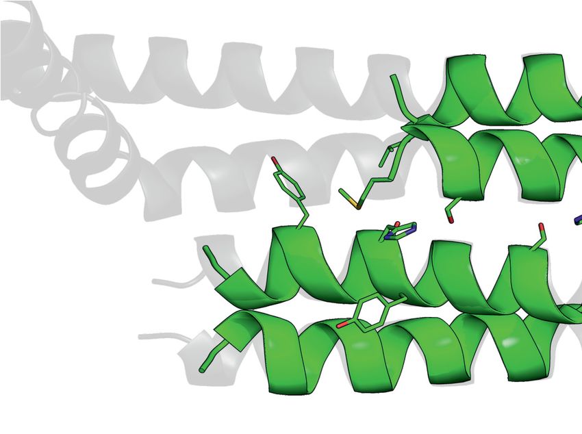
7208 Nucleic Acids Research, 2021, Vol. 49, No. 13

Cln, G5 has been reported to be the snRNA recruiter during         a single T7 promotor and individual ribosome binding sites
UsnRNP assembly (29,34–37).                                        for each gene.
   Consistent with its reported role in RNP biogenesis, sev-
eral factors of the assembly machinery including SMN have
been shown to be essential for viability (38–40). Interest-        Plasmid construction: human
ingly, the human disorder spinal muscular atrophy (SMA) is         Genes encoding human SMN complex components (and
causally linked to reduced levels of functional SMN. SMN           variants thereof) were sub-cloned into either pETM-30 (N-
deficiency alters the stoichiometry of snRNAs in SMN-              terminal His6 -GST-tag), pETM-11 (N-terminal His6 -tag)
deficient mouse tissues and causes widespread and tissue-          or pETM-13 (No tags) from DNA plasmids described pre-
specific pre-mRNA splicing defects in SMA animal mod-              viously (1). Truncation variants were generated with spe-
els. SMA might hence arise from the inefficient splicing of        cific primers and mutants were generated by overlap ex-
pre-mRNAs coding for proteins required for motor neuron            tension PCR. Poly-cistronic plasmids were generated by
function (41–43).

                                                                                                                                    Downloaded from https://academic.oup.com/nar/article/49/13/7207/6179931 by guest on 17 September 2021
                                                                   iterative cloning employing the isocaudomers XbaI and
   Biochemical and genetic studies enabled insight into the        NheI, similar to a strategy described previously (25). For
role of the SMN complex in UsnRNP assembly. Structural             crystallization, the constructs His6 -GST-HsG8190–230 and
insight into the architecture of the SMN complex, however,         HsG61–92 /His6 -HsG746–131 were designed. Similar to S.
is still limited. Thus far, SMN’s Tudor domain (44) and            pombe constructs, each poly-cistronic construct was de-
C-terminal region (45,46), the WD-repeat domain of G5              signed under a single T7 promotor and individual ribosome
(35,36) and parts of a G6/G7 dimer (47) have been deter-           binding sites for each gene. MBP fusion proteins of human
mined by X-ray crystallography or NMR studies. In addi-            YG-box252–284 constructs (and variants thereof) were de-
tion, assembly intermediates encompassing the N-terminus           signed using the pETM-41 vector. All pETM vectors were
of SMN bound to G2 and Sm proteins have been struc-                obtained from EMBL protein expression facility (Heidel-
turally analyzed (29,34), which provided important insight         berg, Germany).
into the mechanism of pICln release and Sm protein ar-
rangements on the complex.
   In this paper, we describe the identification of a simplified   Protein expression in E. coli
version of the SMN complex in the fission yeast Schizosac-
charomyces pombe consisting of five proteins only. The bio-        Recombinant proteins and/or protein complexes were pro-
chemical reconstitution of the yeast SMN (SpSMN) com-              duced either by single expression of plasmids or by co-
plex allowed us to determine its structure by a combination        expression from poly-cistronic constructs using BL21(DE3)
of X-ray crystallography, homology modeling, and small             competent cells (NEB #C2527I). Transformed bacterial
angle X-ray scattering (SAXS) analysis. These studies iden-        cells were cultured in TB medium containing 1× TB buffer
tified the SMN complex as a multivalent hub where Sm pro-          (17 mM KH2 PO4 ; 72 mM K2 HPO4 ), 2 mM MgCl2 , and
teins are collected in its periphery to allow their joining with   appropriate antibiotics until OD600 of 1.0 at 37◦ C and 215
UsnRNA.                                                            rpm. Then, protein expression was induced by adding 0.5
                                                                   mM IPTG and the cultures were left to grow for 18 h at
                                                                   15◦ C and 215 rpm. Cells were harvested by centrifugation
MATERIALS AND METHODS                                              and cell pellets resuspended in either lysis-buffer1 (150 mM
                                                                   NaCl; 50 mM HEPES pH 7.4; 20 mM Imidazole; 2 mM
Plasmid construction: S. pombe                                     2-mercaptoethanol; 10% glycerol) for S. pombe proteins or
Genes encoding SpSMN complex components (and vari-                 in lysis-buffer2 (200 mM NaCl; 50 mM HEPES pH 7.0; 25
ants thereof) were first cloned as mono-cistronic con-             mM Imidazole; 5 mM 2-mercaptoethanol) for human pro-
structs into either pETM-11 (N-terminal His6 -tag) or              teins, each containing protease inhibitors. Cell suspensions
pETM-13 (No tags) vectors. Following this, various poly-           were snap frozen in liquid nitrogen and stored at –20◦ C until
cistronic constructs were generated by iterative cloning           further use.
using the isocaudomers XbaI and NheI restriction sites,
in a strategy similar to what has been previously de-
                                                                   Protein purification
scribed (25). For the determination of the interaction
map and in vitro reconstitution of the SpSMN complex,              Frozen cell suspensions were thawed and subsequently lysed
polycistronic constructs of full-length proteins were de-          by sonication (Branson Sonifier 250). Lysed cell suspension
signed as SpSMN/His6 -SpG2, His6 -SpSMN/SpG2/SpG8,                 was clarified by centrifugation at 30 000 rpm (rotor 45 Ti,
SpG6/SpG7/His6 -SpG8, and SpG6/His6 -SpG7. To inves-               Beckman Coulter) at 4◦ C for 1 h. Cleared lysate was incu-
tigate the interaction between SpG8 and SpSMN, the                 bated with Ni-NTA agarose beads (Qiagen) or Glutathione
constructs His6 -SpSMNYG /SpG2/SpG8 (YG = residues                Sepharose 4B (GE Healthcare) for 2 h at 4◦ C. Following
130–152) and SpG6/SpG7/His6 -SpG8N58 were designed.               this, the beads were washed with 20–40 bed volumes of ly-
For crystallization and SAXS experiments the constructs            sis buffer1/2 and the bound proteins were eluted with 250
SpSMN36–119 /His6 -SpG2N80 , SpSMN/His6 -SpG2N80 ,              mM Imidazole or 20 mM GSH. The eluted proteins were
and SpG6/SpG7/His6 -SpG835–58 were designed. For mu-              supplemented with 1–2% (w/w) TEV protease (for His6 re-
tational analysis, constructs SpSMN36–119 S130D/His6 -            moval) or PreScission™ protease (for His6 -GST removal).
SpG2N80 and SpSMN36–119 A134E/His6 -SpG2N80 were                S. pombe proteins were subsequently dialyzed into gel fil-
designed. Each poly-cistronic construct was designed under         tration buffer (150 mM NaCl; 20 mM HEPES pH 7.4; 2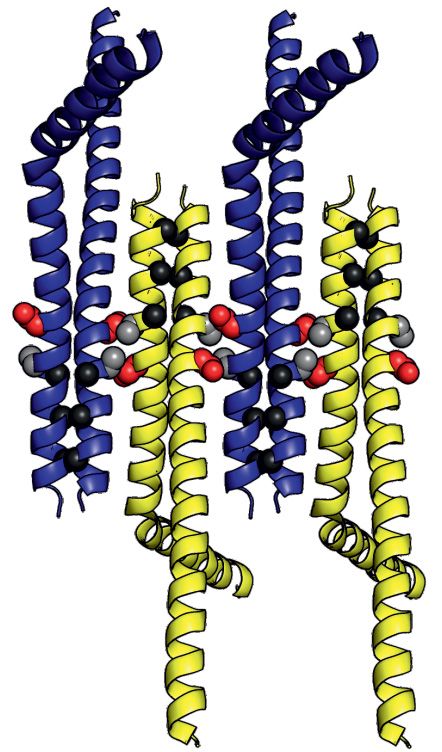
Nucleic Acids Research, 2021, Vol. 49, No. 13 7209

mM DTT) overnight at 4◦ C and the dialysate was concen-         In vitro transcription and translation of human Gemin8
trated for further steps. HsG8190–230 was subsequently incu-
                                                                N-terminal His6 -HsG8 (full length) was in vitro transcribed
bated with Ni-NTA beads and HsG61–92 /His6 -HsG746–131
                                                                and translated with [35 S]-Methionine labeling with the
was used as bait to purify the trimeric complex.
                                                                TNT® T7 Quick coupled Transcription/Translation sys-
                                                                tem (Promega).
Gel filtration and in vitro reconstitution
Purified complexes were further characterized using analyt-     In vitro protein binding assays
ical gel filtration columns Superose 6 10/300, Superdex 75
10/300, and Superdex 200 10/300 (GE Healthcare, Munich,         For the MBP binding assays, MBP fusion proteins im-
Germany). For reconstitution assays, equimolar amounts          mobilized on Amylose resin (NEB) were incubated with
of SpSMN sub-complexes were combined and incubated on           in vitro transcribed translated [35 S]-methionine labeled
ice for 15 min. Hereafter, the samples were briefly placed on   Gemin8 transcripts in binding buffer (HEPES, pH 7.0,

                                                                                                                               Downloaded from https://academic.oup.com/nar/article/49/13/7207/6179931 by guest on 17 September 2021
37◦ C for 5 min followed by an additional 15 min on ice. The    150 mM NaCl, 2 mM DTT and protease inhibitors) at
samples were then centrifuged at 10 000 g for 15 min at 4◦ C    4◦ C for 3 h. The resin was then washed initially with
before applying onto gel filtration columns. Gel filtration     a high salt buffer (HEPES, pH 7.0, 300 mM NaCl, 2
fractions were analyzed by 15% Tris–tricine SDS-PAGE.           mM DTT and protease inhibitors) followed by washes
                                                                with the binding buffer. Bound proteins were then eluted
Crystallization and structure determination                     with 1× SDS sample buffer, resolved by SDS-PAGE (13%
                                                                Bis–Tris) and analyzed by Coomassie staining. Labeled
Crystallization trials were conducted with the                  proteins were detected by autoradiography of the dried
SpSMN36–119 /SpG2N80 complex at a concentration               gel.
of 19.7 mg/ml. Needle shaped crystals of 0.6 mm size
of space group C2 2 21 were obtained with a condition
containing 65% 2-methylpentanediol, 80 mM KCl, and 40           Yeast strains, media and genetic methods
mM HEPES (Natrix HT crystallization screen, Hampton             Standard methods were used for growth and genetic
Research) at different pH values (6.8, 6.9, and 7.2), by        manipulation of S. pombe (50). Cells were grown on YES
the hanging drop vapor diffusion method. The crystals           or minimal EMM2 medium with adequate supplements.
were snap frozen in liquid nitrogen in the mother liquor        Strains carrying null allele of SpG6 (SPAC4D7.15::NatN2)
and X-ray diffraction data were collected. Phases were          and SpG7 (SPBC32F12.16::NatN2) were constructed
determined by molecular replacement using the dimeric           in diploid strain (h+/h+ ade6-M210/ade6-M216 ura4-
SpYG-domain structure (PDB ID: 4RG5 (46)) as a tem-             D18/ura4-D18 leu1–32/leu1–32) by homologous recombi-
plate. Electron density for the globular SpG2N80 could         nation as described previously using appropriate templates
not be assigned. Instead, electron density for helical          and primers (51). The diploid strain heterozygous for
dimers of SpSMN36–119 was clearly observed. The ab-            the null allele of SpG8 (SPBC16H5.15) (h+/h+ ade6-
sence of SpG2N80 from the crystals is attributed to the        M210/ade6-M216 ura4-D18/ura4-D18 leu1–32/leu1–32
denaturation of this compound.                                  SPBC16H5.15/ SPBC16H5.15::KanMX4) was purchased
   HsG61–92 /HsG746–131 /HsG8191–230 crystals were grown        from Bioneer Corporation (Korea). After transformation
at a concentration of 30 mg/mL in 100 mM 2-(N-                  with the sporulation-inducing plasmid pON177 (52),
morpholino) ethanesulfonic acid, 200 mM NaCl and 30%            spores were dissected and germinated at 25◦ C on YES
Jeffamine ED2003 by sitting-drop vapour diffusion at            plates. The temperature-degron tdGemin8 allele was con-
18◦ C. Crystals were transferred into a cryoprotectant solu-    structed using the pSMRG2-nmt41-degron plasmid (53) as
tion containing 100 mM 2-(N-morpholino) ethanesulfonic          described previously (54). A DNA fragment carrying 400
acid, 200 mM NaCl and 35% Jeffamine ED2003 before be-           nucleotides homologies to genomic DNA was amplified
ing snap frozen in liquid nitrogen. The structure of this       and transformed into fission yeast wild-type cells. Correct
trimeric complex was solved using the HsG6/HsG6 (PDB            homologous recombinations of the disrupted and tagged
ID: 1Y96 (47)) dimer as the molecular replacement model         alleles were checked by PCR amplification of genomic
and the HsG8190–230 fragment could be traced from the           DNA.
initial 2Fo – Fc density map. The resulting model could
be refined to an Rfree /Rwork of 0.205/0.250 and included
                                                                Plasmid constructions
residues 1–86, residues 47–131 and residues 191–227 for
HsG6, HsG7 and HsG8 respectively.                               PCR fragments containing the coding sequences of the S.
   The data sets for each of the protein crystals were col-     pombe Gemins were PCR amplified from genomic DNA or
lected at the ID30B beam line of the European Synchrotron       from the pTN-RC5 cDNA library (a gift from T. Naka-
Radiation Facility (ESRF, Grenoble, France) and processed       mura, YGRC, Osaka, Japan) using forward and reverse
with XDS (48). The structures were solved by molecular re-      oligonucleotides carrying adequate restriction sites. Af-
placement with PHASER (49). Automated refinement was            ter separation on agarose gels, DNA fragments were pu-
performed in PHENIX until R/Rfree factors converged. The        rified using the GeneClean procedure and ligated into
crystallographic data processing and refinement parameters      previously cut pREP41/42 or pREP41/42-GFP-N vec-
are summarized in Table 1. The final Figures were gener-        tor (55). The pAS and pACT2st vectors were used
ated using PyMOL Molecular Graphics System, Version             to constructs baits and preys for two-hybrid analyses
2.0, Schrödinger, LLC.                                         (56). PCR amplification were performed from pREP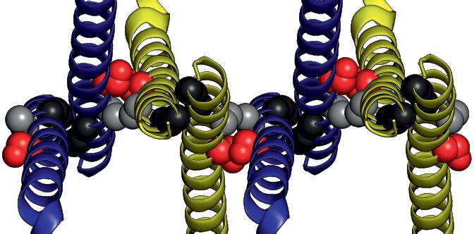
7210 Nucleic Acids Research, 2021, Vol. 49, No. 13
Table 1. Crystallographic data and refinement statistics
Parameter HsG61–92 /HsG746–131 /HsG8191–230 SpSMN36–119
Wavelength (Å) 0.9762 0.9762
Resolution range (Å) 48.07–1.52 (1.57–1.52) 40.49–2.15 (2.23–2.15)
Space group P221 21 C2221
Unit cell a = 59.88, b = 80.59, c = 82.66; ␣ = 90,  = 90, a = 27.19, b = 83.71, c = 160.06;
␥ = 90 ␣ = 90,  = 90, ␥ = 90
Total reflections 223044 (10189) 67776 (7057)
Unique reflections 58263 (4340) 10282 (1010)
Multiplicity 3.8 (2.3) 6.6 (7.0)
Completeness (%) 93.5 (70.8) 99.3 (99.7)
Mean I/sigma(I) 10.83 (1.03) 10.85 (2.25)
Wilson B-factor (Å2 ) 18.6 46.4
Rmeas (%)a 7.6 (95.9) 11.4 (89.0)
Downloaded from https://academic.oup.com/nar/article/49/13/7207/6179931 by guest on 17 September 2021
CC1/2 (%) 99.9 (19.7) 99.8 (70.8)
Reflections used in refinement 58 076 (4340) 10 262 (1010)
Reflections used for R-free 2873 (207) 531 (31)
Rwork (%)b 20.5 (40.9) 25.6 (38.0)
Rfree (%)c 25.0 (41.9) 29.1 (37.8)
No. of non-hydrogen atoms 3735 927
Ligand – 32 (MPD)
Water 363 3
No. of protein residues 418 108
RMSDd bond lengths (Å) 0.010 0.004
RMSDd bond angles (◦ ) 1.020 0.800
Ramachandran favored (%)e 95.8 100
Ramachandran allowed (%)e 4.2 0.0
Ramachandran outliers (%)e 0.0 0.0
Rotamer outliers (%)e 0.6 0.0
Clash scoree 1.64 1.10
Average B-factor (Å2 ) 27.4 69.8
Macromolecules 26.6 69.8
Waters 34.4 54.9
Ligands - 73
PDB code 7BBL 7BB3
meas = h (n/n – 1)
aR 1/2 |I (h) – |/ I (h), where I (h) and are the ith and mean measurement of the intensity of reflection h.
i i h i i i
bR
work = h ||F obs (h)| – |Fcalc (h)||/ h |Fobs (h)|, where Fobs (h) and Fcalc (h) are the observed and calculated structure factors, respectively.
cR
free is the R-value obtained for a test set of reflections consisting of a randomly selected 5% subset of the data set excluded from refinement.
d Root Mean Square Deviation
e Values from Molprobity server (http://molprobity.biochem.duke.edu/)
Values in parenthesis are for the highest resolution shell.
plasmids containing the corresponding genes. Primer se- Purification of endogenous SpSMN complex
quences and PCR regimes are available upon request.
Yeast cells carrying a GFP-SpG6 fusion sequence were
Construction of the SMN-A134E and SMN-S130D mu-
grown in EMM2 -Ura media to an ODA600 of 0.6–0.8
tants was achieved using the QuikChange Site-Directed
and the cell pellet was resuspended in lysis buffer (10 mM
Mutagenesis kit (Stratagene, La Jolla CA, USA) essen-
Tris/Cl pH 7.5; 150 mM NaCl; 0.5 mM EDTA; 0.25% NP-
tially according to the manufacturer’s instructions. All the
40; 1 mM PMSF; 1× Complete protease inhibitors) and
cloning junctions and coding sequences were verified by
frozen. For purification of the endogenous SpSMN com-
sequencing.
plex, frozen cells were ground to fine powder using a Freezer
Mill 6770 grounder (Spex) and after centrifugation at 14 000
Two-hybrid assays for protein–protein interactions rpm for 30 min, the soluble extract was recovered by cen-
trifugation at 49 000 rpm for 1 h at 4◦ C and incubated with
Two-hybrid assays were performed with the CG1945 and GFP-Trap beads (Chromotek, Germany) for 4 h at 4◦ C. The
Y187 strains (57). The CG1945 strain was transformed with beads were then washed four times in wash buffer (10 mM
the pAS– constructs and selected on –Trp plates while Tris/Cl pH 7.5; 150 mM NaCl; 0.5 mM EDTA) and the im-
Y187 was transformed with the pACT2st- constructs and munoprecipitated proteins were separated by SDS-PAGE.
selected on –Leu plates. Strains carrying bait and prey plas-
mids were mated overnight on rich YPD plates and diploids
Northern blot, primer extension and native gel electrophoresis
containing the bait and prey combinations were selected
on –Trp –Leu plates. Diploid yeast cells carrying bait/prey Total yeast RNA was purified from exponentially growing
combinations were cultured in –Trp–Leu media and inter- cells with Tri-Reagent (Sigma) according to the manufac-
actions were screened by spotting serial dilutions on –Trp– turer’s procedure. Primer extension and Northern blot anal-
Leu–His plates. Incubations were performed at 30◦ C for 3–5 yses were performed as described previously (58). For native
days. gel analysis of snRNPs, extracts were prepared from cells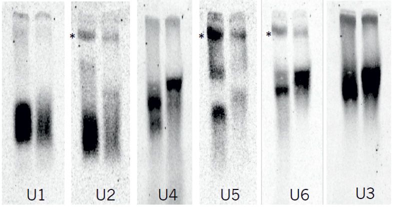
Nucleic Acids Research, 2021, Vol. 49, No. 13 7211
which were resuspended to 1 g/ml in AGK400 buffer (10 RESULTS
mM HEPES–KOH pH 7.9, 400 mM KCl, 1.5 mM MgCl2 ,
Identification of the fission yeast SMN complex
0.5 mM DTT, 1× Complete protease inhibitors and 10%
glycerol). After freezing in liquid nitrogen, cells were ground Only SMN and G2 orthologues of the human SMN com-
to fine powder. After thawing on ice, cells were centrifuged plex have been found thus far in S. pombe (62–64). Using
at 14 000 rpm for 10 min at 4◦ C and the supernatant recov- a bioinformatics approach, we identified putative orthologs
ered and spun at 55 000 rpm for 30 min at 4◦ C in a TLA- of G6 (SpG6), G7 (SpG7) and G8 (SpG8) based on homol-
100.3 rotor. The extract was then dialyzed for 2 h against ogy at the level of amino acid sequence and secondary struc-
buffer D (20 mM HEPES–KOH pH 7.9, 0.2 mM EDTA, ture (Figure 1A and Supplementary Figure S1). Whereas
100 mM KCl, 0.5 mM DTT, 1 mM PMSF, 20% glycerol) the sequence conservation of all three candidates is weak,
and aliquots stored at –80◦ C. Native gel electrophoresis and their predicted secondary structures correspond well to
analysis were performed as previously described (54). their human counterparts. To investigate whether these fac-
tors are part of a larger complex, immunoprecipitation ex-
Downloaded from https://academic.oup.com/nar/article/49/13/7207/6179931 by guest on 17 September 2021
Small angle X-ray scattering data acquisition periments were performed using extracts from strains ex-
pressing either GFP alone or GFP-tagged SpG6 as the sole
Synchrotron SAXS data from solutions of protein com- source of SpG6. As determined by mass spectrometry, the
plexes in 150 mM NaCl, 20 mM HEPES, 1 mM DTT, pH immunoprecipitate contained apart from the tagged SpG6
7.5, were collected at the BM29 beam line of the European bait, SpSMN, SpG2 as well as the newly identified ortho-
Synchrotron Radiation Facility (ESRF, Grenoble, France) logues SpG7 and SpG8 (Figure 1B and Supplementary Ta-
using a PILATUS 1M detector (Dectris) at a distance of ble S1). Importantly, Sm proteins were also found in this
2.867 m from the sample, and a wavelength of 0.9919 Å (I(s) immunoprecipitation albeit in sub-stoichiometric amounts
versus s, where s = 4sin /, and 2 is scattering angle). (Figure 1B (asterisks) and Supplementary Table S1). These
Data collection was done for a scattering vector (s) range findings show that the SpSMN complex consists of SpSMN,
of 0.0032–0.4944 Å−1 . In-line size-exclusion chromatogra- SpG2, SpG6, SpG7 and SpG8, and binds to Sm protein
phy (SEC) was employed for the data collection. Protein so- substrates. However, orthologues of HsG3–5 and unrip are
lutions were injected onto a Superdex 200 10/300 column lacking.
(GE Healthcare, Munich, Germany) at 20◦ C and run at a We next tested whether the SpSMN complex is function-
flowrate of 1 ml/min. A total of 1800 frames spanning the ally related to its human counterpart. Consistent with a
whole elution profile (with 1 s exposure per frame) were col- role in UsnRNP assembly, a tetrad analyses showed that
lected. The data was then normalized to the intensity of deletion of the SpG6–8 genes causes lethality (Supplemen-
the transmitted beam and radially averaged. (see also Sup- tary Figure S2A), as has been shown already for SpSMN
plemetary Table S4). and SpG2, demonstrating that SpG6, SpG7 and SpG8
are essential genes. Furthermore, a yeast strain carrying a
Small angle X-ray scattering data validation and analysis temperature-degron allele of SpG8 (tdSpG8) displays al-
ready a growth defect at permissive-temperature as well as
All data processing was performed using ATSAS 3.0.3 soft- reduced splicing after a shift to non-permissive tempera-
ware package (59). For data shown in Figure 7, 20 frames ture (Figure 1C and D). Lastly, extracts prepared from td-
at the peak of the SEC-SAXS chromatogram were scaled SpG8 cells contained decreased levels of the U1, U2 and
and averaged. Background subtraction was performed us- U5 Sm-class snRNPs while the amount of the U3snoRNP
ing scaled and averaged buffer frames preceding the pro- (an RNP lacking Sm proteins) remained unaffected (black
tein peak. Protein concentrations were obtained from the arrows, Figure 1E). Of note, the mobility of the U4/U6 di-
peak of the UV280 trace. For data shown in Supplemen- snRNP is slightly decreased (blue asterisks, Figure 1E) and
tary Table S3, individual frames at various regions of the the amount of the post-splicing U2/U5/U6 complexes (red
chromatograms were selected and background subtraction asterisks, Figure 1E) is decreased in the mutant, which in-
was performed using buffer frames. Protein concentrations dicates defects in spliceosome activity (see also Figure 1F
at each selected frame was obtained from the UV280 trace for quantification of snRNP levels). Together, our data sug-
of the chromatogram. Quality of each of the final scatter- gest that the SpSMN complex is required for formation of
ing curve was investigated using Guinier plots (60). The ra- Sm-class UsnRNPs and splicing.
dius of gyration (Rg ) was obtained from Guinier approx-
imation: I(s) = I(0) exp(s2 Rg 2 /3), with the limits sRg <
Architecture and in vitro reconstitution of the SpSMN pen-
1.3. The pairwise distance distribution function P(r) and
tameric complex
maximum particle dimension Dmax were obtained from
the GNOM program (61) integrated into the ATSAS soft- The discovery of a simplified SMN complex in S. pombe
ware package. The molecular weights calculated from I(0) enabled its biochemical and structural investigation. Ear-
in Figure 7 and Supplementary Table S3 were obtained lier studies revealed an elaborate interaction network that
by the following formula: (MWu /MWs ) = [I(0)u /Conc.u ]/ ties together the proteins of the human SMN complex
[I(0)s /Conc.s ], where u = unknown and s = standard. SAXS (31). In this network, HsSMN forms the central core
data of SpSMN36–119 S130D/SpG2N80 was used as a stan- onto which HsG2 binds via the N-terminus of HsSMN.
dard. The molecular weight obtained from the Porod vol- The C-terminus of HsSMN, termed the YG-domain, es-
ume (Vp ) was calculated by the following formula: MW = tablishes the connection to HsG8, which in turn re-
Vp /1.66. cruits the HsG6/HsG7 heterodimer. In support of a sim-
7212 Nucleic Acids Research, 2021, Vol. 49, No. 13

                                                                                                                                                             Downloaded from https://academic.oup.com/nar/article/49/13/7207/6179931 by guest on 17 September 2021
Figure 1. Identification of a pentameric SpSMN complex linked to UsnRNP assembly. (A) Secondary structural elements of the human and the S. pombe
SMN complex core subunits (SMN, G2, G6, G7 and G8). Known domain compositions for the subunits are indicated. (B) Immunoprecipitation of
endogenous SpSMN complex using GFP-Trap-A beads from cells expressing GFP-SpG6 as the sole source of SpG6 (lane 2). A control purification with
GFP only is shown in lane 1 (4–12% gradient gel). The asterisks point to Sm proteins. (C) Serial dilutions of wild type and tdSpG8 cells were spotted onto
rich media and grown at the indicated temperature. (D) Splicing inhibition in the tdSpG8 mutant. After growth of wild type cells and cells carrying the
tdSpG8 allele at the indicated temperature for 4 h, total RNA was isolated and used for primer extension. Pre-U6 indicates the species corresponding to
the U6 precursor and U6 indicates the spliced matured U6 RNA. (E) Native gel analysis of snRNPs in tdSpG8 and wild type cells. Extracts were prepared
from cells grown at 25◦ C and similar amounts were separated on 4% native gels. The RNAs were subjected to Northern analysis and hybridized with
oligonucleotide probes for the different snRNAs. The arrows indicate U1, U2, U5 (snRNPs) or U3 (snoRNP). Blue and red asterisks point to U4/U6 di-
snRNPs and U2/U5/U6 post-splicing complexes, respectively. (F) quantification of snRNP levels using ImageJ. Data from two independent experiments
are presented as mean ± SEM. A.U.: arbitrary units.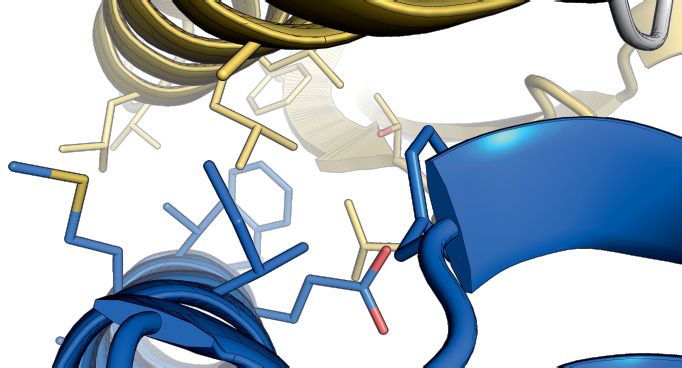
Nucleic Acids Research, 2021, Vol. 49, No. 13 7213

ilar protein network in the S. pombe complex we de-           the mode of HsG8 binding to HsG7. The C-terminus of
tected identical interaction pattern among the yeast ortho-   HsG8 adopts a helix (␣1)-turn-helix (␣2) motif and inter-
logues using yeast two-hybrid assays (Supplementary Fig-      faces with the N-terminal helix of HsG7 (Figure 3C). This
ure S2B). Furthermore, we succeeded in the co-expression      interface comprises several highly conserved hydrophobic
and purification of SpSMN/SpG2, SpSMN/SpG2/SpG8,              residues of HsG7 (A60, L67, L70, L71, F92 and L97) and
SpG6/SpG7/SpG8/ and SpG6/SpG7, providing biochem-             of HsG8 (Y205, I212, M215, A218, V219 and F223) (Figure
ical evidence for the interaction network (Figure 2A and      3E). A hydrogen bond is established between the sidechain
Supplementary Figure S3).                                     amino group of HsG7 Q56 and the carbonyl group of
   The availability of these protein modules enabled          HsG8 R203. In addition, salt bridge interactions between
the reconstitution of the pentameric SpSMN complex            the guanidine group of HsG7 R63 and sidechain carboxyl
in vitro. Equimolar amounts of bacterially expressed          group of HsG8 E216 are also established (Supplemen-
SpSMN/SpG2 and SpG6/SpG7/SpG8 complexes were                  tary Figure S5A). The corresponding S. pombe proteins
mixed and subjected to gel filtration chromatography. All     SpG6, SpG7 and SpG8115–166 share 21%, 29% and 21%

                                                                                                                               Downloaded from https://academic.oup.com/nar/article/49/13/7207/6179931 by guest on 17 September 2021
proteins elute in a single peak near the 669 kDa marker,      sequence identity, respectively, with their human counter-
showing the formation of the pentameric complex (red          parts. This allowed us to build a homology model of the
dashed box, Figure 2B). In the absence of SpG8, how-          S. pombe SpG6/SpG7/SpG8 complex based on our crys-
ever, SpSMN/SpG2 and SpG6/SpG7 fail to form a com-            tal structure (Figure 3D). The homology model showed
plex and are completely separated into two distinct peaks     that many conserved residues are clustered in the hydropho-
(Figure 2C). Interestingly, a trimeric complex lacking the    bic interface between SpG7 and SpG8 (Figure 3F, relevant
first 58 residues of SpG8 (SpG6/SpG7/SpG8N58) also           residues are indicated). We therefore conclude that both
fails to form the pentameric SpSMN complex (Figure 2D).       systems possess a similar mode of interaction. Our results
Furthermore, full-length SpG8 bound to SpSMN/SpG2             thus demonstrate a conserved modular architecture of the
only when the YG-domain of SpSMN was present (Fig-            SpG6/SpG7/SpG8 sub-complex.
ure 2E and F). Thus, SpG8 forms the link between the
SpSMN/SpG2 and SpG6/SpG7 dimers through an inter-
                                                              Structural basis of SMN oligomerization
action of the N-terminus of SpG8 with the YG-domain of
SpSMN (Figure 2G).                                            We next investigated the oligomeric properties of the
   We noted that the hydrodynamic size of the                 SpSMN complex. The C-terminal YG-domain of SMN
SpSMN/SpG2 unit is almost identical to the size of            is homologous across species with the two overlap-
the entire pentameric SpSMN complex with elution peaks        ping sequence elements, YxxGYxxGYxxG (YG-box) and
at approx. 669 kDa on gel filtration columns (Figure          SxxxSWxxSxxxT (serine-motif) being the key features (Fig-
2B–D). In fact, no significant variation in its hydrody-      ure 4A). The crystal structures of the human and S. pombe
namic size was observed when individual SpGemins or           YG-domain had previously been solved (45,46) and re-
subunits thereof were bound onto the SpSMN/SpG2               vealed SMN dimerization via a glycine–zipper interac-
module (Supplementary Figure S4). However, deletion of        tion of the YG-box. To re-evaluate this interaction, an
the YG-domain or the long unstructured region (residues       SpSMN fragment lacking its unstructured middle region
36–119) of SpSMN showed a drastic decrease in size (Sup-      (SpSMN36–119, Figure 4B) was co-expressed with a
plementary Figure S4). Thus, the hydrodynamic properties      fragment of SpG2 lacking the N-terminus (SpG2N80).
of the whole SpSMN complex are primarily a function of        The resulting SpSMN36–119 /SpG2N80 complex was crys-
the core SpSMN subunit.                                       tallized, a 2.16 Å dataset was collected and its struc-
                                                              ture solved by molecular replacement (Figure 4C–G) us-
                                                              ing the YG-domain fragment from the known MBP-SpYG-
Structure of the G6/G7/G8 module
                                                              domain structure (PDB-ID:4RG5) (see Table 1 for crys-
We next focused on the structural investigation of the        tallographic data and refinement statistics). We could de-
SMN complex. The structures of HsG6/G7 (47) and               tect clear electron density for SpSMN36–119 but no elec-
HsSMN/G2 modules are known (29,34) but the ba-                tron density could be assigned to SpG2N80 , suggesting
sis of G8-mediated bridging of both modules has not           that the latter had dissociated and/or precipitated dur-
yet been established. We therefore expressed and pu-          ing crystallization. The structure revealed the SpSMN N-
rified complexes composed of the S. pombe proteins            terminal G2 binding domain (residues 10–35) and the C-
SpG6/SpG7/SpG8115–166 and the corresponding human             terminal YG-domain (residues 120–147) encompassing the
proteins HsG61–92 /HsG746–131 /HsG8190–230 , respectively     YG-box and the serine-motif (Figure 4C). Two molecules of
(Figure 3A and B). The human complex allowed struc-           SpSMN36–119 in the asymmetric unit (termed the glycine–
ture determination by X-ray crystallography and the gen-      zipper dimeric unit), interact via the YG-box residues of in-
eration of a homology model for the S. pombe orthologues      terfacing helices (Figure 4C). This interaction is identical
(Figure 3C and D). The HsG61–92 /HsG746–131 /HsG8190–230      to the previously observed interaction in the MBP-SpYG-
crystals yielded a 1.52 Å dataset and the structure was      domain crystal structure (PDB ID: 4RG5) and exhibits two
solved by molecular replacement using the HsG6/HsG7           sets of hydrophobic interactions. First, interfacing glycine
structure (PDB ID: 1Y96). We obtained a complete atomic       residues (black spheres, Figure 4D) pack tightly against
model (see Table 1 for crystallographic data and refine-      each other. Second, tyrosine and leucine residues of each
ment statistics), which confirms the previously reported      helix (grey sticks, Figure 4D) pack tightly against glycine
Sm-like fold of the HsG6/HsG7 dimer (47) and reveals          residues of the interfacing helix. Interestingly, a closer in-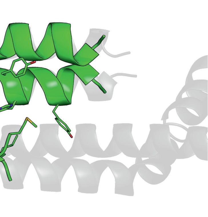
7214 Nucleic Acids Research, 2021, Vol. 49, No. 13

                                                                                                                                                   Downloaded from https://academic.oup.com/nar/article/49/13/7207/6179931 by guest on 17 September 2021
Figure 2. In vitro reconstitution of the pentameric SpSMN complex. (A) Recombinant co-expression and Ni-NTA purifications of SpSMN/His6 -SpG2
(lane 1), His6 -SpSMN/SpG2/SpG8 (lane 2), SpG6/SpG7/His6 -SpG8/ (lane 3), and SpG6/His6 -SpG7 (lane 4) sub-complexes from E. coli. Purified com-
plexes were analyzed by 15% Tris-Tricine SDS-PAGE unless otherwise mentioned. (B–D) Complexation assays. SpSMN/SpG2 was added to equimolar
amounts of SpG6/SpG7/SpG8 (B), SpG6/SpG7 (C), or SpG6/SpG7/SpG8N58 (D), and the resulting mixtures analyzed by gelfiltration (Superose 6
10/300). Fractions under peaks I and II are analyzed by 15% Tris-Tricine SDS-PAGE. The formation of pentameric SpSMN complex is indicated by
red dashed box in (B). (E) Ni-NTA purification of SpSMNYG/His6 -SpG2/SpG8. YG refers to residues 130–152. (F) Ni-NTA purification of His6 -
SpSMN/SpG2/SpG8. Asterisk indicates insoluble SpG8. S = supernatant, P = pellet, FT = flow through, W = wash, E = elution. (G) Interaction map
of the SpSMN complex.

spection of the crystallographic packing showed that each                    Anti-parallel stacking of glycine–zipper dimeric units is
glycine–zipper dimeric unit is stacked upon each other in                 facilitated by the serine-motif where S130 and A134 pack
an anti-parallel fashion around a screw axis between S130                 against A134 and S130, respectively, of the interfacing he-
and A134, leading to an infinite stacking along the crys-                 lix of the adjacent dimeric unit, through mainchain atoms
tallographic A axis (Figure 4E and Supplementary Fig-                     (Figure 4E and F). These reciprocal interactions place in-
ure S5B). This interface, termed the anti-parallel interface,             terfacing serine and alanine residues on opposite sides
buries a surface area of 592 Å2 which is similar to the 620              of the oligomeric stack (Figure 4F). As a consequence,
Å2 buried surface area within the glycine–zipper interface               the serine- and alanine-sides alternate through consecutive
(Figure 4E).                                                              anti-parallel interfaces (Figure 4F). The alanine-side forms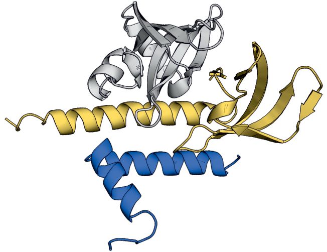
Nucleic Acids Research, 2021, Vol. 49, No. 13 7215
Downloaded from https://academic.oup.com/nar/article/49/13/7207/6179931 by guest on 17 September 2021
Figure 3. Crystal structure and homology model of the G6/G7/G8 module. (A) Representations of G8, G7 and G6 from human and S. pombe depicting
the constructs used for purification, crystallization or homology modeling shown in B–F. (B) Gel filtration of HsG61–92 /HsG746–131 /HsG191–230 (1 and
2) and SpG6/SpG7/SpG8115–166 (3 and 4) complexes. (C) Crystal structure of HsG61–92 /HsG746–131 /HsG191–230 . The C-terminus of HsG8 consisting of
a helix (␣1)-turn-helix (␣2) motif engages with the N-terminal ␣1 helix of HsG7. The dimerization interface between HsG7 (5) and HsG6 (4) strands
forming a continuous 10 sheet -barrel remains intact. The respective N- and C- termini are labeled. (D) A homology model of SpG6/SpG7/SpG8115–166
generated using HsG61–92 /HsG746–131 /HsG191–230 crystal structure as a template. (E) Cluster of hydrophobic residues at the interface between HsG7 and
HsG8. (F) Cluster of hydrophobic residues at the interface between SpG7 and SpG8. The homology model was generated using SWISS-MODEL and
showed a QMEAN score of -1.4 with overall sequence identity of 24.44% between the template and the target sequences. Structures were generated using
PyMOL Molecular Graphics System, Version 2.0, Schrödinger LLC.
crucial interactions necessary for the formation of higher partially buried and therefore accessible for additional in-
order oligomers (Figure 4G). The methyl group of each teractions.
A134 forms hydrophobic contacts with the W131 sidechain
of the interfacing helix. The sidechains of each W131 are
SMN oligomerization determines the SMN complex compo-
stabilized by hydrogen bonding to the interfacing T138
sition
sidechains (Figure 4G). As a result of these interactions, in-
terfacing A134 residues remain fully buried at the center of A set of experiments was performed to test whether the
the anti-parallel interface, while the S130 residues are only newly discovered anti-parallel interface of SMN is physio-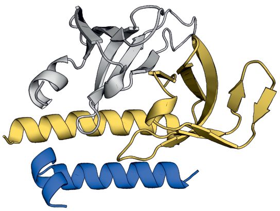
7216 Nucleic Acids Research, 2021, Vol. 49, No. 13

                                                                                                                                                                  Downloaded from https://academic.oup.com/nar/article/49/13/7207/6179931 by guest on 17 September 2021

Figure 4. Crystal structure of SpSMN36–119 . (A) Multiple sequence alignment of SMN YG-domain of various organisms. Conserved motifs are high-
lighted with boxes. (B) Representative diagram showing generation of SpSMN36–119 construct. (C) Crystal structure of SpSMN36–119 showing YG-
domain dimerization resulting in a glycine–zipper dimeric unit (the asymmetric unit). (D) Closeup view of the glycine–zipper dimeric unit showing interac-
tions between YG-box residues of interfacing helices. The glycine residues are shown as black spheres. Tyrosine and Leucine residues are depicted as grey
sticks. (E) Anti-parallel stacking of glycine–zipper dimeric units around a screw axis between S130 (red) and A134 (grey). Alternating dimeric units are
colored yellow for clarity. The two distinct interfaces are indicated. (F) S130 and A134 of one helix pack tightly against A134 and S130, respectively, of the
interfacing helix at the point of closest contact in the anti-parallel interface. The serine- and the alanine-sides alternate through consecutive anti-parallel
interfaces. (G) Residue specific interactions on the alanine-side of the anti-parallel interface. Each W131 residue engages in hydrophobic interactions with
the CH3 group of the interfacing A134. Sidechain conformation of each W131 is stabilized by H-bonding with T138 of the interfacing helix. Structures
were generated using PyMOL Molecular Graphics System, Version 2.0, Schrödinger LLC.
Nucleic Acids Research, 2021, Vol. 49, No. 13 7217
logically relevant. We reasoned that residues with bulkier SMA-causing mutations interfere with SMN oligomerization
sidechains at this interface would prevent oligomer for- and SMN complex composition
mation but would not impact the glycine–zipper interface.
The YG-domain of human SMN is a hotspot for missense
Hence, we substituted either S130 to aspartate (S130D) or
mutations causing the motoneuron disease SMA. In fact,
A134 to glutamate (A134E) and analyzed the oligomeric
nearly 50% of known mutations are located in this region
states of the mutants by small angle X-ray scattering
and have been shown to interfere with SMN oligomeriza-
coupled to size exclusion chromatography (SEC-SAXS).
tion (45). We hence asked whether the oligomerization ob-
Wild-type SpSMN36–119 /SpG2N80 forms oligomers in
served for the S. pombe YG-domain can also occur in hu-
the range of dimers to decamers at low concentrations (peak
man SMN and whether this is affected by SMA-causing
concentrations 5.8–16 M, Figure 5A) but converts entirely
missense mutations. To this end, we first constructed a
to higher order oligomers when the concentration is in-
model of the human YG-domain263–281 . We used the re-
creased (peak concentration 84 M, Figure 5B). Both mu-
ported structure of the human YG-domain263–281 fused to
tants, however, form exclusively dimers at any concentra-
Downloaded from https://academic.oup.com/nar/article/49/13/7207/6179931 by guest on 17 September 2021
MBP (PDB ID: 4GLI), which only forms glycine–zipper
tion (Figure 5A and B), but no higher order oligomers.
dimers due to steric obstruction by the MBP fusion pro-
This suggests that the anti-parallel interface is the ma-
tein (45). In our model, we populated both interfaces by su-
jor determinant for higher order oligomerization in solu-
perposition of the human YG-domain263–281 structure onto
tion but irrelevant for dimerization. Our results thus cor-
the SpSMN36–119 structure and energy minimized the final
roborate the previous notion that the SMN glycine–zipper
model (Figure 6A and Supplementary Figure S6). Of note,
dimers are the fundamental unit of higher order oligomers
the human residues crucial for oligomer formation within
(46) and reveal a novel anti-parallel interface between
the serine-motif (S266, W267, S270 and T274) are located
glycine–zipper dimers required for higher order oligomer
precisely at positions relevant to establish a functional in-
formation.
terface (compare Supplementary Figure S6B and C). The
Next, we tested whether SMN oligomerization is
modeled human YG-domain263–281 structure is thus in per-
relevant for the biochemical composition and/or
fect agreement with higher order oligomer formation as has
function of the SMN complex. Based on our finding
been observed for the yeast system.
that SpSMN/G2 binds SpG6/SpG7/SpG8 and thus
We then asked whether known SMA-causing missense
enables pentamer formation (Figure 2B), we asked
mutations (65) would interfere with SMN oligomerization
whether the SMN mutants S130D and A134E can
and/or G8 binding. To this end, we expressed MBP fused
engage in similar interactions despite their oligomer-
to the YG-domain252–284 containing SMA-causing missense
ization defect. To this end, we analyzed binding of the
mutations M263T, M263R, S266P, Y272C, H273R and
mutant dimers SpSMN36–119 S130D/SpG2N80 and
T274I and analyzed their oligomeric properties by gel filtra-
SpSMN36–119 A134E/SpG2N80 to the trimeric module
tion chromatography (see Supplementary Table S2). With
SpG6/SpG7/SpG8. SpSMN36–119 S130D/SpG2N80
the exception of H273R, all missense mutations showed
failed to form the pentameric SpSMN com-
oligomerization defects to varying degrees. While M263R,
plex completely (compare Figure 5C, E and F).
S266P and Y272C existed predominantly as monomers,
SpSMN36–119 A134E/SpG2N80 in contrast, formed
M263T and T274I existed as multiple oligomeric forms
the pentameric SpSMN complex albeit with much lower
ranging from monomers to tetramers to octamers. A closer
efficiency as compared to the wild type (compare Figure
inspection of our tetrameric model of the human YG-
5D, E and G). Thus, mutations in the YG-domain that
domain263–281 shows that these residues are implicated in
specifically interfere with SMN oligomerization but do not
the glycine–zipper and/or the anti-parallel interface (Fig-
affect dimerization, compromise, or even prevent SMN
ure 6A and Supplementary Figure S6C). While S266 and
complex formation in vitro.
T274 are crucial for the anti-parallel interface and are part
Based on this observation we asked whether the muta-
of the serine-motif, Y272 is implicated in the glycine–zipper
tions S130D and A134E in the YG-domain, impact on the
interface and is part of the YG-box. M263 on the other
viability of S. pombe (Figure 5H). For this, we generated
hand would form important hydrophobic interactions re-
a strain with a chromosomal deletion of SMN comple-
quired for both interfaces (with L264 and Y277). Relative to
mented by a pREP42 plasmid encoding the wild-type SMN
these residues, H273 is oriented away from both interfaces
gene and the URA4 marker. The SMN mutants were sub-
and therefore does not show significant oligomerization de-
cloned into the pREP41 vector carrying a LEU2 marker
fects compared to the wild-type construct. Hence, our anti-
and their phenotypes were determined by spotting cells
parallel oligomeric model of the human YG-domain263–281
onto plates containing 5-fluoroorotic acid (5FOA). Since
supports the oligomerization defects observed for SMA
5FOA selects cells that have lost the URA4 plasmid, the
missense mutations.
phenotype of strains on this media will be due to the
Next, binding of [35 S]-labeled in vitro translated HsG8
SMN mutant genes. Both mutants display a growth de-
to immobilized MBP fusion proteins of human YG-
fect compared to the wild-type SMN gene (Figure 5H).
domain252–284 was analyzed. As shown in Figure 6B,
The S130D mutant is more severe than the A134E mu-
M263R, M263T, Y272C and T274I show slightly reduced
tant, which is consistent with our biochemical analysis.
binding of HsG8 compared to the wild type. HsG8 binding
Together these results show that loss of SMN oligomer-
to mutants S266P and H273R, on the other hand was en-
ization impacts on yeast viability and is thus functionally
tirely abolished. Since H273R does not show any oligomer-
relevant.
ization defects (see Supplementary Table S2), it stands to7218 Nucleic Acids Research, 2021, Vol. 49, No. 13
Downloaded from https://academic.oup.com/nar/article/49/13/7207/6179931 by guest on 17 September 2021
Figure 5. In vitro and in vivo analysis of anti-parallel interface through disruptive mutations. (A and B) Small angle X-ray scattering coupled to
size exclusion chromatography (SEC-SAXS) chromatograms of indicated complexes at low (A) and high concentrations (B). The chromatograms
are represented as (Summed Intensity vs SEC-SAXS frame number). Molecular weights for each frame within the chromatogram are shown as
scatter plots. Peak concentrations for each chromatogram are indicated. (C–E) Control gel filtration runs (using Superdex 75 10/300) and SDS-
PAGE analysis of SpSMN36–119 S130D/SpG2N80 (C), SpSMN36–119 A134E/SpG2N80 (D), and SpG6/SpG7/SpG8 (E). The wild-type (wt) complex
(SpSMN36–119 /SpG2N80 ) is shown as grey dotted chromatogram for comparison. (F and G) Complexation assay. SpG6/SpG7/SpG8 was mixed with
equimolar amounts of either SpSMN36–119 S130D/SpG2N80 (F) or SpSMN36–119 A134E/SpG2N80 (G), and complex formation was monitored by gel
filtration (using Superdex 75 10/300) and SDS-PAGE analysis. The A134E mutant forms a distinct pentameric complex with SpG6/SpG7/SpG8 (peak I).
Excess SpG6/SpG7/SpG8 is separated in peak II. (H) Viability assay of full-length SpSMNwt and oligomerization defective mutants S130D and A134E.
Yeast strain lacking endogenous SMN and carrying a plasmid containing the wild-type version of SpSMN and URA4 marker, was transfected with plas-
mids containing LEU2 marker with either SpSMNwt, SpSMN A134E, SpSMN S130D, or the empty vector. Yeast cells were spotted in 10-fold dilutions
on (–Ura, –Leu) or on (–Leu, +5FOA) plates and incubated at 30◦ C.Nucleic Acids Research, 2021, Vol. 49, No. 13 7219
Downloaded from https://academic.oup.com/nar/article/49/13/7207/6179931 by guest on 17 September 2021
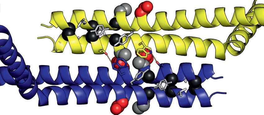
Figure 6. SMA missense mutations at the anti-parallel interface of human YG-domain tetramer. (A) Superimposition of the human YG-domain263–281
glycine–zipper dimeric units (green, PDB ID: 4GLI) onto the anti-parallel tetrameric SpSMN36–119 structure (grey, this work). Residues implicated
in SMA are depicted as sticks. (B) Pulldown assay of MBP-Hs-YG-domain252–284 (wt and SMA mutants). Immobilized MBP-Hs-YG-domain252–284
constructs were incubated with in vitro translated [35 S]-labeled HsG8 (full-length). Eluates were analyzed by SDS-PAGE and Coomassie staining, and
autoradiography. The relative quantifications of [35 S]-labeled HsG8 from each IP are indicated below the respective lanes. Structural models were generated
using PyMOL Molecular Graphics System, Version 2.0 Schrödinger, LLC.
reason that residue H273 is part of an exposed surface re- dimension (Dmax ), and molecular weight. In addition, di-
quired for HsG8 binding. Thus, pathogenic missense muta- mensionless Kratky plots and pairwise distance distribu-
tions cause specific defects in SMN oligomerization, which tion functions [P(r)] derived from SAXS data, illustrated the
results in impaired binding of HsG8. Based on the critical flexibility and disordered properties of the whole SpSMN
role of G8 in the architecture of the SMN complex it is likely complex.
that this defect results in the loss of SMN complex integrity We collected datasets of SAXS coupled to size exclusion
and function. chromatography (SEC-SAXS) for SpSMN/SpG2N80 ,
SpSMN/SpG2N80 /SpG6/SpG7/SpG835–58 ,
SAXS analysis of SpSMN complex SpSMN36–119 /SpG2N80 and SpSMN36–119 S130D/
SpG2N80 (Figure 7A, see also Supplementary Table S3).
With the characterization of the SpSMN complex, insight Note that predicted unstructured regions of SpG2 (N80)
into its structural organization became feasible. We have and SpG8 (35–58) were deleted in these complexes (see
determined the structural basis of SMN oligomerization also Figure 1A). The SpSMN36–119 S130D/SpG2N80
via its YG-box and the serine motif, which showed anti- complex was used in our analyses as a standard for glob-
parallel multimerization of glycine–zipper dimeric units. As ular entities (Figure 7B, see also Supplementary Table
a consequence of this, the N-termini of SMN protrude S3). Compared to the globular standard (red, Figure 7B),
on either side of the central oligomeric core. Such an ar- both complexes with full length SpSMN (black and grey,
rangement would imply a high degree of disorder of the Figure 7B), exhibit dual behavior in the dimensionless
whole SMN complex and explain previously failed attempts Kratky plot. It shows a distinct maximum at the ex-
to solve its structure by X-ray crystallography or cryo- pected value for globular entities (66) (orange crosshair,
EM. We therefore set out to use small angle X-ray scatter- Figure 7B), and a significantly raised signal at higher
ing (SAXS) to generate additional data towards the goal angles (black and grey arrowheads, Figure 7B), which is
of building a holistic model of the SMN complex. SAXS explained by the flexible region of SpSMN. The complex
data provided various biophysical parameters of our com- SpSMN36–119 /SpG2N80 (with wild type YG-domain se-
plexes such as radius of gyration (Rg ), maximum particle quence, expected to form higher order oligomers) exhibited7220 Nucleic Acids Research, 2021, Vol. 49, No. 13
Downloaded from https://academic.oup.com/nar/article/49/13/7207/6179931 by guest on 17 September 2021
Figure 7. SAXS analysis and model of the SpSMN complex. (A) Small angle X-ray scattering curves of respective complexes at indicated concentrations
represented as [I(s) vs s]. The scattering data have been deposited to SASBDB under the following accession√codes: SASDKZ4, SASDK85, SASDK66,
and SASDKF5. (B) Dimensionless Kratky plots [(sRg)2 I(s)/I(0) versus sRg]. The expected maximum at ( 3, 1.104) for globular entities is indicated
by an orange crosshair. Deviation from globularity is indicated by arrowheads. (C) Normalized pairwise distance distribution functions represented as
[Normalized P(r) vs r]. Molecular weights, either calculated from I(0) or from Porod volume (Vp/1.66) are indicated in the inset. For full length SpSMN
complexes, * and ** represent shoulder and extended tail, respectively. (D) An integrative model of the SpSMN complex. The core of the SpSMN complex
is formed by antiparallel multimerization (indicated by alternating SMN C-terminus) of glycine–zipper YG-domain dimeric units. The flexible N-terminal
extensions of SpSMN (dotted lines) facilitate the capture of Sm proteins via the SpG2 subunit. The overall shape, flexibility, and oligomeric state of SpSMN
is influenced by the SpG8/SpG7/SpG6 sub-complex.
a shoulder (blue arrowhead, Figure 7B) typical for multi- ure 7B), suggesting that additional factors control the
domain proteins, but is highly compact in contrast to the conformation of SpSMN.
full length SpSMN complexes. In addition, compared to the Next, using the data from standard, we determined the
SpSMN36–119 /SpG2N80 complex (232 kDa) (blue, Figure molecular weight, radius of gyration (Rg ) and maximum
7C), the normalized P(r) functions for full length SpSMN particle dimension (Dmax ) for both full length SpSMN com-
complexes of comparable molecular weights (black and plexes at various concentrations (see Supplementary Ta-
grey, Figure 7C) showed asymmetric curves with a shoulder ble S3). SpSMN/SpG2N80 was found to exist as a mix-
(*, Figure 7C) indicating multidomain architecture, and an ture of hexameric to octameric species. The oligomeric state
extended tail region (**, Figure 7C) indicating disorder. of SpSMN/SpG2N80 /SpG6/SpG7/SpG835–58 , however,
These observations demonstrate that the SpSMN complex is restricted to a tetrameric species at similar concentrations.
adopts highly extended conformations and behaves as a In addition to this, both the Rg and the Dmax are signifi-
multidomain unit with flexible linkers. Interestingly, sig- cantly reduced in the presence of SpG6/SpG7/SpG835–58 .
nificant compaction was observed for the whole complex These results show that the oligomeric state and flexibility
(SpSMN/SpG2N80 /SpG6/SpG7/SpG835–58 ) compared of the whole complex is influenced by the presence of the
to SpSMN/SpG2N80 (compare grey and black, Fig- SpG6/SpG7/SpG8 module.You can also read