Role of solute carriers in response to anticancer drugs
←
→
Page content transcription
If your browser does not render page correctly, please read the page content below
Li and Shu Molecular and Cellular Therapies 2014, 2:15
http://www.molcelltherapies.com/content/2/1/15

 REVIEW                                                                                                                                       Open Access

Role of solute carriers in response to anticancer
drugs
Qing Li1,2 and Yan Shu1*

  Abstract
  Membrane transporters play critical roles in moving a variety of anticancer drugs across cancer cell membrane, thereby
  determining chemotherapy efficacy and/or toxicity. The retention of anticancer drugs in cancer cells is the result of net
  function of efflux and influx transporters. The ATP-binding cassette (ABC) transporters are mainly the efflux transporters
  expressing at cancer cells, conferring the chemo-resistance in various malignant tumors, which has been well documented
  over the past decades. However, the function of influx transporters, in particular the solute carriers (SLC) in cancer cells, has
  only been recently well recognized to have significant impact on cancer therapy. The SLC transporters not only directly
  bring anticancer agents into cancer cells but also serve as the uptake mediators of essential nutrients for tumor growth
  and survival. In this review, we concentrate on the interaction of SLC transporters with anticancer drugs and nutrients,
  and their impact on chemo-sensitivity or -resistance of cancer cells. The differential expression patterns of SLC transporters
  between normal and tumor tissues may be well utilized to achieve specific delivery of chemotherapeutic agents.
  Keywords: Anticancer drugs, Solute carriers (SLC), Chemosensitivity, Chemoresistance

Review                                                                                 As reviewed elsewhere, the ABC transporters, which
Membrane transporters play critical roles in moving a                               are typical efflux drug transporters using energy from
myriad of endogenous and exogenous substances across                                the hydrolysis of ATP as the driving force to move
cellular and organelle membranes, including the major                               substrates against the electrochemical gradients outward
nutrient metabolites sugars, digested peptides, amino                               or into intracellular organelles. The 49 ABC transporters
acids and nucleosides, rare elements such as hormones                               can be classified into 7 subfamilies from A to G subfamily
and neurotransmitters, and xenobiotics. These transporters                          [2-5]. In the past two decades, accumulating evidence
are encoded by numerous gene families, accounting for ap-                           has shown that enhanced expression of several ABC
proximately 4% of genes in human genome [1]. Clinically                             transporter genes is associated with reduced cellular
used drugs usually have similar physiochemical structures                           accumulation of anticancer drugs and acquired multi-
with certain endogenous substrates. In the last two de-                             drug resistance in many human cancer cells. Those
cades, a variety of membrane transporters have been                                 ABC transporters of importance in cancer therapy have
recognized as drug transporters. These drug transporters                            been well documented for ABCB1 (or MDR1), ABCC1 (or
are of essential importance in determining drug pharmaco-                           MRP1), ABCC2 (or MRP2), and ABCG1 (or BCRP) [6-8].
kinetics, efficacy and adverse effects. Based on the driving                        By contrast, the impact of the solute carriers (SLC), which
force, drug transporters are characterized by two major                             are usually influx or bi-directional transporters, on cancer
superfamilies, the solute carriers (SLC) and the ATP-                               therapy has not been extensively characterized. In this re-
binding cassette (ABC) transporters which can each be                               view, we provide updates on the documented interaction
functionally classified into influx and efflux transporters                         of SLC transporters with anti-cancer drugs.
according to the direction of movement of substrates                                   More than 400 SLC transporter genes have been identi-
(Figure 1).                                                                         fied and grouped into 55 families, including ion coupled
                                                                                    transporters, exchangers and passive transporters located
                                                                                    at the plasma membrane or in intracellular organelles
* Correspondence: yshu@rx.umaryland.edu
1
 Department of Pharmaceutical Sciences, School of Pharmacy, University of           (e.g. mitochondrial or vesicular transporters) [9]. Unlike
Maryland at Baltimore, Baltimore, Maryland, USA                                     ABC transporters which driving energy is provided by
Full list of author information is available at the end of the article

                                       © 2014 Li and Shu; licensee BioMed Central Ltd. This is an Open Access article distributed under the terms of the Creative
                                       Commons Attribution License (http://creativecommons.org/licenses/by/4.0), which permits unrestricted use, distribution, and
                                       reproduction in any medium, provided the original work is properly credited. The Creative Commons Public Domain
                                       Dedication waiver (http://creativecommons.org/publicdomain/zero/1.0/) applies to the data made available in this article,
                                       unless otherwise stated.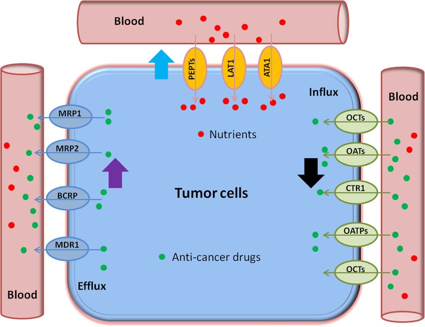
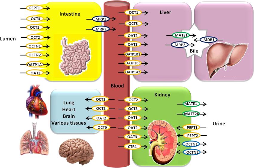
Li and Shu Molecular and Cellular Therapies 2014, 2:15                                                                                           Page 2 of 14
http://www.molcelltherapies.com/content/2/1/15

 Figure 1 Schematic model of the transporters in major organs responsible for drug disposition. The SLC members reviewed here may play an
 important role in determining the pharmacokinetics of anti-cancer drugs. They may be involved in drug absorption at the intestine, drug uptake into the
 liver for metabolism, drug elimination in the kidney, and drug distribution in a variety of additional tissues such as heart, lung, and brain. To facilitate
 understanding of drug transport processes in the organs, important efflux ABC transporters, which are reviewed elsewhere, are also depicted. Influx SLC
 transporters are shown as yellow open ovals and efflux SLC transporters as green open ovals. Some SLC transporters which mediate bi-directional
 transport are depicted by blue open ovals. ABC transporters are shown in black ovals. Black solid arrows indicate the direction of drug transport.

hydrolysis of ATP, SLC proteins work either by facilitating                         To improve outcomes of cancer therapy, it is necessary
passive diffusion along the concentration gradient of the                        to fully characterize the function of SLC drug transporters
substrate or by co-transport and counter-transport against                       in the organs critical to the disposition of anti-cancer
the concentration gradient of another solute. The super-                         drugs, such as intestine, liver and kidney. Moreover, by
family of SLCs is responsible for mediating the transport                        understanding the altered expression of SLC transporters
of a wide spectrum of substrates, including different nutri-                     in various cancer cells, we may develop novel therapeutic
ents as well as drugs [10].                                                      strategies to treat cancers. For instance, transporter-
   The SLC transporters expressed in the small intestine,                        targeted chemotherapy may be achieved via down-
the liver, and the kidney may be of particular importance                        regulation of certain essential transporters for cancer
for the disposition of cancer drugs. Interindividual vari-                       cell survival. The major subfamilies of SLC members,
ation in the activities of these transporters may cause                          which are, to different extents, explored in their associ-
altered pharmacokinetic profiles of anticancer drugs, sub-                       ation with cancer therapy, include the following: folate
sequently leading to variability in the pharmacodynamic                          transporters (SLC19A1 and SLC 46A1), which are par-
effects. The SLC transporters expressed in cancer cells                          ticularly important for antifolate chemotherapy of cancer
play an important role in cellular uptake of cancer drugs,                       and reviewed elsewhere [11-13]; organic cation trans-
which may be a determinant step toward anti-cancer effi-                         porters (OCT) (SLC22A1-3); organic anion transporters
cacy. Indeed, cancer cells are more likely to show substan-                      (OAT) (SLC22A6-8); organic cation/carnitine transporters
tially different expression profiles of SLC transporters as                      (SLC22A4-5); organic anion transporters polypeptides
compared to those of normal cells. Moreover, by mediating                        (OATPs) (SLCO); copper transporters (SLC31A); multi-
the transport of essential nutrient molecules and modulat-                       drug and toxin extrusion proteins (MATEs) (SLC47A),
ing the electrochemical gradient across the membranes,                           which intriguingly function as efflux transporters in cer-
SLC proteins can function to modify the efficiency of drug                       tain tissues; oligopeptide transporters (SLC15A1/2); and
diffusion into cells or alter cell survival pathways, conse-                     amino acid transporters (SLC7A and SLC3A) (Figure 3).
quently influencing chemotherapeutic efficacy (sensitivity
or resistance). In certain cases, cancer cells may possess                       Organic cation transporters (OCT)
enhanced expression of SLC transporters for certain nutri-                       Organic cation transporters consist of three isoforms
tional requirements and take a growth advantage over nor-                        (OCT1/SLC22A1, OCT2/SLC22A2, OCT3/SLC22A3),
mal cells when nutrients become restricted (Figure 2).                           which mediate the transport of various organic cations,
Li and Shu Molecular and Cellular Therapies 2014, 2:15                                                                                   Page 3 of 14
http://www.molcelltherapies.com/content/2/1/15

 Figure 2 Schematic model of development of anticancer drug resistance due to altered expression of transporters. SLC transporters
 (OCTs, OATs, CTR, and OATPs in green open ovals) may take up anticancer drugs into the cancer cells, while some ABC transporters (MDR1,
 MRP1, MRP3, and BCRP in blue open ovals) may extrude them. In addition, certain SLC transporters (PEPTs, LATs, and ATA1 in yellow open ovals)
 mediate the uptake of nutrients (e.g., amino acids and peptides) into cancer cells for their survival. The decreased expression of SLC transporters
 (black bold arrow) responsible for drug uptake or/and the increased expression of ABC transporter (purple bold arrow) for drug efflux make the
 cancer cells more resistant to anticancer drugs. The increased expression of SLC transporters (blue bold arrow) responsible for nutrient uptake
 may also cause more resistant to anticancer drugs as the cancer cells have an advantage of survival from limited nutrient supply.

weak bases, and some neural compounds across plasma                          OCT1
membranes [14,15]. These transporters are facilitative                       Human OCT1 (SLC22A1) is predominantly expressed in
diffusion systems, and the driving force is provided by                      the liver where it is located in the sinusoidal membrane
the electrochemical gradient of the transported com-                         of the hepatocytes [17]. In rat, mouse, and rabbit, be-
pounds [16].                                                                 sides high expression in the liver, strong expression was

 Figure 3 Classified subfamilies of SLC transporters involved in response to anticancer drugs. Only the SLC members and the anticancer
 drugs reviewed in this article are included.
Li and Shu Molecular and Cellular Therapies 2014, 2:15                                                           Page 4 of 14
http://www.molcelltherapies.com/content/2/1/15

also detected in the kidney and it was localized at the         the organic cation 3H-1-methyl-4-pyridinium iodide into
basolateral membranes of S1 and S2 segments of proximal         Chinese hamster ovary cells that are overexpressed with
tubules [18]. Human OCT1 is expressed to a much lesser          human OCT1, with Ki values of 1.7, 85, and 50 μM, re-
extent in various additional organs including kidney, small     spectively [24]. The OCT1-transfected cells also exhibited
intestine, lung, heart, skeletal muscle, brain, placenta,       significantly more susceptible to the cytotoxicity of irino-
mammary gland, adrenal gland, eye, adipose tissue, and          tecan and paclitaxel when compared with mock cells, sug-
immune cells [18-23], and differentially expresses in vari-     gesting that OCT1 may contribute to accumulation of the
ous tumors [18,24,25].                                          selected antineoplastic drugs in cancer cells. The expres-
   OCT1 has been showed to be expressed in colon can-           sion of OCTs in various tumors should be further investi-
cer and polyps [26]. Specifically, it has been reported         gated as it may serve as a biomarker for selecting specific
that OCT1 mRNA level is increased in human colon                antineoplastic agents to tailor cancer therapy for individ-
cancer cell lines and patient-derived colorectal tumor          ual patients.
samples [27]. Platinum-based drugs, including cisplatin
and oxaliplatin, are efficient to induce DNA damage by          OCT2
forming DNA adducts and subsequently cause apoptosis            The cloning of Slc22a2 encoding Oct2 from rat was re-
in colon cancer cells. Cisplatin has shown a high specifi-      ported in 1996 [37]. OCT2 orthologs were later cloned
city and affinity for OCT1 with an IC50 (half maximal in-       from other species including humans [14,38]. Human
hibitory concentration) value of 8.1 uM. However, Zhang         OCT2 (SLC22A2) is mainly expressed at the basolateral
et al. reported that human OCT1 markedly increased oxa-         membrane of renal proximal tubules in the kidney [14],
liplatin, but not cisplatin, accumulation and cytotoxicity in   and its expression is also detectable in small intestine,
the stable cells expressing OCT1 transporter and oxalipla-      lung, placenta, thymus neurons, choroid plexus, inner
tin was an excellent substrate of OCT1 [28]. The cytotox-       ear, and respiratory mucosa [39,14].
icity by oxaliplatin was also much greater than that of            OCT2 mediates the first step of many cationic drugs
cisplatin in the colon cancer cells. By using an OCT in-        in renal excretion via their uptake at the basolateral
hibitor, cimetidine, they demonstrated that the function of     membrane of proximal tubule cells. As for anticancer
OCTs such as OCT1 and OCT2 is a major determinant of            drugs, cisplatin has been demonstrated to be a substrate
the anticancer activity of oxaliplatin, which may be causa-     of OCT2, evidenced by significantly higher uptake of cis-
tive of its specificity of anti-colon cancer. Picoplatin, a     platin in OCT2 overexpressing cells compared with that
third-generation platinum agent, is efficacious against lung    in mock cells [39]. Abundant evidence indicates that cis-
cancers that become refractory to other platinum-based          platin accumulates in the proximal tubules and causes
treatment. More et al. reported that the tumor size of          nephrotoxicity. Human OCT2 transporter has been re-
OCT1-expressing xenografts in mice was significantly            ported to mediate the transport of cisplatin across proximal
reduced by picoplatin treatment as compared to control          tubules membrane from circulation [40], and is critical to
xenografts, which suggested that OCT1 could enhance             the nephrotoxicity of cisplatin. A non-synonymous single-
the antitumor efficacy of picoplatin as well [25].              nucleotide polymorphism (SNP, rs316019) in the OCT2
   OCT1 has also been demonstrated to be significantly          gene has been demonstrated to be associated with de-
expressed in chronic myeloid leukemia (CML) cell lines          creased nephrotoxicity of cisplatin in patients [40].
and primary CML cells [29]. Imatinib, a potent tyrosine         Moreover, the mice lacking of Oct2 function have reduced
kinase inhibitor, is used to treat CML. OCT1 has been           urinary excretion of cisplatin and less cisplatin-induced
reported to mediate the transport of imatinib mesylate          nephrotocixity [40]. In addition, the wild-type mice treated
across cellular membrane. Considerable evidence has             with cisplatin plus the OCT inhibitor cimetidine have a re-
revealed that OCT1 expression level in leukemic cells is        duced nephrotoxicity similar to that seen in Oct1/2-/-
associated with the therapeutic outcome in CML [30-35].         double knockout mice treated with cisplatin alone [41]. It
It has been reported that OCT1 activity may be a key de-        seems that a reduced uptake by transporters such as OCT2
terminant of molecular response to imatinib [36]. By using      at the basolateral membrane in the proximal tubules is
a potent inhibitor such as prazosin, the activity of OCT1-      likely to protect kidney from cisplatin-induced nephrotox-
mediated imatinib transport can be determined in vitro in       icity. Oxaliplatin has also been considered as a substrate of
the isolated peripheral blood leukocytes from CML pa-           OCT2. However, it does not induce severe nephrotoxic ef-
tients. This measurement of OCT1 function may be useful         fects similar to cisplatin [42]. This may be explained by
to individualize dosage regimens for patients with CML in       another protein transporting organic cations, the kidney
order to obtain an optimal outcome in the long-term             isoform of MATE2 (MATE2K). MATE2K (SLC47A2) is
imatinib-treated patients [36].                                 located at the apical membrane of proximal tubules and
   In addition, the antineoplastic agents irinotecan, mitox-    can specifically remove oxaliplatin, not cisplatin, from the
antrone, and paclitaxel were found to inhibit the uptake of     proximal tubules [43-45]. In addition, the third-generation
Li and Shu Molecular and Cellular Therapies 2014, 2:15                                                              Page 5 of 14
http://www.molcelltherapies.com/content/2/1/15

platinum drug, picoplatin, has also been shown to signifi-       diuretics, HMG CoA reductase inhibitors, β-lactam antibi-
cantly enhance cytotoxicity in the presence of OCT2 ex-          otics, antiviral drugs, uricosuric drugs, and antineoplastic
pression with increased DNA adduct formation [25].               drugs. In this review, we focus on OAT1-3, whose role in
These studies underscore the importance of OCT2 func-            drug disposition and cancer therapy has been investigated
tion in renal disposition and possible tumor accumulation        relatively well.
of platinum compounds in platinum-based chemotherapy.
                                                                 OAT1
OCT3                                                             The organic anion transporter 1, OAT1 (SLC22A6), was
The SLC22A3 gene encoding OCT3 was independently                 the first OAT member which was cloned from rat [54],
cloned as the extraneuronal monoamine transporter by             mouse [55], and later human [56,57]. Human OAT1 was
different groups [46,47]. Unlike human OCT1 and OCT2             localized to the basolateral membrane of renal proximal
with high expression in limited tissues, human OCT3 is           tubule cells by immunocytochemistry and was found
ubiquitously expressed [15,18,48] with relatively high           along the whole proximal tubule [57-59]. This expres-
expression in the heart, skeletal muscle, brain, liver, and      sion pattern is consistent with the role of OAT1 in the
kidney [14,47,49]. Specifically, human OCT3 is located           uptake of anionic drugs from the circulation into prox-
to the basolateral membrane of placental epithelium              imal tubule cells. OAT1 has also been detected in the
[50], the sinusoidal membrane of hepatocytes [17], and           brain, specifically in the choroid plexus [60].
the luminal membrane of bronchial epithelial cells [18].            Human OAT1 has been reported to be responsible for
  Human OCT3 has differential expression between nor-            renal tubular secretion of methotrexate which is an antifo-
mal and cancerous tissues. An upregulated OCT3 expres-           late used in the treatment of malignancies [61]. Metho-
sion was observed in the cancerous tissues of colon              trexate is also transported by rat and mouse Oat1 with a
rectum and stomach as compared to the normal tissues,            moderate to high affinity [62,63]. Clinical interaction be-
however, significantly decreased expression was found            tween methotrexate and other drugs such as loxoprofen
in several other cancerous tissues, including uterus,            has been well documented. Interestingly, loxoprofen and
breast, ovary and lung cancers [51]. It has been reported        its trans-OH metabolite, the major active metabolite,
that oxaliplatin is a substrate of OCT3, in contrast to          markedly inhibit the transport of methotrexate by OAT1.
carboplatin, which are not substrate of OCT3 [43,44].            In clinical chemotherapy, high doses of methotrexate are
OCT3 moderately transports oxaliplatin and can medi-             frequently used, and methotrexate-induced severe nephro-
ate oxaliplatin-induced cytotoxicity in cell cultures [51].      toxicity has been well documented. OAT-mediated uptake
Yokoo et al. reported that the effect of oxaliplatin             of methotrexate into tubule cells may significantly contrib-
against colorectal cancer was superior to that of cisplatin      ute to its nephrotoxicity. Reports are also available suggest-
because OCT3 was highly expressed when compared with             ing that the cytotoxicity and anticancer effects of several
other organic cation transporters [51]. Recent studies have      other antineoplastic agents, including 6-mercaptopurine,
showed that other cytostatics, such as melphalan, irinote-       azathioprin, cisplatin, imatinib, cytarabine, vinblastine, vin-
can, and vincristine [52], may interact with OCT3 as well.       cristine, hydrocortisone, and mitoxantrone, may be associ-
Irinotecan, which is well used to treat colon carcinoma,         ated with OAT1 expression [64,65].
exhibits a high affinity for OCT3, and its increased cyto-
toxicity in kidney carcinoma cell lines was associated with      OAT2
OCT3 expression. These results suggested that human              Liver and kidney have the highest expression level of
OCT3 expression may be further evaluated as a potential          OAT2, and relatively low expression level of OAT2 was
biomarker for the efficacy of cancer chemotherapy.               found in pancreas, small intestine, lung, brain, spinal cord,
                                                                 and heart. [66-69]. However, the expression of OAT2
Organic anion transporters (OAT)                                 mRNA is much lower than those of OAT1 and OAT3
Organic anion transporters (OATs), also members of the           mRNAs In human kidneys. [67]. OAT2 is located in the
solute carrier family 22, play a key role in renal excretion     sinusoidal membrane of hepatocytes [70] and the basolat-
of water-soluble, negatively charged organic compounds           eral membrane of proximal tubule cells [71].
[53]. Renal OATs are expressed in the baselateral mem-             As a major hepatic transporter, OAT2 plays an important
branes of proximal tubular cells, the major site for secreting   role in hepatic drug uptake, which may be a determinant of
organic anions in the kidney. OATs are also expressed in         metabolism for various anticancer drugs. 5-fluorouracil,
other tissues, including liver, placenta, nasal epithelium,      which is used in colorectal and pancreatic cancer therapy,
and blood-brain barrier. In humans, OAT subfamily con-           is a verified substrate of OAT2 [72]. In the same study,
sists of OAT1–4, OAT7, OAT10, and URAT1 (SLC22A16).              Kobayashi et al. also reported that paclitaxel was an OAT2
Several classes of drugs interact with human OAT1-3, in-         substrate [72]. Tanino et al. later showed a concentration-
cluding ACE inhibitors, angiotensin II receptor antagonists,     dependent uptake of paclitaxel in rat hepatocytes [73].
Li and Shu Molecular and Cellular Therapies 2014, 2:15 Page 6 of 14
http://www.molcelltherapies.com/content/2/1/15
However, indomethacin, a representative inhibitor of Oat2, neuronal accumulation and treatment-limiting neurotox-
did not significantly inhibited the uptake of paclitaxel, icity [83].
which suggested that Oat2 was not a significant transporter OCTN2 has been recently suggested by Hu et al. as an
for the hepatic uptake of paclitaxel, at least in rat hepato- efficient and capable transporter for imatinib at a con-
cytes [73]. centration (e.g., 0.2 μmol/L) [84] that is readily achiev-
able in human patients [85]. Furthermore, among the
OAT3 patients of gastrointestinal stromal tumors (GIST) who
OAT3 (SLC22A8) has been cloned from human, monkey, received imatinib treatment, the time to progression was
pig, rabbit, rat, and mouse. In these species, OAT3 mRNA recently shown to be significantly improved in the carriers
is detected in a variety of tissues or organs including of the C allele of an OCTN1 polymorphism (rs1050152)
kidney, liver, brain, skeletal muscle, and adrenal glands as well as in carriers of minor alleles of two OCTN2 poly-
[65,74]. In all tested species, the kidney has the highest morphisms (rs2631367 and rs2631372), suggesting the
mRNA expression of OAT3 among all organs [21,74]. activities of OCTN1 and OCTN2 as a predictor of chemo-
The transporter protein is located at the basolateral mem- therapeutic efficacy of imatinib [86].
brane of proximal tubule cells in the kidney [38,75]. Doxorubicin is a widely used anticancer drug for
Methotrexate (MTX) has a 30-fold stronger affinity to hematological malignancies, particularly for acute lympho-
OAT3 with the Km value of 17.2 μM as compared with cytic leukemia (ALL) and acute myeloid leukemia (AML).
OAT1 (Km: 724 μM) [61]. Thus, human OAT3 could be OCT6 has been found by Okabe et al. to mediate the
mainly responsible for the renal MTX excretion and uptake of doxorubicin in leukemic cells and the stable
MTX-induced nephrotoxicity during cancer therapy. leukemic Jurkat cells with over-expression of OCT6 gene
Among interactions between anticancer drugs and OAT3, became more sensitive to doxorubicin. In the same report,
6-thioguanine was found to inhibit rat Oat3 (IC50 172 OCT6 expression was detected in the primary blood cells
μM), 6-mercaptopurine could be transported by both rat collected from the patients with acute leukemia [81]. Bleo-
and mouse Oat3 (Km: 50.5 μM and 4 μM, respectively), mycin is widely used in combination with other antineo-
and 5-fluorouracil was substrate of mouse Oat3 (Km: plastic agents to effectively treat lymphomas, testicular
0.054 μM) [76,64]. In addition, rat Oat3 (Km: 21.9 μM) carcinomas, and squamous cell carcinomas of the cervix,
and human OAT3 (Km: 56.5 μM) translocated topotecan head, and neck. OCT6 has also been found to be involved
with a moderate affinity [77]. in the uptake of bleomycin-A5 as well as polyamines.
Human testicular cancer cells overexpressing OCT6 were
Organic cation/carnitine transporters (OCTN1-2 and OCT6) extremely sensitive to bleomycin-A5 and the siRNA tar-
Organic cation/carnitine transporters comprise the trans- geted OCT6 induced significant resistance to bleomycin-
porters OCTN1, OCTN2 and OCT6, encoded by the A5-dependent genotoxicity. The knowledge of OCT6
genes SLC22A4, SLC22A5 and SLC22A16, respectively function remains relatively limited. These recent findings
[18]. The three transporter genes have a broad expression suggest that characterizing OCT6 expression in human
pattern in normal tissues. Some human tumor-derived cell cancer cells may be valuable for us to improve current
lines, including lung carcinoma A549, colorectal carcin- chemotherapy and explore novel cancer therapeutic strat-
oma SW480, and cervix carcinoma Hela S3 cells, have egies [87].
been detected to express high levels of OCTN1 and
OCTN2 transcripts [78,79]. Human OCT6 expression has Organic anion transporting polypeptides (OATP, SLCO)
been determined from testis, hematopoietic cells, and There are 11 known human OATPs which have been
leukemia lines HL-60 and MOLT4 [80-82]. divided into six subfamilies based on their amino acid
Human OCTN1 mediates uniport of organic cations sequence similarities [88-91]. OATPs expression can be
such as the antioxidant ergothioneine as well as H+/or- either tissue-specific or ubiquitous in multiple tissues
ganic cation antiport, whereas human OCTN2 and OCT6 throughout the body. Among the OATP family mem-
are Na+/carnitine cotransporters as well as organic cation bers, OATP1A2, OATP1B1, OATP1B3 and OATP2B1
uniporters [18]. Recently, by conducting uptake studies in have broad substrate specificity and accept a number of
the HEK293 cells over-expressing rat Octn1, rat Octn2, therapeutic agents. Latest studies have demonstrated that
human OCTN1, and human OCTN2, Jong et al. reported OATP expression is differently regulated in certain cancer
that the transport of oxaliplatin across cellular membrane tissues as compared to normal tissues. The OATP1 sub-
could be mediated by OCTN1 and OCTN2. Both the up- family is the most characterized among the OATPs. It
take and cytotoxicity of oxaliplatin were well inhibited by should be pointed out that very different subfamily mem-
ergothioneine and L-carnitine. Importantly, they provided bers of SLCO/Slco have been evolved between humans
evidence supporting that the transport of oxaliplatin me- and rodents. This is particularly true within the OATP1
diated by OCTN1 appeared to mainly contribute to its family as there are no clear orthologs between individualLi and Shu Molecular and Cellular Therapies 2014, 2:15 Page 7 of 14
http://www.molcelltherapies.com/content/2/1/15
human and mouse OATP1s. Humans have only one demonstrated as an OATIP1B3 substrate as OATP1B3
OATP1A transporter (OATP1A2), but mice have at least transports methotrexate in a saturable and dose-dependent
4 (Oatp1a1, Oatp1a4, Oatp1a5, and Oatp1a6) [89,92]. In manner. The introduction of the OATP1B3 gene into mam-
addition, while humans have 2 OATP1B transporters malian cells potentiates their sensitivity to methotrexate
(OATP1B1 and OATP1B3), mice only have Oatp1b2 [92]. [106]. In Xenopus laevis oocytes injected with OATP1B3
In this section, we focus on the interaction of OATP1 with cRNA, the uptake of docetaxel and paclitaxel were 2.2-fold
anticancer drugs. and 3.3-fold higher, respectively, than that of water-injected
control oocytes [107,108], suggesting both drugs as
OATP1B1 OATP1B3 substrates. However, in screening interaction
OATP1B1 (SLCO1B1) is highly expressed in human liver between OATP1B3 and a variety of compounds, although
[93-95]. Further, the transporter gene profiling assay in docetaxel, paclitaxel, and three other antineoplastic
NCI-60 cancer cell lines showed relatively high expres- agents, actinomycin D, mitoxantrone, and SN-38 exhibited
sion of OATP1B1 in the cells derived from lung cancers, potent inhibitory effects on OATP1B3-mediated transport
such as A549 and EKVX cells, and colon cancers, such of CDCA-NBD (chenodeoxycholyl-(Nepsilon-NBD)-lysine,
as HCT-15 and KM12 cells [96]. In addition, the mRNA a fluorescent substrate of OATP1B3)) [109], only SN-38
level of OATP1B1 was found to be higher in colon was further determined as a novel substrate for OATP1B3
polyps and cancer tissues as compared with normal [110]. Overall, OATP1B3 may be a clinically relevant
colon tissues [26]. transporter responsible for hepatic disposition and the
OATP1B1 activity may be important in hepatic dispos- chemotherapeutic response in cancer tissues of certain
ition of anticancer drugs. Nozawa et al. firstly reported anticancer drugs.
that OATP1B1 is responsible for hepatic uptake of SN-38,
the major active metabolite of irinotecan and that the gen- OATP1A2
etic polymorphisms of OATP1B1 may contribute to the Among normal tissues, OATP1A2 (SLCO1A2) is expressed
well-known individual variation in the disposition of iri- in the intestinal epithelium [111], the renal epithelium,
notecan [97]. Later, in a case report, severe irinotecan and highly in brain capillary endothelial cells. [112]
toxicity was observed in a 66-year-old Japanese with Altered OATP1A2 expression has been detected in
dysfunctional alleles of OATP1B1 [98]. The toxicity is glioma, colon polyps and cancers, and breast cancers
possibly due to a decreased uptake of active metabolite as compared to normal tissues. In addition, OATP1A2
(SN-38) of irinotecan into the liver and subsequently mRNA were found both in bone metastases from pri-
reduced hepatic metabolism. It has been demonstrated mary kidney cancer and in the malignant osteosarcoma
that in OATP1B1 transgenic mice, the hepatic accumu- cell lines HOS and MG-63 [113].
lation of MTX was significantly higher (approximately Paclitaxel has been characterized as an OATP1A2 sub-
2-fold) compared with wild-type mice after MTX treat- strate. When compared to wild-type mice, the transgenic
ment, resulting in 2- to 4-fold higher liver-plasma ratios mice overexpressing human OATP1A2 had a remarkable
of MTX. The findings suggest a marked and possibly increased hepatic uptake of paclitaxel [114]. Further-
rate-limiting role for human OATP1B1 in MTX elimin- more, the systemic exposure of paclitaxel after an intra-
ation in vivo [99]. In addition, polymorphisms in the venous dose (10 mg/kg) was increased by greater than
OATP1B1 gene were found to be associated with the 2-fold in Slco1a/1b (-/-) mice compared with wild-type
disposition and therapeutic outcomes of flavopiridol and mice, whereas its hepatic uptake was reduced by about
atrasentan [100,101]. 2-fold [115]. There is in vivo evidence supporting
methotrexate (MTX) as a substrate of OATP1A2 as
OATP1B3 well. When compared with wild-type mice, Slco1a/1b
OATP1B3 (SLCO1B3), normally and specifically expressed (-/-) mice exhibited a 3.4-fold increase in plasma and
in liver, has been found in different cancer tissues [102]. 30-fold decrease in hepatic levels of MTX after received a
Specifically, OATP1B3 is upregulated in gastrointestinal high dose of 500 mg/kg, suggesting an overall role of
cancer cell lines, pancreatic cancer cell lines, and gallblad- OATP function in MTX disposition [115]. Moreover, hu-
der cancer cell lines. OATP1B3 expression was dramatic- manized OATP1A2 transgenic mice showed significant
ally higher in colorectal adenocarcinoma tissues and in rescue of the increased plasma levels and decreased liver
prostate cancer tissues as compared with their corre- and small intestinal accumulation of MTX that were ob-
sponding normal tissues [103-105]. served in Slco1a/1b (-/-) mice [114], confirmed MTX as
Hu et al. found a significantly higher uptake rate for an OATP1A2 substrate. OATP1A2 has been extensively
[3H] imatinib in HEK293 cells transfected with human detected in primary and metastatic liver cancers. As
OATP1B3 gene and in Xenopus laevis oocytes injected the transporter could be a critical mediator of drug up-
with OATP1B3 cRNA [84]. Methotrexate has also been take in the liver, OATP1A2 may be further exploitedLi and Shu Molecular and Cellular Therapies 2014, 2:15 Page 8 of 14
http://www.molcelltherapies.com/content/2/1/15
for the delivery of chemotherapeutic agents to treat liver organisms, including prokaryotes, plants and mammals,
cancers [116]. and they are responsible for the transport of various
endo-/exogenous substrates. In humans, MATE1 is highly
Copper transporters (CTR, SLC31A) expressed in the kidney and liver, which is localized at the
Copper is a vital mineral for humans as demonstrated by apical membrane of proximal tubules and hepatocytes.
serious health concerns associated with its deficiency or Human MATE1 is also detectable in other tissues, in-
excess accumulation. Copper transporter 1 (CTR1) is the cluding adrenal gland, skeletal muscle, testis, and first
major high-affinity copper uptake transporter in mammals trimester placenta [129,130]. MATE2 and MATE2-K are
[117]. CTR1, encoded by the SLC31A1 gene, is localized mainly expressed in the kidney [131].
in the plasma membrane and intracellular membranes Otsuka et al. reported that MATE1 mediates the ex-
[118,119]. CTR1 protein is ubiquitously expressed in cretion of cisplatin in the kidney [128]. It has been lately
many tissues such as hepatocytes, α cells of the pancreatic reported by Nakamura et al. that cisplatine significantly
islets, enteroendocrine cells of the gastric mucosa and increased the levels of plasma creatinine and blood urea
bronchioles, c cells of the thyroid, and a subsets of cells nitrogen (BUN), two major biomarkers for renal injury,
in the anterior pituitary [120]. It is also observed to be in Mate1-deficient (Mate1-/-) mice when compared with
expressed in both normal colonic epithelium and colon wild-type mice [132]. Further, the levels of creatinine
carcinomas [120]. In addition, strong expression has and BUN in the mice treated with cisplatin were signifi-
been found in a few cases of carcinoid tumors, Ewing’s cantly enhanced by pyrimethamine, a potent MATE in-
sarcoma, undifferentiated carcinomas, and enteroen- hibitor [132]. Li et al. later reported that much severer
docrine cells [120]. nephrotoxicity of cisplatin was observed in Mate1-/- mice
Abundant evidence has been consistently showed that than in wild-type mice [133]. Thus, reduced function of
CTR1 is an important determinant of cellular accumula- MATEs, which serves as efflux transporters for cisplatin
tion and toxicity of platinum-based anti-cancer drugs, elimination in the kidney, may be responsible for
such as cisplatin [121-123]. Stable overexpression of hu- cisplatin-induced nephrotoxicity. In addition to cisplatin
man CTR1 significantly increased the uptake of cisplatin, as a substrate of MATE1, oxaliplatin was reported to be
carboplatin and oxaliplatin in human small cell lung transported by rat Mate1 as well as human MATE1 and
cancer cell lines and rendered the cells more sensitive MATE2-K [134].
to the cytoxicity of these compounds [124,125]. In con-
trast, small interfering RNA (siRNA)-mediated knock- Oligopeptide transporters (PEPT1/2, SLC15A1/2)
down of CTR1 was shown to be able to reduce both The oligopeptide transporters (PEPTs, SLC15A) serve as
cellular platinum accumulation and cytotoxicity of cis- integral membrane proteins for mediating the cellular
platin in human embryonic kidney 293 cells [126]. In uptake of di- and tripeptides into cells. Their driving
addition, mouse embryonic fibroblasts (MEF) of heterozy- force comes from an inwardly directed H+ gradient
gous (+/-) and homozygous (-/-) Ctr1 gene deletion accu- across the membrane [135]. PEPT1 and PEPT2 are two
mulated 35% and 70% less platinum and exhibited 4- and major PEPTs that have been cloned. Physiologically,
8-fold more resistant to cisplatin cytotoxicity after a 2 PEPTs are localized at brush-border membranes of in-
hour cisplatin exposure, respectively, than the wild-type testinal and renal epithelial cells, and play important
MEF cells [121]. Taken together, these studies have pro- roles in protein absorption and the conservation of
vided strong evidence in support of CTR1 role in mediat- peptide-bound amino nitrogen. Peptide-like drugs with
ing the uptake of cisplatin into normal and tumor cells. structural similarities to di- and tripeptides are also
Further clinical studies are needed to clarify how import- transported by PEPTs [136]. PEPT1, a high-capacity
ant the transporter function is in determining the antican- but low affinity transporter, mainly expressed in the
cer efficacy and the toxic side effects of platinum-based small intestine, whereas PEPT2, a high-affinity but
treatment in patients. low-capacity transporter, broadly expressed in a variety
of tissues [135].
Multidrug and toxin extrusion (MATE) proteins (SLC47A) PepT1 and PepT2 were interestingly found to be
Multidrug and toxin extrusion (MATE) proteins were expressed in fibroblast-derived tumor cells but not in
first found in bacteria in 1998 [127]. In 2005, the human normal fibroblasts [137]. High levels of PEPT 1 protein
ortholog, MATE1, was first cloned and characterized as were also detected in two human pancreatic cancer cell
an efflux transporter mediating the excretion of organic lines, AsPc-1 and Capan-2 [138]. In addition, PEPT1
cations from the kidney [128]. MATE2 and MATE2-K mRNA was increased 2.3-fold in colon cancer tissues
were identified shortly thereafter. The driving force for as compared to normal tissues [139]. Many peptide-
MATE is provided by the oppositely directed proton mimetic agents are the substrates of PEPTs. The higher
gradient [128]. MATE proteins exist in various living expression of PEPTs in cancer cells may serve as the basisLi and Shu Molecular and Cellular Therapies 2014, 2:15 Page 9 of 14
http://www.molcelltherapies.com/content/2/1/15
of a novel strategy for specific delivery of oligopeptide- interestingly shown to be transported by the bidirectional
mimetic anticancer drugs into tumors. transporters SLC1A5 and SLC7A5 (LAT1) [150]. Regula-
Bestatin, a potent aminopeptidase inhibitor and a tion of mTOR signaling pathway by modulating the activ-
known substrate of PEPT1, suppressed the growth of ity of these amino acid transporters could be an attractive
tumor (Hela cells) xenografts overexpressing PEPT1 by strategy to control tumor cell survival and progression.
4-week consecutive oral administration [140]. The high Amino acid transporter system A1 (ATA1), encoded by
expression of PEPTs in various cancer tissues has been SLC38A2, mediates the transport of most small neutral
attributed to increased nutrients demand by fast tumor amino acids, including alanine, serine, and glutamine. The
growth, and thus inhibition of the activity of PEPTs mRNA level of ATA1 was markedly induced in human
might be also a novel targeting strategy to delay or stop hepatoma cancer cell lines and in patient-derived hepato-
tumor growth. Mitsuoka et al. found that a newly syn- cellular cancer tissues as well. Enhanced expression of
thesized dipeptide, 4-(4-methoxyphenyl)-L-phenylalanyl ATA1 is also positively related to the progression of chol-
sarcosine which was an inhibitor of PEPT1, resulted in angiocarcinoma. Furthermore, silencing ATA1 mRNA ex-
nearly complete suppression of the xenograft growth of hu- pression decreased the viability of HepG2 cells, suggesting
man pancreatic cancer AsPC-1 cells that highly expressed that ATA1 is likely essential to tumor survival [151]. These
PEPT1 [141]. studies have indicated the prognostic significance of ATA1
Floxuridine, a clinically proven anticancer drug, is in cancer development and progression and provided
commonly used for the treatment of metastases from rationales to target ATA1 for cancer therapy.
colon carcinomas and hepatocellular carcinoma [142]. L-type amino acid transporter 1 (LAT1), encoded by
In order to improve its selectivity and to reduce undesir- SLC7A5, is responsible for the transport of large neutral,
able toxic effects, a series of prodrugs of floxuridine has aromatic or branched amino acids from extracellular
been developed [143,144]. These amino acid ester pro- fluids into the cells. Acivicin is an antineoplastic antibiotic
drugs have been shown to target the PEPT1 transporter that targets glutamine-dependent amidotransferases in the
[145]. MDCK cells stably transfected with the human biosynthesis of purines and pyrimidines [152]. The IC50
PEPT1 (MDCK/hPEPT1) demonstrated enhanced cell values for acivicin to inhibit the gabapentin (a LAT1 sub-
growth inhibition in the presence of these prodrugs [146]. strate) accumulation in the stable HEK-LAT1 cells ranged
This prodrug strategy to modify nucleoside drugs seems from 7.9 μM to 340 μM [153]. The experiments of trans-
to have great potential to improve their tumor selectivity stimulation and cell-proliferation have demonstrated that
and drug efficacy. acivicin is likely to be a substrate for LAT1, suggesting
that LAT1 may be targeted for acivicin delivery into tumor
Amino acid transporters cells [153]. Petel et al. have recently demonstrated that
The transporter-mediated entry of amino acids into cells LAT1 is functionally active in prostate cancer cells (PC-3).
is necessary for various cellular functions including pro- Hence, LAT1 transporter may be used as a target for
tein synthesis, energy metabolism, glutathione synthesis, improving the availability of poorly permeable but highly
and others. Moreover, amino acids transporters play im- potent anticancer drugs at least in prostate cancer cells
portant roles in conferring not only drug sensitivity by [154].
mediating the uptake of amino acid analog drugs), but
also drug resistance by promoting the uptake of essential Conclusions
amino acids for tumor growth and survival. There are 6 It is critical to target drugs to tumor cells in order to
major families of amino acid transporters in the solute improve the clinical efficacy and avoid the adverse ef-
carrier (SLC) gene superfamily (SLC1, SLC6, SLC7, SLC36, fects of anticancer drugs. The efficacy of chemotherapy
SLC38, and SLC43 families) and the orphan SLC16 mono- may be largely dependent on the relative activity of
carboxylate transporter which transports aromatic amino transporters in normal and cancer tissues. In addition
acids [147]. to already extensively investigated efflux transporters,
Accumulating evidence in the past two decades has multiple types of membrane influx transporters, in par-
indicated that amino acid availability controls cellular ticular the SLC superfamily members play very import-
physiology by altered gene expression levels and signal ant roles in conferring sensitivity and resistance to
transduction pathways in cancer cells. For example, the anticancer agents. These SLC transporters not only dir-
mammalian target of rapamycin (mTOR) is a serine/ ectly bring anticancer agents into cancer cells but also
threonine kinase that regulates fundamental biological serve as the uptake mediators of essential nutrients for
processes and plays critical roles in cell growth regulation tumor growth and survival. The differential expression
and tumorigenesis [148,149]. The activation of mTOR and patterns of SLC transporters between normal and tumor
subsequent regulation of mTORC1 is regulated by the up- tissues may be well utilized to achieve specific delivery of
take of amino acids such as L-glutamine which has been chemotherapeutic agents. The transporters may be alsoLi and Shu Molecular and Cellular Therapies 2014, 2:15 Page 10 of 14
http://www.molcelltherapies.com/content/2/1/15
directly targeted in development of anticancer drugs to in- 8. Ross DD, Nakanishi T: Impact of breast cancer resistance protein on
crease chemosensitivity, for example, via limiting nutrient cancer treatment outcomes. Methods Mol Biol 2010, 596:251–290.
9. He L, Vasiliou K, Nebert DW: Analysis and update of the human solute
supply to cancer cells and regulating their apoptosis and carrier (SLC) gene superfamily. Hum Genomics 2009, 3(2):195–206.
electrochemical gradients. The SLC transporters expressed 10. Rask-Andersen M, Masuram S, Fredriksson R, Schioth HB: Solute carriers as
in the intestine, liver and kidney are of particular import- drug targets: current use, clinical trials and prospective. Mol Aspects Med
2013, 34(2–3):702–710.
ance as their activity may be critical to systemic exposure 11. Matherly LH, Wilson MR, Hou Z: The Major Facilitative Folate Transporters
and disposition of various anticancer agents, serving as a SLC19A1 and SLC46A1: Biology and Role in Antifolate Chemotherapy of
common basis or determinant for drug-drug interaction, Cancer. Drug Metab Dispos 2014, 42(4):632–649.
12. Desmoulin SK, Hou Z, Gangjee A, Matherly LH: The human proton-coupled
pharmacological effects, and side effects. The function of folate transporter: Biology and therapeutic applications to cancer. Cancer
SLC transporters in anticancer drug disposition and action Biol Ther 2012, 13(14):1355–1373.
has been increasingly recognized. However, the major bio- 13. Trippett TM, Bertino JR: Therapeutic strategies targeting proteins that
regulate folate and reduced folate transport. J Chemother 1999, 11(1):3–10.
logical implication and pathophysiological function of these 14. Koepsell H, Schmitt BM, Gorboulev V: Organic cation transporters. Rev
membrane proteins are far from clear and under extensive Physiol Biochem Pharmacol 2003, 150:36–90.
exploration. With advanced knowledge of SLC trans- 15. Nies AT, Schwab M: Organic cation transporter pharmacogenomics and
drug-drug interaction. Expert Rev Clin Pharmacol 2010, 3(6):707–711.
porters, their role in the development, optimization, and
16. Koepsell H: Substrate recognition and translocation by polyspecific
personalization of anticancer medicine will be further organic cation transporters. Biol Chem 2011, 392(1–2):95–101.
underscored and merited. 17. Nies AT, Koepsell H, Winter S, Burk O, Klein K, Kerb R, Zanger UM, Keppler D,
Schwab M, Schaeffeler E: Expression of organic cation transporters OCT1
Competing interests (SLC22A1) and OCT3 (SLC22A3) is affected by genetic factors and
The authors declare that they have no competing interests. cholestasis in human liver. Hepatology 2009, 50(4):1227–1240.
18. Koepsell H, Lips K, Volk C: Polyspecific organic cation transporters:
structure, function, physiological roles, and biopharmaceutical
Authors’ contributions implications. Pharm Res 2007, 24(7):1227–1251.
QL wrote the draft, and YS revised the manuscript. Both authors read and 19. Gilchrist SE, Alcorn J: Lactation stage-dependent expression of transporters
approved the final manuscript. in rat whole mammary gland and primary mammary epithelial organoids.
Fundam Clin Pharmacol 2010, 24(2):205–214.
Acknowledgements 20. Minuesa G, Purcet S, Erkizia I, Molina-Arcas M, Bofill M, Izquierdo-Useros N,
Qing Li received research support from National Natural Science Foundation Casado FJ, Clotet B, Pastor-Anglada M, Martinez-Picado J: Expression and
(NNSF) of China (81001445). The present study was partly supported by the functionality of anti-human immunodeficiency virus and anticancer drug
National Institute of General Medical Sciences of the US National Institutes of uptake transporters in immune cells. J Pharmacol Exp Ther 2008,
Health (NIH) under Award R01GM099742, and by the US Food and Drug 324(2):558–567.
Administration (FDA) under Award U01FD004320. The content is solely the 21. Nishimura M, Naito S: Tissue-specific mRNA expression profiles of human
responsibility of the authors and does not necessarily represent the official ATP-binding cassette and solute carrier transporter superfamilies. Drug
views of the NNSF, NIH and FDA. Metab Pharmacokinet 2005, 20(6):452–477.
22. Zhang T, Xiang CD, Gale D, Carreiro S, Wu EY, Zhang EY: Drug transporter
Author details and cytochrome P450 mRNA expression in human ocular barriers:
1
Department of Pharmaceutical Sciences, School of Pharmacy, University of implications for ocular drug disposition. Drug Metab Dispos 2008,
Maryland at Baltimore, Baltimore, Maryland, USA. 2Institute of Clinical 36(7):1300–1307.
Pharmacology, Central South University, Changsha, Hunan 410078, China. 23. Koepsell H: The SLC22 family with transporters of organic cations, anions
and zwitterions. Mol Aspects Med 2013, 34(2–3):413–435.
Received: 17 February 2014 Accepted: 14 May 2014 24. Gupta S, Wulf G, Henjakovic M, Koepsell H, Burckhardt G, Hagos Y: Human
Published: 27 May 2014 organic cation transporter 1 is expressed in lymphoma cells and
increases susceptibility to irinotecan and paclitaxel. J Pharmacol Exp Ther
References 2012, 341(1):16–23.
1. Venter JC, Adams MD, Myers EW, Li PW, Mural RJ, Sutton GG, Smith HO, 25. More SS, Li S, Yee SW, Chen L, Xu Z, Jablons DM, Giacomini KM: Organic
Yandell M, Evans CA, Holt RA, Gocayne JD, Amanatides P, Ballew RM, Huson cation transporters modulate the uptake and cytotoxicity of picoplatin, a
DH, Wortman JR, Zhang Q, Kodira CD, Zheng XH, Chen L, Skupski M, third-generation platinum analogue. Mol Cancer Ther 2010,
Subramanian G, Thomas PD, Zhang J, Gabor Miklos GL, Nelson C, Broder S, 9(4):1058–1069.
Clark AG, Nadeau J, McKusick VA, Zinder N, et al: The sequence of the 26. Ballestero MR, Monte MJ, Briz O, Jimenez F, Gonzalez-San Martin F, Marin JJ:
human genome. Science 2001, 291(5507):1304–1351. Expression of transporters potentially involved in the targeting of
2. Dean M, Rzhetsky A, Allikmets R: The human ATP-binding cassette (ABC) cytostatic bile acid derivatives to colon cancer and polyps. Biochem
transporter superfamily. Genome Res 2001, 11(7):1156–1166. Pharmacol 2006, 72(6):729–738.
3. Ross DD, Doyle LA: Mining our ABCs: pharmacogenomic approach for 27. Nakanishi T, Tamai I: Solute carrier transporters as targets for drug
evaluating transporter function in cancer drug resistance. Cancer Cell delivery and pharmacological intervention for chemotherapy. J Pharm Sci
2004, 6(2):105–107. 2011, 100(9):3731–3750.
4. Choi YH, Yu AM: ABC Transporters in Multidrug Resistance and 28. Zhang S, Lovejoy KS, Shima JE, Lagpacan LL, Shu Y, Lapuk A, Chen Y,
Pharmacokinetics, and Strategies for Drug Development. Curr Pharm Des Komori T, Gray JW, Chen X, Lippard SJ, Giacomini KM: Organic cation
2013, 20(5):793–807. transporters are determinants of oxaliplatin cytotoxicity. Cancer Res 2006,
5. Sohma Y: [ABC transporter superfamily]. Nihon Yakurigaku Zasshi 2013, 66(17):8847–8857.
141(4):222–223. 29. Thomas J, Wang L, Clark RE, Pirmohamed M: Active transport of imatinib
6. Deeley RG, Westlake C, Cole SP: Transmembrane transport of endo- and into and out of cells: implications for drug resistance. Blood 2004,
xenobiotics by mammalian ATP-binding cassette multidrug resistance 104(12):3739–3745.
proteins. Physiol Rev 2006, 86(3):849–899. 30. Koren-Michowitz M, Buzaglo Z, Ribakovsky E, Schwarz M, Pessach I, Shimoni
7. Gottesman MM, Ling V: The molecular basis of multidrug resistance in A, Beider K, Amariglio N, Le-Coutre P, Nagler A: OCT1 genetic variants are
cancer: the early years of P-glycoprotein research. FEBS Lett 2006, associated with long term outcomes in imatinib treated chronic myeloid
580(4):998–1009. leukemia patients. Eur J Haematol 2014, 92(4):283–288.Li and Shu Molecular and Cellular Therapies 2014, 2:15 Page 11 of 14
http://www.molcelltherapies.com/content/2/1/15
31. Rumjanek VM, Vidal RS, Maia RC: Multidrug resistance in chronic myeloid 51. Yokoo S, Masuda S, Yonezawa A, Terada T, Katsura T, Inui K: Significance of
leukaemia: how much can we learn from MDR-CML cell lines? Biosci Rep organic cation transporter 3 (SLC22A3) expression for the cytotoxic
2013, 33(6):e00081. effect of oxaliplatin in colorectal cancer. Drug Metab Dispos 2008,
32. Wang L, Giannoudis A, Austin G, Clark RE: Peroxisome proliferator-activated 36(11):2299–2306.
receptor activation increases imatinib uptake and killing of chronic myeloid 52. Shnitsar V, Eckardt R, Gupta S, Grottker J, Muller GA, Koepsell H, Burckhardt
leukemia cells. Exp Hematol 2012, 40(10):811–819. e812. G, Hagos Y: Expression of human organic cation transporter 3 in kidney
33. Sacha T, Czekalska S, Foryciarz K, Zawada M, Florek I, Cwynar D, Wator G, carcinoma cell lines increases chemosensitivity to melphalan, irinotecan,
Balwierz W, Skotnicki AB: H-oCT1 gene expression as a predictor of major and vincristine. Cancer Res 2009, 69(4):1494–1501.
and complete molecular response to imatinib of chronic myeloid 53. Koepsell H, Endou H: The SLC22 drug transporter family. Pflugers Arch
leukemia. Single center experience. Przegl Lek 2011, 68(4):191–195. 2004, 447(5):666–676.
34. Jiang X, Zhao Y, Smith C, Gasparetto M, Turhan A, Eaves A, Eaves C: Chronic 54. Sekine T, Watanabe N, Hosoyamada M, Kanai Y, Endou H: Expression
myeloid leukemia stem cells possess multiple unique features of cloning and characterization of a novel multispecific organic anion
resistance to BCR-ABL targeted therapies. Leukemia 2007, 21(5):926–935. transporter. J Biol Chem 1997, 272(30):18526–18529.
35. Eechoute K, Sparreboom A, Burger H, Franke RM, Schiavon G, Verweij J, Loos 55. Lopez-Nieto CE, You G, Bush KT, Barros EJ, Beier DR, Nigam SK: Molecular
WJ, Wiemer EA, Mathijssen RH: Drug transporters and imatinib treatment: cloning and characterization of NKT, a gene product related to the
implications for clinical practice. Clin Cancer Res 2011, 17(3):406–415. organic cation transporter family that is almost exclusively expressed in
36. White DL, Saunders VA, Dang P, Engler J, Venables A, Zrim S, Zannettino A, the kidney. J Biol Chem 1997, 272(10):6471–6478.
Lynch K, Manley PW, Hughes T: Most CML patients who have a 56. Reid G, Wolff NA, Dautzenberg FM, Burckhardt G: Cloning of a human
suboptimal response to imatinib have low OCT-1 activity: higher doses renal p-aminohippurate transporter, hROAT1. Kidney Blood Press Res 1998,
of imatinib may overcome the negative impact of low OCT-1 activity. 21(2–4):233–237.
Blood 2007, 110(12):4064–4072. 57. Hosoyamada M, Sekine T, Kanai Y, Endou H: Molecular cloning and
37. Okuda M, Saito H, Urakami Y, Takano M, Inui K: cDNA cloning and functional expression of a multispecific organic anion transporter from
functional expression of a novel rat kidney organic cation transporter, human kidney. Am J Physiol 1999, 276(1 Pt 2):F122–F128.
OCT2. Biochem Biophys Res Commun 1996, 224(2):500–507. 58. Tahara H, Shono M, Kusuhara H, Kinoshita H, Fuse E, Takadate A, Otagiri M,
38. Motohashi H, Sakurai Y, Saito H, Masuda S, Urakami Y, Goto M, Fukatsu A, Sugiyama Y: Molecular cloning and functional analyses of OAT1 and
Ogawa O, Inui K: Gene expression levels and immunolocalization of OAT3 from cynomolgus monkey kidney. Pharm Res 2005, 22(4):647–660.
organic ion transporters in the human kidney. J Am Soc Nephrol 2002, 59. Nomura M, Motohashi H, Sekine H, Katsura T, Inui K: Developmental
13(4):866–874. expression of renal organic anion transporters in rat kidney and its
39. Ciarimboli G, Deuster D, Knief A, Sperling M, Holtkamp M, Edemir B, effect on renal secretion of phenolsulfonphthalein. Am J Physiol Renal
Pavenstadt H, Lanvers-Kaminsky C, Am Zehnhoff-Dinnesen A, Schinkel AH, Physiol 2012, 302(12):F1640–F1649.
Koepsell H, Jurgens H, Schlatter E: Organic cation transporter 2 mediates 60. Hasannejad H, Takeda M, Taki K, Shin HJ, Babu E, Jutabha P, Khamdang S,
cisplatin-induced oto- and nephrotoxicity and is a target for protective Aleboyeh M, Onozato ML, Tojo A, Enomoto A, Anzai N, Narikawa S, Huang
interventions. Am J Pathol 2010, 176(3):1169–1180. XL, Niwa T, Endou H: Interactions of human organic anion transporters
40. Filipski KK, Mathijssen RH, Mikkelsen TS, Schinkel AH, Sparreboom A: with diuretics. J Pharmacol Exp Ther 2004, 308(3):1021–1029.
Contribution of organic cation transporter 2 (OCT2) to cisplatin-induced 61. Uwai Y, Taniguchi R, Motohashi H, Saito H, Okuda M, Inui K: Methotrexate-
nephrotoxicity. Clin Pharmacol Ther 2009, 86(4):396–402. loxoprofen interaction: involvement of human organic anion transporters
41. Franke RM, Kosloske AM, Lancaster CS, Filipski KK, Hu C, Zolk O, Mathijssen hOAT1 and hOAT3. Drug Metab Pharmacokinet 2004, 19(5):369–374.
RH, Sparreboom A: Influence of Oct1/Oct2-deficiency on cisplatin- 62. Nozaki Y, Kusuhara H, Endou H, Sugiyama Y: Quantitative evaluation of the
induced changes in urinary N-acetyl-beta-D-glucosaminidase. Clin Cancer drug-drug interactions between methotrexate and nonsteroidal
Res 2010, 16(16):4198–4206. anti-inflammatory drugs in the renal uptake process based on the
42. Raymond E, Lawrence R, Izbicka E, Faivre S, Von Hoff DD: Activity of contribution of organic anion transporters and reduced folate carrier.
oxaliplatin against human tumor colony-forming units. Clin Cancer Res J Pharmacol Exp Ther 2004, 309(1):226–234.
1998, 4(4):1021–1029. 63. Kaler G, Truong DM, Khandelwal A, Nagle M, Eraly SA, Swaan PW, Nigam SK:
43. Yokoo S, Yonezawa A, Masuda S, Fukatsu A, Katsura T, Inui K: Differential Structural variation governs substrate specificity for organic anion
contribution of organic cation transporters, OCT2 and MATE1, in platinum transporter (OAT) homologs. Potential remote sensing by OAT family
agent-induced nephrotoxicity. Biochem Pharmacol 2007, 74(3):477–487. members. J Biol Chem 2007, 282(33):23841–23853.
44. Yonezawa A, Masuda S, Yokoo S, Katsura T, Inui K: Cisplatin and oxaliplatin, 64. Mori K, Ogawa Y, Ebihara K, Aoki T, Tamura N, Sugawara A, Kuwahara T,
but not carboplatin and nedaplatin, are substrates for human organic Ozaki S, Mukoyama M, Tashiro K, Tanaka I, Nakao K: Kidney-specific
cation transporters (SLC22A1-3 and multidrug and toxin extrusion expression of a novel mouse organic cation transporter-like protein. FEBS
family). J Pharmacol Exp Ther 2006, 319(2):879–886. Lett 1997, 417(3):371–374.
45. Chen Y, Teranishi K, Li S, Yee SW, Hesselson S, Stryke D, Johns SJ, Ferrin TE, 65. Burckhardt G, Burckhardt BC: In vitro and in vivo evidence of the
Kwok P, Giacomini KM: Genetic variants in multidrug and toxic importance of organic anion transporters (OATs) in drug therapy. Handb
compound extrusion-1, hMATE1, alter transport function. Exp Pharmacol 2011, 201:29–104.
Pharmacogenomics J 2009, 9(2):127–136. 66. Fork C, Bauer T, Golz S, Geerts A, Weiland J, Del Turco D, Schomig E,
46. Grundemann D, Schechinger B, Rappold GA, Schomig E: Molecular Grundemann D: OAT2 catalyses efflux of glutamate and uptake of orotic
identification of the corticosterone-sensitive extraneuronal catecholamine acid. Biochem J 2011, 436(2):305–312.
transporter. Nat Neurosci 1998, 1(5):349–351. 67. Hilgendorf C, Ahlin G, Seithel A, Artursson P, Ungell AL, Karlsson J: Expression
47. Kekuda R, Prasad PD, Wu X, Wang H, Fei YJ, Leibach FH, Ganapathy V: Cloning of thirty-six drug transporter genes in human intestine, liver, kidney, and
and functional characterization of a potential-sensitive, polyspecific organic organotypic cell lines. Drug Metab Dispos 2007, 35(8):1333–1340.
cation transporter (OCT3) most abundantly expressed in placenta. J Biol 68. Sun W, Wu RR, van Poelje PD, Erion MD: Isolation of a family of organic
Chem 1998, 273(26):15971–15979. anion transporters from human liver and kidney. Biochem Biophys Res
48. Nies AT, Koepsell H, Damme K, Schwab M: Organic cation transporters Commun 2001, 283(2):417–422.
(OCTs, MATEs), in vitro and in vivo evidence for the importance in drug 69. Cropp CD, Komori T, Shima JE, Urban TJ, Yee SW, More SS, Giacomini KM:
therapy. Handb Exp Pharmacol 2011, 201:105–167. Organic anion transporter 2 (SLC22A7) is a facilitative transporter of
49. Shang T, Uihlein AV, Van Asten J, Kalyanaraman B, Hillard CJ: 1-Methyl-4- cGMP. Mol Pharmacol 2008, 73(4):1151–1158.
phenylpyridinium accumulates in cerebellar granule neurons via organic 70. Simonson GD, Vincent AC, Roberg KJ, Huang Y, Iwanij V: Molecular cloning
cation transporter 3. J Neurochem 2003, 85(2):358–367. and characterization of a novel liver-specific transport protein. J Cell Sci
50. Sata R, Ohtani H, Tsujimoto M, Murakami H, Koyabu N, Nakamura T, 1994, 107(Pt 4):1065–1072.
Uchiumi T, Kuwano M, Nagata H, Tsukimori K, Nakano H, Sawada Y: 71. Rizwan AN, Burckhardt G: Organic anion transporters of the SLC22 family:
Functional analysis of organic cation transporter 3 expressed in human biopharmaceutical, physiological, and pathological roles. Pharm Res 2007,
placenta. J Pharmacol Exp Ther 2005, 315(2):888–895. 24(3):450–470.You can also read