Bacterial Endotoxin Test and Sterility Test for Radiopharmaceuticals
←
→
Page content transcription
If your browser does not render page correctly, please read the page content below
.::VOLUME 14, LESSON 5::.

Bacterial Endotoxin Test and Sterility Test for
           Radiopharmaceuticals

                Continuing Education for Nuclear Pharmacists
                                    and
                      Nuclear Medicine Professionals

                                                By

                        James F. Cooper, Pharm.D., FAPhA
                                       And
                  Joseph C. Hung, Ph.D., BCNP, FASHP, FAPhA
                       Professor of Pharmacy and Radiology
                    Director of Nuclear Pharmacy Laboratories
                     PET Radiochemistry Facility Mayo Clinic

The University of New Mexico Health Sciences Center College of Pharmacy is accredited by the Accreditation
Council for Pharmacy Education as a provider of continuing pharmaceutical education. Program No. 039-000-08-
006-H04-P and 039-000-08-006-H04-T. 4.0 Contact Hours or .4 CEUs.

                                          -Page 1 of 54-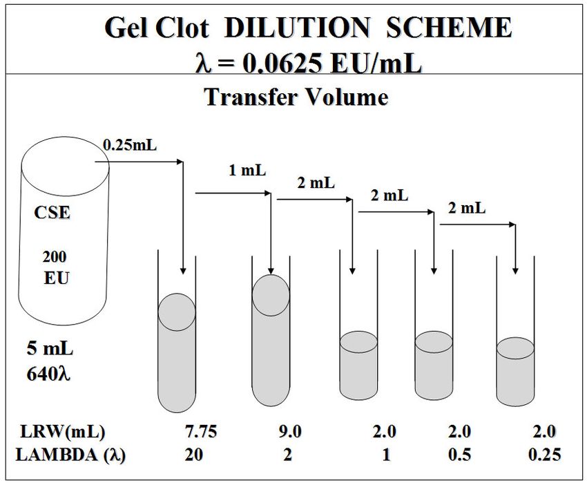
-- Intentionally left blank --

         -Page 2 of 54-
Bacterial Endotoxin Test and Sterility Test for
Radiopharmaceuticals
By
James F. Cooper and Joseph C. Hung
Editor, CENP
Jeffrey Norenberg, MS, PharmD, BCNP, FASHP, FAPhA
UNM College of Pharmacy
Editorial Board
Stephen Dragotakes, RPh, BCNP, FAPhA
Neil Petry, RPh, MS, BCNP, FAPhA
James Ponto, MS, RPh, BCNP, FAPhA
Tim Quinton, PharmD, MS, FAPhA
S. Duann Vanderslice, RPh, BCNP, FAPhA
John Yuen, PharmD, BCNP
Advisory Board
Dave Abbott, RPh., BCNP
Mark Gurgone, BS, RPh.
Vivian Loveless, PharmD., BCNP, FAPhA
Lisa Marmon, RPh, BCNP
Michael Mosley, RPh, BCNP
Janet Robertson, BS, RPh, BCNP
Brantley Strickland, BCNP
Scott Knishka, RPh, BCNP
Dave Engstrom, PharmD., BCNP
Brigette Nelson, MS, PharmD., BCNP
Samuel Ernesto, RPh, MBA
Director, CENP CE Administrator & Web Publisher
Kristina Wittstrom, RPh, FAPhA, BCNP Christina Muñoz, B.S.
UNM College of Pharmacy UNM College of Pharmacy
While the advice and information in this publication are believed to be true and accurate at the time of press, the author(s), editors, or the
publisher cannot accept any legal responsibility for any errors or omissions that may be made. The publisher makes no warranty,
expressed or implied, with respect to the material contained herein.
Copyright 2008
University of New Mexico Health Sciences Center
Pharmacy Continuing Education
Albuquerque, New Mexico
-Page 3 of 54-BACTERIAL ENDOTOXIN TEST AND STERILITY TEST FOR
RADIOPHARMACEUTICALS
STATEMENT OF LEARNING OBJECTIVES:
Upon successful completion of this CE course, the participant should be able to discuss the general
concepts and processes associated with the bacterial endotoxin test (BET) and sterility test, and apply
them in their daily practice. Specifically, the participant should be able to:
1. Distinguish the requirements for BET and sterility test.
2. Understand the basic principles and procedure for BET and sterility test.
3. Observe, report, and interpret the results of BET and sterility test.
4. Develop corrective action(s) for out-of-specification finding(s) of BET and sterility test.
5. Identify the personnel and facility requirements for the performance of BET and sterility test.
6. Define the term bacterial endotoxin and describe its toxic effects.
7. Identify the sources of bacterial endotoxin in the preparation of injectable radiopharmaceuticals
such as an F-18 product.
8. Describe the reagents, materials and equipment needed for the BET.
9. Understand how to select and establish a BET method.
10. Define volumes, solutions and containers needed to prepare a radiopharmaceutical for a BET.
11. Explain the calculations for the endotoxin limit and limit of detection (LOD).
12. Identify the advantages of the photometric-BET in contrast with the gel-clot BET.
13. Describe the procedure for qualifying a LAL reagent and an analyst for the BET.
14. Describe how glassware is rendered endotoxin-free for PET drug preparation and endotoxin
testing.
15. Identify the principle cause of invalidity in a photometric or gel-clot BET.
16. Discuss the limitations of sterility test.
17. Compare and contrast various regulations and standards associated with sterility test
requirements.
18. Select and validate media for sterility test.
19. List the unique aspects of sterility test for radiopharmaceuticals.
20. Provide the rationale for the filter membrane integrity test.
-Page 4 of 54-COURSE OUTLINE
OVERVIEW: ENDOTOXIN DETECTION........................................................................................................................ 8
NATURE OF PYROGENS ................................................................................................................................................... 8
PRINCIPLE OF THE BACTERIAL ENDOTOXINS TEST (BET) ................................................................................. 9
LIMULUS AMEBOCYTE LYSATE (LAL) REAGENT ............................................................................................................... 10
ENDOTOXIN AND ENDOTOXIN STANDARDS ........................................................................................................................ 11
ENDOTOXIN LIMITS ............................................................................................................................................................ 11
ENDOTOXIN TEST METHODS ....................................................................................................................................... 12
PHARMACOPEIAL REQUIREMENTS ...................................................................................................................................... 12
LAL REAGENT CARTRIDGE METHOD ................................................................................................................................ 13
GEL CLOT METHOD ........................................................................................................................................................... 14
CREATING A VALID BET PROCEDURE...................................................................................................................... 16
BET VERIFICATION ............................................................................................................................................................ 16
TEST FOR INTERFERING FACTORS AND MAXIMUM VALID DILUTION (MVD) .................................................................... 17
SPECIFYING A TEST DILUTION AND LIMIT OF DETECTION .................................................................................................. 18
STANDARD OPERATING PROCEDURE (SOP) AND VALIDATION REPORT ............................................................................. 19
DEPYROGENATION AND VALIDATION OF DEPYROGENATION PROCESSES ............................................... 19
INVALIDITY AND OUT-OF-SPECIFICATION (OOS) INVESTIGATIONS ............................................................. 20
RISK ASSESSMENT ........................................................................................................................................................... 21
CALCULATIONS ................................................................................................................................................................ 21
ENDOTOXIN LIMIT .............................................................................................................................................................. 21
MAXIMUM VALID DILUTION (MVD) ................................................................................................................................. 21
LIMIT OF DETECTION (LOD) .............................................................................................................................................. 22
DEPYROGENATION ............................................................................................................................................................. 22
THE FORMULA FOR DETERMINING LOG REDUCTION VALUE (LRV) FOR A DEPYROGENATION PROCESS IS AS FOLLOWS: ..... 22
STERILITY TEST ............................................................................................................................................................... 23
INTRODUCTION ................................................................................................................................................................... 23
ISSUES RELATED TO STERILITY TESTING .............................................................................................................. 24
ABSOLUTE STERILITY ........................................................................................................................................................ 24
UNDETECTED MICROORGANISMS ....................................................................................................................................... 24
RADIOPHARMACEUTICAL UNIQUENESS .............................................................................................................................. 24
MINIMAL TESTING ............................................................................................................................................................. 25
USP GENERAL CHAPTER “RADIOPHARMACEUTICALS FOR POSITRON EMISSION TOMOGRAPHY – COMPOUNDING” 26
USP GENERAL CHAPTER “PHARMACEUTICAL COMPOUNDING – STERILE PREPARATIONS” .................................... 26
High-Risk Level CSPs ................................................................................................................................................... 27
TESTING PREREQUISITES ............................................................................................................................................. 28
TESTING ENVIRONMENT ..................................................................................................................................................... 28
PERSONNEL QUALIFICATION .............................................................................................................................................. 28
TYPES OF MEDIA .............................................................................................................................................................. 29
FLUID THIOGLYCOLLATE MEDIUM (FTM) ......................................................................................................................... 29
SOYBEAN-CASEIN DIGEST MEDIUM (SDM) ....................................................................................................................... 30
STORAGE AND SHELF-LIFE OF THE CULTURE MEDIA ......................................................................................................... 30
COMPOSITION OF FTM AND SDM ...................................................................................................................................... 30
GROWTH PROMOTION TEST ............................................................................................................................................... 30
-Page 5 of 54-STERILITY TESTING PROCEDURES ........................................................................................................................... 31
MEMBRANE FILTRATION .................................................................................................................................................... 31
DIRECT INOCULATION ........................................................................................................................................................ 32
NEGATIVE CONTROL .......................................................................................................................................................... 32
VALIDATION TEST .............................................................................................................................................................. 33
Membrane Filtration..................................................................................................................................................... 33
Direct Inoculation ......................................................................................................................................................... 33
INITIATION TIME ................................................................................................................................................................ 34
POOLED SAMPLES .............................................................................................................................................................. 34
QUANTITY TO BE TESTED ................................................................................................................................................... 34
NUMBER OF ARTICLES TO BE TESTED ................................................................................................................................ 35
OBSERVATION AND INTERPRETATION OF RESULTS .......................................................................................... 35
FIRST STAGE ...................................................................................................................................................................... 36
SECOND STAGE .................................................................................................................................................................. 36
OUT-OF-SPECIFICATION INVESTIGATION ............................................................................................................................ 37
MEMBRANE FILTER INTEGRITY .......................................................................................................................................... 38
BUBBLE-POINT TEST .......................................................................................................................................................... 38
“Limitus Test” .............................................................................................................................................................. 40
Reprocessing ................................................................................................................................................................. 40
SUMMARY .......................................................................................................................................................................... 40
APPENDIX ........................................................................................................................................................................... 42
REFERENCES ..................................................................................................................................................................... 46
GLOSSARY .......................................................................................................................................................................... 48
ABBREVIATIONS AND ACRONYMS ............................................................................................................................ 49
ASSESSMENT QUESTIONS ............................................................................................................................................. 50
-Page 6 of 54--- Intentionally left blank --
-Page 7 of 54-BACTERIAL ENDOTOXIN TEST AND STERILITY TEST FOR
RADIOPHARMACEUTICALS
James F. Cooper, Pharm.D., FAPhA
And
Joseph C. Hung, Ph.D., BCNP, FASHP, FAPhA
Professor of Pharmacy and Radiology
Director of Nuclear Pharmacy Laboratories
PET Radiochemistry Facility Mayo Clinic
OVERVIEW: ENDOTOXIN DETECTION
Pyrogen (fever-inducing agent), principally known as bacterial endotoxin, is one of the most potent
bacterial toxins. Its only source is Gram (-) bacteria (GNB), where endotoxin comprises about 75% of
the GNB cell wall. Bacterial contamination (bioburden) in water and on surfaces normally contains
GNB, so endotoxin is ubiquitous in nature.1,2 This unit describes how to apply the Bacterial
Endotoxins Test (BET) to assure the absence of unsafe levels of bacterial endotoxin pyrogen in
compounded or manufactured radioactive drugs.
The first test for endotoxin pyrogen was an expensive, time-consuming rabbit fever assay. The rabbit
test was replaced in recent years by the Bacterial Endotoxins Test, an in vitro test that uses a highly
sensitive reagent called Limulus amebocyte lysate (LAL).1 Chapter of the U.S. Pharmacopeia
describes how to conduct the BET with LAL reagent and qualify an analyst for routine testing.3 The
Food and Drug Administration (FDA) requires radiopharmaceutical producers to test all injectable
products with FDA-licensed LAL reagent to meet predetermined endotoxin limits.4,5 The BET is
relatively simple and does not require highly specialized equipment. However, the preparation of
controls for some BET methods is tedious and demands attention to detail in order to complete the test
accurately and reproducibly. This CE unit emphasizes a simplistic, automated BET method.
NATURE OF PYROGENS
Due to its potency and ubiquity in nature, bacterial endotoxin is the only significant pyrogen of
concern to the parenteral drug industry.6,7 Endotoxins are large complexes from GNB bacterial cell
wall that are constantly shed into the environment when the bacteria disintegrate or multiply.
Endotoxins contain lipid, carbohydrate, protein, and are composed of a common structure of a
-Page 8 of 54-hydrophilic polysaccharide covalently bound to a hydrophobic region known as Lipid A. The latter
component causes the toxicity and LAL activation related to endotoxin. Endotoxins are negatively
charged macromolecules that are not removed by sterilizing membrane filters, such as those used in
aseptic processing of a Positron Emission Tomography (PET) drug. It is quite possible to have a sterile
solution that has sufficient endotoxin to be pyrogenic.
The most likely sources of endotoxins in PET radiopharmaceuticals are containers, tubing and non-
sterile water and chemicals used in the preparation of the product. Endotoxin is difficult and expensive
to eliminate after the product is made. The only practical way to avoid endotoxin contamination is to
eliminate it at the outset by using endotoxin-free materials and aseptic technique for all critical steps in
the production of a Compounded Sterile Preparation (CSP) in the form of a radiopharmaceutical or
PET drug.
The skin and gastrointestinal tract are barriers to endotoxin. However, when endotoxin gains access to
blood or tissues by injection, nanogram (parts per billion) quantities can cause fever and hypotension
while large amounts can lead to irreversible shock and death.6,7 Signs of pyrogenic reactions caused
by endotoxin contamination include chills, fever, rigors, tachycardia, hypotension and respiratory
distress. Although advances have been made in parenteral drug production, pyrogenic reactions still
occur, as evidenced by a pyrogen outbreak to endotoxin-contaminated gentamicin in 1998 and 1999
that produced at least 155 patient reactions.7 Signs of intrathecal toxicity are more serious with morbid
symptoms of aseptic meningitis.2,7
PRINCIPLE OF THE BACTERIAL ENDOTOXINS TEST (BET)
The Atlantic horseshoe crab (Limulus polyphemus) has an enzymatic blood coagulation system that is
specific and highly sensitive to endotoxin. The development of the in vitro endotoxins test began
when Levin and Bang8 proved that the blood cells (amebocytes) from the horseshoe crab responded to
endotoxin on the cell wall of Gram-negative bacteria. Cooper, Levin, and Wagner9 expanded this
concept into an endotoxin test for radiopharmaceuticals and other drugs. The new test was known as
the LAL test until it was adopted into the USP. The name was changed to the Bacterial Endotoxins
Test (BET) to reflect its purpose. The industry uses the terms BET and LAL test interchangeably, but
BET is the official acronym.
-Page 9 of 54-The development of radiotracers and endotoxin testing has remarkable parallels. The need for an
alternative to the rabbit pyrogen test for radiotracer development led us to define the usefulness of
LAL for testing injectables for endotoxin. The FDA began approving LAL reagents in the 1970s about
the time radiopharmaceuticals came under the purview of the FDA. Aseptic meningitis caused by
endotoxin in cerebrospinal-fluid imaging agents led to a strict USP endotoxin limit for intrathecal
drugs. Replacement of the pyrogen test with the BET began in 1983 with introduction into the USP 29
monographs for radiopharmaceuticals and for five USP pharmaceutical waters. The innovation of the
LAL cartridge system provides a realistic way to test PET products for endotoxin.
Limulus Amebocyte Lysate (LAL) Reagent
FDA regulated parenteral products must be tested by FDA-approved biological Limulus amebocyte
lysate (LAL) reagent, before released for patient use.4,5 The BET can be conducted either by the
traditional gel-clot method or photometric tests done with spectrophotometric readers.2,3,6 The basis of
the endotoxin-LAL reaction is that endotoxin activates an enzymatic cascade in LAL that modifies a
clotting protein to yield a turbid gelatinous endpoint. The BET is standardized for incubation of equal
parts of test solution and LAL. Optimum reactivity requires pH neutrality, optimum levels of divalent
cations (Mg and Ca) and sodium, low salt strength and generally less-than-milligram concentrations of
drug products. Therefore, compatibility testing is required to identify valid test conditions.6,7 The
basic test in the BET is termed a limit test because it is designed to reveal when an endotoxin limit is
exceeded. Positive controls are included to assure that results are valid. The test may be done on a
clean bench top; a laminar air flow hood is not necessary. Touch contamination of LAL solution, LAL
vial or rehydration water is a principal source of false positives.2,3 The reagent is somewhat labile in
solution, and as such requires limited storage at ambient temperature and only one freeze-and-thaw
cycle.
A unique terminology for BET applications has emerged (see attached glossary). Some of the
prominent terms deserve an explanation. The first LAL document was the FDA’s LAL-test Guideline
for injectables.10 This guide defined the symbol lambda (λ) as the LAL reagent sensitivity in EU/mL.
It introduced the concept of the Endotoxin Limit (EL), based on patient dose, to define a safe level of
endotoxin. It also provided a formula for the use of dilution (MVD, maximum valid dilution) to
overcome interference mechanisms for the LAL-endotoxin reaction. The first USP gel-clot test
required an endotoxin dilution series, from 2λ through ¼λ, to bracket an expected endpoint of the LAL
-Page 10 of 54-reagent and confirm that a test was in control. An operator is deemed to be “qualified” to conduct the
BET assay by preparing and testing dilutions of an endotoxin standard with LAL and achieving
recovery of positive controls at the expected levels.3
A revision of the BET chapter was introduced into the USP in 2001 that simplified procedures and
introduced photometric methods. The BET is now the minimum standard for endotoxin testing, so
standard procedures must reference USP Bacterial Endotoxins Test.
Endotoxin and Endotoxin Standards
Environmental (unpurified) endotoxins contain lipid, carbohydrate, protein, and are readily dispersed
in water; they are remarkably stable. However, the purified endotoxins that are used for standards and
positive controls are quite different. Purification processes remove protein units from endotoxin to
yield lipopolysaccharide (LPS), which is poorly dispersible and less stable in potency. The Control
Standard Endotoxins (CSE) that are supplied by LAL reagent vendors serve as positive controls for
routine endotoxin tests. Alas, the low-concentration CSE solutions are susceptible to absorption and
poorly understood aggregation phenomena. With time, activity of endotoxin standards, such as CSE,
seem to disappear unless subjected frequently to vortex mixing. The most common problem in routine
gel-clot methods is loss of endotoxin potency and invalidity due to failure to recover the positive
controls. The potency of a CSE preparation is specified in Endotoxin Units (EU) when assigned to a
specific lot of LAL reagent through a Certificate of Analysis (CoA). The LAL-reagent suppliers
calibrate the sensitivity of their reagents using an international reference standard endotoxin (RSE).
Endotoxin Limits
An endotoxin unit (EU) is a unit of biological activity established by the USP Endotoxin RS
(Reference Standard). The adverse effects of endotoxin are dependent on dose, route and rate of
administration. A general endotoxin limit of 5 EU/kg/hr or 350 EU per adult (70 kg) was scientifically
established to avoid the fever and hypotension from IM or IV injection of endotoxin contamination.6
Time is a factor because there are mechanisms in the liver and blood that neutralize endotoxin.
However, there are no clearance mechanisms in intraspinal spaces, so the IT (intrathecal) endotoxin
limit is much more stringent.2,11 If an IV drug is infused over a period of time that exceeds one hour,
the dose may be divided by the number of hours to determine the dose-per-hour value. (See example
in the section on calculations).
-Page 11 of 54-When endotoxin limits for radiopharmaceuticals were first approved in 1983 as a replacement for the
rabbit test, the limit was arbitrarily set at half the general limit. Therefore, the radiopharmaceutical
limit was set at 175 EU per adult dose for IV or IM injection and 14 EU/dose for intrathecal
administration. Validation of BET methods, including extent of pre-test dilution, is performed after
endotoxin limits are established, based on factors such as dose, infusion rate and route of
administration. For traditional drugs, endotoxin limits may be expressed in EU/mg of a specific
medication or EU/mL for infusion solution or a mixture of medications in solution. The term pyrogen
free may be applied to a CSP or finished manufactured pharmaceutical product that contains endotoxin
less than the endotoxin limit as specified for each product.
ENDOTOXIN TEST METHODS
Pharmacopeial Requirements
As introduced above, endotoxin activates a cascade of serine-protease enzymes in LAL that alters the
clotting protein. Macromolecules of this protein aggregate and cross-link to eventually produce a
reversible, opaque gel. Spectrophotometric measurement of increasing optical density (OD) with time
and endotoxin concentration describes endotoxin analyses by kinetic LAL methods. A photometer
with endotoxin-specific software passes a optical signal through the sample at one-minute-or-less
intervals to generate a reaction curve for each sample. The software assigns a reaction time when the
optical density of a sample exceeds a preset OD. Then, reaction times of unknowns and positive
controls are interpolated against a standard curve that is generated by at least three endotoxin standard
concentrations, made at 10-fold intervals. Increases in OD produced by absorption and light-scattering
may be monitored with time to produce kinetic turbidimetric assays (KTA). Increases in OD, due to
cleavage of a colorless substrate to yield a yellow color, may be monitored with time to yield kinetic
chromogenic assays (KCA). The parenteral drug industry primarily uses kinetic LAL methods, with
the aid of an incubating 96-well microplate reader, to conduct up to 20 assays of test samples,
simultaneously.
The two types of endotoxin tests that are described in the BET require substantially different reagents
and equipment. Gel-clot methods require a dry-heat block, calibrated pipettes and thermometer, vortex
mixer, freeze-dried LAL reagents, LAL Reagent Water (LRW) for hydrating reagents and
depyrogenated assay tubes. In the Gel-clot method test, diluted sample and liquid reagents require
about an hour for sample and positive-control preparation and an hour’s incubation in a heat block;
-Page 12 of 54-results are recorded manually. In contrast, photometric tests require a more highly-processed reagent,
a spectrophotometer, endotoxin-specific software and printout capability. In its most simplistic form,
the simplest photometric system exists as a handheld unit employing a single-use, LAL cartridge that
contains dried, pre-calibrated reagents; no liquid reagents or standards are needed. A FDA-approved
hand held system unit was recently introduced under the trade name of Endosafe PTS™ (Charles River
Laboratories, Charleston, SC). The device requires about 15 minutes to analyze small amounts of
sample, a 100-µL aliquot from a Compounded Sterile Product (CSP) diluted with sterile, pyrogen-free
water. The simplicity and speed of this new automated system make it ideally suited for PET drug
products.
LAL Reagent Cartridge Method
The Endosafe PTS™ (Portable
PTS™ Cartridge Technology Test System) is a handheld
Charles River Laboratories
spectrophotometric reader with
endotoxin specific software that
The reagents are
filled and dried is designed to measure a kinetic
on disposable chromogenic assay that occurs
polystyrene
cartridges: within a uniquely designed
polystyrene cartridge. (Figure 1)
The cartridge has four channels
with kinetic LAL reagents dried
in place. Two channels have
LAL reagent and color
Figure 1. LAL Reagent Cartridge. Kinetic chromogenic reagents and
endotoxin controls are dried in the channels of a polystyrene cartridge. substrate; the other two channels
Diluted product pumped into the channels where it is mixed with
reagents, monitored for changes in optical density and results are have LAL reagent.
interpolated against an archived endotoxin standard curve.
The archived curve for each batch of cartridges is embedded in a calibration code that is entered into
the software upon first use of the batch by an analyst. The dry cartridges are thus pre-calibrated so that
there is no need for handling liquid reagent or standards. An endotoxin test is conducted on a verified
test dilution by entering identifying test and product information into the hand held unit, inoculating
each of the four cartridge reservoirs with 25 μL of diluted product, incubation for 15 minutes and
printing of results by a suitable means. (See procedure in the Appendix). A study is valid if the
-Page 13 of 54-positive control is recovered within a range of 50-200% and there is agreement between each pair of
channels.
Gel Clot Method
The traditional gel clot method is more complicated, time-consuming and technique-dependent than
other methods. In its simplest form, a gel-clot BET is conducted by mixing 100 μL (0.1 mL) of
sample or positive (endotoxin) control with 100 μL of LAL reagent, using aseptic technique, and
incubating the mixture undisturbed at 37°±1C for one hour (60±2 minutes). If endotoxin is present in a
concentration greater than the labeled LAL sensitivity, an opaque gel will form that remains firm when
the assay tube is carefully inverted. Gel-clot reagents are available in single and multi-test vials; single
test vials eliminate handling liquid LAL reagent. The labeled sensitivity of the LAL reagent, lambda
(λ), is the minimum concentration of endotoxin that will produce a gel endpoint under standardized test
conditions; a 0.06 EU/mL reagent is most frequently used. The assay is time and temperature
sensitive, so attention to test conditions is important to achieve meaningful results.
Table 1
COMPONENTS OF A GEL-CLOT LIMITS TEST BY USP
Solution Endotoxin Sample Solution No. Replicates Expected Result
A (Product) None Diluted PET drug 2 Negative
B (PPC) ~2λ Diluted PET drug 2 Positive
C (PWC) ~2λ LAL Reagent Water 2 Positive
D (NC) None LAL Reagent Water 2 Negative
Note: Lambda (λ) is the LAL Reagent sensitivity; PPC is the positive product control, PWC is a Positive Water Control;
NC is a negative control. The PPC and PWC tubes contain co-lyophilized LAL and endotoxin control. A result is reported
as positive if the gel holds firm through a 180° rotation, whereas all other results are negative.
The gel-clot test has four components tested in duplicate, as listed in Table 1. The test sample is drug
product that has been diluted to avoid interfering test conditions and unnecessary radiation exposure.
Component A is diluted product and B is diluted product with a positive control. Component C is
sterile, pyrogen-free water with the positive control and D is a negative control; C and D tubes are
optional, but should be done once daily with the first set of tests to show that the positive controls have
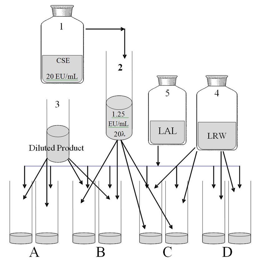
sufficient potency and that sterile water used for dilution is not contaminated. Figure 2 illustrates
preparation of reagents and assay tubes for a BET.
-Page 14 of 54-The most challenging part of this method is preparation of the solutions for the Positive Product
Control (PPC). The components needed to make positive controls are 1) a CSE vial that is rehydrated
and stored according to instructions, and 2) a 20-λ CSE tube for preparation of 2λ positive controls in
situ for the BET limits test. (Figure 2) The key to efficiency in this method is use of a 20-λ CSE tube
that allows the positive controls to be made in the assay tubes rather than making a separate tube; this
approach is known as the ‘hot spike’ technique. The USP allows the positive water controls (PWCs) to
substitute for an endpoint dilution series in a limit test. The purpose of the PWC is to indicate that the
PPC was made properly.
Gel-clot LAL Method
1. Prepare 30-λ SCE Tube,
Where λ - 0.06 EU/mL
2. 0.01 mL (10µL) CSE into
tubes B & C
3. 0.1 mL of LRW into tubes C
& D*
4. 0.1 mL of diluted PET drug
into tubes A & B
5. 0.1 mL of LAL into ALL tubes,
mix and incubate
6. Read and record results
7. *Only one set of C&D tubes
needed when testing multiple
samples
Figure 2. Scheme for USP Bacterial Endotoxins Test (BET) by a gel-clot method. Test preparation
for a BET requires dilutions of endotoxin control (2) and PET drug (4). Four sets of solutions are
made for testing in duplicate. Two sets contain the diluted product either alone (A) or with
endotoxin control (B), and water tubes (3) either with endotoxin control (C) or without (D). A 10-μL
inoculum of endotoxin (4) produces the positive product control (B) and positive water control (C).
LAL reagent (5) is added to all tubes prior to incubation in a heating block for 60 ± 2 minutes at 37 ±
1°C. Results are read and recorded after an hour. The drug is within the endotoxin limit and the
test is valid if tubes A & D are negative (no gel) and B & C are positive (gel). Figure only applies to
BET where λ equals 0.06 EU/mL.
The 20-λ CSE solution is made at least weekly in order to prepare the positive controls in the limit test.
As an example, for an LAL reagent with a sensitivity of 0.06 EU/mL, the 20-λ tube is 1.25 EU/mL,
which is twenty times lambda. (Figure 2) Preparation of the 20-λ tube, in polystyrene, requires vortex
mixing and hydration of a CSE vial according to the vendor’s direction to make 20 EU/mL CSE.
-Page 15 of 54-Vigorous and frequent vortex mixing of the tube is needed to maintain its potency. Inadequate vortex
mixing is the principal cause of invalidity, which is a failed positive control (no gel). A different
dilution scheme is required for LAL reagent with sensitivity other than 0.06 EU/mL.
Potency of a 20-λ CSE tube is verified on the
day it is made by preparing a CSE dilution series
to assay its potency. The assay is an endpoint
test with four sterile polystyrene tubes consisting
of a serial two-fold dilution of 2λ, λ, 1/2λ and
1/4λ that bracket labeled sensitivity of the LAL
reagent. The 2-λ tube is made by creating a 1:10
dilution from 20-λ tube, and the other tubes are
made by two-fold dilution. (Figure 3) The 20-λ
CSE tube should be able to produce an endpoint
Figure 3. Endpoint Assay for Endotoxin in a 20-λ
(potency) of between 2λ and 1/2λ after serial positive control tube. Ideally, potency is confirmed by
endpoints at the λ and ½λ endotoxin levels. Scheme
dilution. Any other result suggests preparation will differ for LAL reagents other than 0.06 EU/mL.
of a new 20-λ CSE tube and re-assay for 20-λ
CSE potency verification. This assay is also an alternative for the PWC tubes (Tubes C in Table 1) in
the limit test. This same endpoint assay is used to qualify an analyst for the BET.
CREATING A VALID BET PROCEDURE
BET Verification
Validation is a term often used to describe the creation of a BET method for a specific drug. For USP
methods such as the Chapter BET, one is not required to validate the accuracy and reliability of
the method. Rather, one is expected to verify the suitability of a test method under actual conditions of
use. Verification conditions are described under “Interfering Factors Test” in USP BET. The
objective of method development and subsequent verification is to generate a standard procedure that
has the following attributes:
1. The product under test conditions described in a test procedure is non-interfering (positive
controls are positive and negatives are negative);
-Page 16 of 54-2. A suitable product dilution or solute concentration is determined by using a published or
calculated endotoxin limit;
3. The assay is unaffected by significant batch or laboratory test variability, as demonstrated by
the requirement for verification using three batches of product;
4. The combination of LAL and prepared product will have a neutral pH (6-8);
5. The laboratory processes are supported by appropriate user procedures and training, instrument
qualifications, and preventive maintenance.
There are questions that should be addressed before developing methods for a new or investigational
radiopharmaceutical. For example, what is the maximum human dose, route of administration, product
pH and formulation? A unique property of radioactive drugs is that the dose volume increases with
time in order to deliver a prescribed dose of activity. Therefore, the dose volume at time of expiration
is used for calculations. An unusual route of administration, such as intrasynovial spaces, may need a
stricter limit, similar to IT administration. Understanding and knowledge of the final product
formulation may lead to anticipation of interference conditions, such as pH.
Test for Interfering Factors and Maximum Valid Dilution (MVD)
The four principal causes of inhibitory conditions for a BET concern divalent cations, pH, enzyme
modifiers and loss of potency in endotoxin standards.6 Non-neutral pH is a common problem because
most LAL reagents have modest buffer capacity in their formulation; a mixture of LAL and diluted
drug product should have a pH very near 7. Also, the presence of divalent-cation chelating agents,
such as citrate, can deplete the magnesium and calcium ions that are needed for the LAL reaction with
endotoxin. Chemicals that denature or alter enzyme function, such as ethanol or chromic ion, will
inhibit the enzymatic cascade. Finally, inadequate vortex mixing or prolonged storage of CSE
endotoxin standards results in loss of potency and failure of positive controls. Sources of enhancement
or false positives include LAL reactive glucans and exposure to cellulose.6
Dilution should always be applied to resolve the first three inhibitory factors. The extent of dilution is
defined by the formula for MVD in the BET chapter of the USP. MVD is calculated by dividing the
endotoxin limit of a drug by the sensitivity (lambda) of the endotoxin test method or reagent. (See
section on calculations for examples).
-Page 17 of 54-Specifying a Test Dilution and Limit of Detection
The low administration volume of imaging agents invariably yields an MVD calculation that is quite
high. It is prudent to assign a valid test dilution for a product that is less than the maximum but
sufficiently high to avoid the risk of interfering conditions. For example, it is commonplace for the
MVD for Fludeoxyglucose 18F (18F-FDG) to be at least 1:350, as shown in the calculations below. On
the other hand, inhibitory conditions can be detected at 1:10 dilutions of 18F- FDG due to the presence
of citrate and other factors associated with a given synthesis unit. Therefore, the choice of 1:50 as the
test dilution for this product is advisable.
A survey of radiopharmaceuticals, including selected PET drugs, found very little dilution was needed
for the BET because their formulations were generally neutral and contain little solute.11 Table 2
shows validated test dilutions for I 123 MIBG and radiotracers labeled with 18F, 11C, and 13N.
After the test dilution is selected, the Limit of Detection (LOD) is determined for the purpose of
uniformly reporting the results. The LOD is the sensitivity of the BET method times the extent of
dilution, as further described in the section of calculations, below. Invariably, the BET for a
radiopharmaceutical will be negative, so the analysis is reported as less than the LOD.
Table 2
VALIDATION OF SHORT-LIVED RADIOPHARMACEUTICALS
WITH THE LAL CARTRIDGE
PET Medication Dilution LOD (EU/mL) PC Recovery (Mean %)
F 18 FDG 10 1 91
F 18 Fluoride 10 1 120
F 18 FLT 10 1 103
C 11 Acetate 20 2 150
C 11 Methionine 20 2 95
N 13 Ammonia 20 2 108
I 123 MIBG 50 5 90
Note: LOD is the Limit of Detection when using cartridges with a sensitivity (λ) of 0.1 EU/mL. PC is positive
control recovery of 1 EU of endotoxin standard in the cartridge.
-Page 18 of 54-Standard Operating Procedure (SOP) and Validation Report
No test method is complete until there is a validation report and operating procedure approved and
placed in document control. As in potty training, the job isn’t done until the paperwork is completed.
The SOP should be sufficiently clear and complete to serve as a training document for operators as
well as conducting tests and interpreting results. The SOP should describe how the results are
recorded, including the LOD, and how validity and invalidity is handled in practice. (Refer to the
Appendix for a representative SOP).
The validation report documents the suitability of written test conditions for each radiopharmaceutical.
The report should include calculation of the endotoxin limit and MVD, pH measurement, and the
results of valid recovery of positive controls for three batches of product. The report may show
validation with both gel-clot and photometric methods.
DEPYROGENATION AND VALIDATION OF DEPYROGENATION PROCESSES
Elimination of endotoxin by separation or destruction is called depyrogenation. Distillation, copious
rinsing of plasticware with SWI and passage of solutions through certain surface-active materials, such
as alumina and positive-ion filters, are common separation methods. Incineration of endotoxin in dry-
heat ovens is the definitive method for depyrogenating glassware used in production and for gel-clot
assay tubes. Chemical depyrogenation is accomplished by basic hydrolysis through exposure to
sodium or potassium hydroxide, or by oxidation with peroxide.6
Endotoxin Indicators (EI) are vials that contain known amounts of LPS, such as 2,000 or 10,000 EU
per vial, in a dry vial. An EI is exposed to a depyrogenation method, such as a dry-heat sterilizing
oven, to document effectiveness. Endotoxin assays of an EI are made before and after a
depyrogenating step to determine if there was at least a 3-log reduction in endotoxin content, the
criterion for depyrogenation. An oven can be qualified by exposing four or more EI vials, embedded
in a typical load of glassware, and quantifying the recovery of endotoxin residues.6 When the recovery
is non-detectable, lambda is used for the exposed EI value (See section on calculations below).
It is ironic that the FDA places such emphasis on testing 18F-FDG because the Al-N cartridge in a
typical 18F-FDG synthesis unit completely removes endotoxin during processing. In our experiments,
the contents of a 2000 EU vial were passed through an Al-N cartridge and analyzed in a system with a
-Page 19 of 54-limit of detection at 0.05 EU/mL. The depyrogenation effect of alumina was confirmed when no
endotoxin was recovered in the eluate of the cartridge. Note that the cartridge eluate was passed
through a sterilizing membrane filter to trap trace alumina particles, which could inhibit the recovery
analysis by absorbing the endotoxin in the positive control for the eluate. In Europe, there is a more
realistic, skip-test policy of endotoxin tests of 18F-FDG, such as testing for one-in-ten preparations.
INVALIDITY AND OUT-OF-SPECIFICATION (OOS) INVESTIGATIONS
Invalidity occurs when positive and negative controls do not react as expected. In photometric assays,
there must be recovery of the positive control between 50-to-200% of positive control. The principal
cause of invalid recovery in a LAL cartridge system is pipetting inaccurate volumes into the cartridge.
Over-pipetting can cause inhibition (200% recovery). For example, a 20-µL inoculation of SWI into a LAL cartridge produced only 46%
recovery, which is out of range and mimics inhibition. The remedy requires addressing causes for
inaccurate volumes, such as training and re-calibration of the pippetor. The most common invalidity in
gel-clot method tests is failure to observe a gel in the PPC and PWC tubes (Table 1), which invariably
is due to loss of CSE potency associated with inadequate vortex mixing of the standard solutions. The
remedy for positive-control failures is usually preparation of new CSE standards. In contrast, gelation
in the NC and diluted product tubes suggests that the reagent is contaminated; the remedy here is new
LRW and LAL reagent.
In the event of an OOS result, where the endotoxin limit of a test was exceeded, it is prudent to
conduct an investigation in a timely fashion in order to determine if the result was a true positive, an
invalid test or a false-positive where contamination was introduced by the analyst. For example, if all
eight tubes in a gel-clot limit test were positive with firm gels, then the test was invalid because the
positive result in the negative control indicated that either the water diluent or LAL reagent was
contaminated by the analyst.
There was an incident where an 18F-FDG batch was recalled due to endotoxin contamination near the
endotoxin limit. The investigation revealed that the operator inadvertently delivered the batch to a
non-sterile beaker instead of the final product vial. The recovered solution was passed through a
sterilizing filter to yield a sterile solution, but of course, endotoxin was not removed. This incident
reaffirms that risk of contamination is extremely low and only occurs when there is a catastrophic
failure in procedure, process or operator error.
-Page 20 of 54-RISK ASSESSMENT
The greatest risk for pyrogenic reactions involves the intrathecal (IT) route of administration.2,7 In the
early 1970s, endotoxin-contaminated imaging agents for radionuclide cisternography caused a large
outbreak of aseptic meningitis.12 Radioiodinated albumin was contaminated by passing the product
over an anion exchange column that had not been depyrogenated; the corrective action evolved to
treatment of in-process columns with concentrated sodium or potassium hydroxide to render them
pyrogen free. Also, In 111 Indium drug was contaminated by pH measurement when using non-sterile,
endotoxin-laden buffer solutions for meter calibration. There were frequent reports of pyrogenic
reactions in the early days of nuclear pharmacy when imaging agents were produced in house. The use
of FDA-approved radiopharmaceuticals and better awareness and control of bioburden in
compounding areas has virtually eliminated pyrogenic reactions in current practice.
CALCULATIONS
Endotoxin Limit
The endotoxin limit is calculated from the K/M formula in the BET, as such:
Endotoxin Limit (EL) = K (tolerance limit)
M (maximum dose/kg)(hr)
Radiopharmaceutical EL = 175 EU
V (mL)
A unique feature of radiopharmaceuticals is that the dose-volume increases with time because of
radioactive decay. Therefore, M is generally the volume of imaging agent at the expiration time that is
required to administer the prescribed dose of radioactivity. Another approach often used for 18F-FDG
is to assign M to the maximum volume of administration. For example, a firm had a procedure that
limited a patient dose to 10 mL. In this case, the endotoxin limit was 17.5 EU/mL when the
radiopharmaceutical limit of 175 EU/dose was divided by the 10 mL/dose.
Maximum Valid Dilution (MVD)
The MVD is used to determine the extent of dilution that may be used to avoid an inhibitory condition
and reduce radiation exposure to the analyst.
Maximum Valid Dilution (MVD) = EL
λ
-Page 21 of 54-The MVD is calculated by dividing the endotoxin limit by the sensitivity (λ) of the BET method. For
example, in the paragraph above, the endotoxin limit was determined to be 17.5 EU/mL. An LAL
cartridge system with a labeled sensitivity of 0.05 EU/mL was used as the test medium. Therefore, the
MVD is 350, expressed as 1:350, when the endotoxin limit of 17.5 EU/mL is divided by the cartridge
sensitivity of 0.05 EU/mL. If a 0.06 EU/mL gel-clot LAL reagent was used instead, the MVD would
be 290.
Limit of Detection (LOD)
The LOD represents the lowest concentration of endotoxin that may be detected under specific test
conditions and specific method. The sensitivity of a BET, or limit of detection (LOD), for a
pharmaceutical is the dilution of the drug multiplied by the labeled sensitivity (lambda) of the LAL
reagent.
Limit of Detection (LOD) = Lambda (λ) x Dilution Factor
For example, the LOD of a BET is 1.25 EU/mL when the product was diluted 1:20 and tested with a
gel-clot reagent with a lambda of 0.0625 EU/mL. In the PTS™ system, the LOD is 1 EU/mL, where
labeled sensitivity (lambda, lowest point on the archived standard curve) is 0.05 EU/mL with a 1:20
dilution.
Depyrogenation
The formula for determining log reduction value (LRV) for a depyrogenation process is as follows:
Log reduction value (LRV) = log10 unexposed EI
Log10 exposed EI
As an example, EIs with approximately 1000 EU was exposed to a 250°C cycle for one hour, in a
representative oven load of glassware, to yield EI vials that had non-detectable endotoxin assay on the
contents of the vials post pyrogen burn . The standard curve for the photometric assay had a lambda of
0.1 EU/mL, so the difference of 1000 andSTERILITY TEST
Introduction
Sterility is the most important quality of injectable because these drugs bypass most of the body’s
defense mechanisms. If radiopharmaceuticals are formulated from raw materials, sterile glassware,
syringes, and other components should be used to lessen the chance of introducing microorganisms
and pyrogenic material into the product. A sterile solution is one that contains no living organisms,
pathogenic or nonpathogenic.
All injectable products must be sterilized, and there are five methods of terminal sterilization (i.e.,
steam sterilization, dry-heat sterilization, gas sterilization, sterilization by ionizing radiation, and
sterilization by filtration) described in USP General Chapter “Sterilization and Sterility
Assurance of Compendial Articles”.13 Sterilization with a 0.22-μm membrane filter is frequently
employed for sterilization of compounded radiopharmaceuticals due to its ease of use. Sterilizing
membrane filtration is also the method of choice for heat-labile drug products.
Terminal sterilization of the final dosage form is generally a preferred step to ensure the minimal risk
of microbial contamination in the finished drug preparation. However, it is vital that, prior to the
terminal sterilization process, the parenteral drug is prepared by trained operating personnel who are
adequately cleansed and gowned, in a certified aseptic processing facility, and via a validated aseptic
SOP. These “aseptic processing” steps (i.e., trained personnel, certified facility, and validated SOP for
aseptic processing) are good practice to minimize the bioburden of the finished lot of final dosage
form. Coupling aseptic processing with the terminal sterilization would further minimize the risk of
microbial contamination in the finished drug product. Aseptic processing is also a “must” for those
products that cannot be terminally sterilized. These “aseptically processed” products can be prepared
within a controlled environment by a series of aseptic steps using the component(s) and container(s)
that have been sterilized by one of the five terminal sterilization methods mentioned earlier in this
section.
A Sterility test is performed to determine whether a product (especially a drug listed in the USP)
purporting to be sterile complies with the requirements as stipulated in the test for sterility in the
individual USP monograph. The test for sterility should be carried out in accordance with USP
General Chapter “Sterility Tests”. 14 The incubation and observation process of the USP sterility
-Page 23 of 54-test takes 14 days.14 Due to the short physical half-life of radionuclides used in nuclear medicine, the
release of prepared radiopharmaceuticals for parenteral administration in human subjects is not hinged
on the completion of the sterility test.15-18 Nevertheless, the sterility test must still be performed
especially for positron emission tomography (PET) radiopharmaceutical that is intended for parenteral
administration.15-18
ISSUES RELATED TO STERILITY TESTING
Absolute Sterility
Within the strictest definition of “sterility”, a specimen would be deemed “sterile” only when there is
complete absence of viable microorganisms from it. However, this absolute definition cannot be
applied to an entire lot of finished drug products since the specified number of units to be tested only a
small percentage of entire lot of the finished drug products. The “absolute sterility” confirmation
cannot be practically achieved without complete destruction of every finished drug product. Currently
there is no non-destructive technology for the sterility testing. To significantly increase the number of
specimens for testing in order to augment the probabilistic prediction of the test outcome is not a
feasible option.
Undetected Microorganisms
Sterility test is also limited in that it can only recognize organisms that are able to grow under the
conditions (e.g., medium, incubation temperature, and incubation time, etc.)19,20 There are a large
number of microorganisms that are unable to replicate under standard testing conditions.21-23 Even
with the longer incubation period there is no assurance that all microorganisms can grow under these
conditions, but are still metabolically active.
Radiopharmaceutical Uniqueness
Special difficulties arise with radiopharmaceutical preparations because of the short half-life of some
radionuclides (especially those used for diagnostic purposes), small size batches, low production
volume, and radiation hazards. The short half-lives of most radiopharmaceuticals used in nuclear
medicine studies prohibit completion of the sterility testing before the release of radiopharmaceutical
products. In addition, when the half-life of the radionuclide is very brief (e.g., less than 20 minutes),
administration of the radiopharmaceutical preparation to the patient is generally on-line with a
-Page 24 of 54-validated production system. It is justifiable to dispense radioactive drug products before completion of
the sterility test if the radiopharmaceutical is prepared by a validated aseptic process.
Sterility testing can usually be conducted in a hospital’s microbiology laboratory. For safety reasons
(i.e., high levels of radioactivity), it may be not possible to use the quantity of a radiopharmaceutical
preparation stipulated in the USP sterility testing chapter.14 To limit radiation exposure of personnel,
the product should be diluted or allowed to decay to a safe working level before sterility tests are
conducted.
There is another aspect of the prepared radiopharmaceutical that is quite different from the
conventional drug product. The preparation of radiopharmaceuticals (e.g., 18F-fludeoxyglucose [FDG]
injection) usually results in one lot of a multiple-dose vial containing a homogeneous solution of the
radiopharmaceutical. Quality control (QC) results from end-product testing (including sterility test) of
samples drawn from the above-mentioned multiple-dose vial have the maximum probability of being
representation of all doses to be administered to patients from that vial. Thus, the referee sterility
testing (as well as other QC tests) performed on prepared radiopharmaceutical would undoubtedly
offer a near “absolute sterility” confirmation.
Minimal Testing
“The extensive aseptic manipulations required to perform sterility testing may result in a probability of
non-product-related contamination of the order of 10–3, a level similar to the overall efficiency of an
aseptic operation and comparable to the microbial survivor probability of aseptically processed
articles. This level of probability is significantly greater than that usually attributed to a terminal
sterilization process, namely, 1 in 1 million or 10 –6 microbial survivor probability.”13 These facts as
stated in the USP General Chapter “Sterilization and Sterility Assurance of Compendial
Articles” seems to suggest that the lower microbial survival probability from an effectively terminally
sterilized products may preclude altogether the necessity for performing sterility testing. However, a
successful terminal sterilization process depends mostly on whether the “microorganism-elimination”
capability of the above sterilization process can effectively handle the bioburden level of the finished
drug product. Thus, the reliability of sterility assurance of terminal sterilization heavily hinges on a
properly validated and documented aseptic technique and sterilization process.
-Page 25 of 54-You can also read