REVIEW Hedgehog signalling in endocrine development and disease
←
→
Page content transcription
If your browser does not render page correctly, please read the page content below
439

REVIEW

Hedgehog signalling in endocrine development and disease
Peter J King, Leonardo Guasti and Ed Laufer1
Barts and the London School of Medicine, Centre for Endocrinology, London EC1M 6BQ, UK
1
    Department of Pathology and Cell Biology, Columbia University Medical Center, New York, New York 10032, USA
(Correspondence should be addressed to P J King; Email: p.j.king@qmul.ac.uk)

Abstract
The hedgehog (Hh) pathway is an evolutionarily conserved                       provide an update on recent findings in the field of vertebrate
signalling pathway that is required for many essential tissue                  Hh signalling and also describe contributions of Hh signalling
and cellular properties such as patterning fields of cells or                  to the development, maintenance and pathology of endocrine
regulating cell differentiation and proliferation. Disruption of               tissues.
the pathway results in serious pathologies. In this review, we                 Journal of Endocrinology (2008) 198, 439–450

Hedgehog signalling                                                            amino-terminal cysteine residues (Pepinsky et al. 1998) by the
                                                                               acyltransferase Skinny Hh (Chamoun et al. 2001). This dual
The hedgehog (Hh) gene was originally identified in the                        lipidation makes Hh proteins highly hydrophobic, and the
seminal work of Nusslein-Volhard & Wieschaus (1980) in                         release of processed Hh from the plasma membrane is thus an
which they performed mutagenesis screens of Drosophila and                     active process. Secretion is mediated by 12 pass transmembrane
isolated and characterised mutants defective in embryonic                      protein Dispatched (Disp), which is required for signalling to
cuticle formation. One of these they named Hh because                          non-adjacent cells (Burke et al. 1999). The majority of secreted
mutation of the gene resulted in the disorganised denticles                    Hh found in soluble multimeric forms thought to assume
forming a spiky pattern. Subsequent investigations demon-                      micelle-like structures (Chen et al. 2004, Goetz et al. 2006). Hhs
strated that Hh encodes an intercellular signal with multiple                  are the only known proteins that are covalently linked to
critical functions as a regulator of embryonic development.                    cholesterol in this way. The requirement for cholesterol
Hh homologues have since been identified in many                               modification partially underlies the spectrum of developmental
invertebrates and vertebrates, and found to play similar vital                 disorders observed in Smith-Lemli-Opitz syndrome (SLOS,
roles controlling tissue patterning, and cellular differentiation              OMIM 270400), a condition in which defects in
and proliferation both during embryonic development and                        7-dehydrocholesterol reductase (DH7CR), an enzyme required
in the control of stem cell behaviour and homeostasis in                       for the formation of cholesterol from 7-dehydrocholestrol, leads
the adult.                                                                     to abnormalities that overlap those seen in cases of impaired Hh
                                                                               signalling (Kelley & Hennekam 2000).
Ligands
                                                                               Signal transduction pathway
The three mammalian homologues of Drosophila Hh are Sonic
Hh (Shh), Indian Hh (Ihh) and Desert Hh (Dhh). Each of the Hh                  Hh proteins bind 12 pass transmembrane receptors Patched1
proteins is processed post-translationally, and the precursor                  (Ptch1) and Ptch2 on responding cells (Fig. 1). Binding to these
forms undergo multiple covalent modifications to produce                       receptors promotes Ptch internalisation and derepresses the
mature signalling molecules. Hh proteins undergo autocatalytic                 activity of Smoothened (Smo), a seven pass transmembrane
cleavage that removes the carboxyl-terminal domain (Lee et al.                 protein of the G protein-coupled receptor family that is related
1994, Bumcrot et al. 1995) and simultaneously modifies the                     to the frizzled class of Wnt receptors. Following Hh binding
newly exposed carboxyl-termini of the amino-terminal                           to Ptch, Smo is phosphorylated on its intracellular carboxyl-
signalling molecules by the addition of cholesterol (Porter                    terminal tail (Chen et al. 2004) and undergoes a reciprocal
et al. 1996). These proteins are further modified into their                   translocation from intracellular vesicles to the plasma
mature forms by signal peptide cleavage and palmitoylation at                  membrane (for a review, see Huangfu & Anderson 2006).

Journal of Endocrinology (2008) 198, 439–450                                                                                DOI: 10.1677/JOE-08-0161
0022–0795/08/0198–439 q 2008 Society for Endocrinology        Printed in Great Britain         Online version via http://www.endocrinology-journals.org
440 P J KING and others . Hedgehog signalling in endocrine development
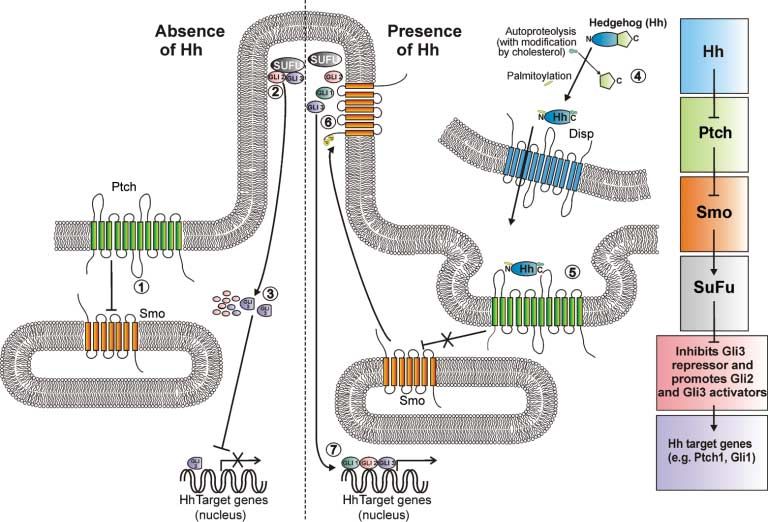
Figure 1 A scheme for Hh signalling. In the absence of Hh ligand, Ptch, located at the base of the cilium,
inhibits Smo, possibly via a small molecule (1). Gli2 and 3, along with SuFu, are located at the tip of the cilium
(2). IFT is required for movement of these molecules in and out of the cilium and they are proteasomally
processed outside the organelle (3). Processing cleaves Gli3 (and Gli2) to produce a transcriptional repressor
that inhibits the expression of Hh target genes and degrades Gli2. Hh is synthesised in signal-producing cells
as a precursor molecule that is cleaved to remove the C terminus and is simultaneously modified by
cholesterol addition. Following palmitoylation at the N terminus, it is secreted via Disp (4). Hh binding to Ptch
on target cells promotes internalisation and relieves the repression Ptch exerts on Smo (5). Smo then
translocates from endosomes to the tip of the cilium (6) resulting in the inhibition of processing of the Glis
which are then able to activate transcription of target genes, including Gli1 (7). A simplified pathway is shown
on the right-hand side of the panel. For details see text.
The inhibition of Smo by Ptch is sub-stoichiometric, and (Kuwabara & Labouesse 2002), underscore the importance
the two proteins do not appear to physically interact, which of cholesterol synthesis both in cells that produce Hh and
raises the still unanswered question of how Ptch inhibits those which receive Hh signals.
Smo activity. Both Ptch and Disp have homology to Ptch and Smo act through a signal transduction cascade that
bacterial small molecule pumps and the Niemann-Pick C1 culminates in the modulation of the activity of the Gli family
cholesterol transporter (Carstea et al. 1997, Loftus et al. of zinc finger transcription factors. There are three Gli genes
1997), which has led to the suggestion that, in an Hh in mammalian cells, Gli1, Gli2 and Gli3, which have partially
binding-modulated fashion, Ptch might transport small overlapping functions. In the absence of Hh signalling, Gli2
molecule regulators of Smo activity. Adding support to this and Gli3, but not Gli1, are expressed at significant levels.
idea, the sterol-related alkaloid cyclopamine and the purine In uninduced cells, these factors are phosphorylated by
homologue purmorphamine can bind Smo and inhibit or protein kinase A (PKA), casein kinase 1 (CK1) and glycogen
activate it respectively. One candidate for an endogenous synthase kinase 3 b (GSK3b) (Sheng et al. 2006, Tempe et al.
negative regulator of Smo is 7-dehydrocholesterol ((pro-) 2006, Wang & Li 2006) and ubiquitinated via interaction
vitamin D3), which can be transported out of the cell by with the E3 ligase b transducin repeat containing protein
Ptch1, and accumulates in the plasma of SLOS patients (bTrCP) (Wang & Li 2006). These modifications lead to
(Bijlsma et al. 2006). Interestingly, oxysterols, which are proteasomal processing that degrades the majority of Gli2,
cholesterol oxidation products, can activate the Hh signalling while limited proteolysis of Gli3 and Gli2 removes a carboxyl-
pathway by binding to Smo, raising the additional possibility terminal activation domain, producing truncated repressor
that oxysterols or related molecules are natural intracellular forms that inhibit the expression of Hh target genes.
Smo agonists (Corcoran & Scott 2006, Dwyer et al. 2007). Activation of Smo via Hh-mediated inhibition of Ptch
These data, together with the modification of Hh and the activity inhibits proteolytic processing of Gli2 and Gli3, and
presence of sterol-sensing domains in Ptch and Disp leads to the production of full-length transcriptional activator
Journal of Endocrinology (2008) 198, 439–450 www.endocrinology-journals.orgHedgehog signalling in endocrine development . P J KING and others 441
forms of these proteins. This changes the ratio of activator and Svard et al. 2006). Fused (Fu) is crucial for Hh signalling in
repressor Gli isoforms in the nucleus, and thus alters the Drosophila but in mammalian cells the importance of its role is
expression of Hh target genes by a combination of as yet unclear (Chen et al. 2005, Merchant et al. 2005). In the
transcriptional derepression and activation. Gli1 transcription absence of ligand, Gli2 and Gli3 are processed by the
is induced as a primary response to Hh signalling, and Gli1 proteasome at the base of the cilium whereupon they can
acts solely as a transcriptional activator to prolong or increase translocate to the nucleus and repress target gene
Hh target gene expression. Gli1 thus acts in conjunction with transcription.
Gli2 and Gli3, although the loss of Gli1 alone does not cause Upon binding Hh, Ptch1 is internalised from its position at
Hh signalling defects (Bai et al. 2002). Gli3 mRNA expression the base of the cilium in uninduced cells and Smo becomes
is also regulated by Hh signalling, although in a negative localised to the cilium tip (Corbit et al. 2005, May et al. 2005,
fashion (Marigo et al. 1996). Thus, pathway activation leads Rohatgi et al. 2007) where it promotes the formation of
not only to the induction of positively acting transcription activators and inhibits formation of repressors in an as yet
factors, but also to the inhibition of negatively acting ones. unexplained manner. Part of the inhibition of Smo by Ptch is
Among the Hh targets are several components of the Hh thus mediated by preventing entry of Smo into the cilium.
signalling system, including Ptch1 and Hh interacting protein This organelle is utilised apparently as a centre for
(Hip). Hip is a transmembrane glycoprotein that binds all accumulating pathway components that link Smo to Gli
forms of Hh with similar affinity to Ptch1 and acts to sequester either in order to promote efficient signal transduction, or
Hh and inhibit signalling in a negative feedback loop (Chuang perhaps to allow for an ordered and vital sequence of protein
& McMahon 1999, Bak et al. 2001). The expression of these interactions and modifications (Caspary et al. 2007).
genes, in particular Gli1, which is not observed in the absence
of Hh, is therefore a useful marker of cells actively transducing
Hh signalling during development, in the adult and in disease
an Hh signal (Ahn & Joyner 2004, Vokes et al. 2007).
Hh signalling plays many crucial roles during embryonic
development, with the Gli family of transcription factors
Signalling and cilia
activating and repressing transcription of target genes in a
Recent work has highlighted the importance of primary cilia, multitude of settings. These include the notochord and floor
which had previously been thought to be functionless plate which are required for neural tube patterning and the
vestigial organelles, for Hh signalling (for a recent review, zone of polarising activity which is involved in limb
see Eggenschwiler & Anderson 2007). Primary cilia are non- development, and in many other developing organs and
motile and are found on the surface of most, if not all, tissues (for review see Ingham & McMahon 2001). Hh
vertebrate cells as solitary microtubular protrusions extending signalling also has importance beyond development and is
from the centriole or basal body. Defects in intraflagellar required for tissue maintenance and differentiation in the
transport (IFT), which is required for the formation and adult, for example in the gastrointestinal tract (Parkin &
function of primary cilia, present with a spectrum of defects Ingham 2008), T-cell activation (Outram et al. 2000),
that overlaps those seen in Hh signalling mutants (see below). haematopoiesis (Bhardwaj et al. 2001) and skin (Athar et al.
Examples of ciliopathies with Hh mutant markers include 2006), with the control of stem cell behaviour being a
Bardet–Biedl syndrome (BBS, OMIM 209900) and Meckel– common observation (Lai et al. 2003, Machold et al. 2003,
Gruber syndrome (OMIM 249000), which are complex Palma et al. 2005, Trowbridge et al. 2006, Zhou et al. 2006,
disorders involving multiple developmental defects including Peacock et al. 2007).
polydactyly, situs inversus and hallmarks of holoprosencephaly Hh can act as a morphogen, a simple inductive signal, a
(for reviews see, Davis et al. 2006, Tobin & Beales 2007). mitogen, a survival factor or a chemoattractant, with the
A rationale for these observations was provided in a series relevant modality depending on the specific tissue and
of studies that revealed that IFT proteins and other ciliary developmental context. When Hh patterns a tissue as a
components are required for Hh signalling. In the absence of morphogen, such as during ventral neural tube development,
IFT, there is no Gli activator or repressor activity with the a gradient of ligand is established and cells respond to
result that there is no response to ligand (Huangfu et al. 2003, variations in the local concentration of ligand by differentially
Haycraft et al. 2005, Huangfu & Anderson 2005, Liu et al. regulating target gene expression (Ingham & McMahon 2001,
2005, May et al. 2005). IFT mutations do not completely Ashe & Briscoe 2006). It is thus perhaps not surprising that
phenocopy mutations that inactivate the Hh pathway because many genes are involved in establishing and shaping the Hh
the contribution of repression and activation of gene ligand gradient. The Hh lipid modifications lead to
expression varies between cell types and both processes are interaction with the extracellular matrix and thus restrict
affected in most ciliopathies. IFT is required for the transport diffusion (for review, see Guerrero & Chiang 2007). Several
of Gli1, Gli2 and Gli3, along with Suppressor of Fused extracellular Hh-binding proteins such as Gas1, Cdo and Boc
(SuFu), into the cilium where they localise at the tip (Haycraft facilitate Hh signalling by increasing sensitivity to low
et al. 2005) and are modified in a poorly understood manner concentrations of ligand, while others, such as Hip, sequester
by a complex that includes SuFu (Kogerman et al. 1999, Hh and reduce signalling (Tenzen et al. 2006). Hh signalling
www.endocrinology-journals.org Journal of Endocrinology (2008) 198, 439–450442 P J KING and others . Hedgehog signalling in endocrine development
regulates the expression of many of these Hh-binding factors, the Hh pathway is associated with lung cancer (Watkins et al.
including Ptch1 itself, and thus activation of the pathway can 2003), glioma (Dahmane et al. 2001), gastric tumours
lead to subsequent modification in the shape and steepness of (Berman et al. 2003) and others discussed below (see Table 1).
the gradient (Martinelli & Fan 2007a,b). Modulation of the
extracellular concentration of Hh might similarly be
important for establishing chemoattractant Hh gradients,
Hh signalling in endocrine tissues
such as that which is sensed by the growing axons of dorsal
commissural neurons as they extend towards the ventral
The pancreas
midline of the neural tube (Charron et al. 2003).
The differential response of cells to variations in the Hh The developing gut is formed from a primitive endodermal
ligand gradient appears to be exhibited through graded tube that gives rise to the entire digestive system and
alterations in the activity of the Gli transcription factors. associated organs from the pharynx to the colon. During
Thus, a low level of Hh response might be achieved through development organs such as the pancreas, lungs and liver are
reduced production of Gli repressor protein, while a higher derived from endodermal buds that grow out of the walls of
level might be achieved by producing Gli activator from Gli the gut tube (for review see, Kiefer 2003). Studies on Hh
repressor, or inducing additional activator expression de novo. signalling during gut development have elucidated a general
By modulating both the type of Gli protein produced as well mechanism whereby endodermal Hh signals act on the
as utilising multiple Gli genes, cells can achieve a wide range surrounding mesenchyme via an epithelial–mesenchymal
of Hh responses. It is now clear that these responses vary with inductive mechanism. One example of this is seen in
tissue context. Thus, for example, in the spinal cord, Gli2 pancreatic development, and although space precludes the
appears to be the most significant family member, while discussion of similar crosstalk mechanisms that control the
in the limb Gli3 is most significant (Ahn & Joyner 2004, lung and liver development, these are well covered in other
Bai et al. 2004). reviews (Warburton et al. 2000, Shannon & Hyatt 2004,
Developmental disorders of Hh signalling can be separated Watkins & Peacock 2004).
into those that result primarily from either inactivation The pancreas contains two primary tissues, comprising the
(constitutive repression) or overactivation of the pathway. exocrine and endocrine pancreas. The exocrine pancreas is
Holoprosencephaly (HPE) results from inactivation of the made up of acinar cells, which constitute the majority of cells in
pathway, with defects observed in Shh (HPE3, OMIM the pancreas, and ductal cells. Acinar cells produce digestive
142945) (Nanni et al. 1999), Ptch1 (HPE7, OMIM 610828) enzymes that drain into the duodenum via the ductal tissue. The
(Ming et al. 2002) and Gli2 (HPE9, OMIM 610829) islets of Langerhans are clusters of cells found within the exocrine
(Roessler et al. 2003), as well as DH7CR (SLOS, OMIM pancreas and constitute the endocrine pancreas. These
270400). The commonly used Smo binding molecule and Hh endocrine islets are themselves composed of five different cell
pathway inhibitor cyclopamine, a teratogenic alkaloid derived types: a-cells (which produce glucagon), b-cells (insulin),
from the corn lily (Veratrum californicum), was discovered after d-cells (somatostatin), 3-cells (ghrelin) (Prado et al. 2004) and
it was observed that the offspring of ewes that grazed on the PP-cells (pancreatic polypeptide), and the hormones these cells
lilies during pregnancy were born with holoprosencephaly produce regulate blood glucose levels (for reviews, see Slack
(Keeler & Binns 1968). Pallister–Hall syndrome (PHS, 1995, Docherty 2001, Murtaugh 2007).
OMIM 146510), which encompasses a number of symptoms The pancreas develops from two buds forming on the
including polysyndactyly, hypothalamic hamartoma and dorsal and ventral sides of the posterior foregut. These buds
internal organ malformations (see below), is caused by come into contact and fuse after stomach rotation (for review,
truncations of the GLI3 gene which mimic the repressor see Murtaugh 2007). The gut and pancreatic development
form of the transcription factor resulting in constitutive proceeds under the control of pancreatic and duodenal
inhibition of Hh signalling (Bose et al. 2002, Hill et al. 2007). homeobox 1 (Pdx1), a homeodomain-containing transcrip-
Inappropriate activation of Hh signalling contributes to tion factor essential for the development and function of the
several cancers. Gorlin syndrome (basal cell nevus syndrome, gland ( Jonsson et al. 1994, Offield et al. 1996). Shh and Ihh are
OMIM 109400) is a disorder with predisposition to basal cell expressed initially throughout the gut including within the
carcinoma (BCC), medulloblastoma and rhabdomyosarcoma presumptive pancreas region during early mouse develop-
(Gorlin 1995), as well as skeletal and other abnormalities. ment (Bitgood & McMahon 1995, Ramalho-Santos et al.
Inactivating mutations in PTCH1 are responsible for this 2000, Spence & Wells 2007), but both are repressed in this
syndrome (Hahn et al. 1996, Johnson et al. 1996) and region by activin B and Fgf2 signals derived from the adjacent
inactivating mutations in PTCH1 (Gailani & Bale 1997), or notochord (Hebrok et al. 1998). Ectopic activation of Hh
activating mutations in SMO (Lam et al. 1999) and other signalling in the pancreas, achieved by the overexpression of
pathway components (Reifenberger et al. 2005) are estimated Shh under the control of the Pdx1 promoter (Apelqvist et al.
to be present in up to 76% of sporadic BCC. Hh pathway 1997) or targeted deletion of Ptch1 or Hip (Kawahira et al.
mutations have also been described in 25% of sporadic 2003), results in severely disrupted endocrine and exocrine
medulloblastomas (Zurawel et al. 2000). Adult reactivation of pancreatic development. By contrast, disruption of the Shh or
Journal of Endocrinology (2008) 198, 439–450 www.endocrinology-journals.orgHedgehog signalling in endocrine development . P J KING and others 443
Table 1 Hedgehog family members expressed in, or responsible for the development of, the endocrine organs discussed in the text are shown
together with the role these factors play in the development and function of the organ and their contribution to endocrine pathologies
Hedgehogs Function Pathology
Organ
Prostate Shh Required for bud formation and subsequent branching Shh and pathway component expression predic-
morphogenesis during development tive of pancreatic cancer and metastasis
Adrenal Shh Required for normal adrenal development Adrenal insufficiency observed in Hh-associated
conditions Pallister–Hall syndrome and holo-
prosencephalya
Testes Dhh Required for organogenesis, Leydig cell formation and DHH mutations cause testicular dysgenesis
sex cord formation
Ovary Dhh Ihh Hh pathway activation increases granulosa cell Overexpression contributes to ovarian tumours?
proliferation and inhibition increases progesterone
production
Pituitary Shh Shh signal from oral ectoderm required for develop- Agenesis observed in holoprosencephalya. Loss of
ment. Shh promotes proliferation and differen- Shh expression may contribute to adenoma in
tiation of gonadotrophs and thyrotrophs. Possible the adult
post-natal expression and signalling in cortico-
trophs
Hypothalamus Shh Patterning ventral neural tissue Shh null mice show hypothalamic dysgenesis
Bone Ihh Controls chondrocyte proliferation and progression to Ihh null mice have malformed limbs
hypertrophy both directly and via secondary
signalling in the growth plate; controls osteogenesis
during bone formation
Adipocytes Ihh Dhh Hh signalling inhibitory to adipogenesis Ptch1 mutant mice have decreased white adipose
tissue. BBS patients exhibit truncal obesity
Pancreas Shh Ihh Dhh Repression of Shh and Ihh in the developing gut is Disruption of Hh expression causes annular
required for pancreas formation. Ihh and Dhh are pancreas-like condition. Activation of Hh
expressed in islets at later stages signalling in adults is associated with chronic
pancreatitis and cancer
a
Holoprosencephaly phenotypes are possibly secondary to ventral midline patterning defects independent of specific activity in endocrine tissue. See text
for details.
Ihh genes in mice (Ramalho-Santos et al. 2000) or inhibition 2000, 2001). Reduced Pdx1 transcription results in adult
of Hh signalling in chicks with cyclopamine (Kim & Melton hyperglycaemia in mice and PDX1 has been identified as a
1998) leads to extension of the pancreatic anlagen. This latter maturity onset of diabetes in the young (OMIM 606391)
case is similar to the rare annular pancreas condition in which gene in humans, raising the possibility that impaired Hh
a ring of pancreatic tissue encircles the duodenum, and can signalling could be a cause of type 2 diabetes in humans.
inhibit intestinal transit in severe cases. Taken together these Chronic pancreatitis (CP) is an inflammatory condition in
data indicate that the pattern of Shh and Ihh expression in the which, the first exocrine and later endocrine tissue is
developing gut creates boundaries between the pancreatic compromised, with both a reduction in islet cell number and
anlagen and those of the stomach and the duodenum, and that changes in their morphology. In CP, expression of IHH, HIP
continuous Hh family gene expression within the anlagen is and PTCH1 is upregulated in the islet cells and is also detectable
inhibitory to pancreatic development. Indeed, a recent report in the exocrine cells (Kayed et al. 2003). CP is a risk factor for
describing the differentiation of embryonic stem cells into pancreatic cancer, and aberrant Hh signalling has been observed
insulin-secreting islet cells for treatment of diabetes used Shh in the majority of pancreatic tumours (Berman et al. 2003,
pathway inhibitors to direct the differentiation of endoderm Thayer et al. 2003). Sonic hedgehog homolog (SHH), which is
precursors towards a pancreatic lineage (D’Amour et al. 2006). undetectable in the normal adult gland, is expressed in 70% of
Ihh and Dhh, along with Ptch1 and Smo, are expressed later adenocarcinomas (Thayer et al. 2003). In pancreatic tumours,
in the developing murine pancreas, by 13.5 days post coitum IHH expression is elevated in the islets, as in CP, and displays
(dpc). While the cell types that express these genes are both a more diffuse pattern of expression than in normal islet
undefined, expression increases throughout gestation cells, and ectopic expression in surrounding tissue, as do PTCH1
(Hebrok et al. 2000) and persists in the islet cells of the and SMO. SHH is strongly expressed in pancreatic cancer cells
adult organ (Thomas et al. 2000). Studies on islet cells using but is not expressed in the islet cells (Kayed et al. 2004).
the rat b-cell line INS-1 have found that insulin production is Inhibition of Hh signalling is inhibitory to growth of pancreatic
controlled by Hh signalling. Cyclopamine treatment inhibits tumours and cells derived from them (Thayer et al. 2003,
insulin secretion and Shh administration activates the insulin Feldmann et al. 2007), indicating that Hh signalling might be
promoter indirectly by increasing Pdx1 levels (Thomas et al. required for the development of some pancreatic cancers.
www.endocrinology-journals.org Journal of Endocrinology (2008) 198, 439–450444 P J KING and others . Hedgehog signalling in endocrine development
The prostate GH), lactotrophs (prolactin), thyrotrophs (thyrotrophin) and
gonadotrophs (luteinizing hormone, LH and follicle-stimu-
The prostate gland is specified by the prostatic anlagen of the
lating hormone), and in the intermediate lobes the
urogenital sinus (UGS) from which it develops in the form of
melanotrophs (melanocyte-stimulating hormone, MSH).
epithelial buds, under the control of androgen produced by
Shh is expressed throughout the ventral diencephalon and
foetal Leydig cells. These buds elongate and canalise during
the oral ectoderm at 8 dpc in the mouse. Its expression is
branching morphogenesis to form ductal structures that downregulated in the region of the oral ectoderm that gives
contain the main prostate cell types, neuroendocrine, basal rise to the invaginating pituitary placode by 9 dpc (Treier et al.
and secretory cells (Cunha et al. 1987). Studies on prostate 1998, 2001). As in the early stages of pancreas development,
development in mice have shown that increased expression of Shh expression thus demarcates a molecular boundary that
Shh in the UGS is observed at the onset of prostate ductal defines the developing pituitary. Ptch1 is expressed through-
budding and remains elevated during the formation of the out Rathke’s pouch indicating that these cells are receiving
main prostatic ducts (15 dpc to post-natal day 5). Dhh and Ihh Hh signals. Shh null mice (Chiang et al. 1996), and mutations
are either undetectable or present at very low levels. in human SHH or the Hh pathway that cause holoprosence-
Equivalent increases in Shh are not observed in the analogous phaly (Odent et al. 1999, Roessler et al. 2003), display
regions in female mice and it has been determined that Shh pituitary agenesis. While this might reflect an indirect
upregulation in the UGS is dependent upon androgen requirement for Shh, as Shh null mice have a disrupted
(Podlasek et al. 1999). Initial experiments in which embryonic ventral diencephalon, which is required for pituitary
UGS tissues were grafted into host animals demonstrated that development (Kimura et al. 1996, Pabst et al. 2000), other
anti-Shh antibodies block prostate development (Podlasek experiments indicate Hh signalling is directly required for the
et al. 1999). However, in organ culture experiments using pituitary development. Attenuation of Shh signalling by
UGS explants from Shh null mice, prostate buds formed forced expression of the Hh antagonist Hip under the control
but subsequent branching morphogenesis was disrupted of the Pitx1 enhancer, which is active in the oral ectoderm
(Freestone et al. 2003, Berman et al. 2004). Further studies and Rathke’s pouch, results in pituitary agenesis with the
have suggested that these contradictory data can be reconciled formation of only a rudimentary pouch, and normal
by functional redundancy with Ihh, which is upregulated in development of the ventral diencephalon (Treier et al.
the absence of Shh, maintaining the developmental pathway 2001). Overexpression of Shh in the pituitary under the
(Doles et al. 2006). Because combined Shh and Ihh null control of the a glycoprotein (aGSU) promoter, which is
embryos do not survive to the time of prostate budding, this expressed throughout Rathke’s pouch, leads to pituitary
issue has not been formally resolved. Nonetheless, Hh hyperplasia and premature appearance of LHb expression,
signalling is clearly important for prostate development and indicating that Shh promotes proliferation and differentiation
differentiation. Examination of human prostate cancers has of the ventral pituitary cell types (gonadotrophs and
shown upregulation of GLI1 and PTCH1 expression thyrotrophs) (Treier et al. 2001). It is unclear as yet whether
indicating active Hh signalling, and high SHH levels Shh induces different ventral cell types in a dose-dependent
in prostatic epithelial tissue are correlated with tumours manner. Some evidence suggests that Shh might regulate
with metastatic potential (Karhadkar et al. 2004, Sanchez ventral pituitary cell specification and proliferation at least
et al. 2004). The possibility of targeting the Hh pathway partially through a secondary bone morphogenetic protein
for therapeutic benefit has also been demonstrated, as (BMP)2 signal induced at the Shh expression boundary in the
anti-Shh antibodies and cyclopamine inhibit the growth of oral ectoderm (Treier et al. 1998).
both prostate cell lines and tumours (Karhadkar et al. 2004, Studies of the adult human pituitary suggest possible roles
Sanchez et al. 2004). for Shh signalling in the mature organ. SHH is apparently
expressed almost exclusively in corticotrophs in normal tissue,
The pituitary which also express PTCH2 and GLI1 (Vila et al. 2005a).
However, SHH is not expressed in corticotrophin-secreting
Pituitary organogenesis begins with an invagination of the adenomas (Vila et al. 2005b). Treating humans corticotrophi-
anterior pituitary placode within the oral ectoderm to form noma- (Vila et al. 2005a), somatotrophinoma- or prolacti-
Rathke’s pouch. The dorsal region of this structure makes noma- (Vila et al. 2005b) derived cell cultures with
direct contact with a region of the ventral diencephalon, the recombinant Shh led to the increased production of
infundibulum, and this interaction, along with regions within ACTH, GH and prolactin respectively. In rodent pituitary
Rathke’s pouch and the surrounding mesenchyme, is required cell cultures, Shh-stimulated ACTH secretion occurs via Gli-
for the specification of the pituitary endocrine cell types from dependent transcriptional activation of pro-opiomelanocortin
progenitor cells within the pouch (for reviews, see Dasen & (POMC), which is enhanced by co-stimulation with
Rosenfeld 1999, Scully & Rosenfeld 2002). These cell types corticotrophin-releasing hormone (CRH). Shh also increased
are defined by the particular hormones they produce, which the expression of the CRH receptor CRH-R1, and CRH was
in the anterior pituitary are the corticotrophs (adrenocortico- shown to stimulate Gli1-dependent transcription (Vila et al.
trophin, ACTH secreting), somatotrophs (growth hormone, 2005a). It was also reported that Shh treatment of the mouse
Journal of Endocrinology (2008) 198, 439–450 www.endocrinology-journals.orgHedgehog signalling in endocrine development . P J KING and others 445
corticotrophinoma AtT-20 cell line could inhibit its in mouse ovaries express both Dhh and Ihh, while Ptch1 and
proliferation (Vila et al. 2005b). These data indicate that the Gli1 mark the differentiating theca cells in the surrounding
loss of SHH expression in the adult pituitary could play a role stroma. Both Dhh and Ihh expression in follicles and Ptch1/
in tumour formation and represent a novel avenue of Gli1 expression in surrounding theca cells are lost during
investigation for the treatment of pituitary adenomas. ovulation (Wijgerde et al. 2005). A specific role for this
Shh null mice lack a hypothalamus as well as a pituitary signalling in ovarian development or function has not been
(Chiang et al. 1996). Studies in zebrafish and chicks indicate defined, although Ihh produced by the granulosa cells may
that Hh signalling promotes the development of the anterior complement Dhh function, and thus explain the normal
dorsal hypothalamus and inhibits the development of the phenotype of Dhh null animals (Wijgerde et al. 2005).
posterior ventral hypothalamus (Mathieu et al. 2002, Manning Recombinant Shh increased the rate of proliferation of
et al. 2006). Thus, the importance of Shh for hypothalamic murine granulosa cells in culture and their progesterone
development can further compound the defects in the production was increased by cyclopamine treatment, indicat-
pituitary and endocrine organs under its control. ing that granulosa cells may be a target for Hh signalling
(Russell et al. 2007). Epithelial ovarian tumours develop from
the surface epithelium, which does not normally express Hh
The gonads
signalling pathway ligands or target genes. However, SHH
The initial phase of gonad development is the formation of and DHH are frequently upregulated in ovarian neoplasias.
the bipotential or indifferent gonads, which occurs in both The level of expression of these genes, as well as PTCH1,
sexes. The bipotential gonads arise from the mesonephric GLI1 and SMO, correlates well with the aggressiveness of the
mesenchyme and coelomic epithelium of the urogenital tumour, with DHH expression levels providing the best
ridge, and the mesenchyme also produces the Wolffian and prognosis. Cell lines derived from these tumours were growth
Mullerian ductal structures. The primordial germ cells inhibited by blockading Hh signalling, indicating that Hh
migrate from the base of the allantois along the hindgut and pathway activation is likely to be critical for their growth
then into the developing gonads, and remain bipotential until (Chen et al. 2007).
13 dpc in the mouse (for a review, see Wilhelm et al. 2007).
Testis development is dependent upon the expression of Sry
The adrenal cortex
from the Y chromosome at around 10.5 dpc in the mouse,
which triggers all the subsequent steps of testis formation The adrenal cortex and the gonads share a common
including Sertoli cell differentiation. Sertoli cell precursors primordium and this cell population, derived from the
express Dhh from 11.5 dpc in the mouse testis and Dhh null mesonephric mesenchyme and overlying coelomic epithelium
male mice exhibit defects in spermatogenesis, abnormal sex of the urogenital ridge (see above), is referred to as the
cord organisation and steroid-producing Leydig cell differ- ‘adrenogenital primordium’ (AGP) (Hatano et al. 1996). At
entiation (Bitgood et al. 1996, Clark et al. 2000, Pier- around 10 dpc in the mouse, these cells begin to express the
ucci-Alves et al. 2001, Yao et al. 2002). Leydig cells express transcription factor SF-1, which is essential for both adrenal
elevated levels of Ptch1 indicating that it is these cells within and gonadal development (Luo et al. 1994). At 11 dpc, the
the testes that are the primary target of Dhh signalling. In the anterior end of the AGP splits into the bipotential gonad and
absence of Dhh signalling, the size of the precursor Leydig cell the adrenocortical primordium, which is located between the
population is unaffected but these cells have reduced gonad and the dorsal aorta. These cells are then invaded by a
expression of the transcription factor steroidogenic factor 1 group of migrating sympathetic neural crest cells that
(SF-1). This is the likely cause of impaired expression of ultimately form the adrenal medulla (Hatano et al. 1996), and
steroidogenic enzymes and ultimately testosterone, which is the entire cortical plus medullary unit becomes encapsulated
required for virilisation and spermatogenesis (Yao et al. 2002). by mesenchymal cells. The majority of factors known to affect
While this work has been performed solely in the mouse, in adrenal development also affect gonadal development and are
humans disrupted DHH genes are also correlated with thus required for the formation of the AGP. These include the
gonadal dysgenesis (Umehara et al. 2000, Canto et al. 2004, transcription factors SF-1, Wilms tumor 1 (WT-1) and Dax-1
2005). Interestingly, reduced testis size and function observed (Luo et al. 1994, Muscatelli et al. 1994, Zanaria et al. 1994,
in offspring exposed to maternal smoking during pregnancy Morohashi 1997). Surprisingly, little is known about the signals
might be linked to significantly decreased DHH expression in that specifically control adrenal primordium specification and
foetal Leydig cells (Fowler et al. 2008). development or the subsequent zonation of the cortex, but
In the absence of Sry, the gonads develop into ovaries, some data implicate a role for the Hh pathway. In situ
accompanied by the production of granulosa and sub- hybridisation experiments showed Shh mRNA expression
sequently theca cells, which are required for steroidogenesis restricted to subcapsular adrenocortical cells after segregation
and gametogenesis (Wilhelm et al. 2007). Dhh null female of the adrenal and gonadal anlagen in mice (Bitgood &
mice are fertile and viable (Bitgood et al. 1996), suggesting McMahon 1995). In humans, holoprosencephaly frequently
that Hh signalling is not important for female gonad presents with adrenal hypoplasia and hypoadrenalism was
development. However, shortly after birth, granulosa cells noted in the original description of PHS (Hall et al. 1980),
www.endocrinology-journals.org Journal of Endocrinology (2008) 198, 439–450446 P J KING and others . Hedgehog signalling in endocrine development
which is caused by a protein-truncating mutation in GLI3 Ihh expression (Karaplis et al. 1994, Lanske et al. 1996,
(Hall et al. 1980). The introduction of a similar Gli3 mutation Vortkamp et al. 1996, St Jacques et al. 1999). In addition
into mice results in adrenal agenesis at late gestation (Bose et al. to controlling chondrocyte proliferation and hypertrophy,
2002). In addition, SLOS is also associated with adrenal Ihh is also required independently of PTHrP for proper
insufficiency (Andersson et al. 1999, Chemaitilly et al. 2003). formation of the perichondrium, and hence osteoblast
These observations are all consistent with a requirement for formation, and for vascularisation of the bone (Colnot et al.
Shh in early adrenal development. However, as described 2005). The crucial roles Ihh plays in chondrocyte prolife-
above, defective Shh signalling also causes pituitary agenesis ration and osteogenesis are dramatically demonstrated in Ihh
and thus can lead to the loss of POMC-derived pituitary null mice, which have markedly malformed long bones that
hormones that are required for the trophic support of adrenal are one-third normal size at birth (St Jacques et al. 1999).
growth in post-natal animals. While the adrenal insufficiency Mesenchymal stem cells are pluripotent cells that can
observed in these cases might thus be secondary to defective differentiate into osteoblasts, osteoclasts, chondrocytes,
pituitary function, adrenal glands develop normally in adipocytes and other cell types (Tocci & Forte 2003). The
POMC null mice (Karpac et al. 2006) indicating that pituitary control of these alternative differentiation pathways is crucial
function is likely to be dispensable for prenatal adrenal for both bone and fat formation and recent data suggest that
development. Our unpublished experiments also indicate that Hh signalling plays an important role in these processes. Shh,
adrenal glands form in Shh null mice but their development is in concert with BMPs, can promote osteoblastic differen-
already defective by 12.5 dpc, when pituitary POMC tiation of the murine mesenchymal C3H10T1/2 (Zehentner
expression begins (Liu et al. 2001). Taken as a whole, these et al. 2000, Spinella-Jaegle et al. 2001) and KS483 (van der
data indicate the likelihood of a direct role for Shh in Horst et al. 2003) cell lines, as well as primary calvaria cell
controlling adrenal development. Thus, it is possible that cultures (Spinella-Jaegle et al. 2001). They also block
disruption of the Shh signalling pathway is a cause of adipogenesis in C3H10T1/2 cells, primary calvaria cells
unassigned cases of adrenal hypoplasia. (Spinella-Jaegle et al. 2001), the model adipogenesis cell line
3T3-L1 (Suh et al. 2006) and human mesenchymal stem cells
Osteogenesis and adipogenesis (Fontaine et al. 2008). Furthermore, a Shh transgene blocks
fat body formation in Drosophila (Suh et al. 2006). Taken
During embryonic development, endochondral bone forma-
together, these studies provide support for a model in which
tion is initiated by the condensation of clusters of
Hh provides a pro-bone and anti-fat signal. Further evidence
mesenchymal cells that differentiate into chondrocytes to
comes from mouse obesity models in which Hh signal
form the cartilage anlagen. This is followed by chondrocyte
components are reduced in the fat deposits (Suh et al. 2006).
proliferation to expand the cartilage and the subsequent
BBS patients can present with increased truncal obesity (Tobin
differentiation, growth arrest and hypertrophy of the more
& Beales 2007), and recent data indicate that mice with an
medial chondrocytes. Osteoblasts are recruited from the
perichondrium and promote ossification at the medial extent inactivating mutation in Ptch1, and hence activated Hh
of the growth plate. The rates of chondrocyte proliferation signalling, have significantly less white adipose tissue than
and hypertrophy govern the elongation rate of the bone and wild-type litter mates (Li et al. 2008). However, some data
are processes under complex control, including systemic show a positive relationship between Hh signalling and
hormonal signals from the GH–insulin-like growth factor adipogenesis. For example, toxicology studies showed that an
system, glucocorticoids, thyroid hormone and sex hormones Shh fusion protein increased fat mass in a reversible fashion
and local signals that include Ihh- and parathyroid-related (Martin et al. 2002), while signal blocking anti-Shh antibodies
peptide (PTHrP) (for reviews, see van der Eerden et al. 2003, can protect mice from diet-induced weight gain (Buhman
Kronenberg 2003). Ihh is expressed in prehypertrophic et al. 2004).
chondrocytes (Bitgood & McMahon 1995), whereas Ptch1, It should be noted, however, that Shh signalling has effects
the Gli genes and Smo are expressed in the distal proliferating at several levels of metabolic control which may confound
chondrocytes as well as in the perichondrium, marking these efforts to see specific effects on adipogenesis in vivo. Never-
as the signal receiving cells. Ihh signals directly to less mature theless, there is an inverse relationship between osteogenesis
chondrocytes where it promotes their proliferation (Long and adipogenesis in several mesenchymal stem cell models
et al. 2001) indirectly to these cells by inducing PTHrP that are controlled, at least in vitro, by Hh signalling. This
expression in distal periarticular chondrocytes near the ends of suggests a possible role for this signalling pathway in human
the growing bone. PTHrP signalling then acts to delay the conditions of imbalance between osteogenesis and adipogen-
differentiation of the proliferating chondrocytes. Ihh and esis, such as the increase in marrow adipose tissue and decrease
PTHrP together establish a negative feedback circuit as in bone seen in osteoporosis (Meunier et al. 1971, Verma et al.
cartilage growth increases the distance between the distal 2002, Nuttall & Gimble 2004). Furthermore, Hh pathway
periarticular PTHrP-expressing cells and the more medial activation in human mesenchymal stem cells undergoing
proliferating chondrocytes, which leads to the latter cells adipogenesis leads to insulin resistance, suggesting a potential
differentiating to a pre-hypertrophic state and upregulating role for Hh signalling in this pathology (Fontaine et al. 2008).
Journal of Endocrinology (2008) 198, 439–450 www.endocrinology-journals.orgHedgehog signalling in endocrine development . P J KING and others 447
Summary Berman DM, Desai N, Wang X, Karhadkar SS, Reynon M, Abate-Shen C,
Beachy PA & Shen MM 2004 Roles for Hedgehog signaling in androgen
production and prostate ductal morphogenesis. Developmental Biology
We have described the current understanding of Hh signalling 267 387–398.
and highlighted its role in several endocrine settings. Hh Bhardwaj G, Murdoch B, Wu D, Baker DP, Williams KP, Chadwick K, Ling LE,
signalling is required for the development of all the endocrine Karanu FN & Bhatia M 2001 Sonic hedgehog induces the proliferation of
organs derived from the foregut, as well as the development of primitive human hematopoietic cells via BMP regulation. Nature Immunology
the hypothalamo-pituitary–gonadal and hypothalamo-pitu- 2 172–180.
Bijlsma MF, Spek CA, Zivkovic D, van de WS, Rezaee F & Peppelenbosch MP
itary–adrenal axes, and other organs not discussed here.
2006 Repression of smoothened by patched-dependent (pro-)vitamin D3
Defective Hh signalling during embryogenesis is the under- secretion. PLoS Biology 4 e232.
lying cause of many endocrine malformation syndromes, and Bitgood MJ & McMahon AP 1995 Hedgehog and Bmp genes are coexpressed
Hh signalling is important in the aetiology of several adult at many diverse sites of cell–cell interaction in the mouse embryo.
endocrine disorders. These include diabetes, with effects on Developmental Biology 172 126–138.
adiposity, islet b-cell function and potentially insulin resistance, Bitgood MJ, Shen L & McMahon AP 1996 Sertoli cell signaling by desert
hedgehog regulates the male germline. Current Biology 6 298–304.
and osteoporosis, as a consequence of its effect on bone Bose J, Grotewold L & Ruther U 2002 Pallister–Hall syndrome phenotype in
formation. The identification of Hh signalling in an increasing mice mutant for Gli3. Human Molecular Genetics 11 1129–1135.
number of adult stem cells suggests the potential for Buhman KK, Wang LC, Tang Y, Swietlicki EA, Kennedy S, Xie Y, Liu ZY,
therapeutic interventions in some or all of these processes in Burkly LC, Levin MS, Rubin DC et al. 2004 Inhibition of Hedgehog
the future. In summary, the Hh pathway is a fascinating signaling protects adult mice from diet-induced weight gain. Journal of
Nutrition 134 2979–2984.
signalling mechanism controlling many fundamental cell
Bumcrot DA, Takada R & McMahon AP 1995 Proteolytic processing yields
processes that are likely to become increasingly studied in the two secreted forms of sonic hedgehog. Molecular and Cellular Biology
search for the causes of common endocrine disorders and may 15 2294–2303.
be explored as a novel avenue for clinical management. Burke R, Nellen D, Bellotto M, Hafen E, Senti KA, Dickson BJ & Basler K 1999
Dispatched, a novel sterol-sensing domain protein dedicated to the release of
cholesterol-modified hedgehog from signaling cells. Cell 99 803–815.
Declaration of Interest Canto P, Soderlund D, Reyes E & Mendez JP 2004 Mutations in the desert
hedgehog (DHH) gene in patients with 46,XY complete pure gonadal
dysgenesis. Journal of Clinical Endocrinology and Metabolism 89 4480–4483.
The authors declare that there is no conflict of interest that could be perceived
Canto P, Vilchis F, Soderlund D, Reyes E & Mendez JP 2005 A heterozygous
as prejudicing the impartiality of the research reported.
mutation in the desert hedgehog gene in patients with mixed gonadal
dysgenesis. Molecular Human Reproduction 11 833–836.
Carstea ED, Morris JA, Coleman KG, Loftus SK, Zhang D, Cummings C, Gu J,
Funding Rosenfeld MA, Pavan WJ, Krizman DB et al. 1997 Niemann-Pick C1
disease gene: homology to mediators of cholesterol homeostasis. Science
This research did not receive any specific grant from any funding agency in the 277 228–231.
public, commercial or not-for-profit sector. Caspary T, Larkins CE & Anderson KV 2007 The graded response to Sonic
Hedgehog depends on cilia architecture. Developmental Cell 12 767–778.
Chamoun Z, Mann RK, Nellen D, von Kessler DP, Bellotto M, Beachy PA &
References Basler K 2001 Skinny hedgehog, an acyltransferase required for
palmitoylation and activity of the hedgehog signal. Science 293 2080–2084.
Ahn S & Joyner AL 2004 Dynamic changes in the response of cells to positive Charron F, Stein E, Jeong J, McMahon AP & Tessier-Lavigne M 2003 The
hedgehog signaling during mouse limb patterning. Cell 118 505–516. morphogen sonic hedgehog is an axonal chemoattractant that collaborates
Andersson HC, Frentz J, Martinez JE, Tuck-Muller CM & Bellizaire J 1999 with Netrin-1 in midline axon guidance. Cell 113 11–23.
Adrenal insufficiency in Smith-Lemli-Opitz syndrome. American Journal of Chemaitilly W, Goldenberg A, Baujat G, Thibaud E, Cormier-Daire V &
Medical Genetics 82 382–384. Abadie V 2003 Adrenal insufficiency and abnormal genitalia in a 46XX
Apelqvist A, Ahlgren U & Edlund H 1997 Sonic hedgehog directs specialised female with Smith-Lemli-Opitz syndrome. Hormone Research 59 254–256.
mesoderm differentiation in the intestine and pancreas. Current Biology 7 801–804. Chen MH, Li YJ, Kawakami T, Xu SM & Chuang PT 2004 Palmitoylation is
Ashe HL & Briscoe J 2006 The interpretation of morphogen gradients.
required for the production of a soluble multimeric Hedgehog protein
Development 133 385–394.
complex and long-range signaling in vertebrates. Genes and Development
Athar M, Tang X, Lee JL, Kopelovich L & Kim AL 2006 Hedgehog signalling
18 641–659.
in skin development and cancer. Experimental Dermatology 15 667–677.
Chen MH, Gao N, Kawakami T & Chuang PT 2005 Mice deficient in the
Bai CB, Auerbach W, Lee JS, Stephen D & Joyner AL 2002 Gli2, but not Gli1,
fused homolog do not exhibit phenotypes indicative of perturbed hedgehog
is required for initial Shh signaling and ectopic activation of the Shh
pathway. Development 129 4753–4761. signaling during embryonic development. Molecular and Cellular Biology
Bai CB, Stephen D & Joyner AL 2004 All mouse ventral spinal cord patterning 25 7042–7053.
by hedgehog is Gli dependent and involves an activator function of Gli3. Chen X, Horiuchi A, Kikuchi N, Osada R, Yoshida J, Shiozawa T & Konishi I
Developmental Cell 6 103–115. 2007 Hedgehog signal pathway is activated in ovarian carcinomas, correlating
Bak M, Hansen C, Friis HK & Tommerup N 2001 The human hedgehog- with cell proliferation: it’s inhibition leads to growth suppression and apoptosis.
interacting protein gene: structure and chromosome mapping to Cancer Science 98 68–76.
4q31.21/q31.3. Cytogenetics and Cell Genetics 92 300–303. Chiang C, Litingtung Y, Lee E, Young KE, Corden JL, Westphal H & Beachy PA
Berman DM, Karhadkar SS, Maitra A, Montes DO, Gerstenblith MR, Briggs K, 1996 Cyclopia and defective axial patterning in mice lacking Sonic hedgehog
Parker AR, Shimada Y, Eshleman JR, Watkins DN et al. 2003 Widespread gene function. Nature 383 407–413.
requirement for Hedgehog ligand stimulation in growth of digestive tract Chuang PT & McMahon AP 1999 Vertebrate Hedgehog signalling modulated
tumours. Nature 425 846–851. by induction of a Hedgehog-binding protein. Nature 397 617–621.
www.endocrinology-journals.org Journal of Endocrinology (2008) 198, 439–450448 P J KING and others . Hedgehog signalling in endocrine development
Clark AM, Garland KK & Russell LD 2000 Desert hedgehog (Dhh) gene is Hahn H, Wicking C, Zaphiropoulous PG, Gailani MR, Shanley S,
required in the mouse testis for formation of adult-type Leydig cells and Chidambaram A, Vorechovsky I, Holmberg E, Unden AB, Gillies S et al.
normal development of peritubular cells and seminiferous tubules. Biology of 1996 Mutations of the human homolog of Drosophila patched in the nevoid
Reproduction 63 1825–1838. basal cell carcinoma syndrome. Cell 85 841–851.
Colnot C, de la FL, Huang S, Hu D, Lu C, St Jacques B & Helms JA 2005 Hall JG, Pallister PD, Clarren SK, Beckwith JB, Wiglesworth FW, Fraser FC,
Indian hedgehog synchronizes skeletal angiogenesis and perichondrial Cho S, Benke PJ & Reed SD 1980 Congenital hypothalamic
maturation with cartilage development. Development 132 1057–1067. hamartoblastoma, hypopituitarism, imperforate anus and postaxial poly-
Corbit KC, Aanstad P, Singla V, Norman AR, Stainier DY & Reiter JF 2005 dactyly – a new syndrome? Part I: clinical, causal, and pathogenetic
Vertebrate Smoothened functions at the primary cilium. Nature considerations American Journal of Medical Genetics 7 47–74.
437 1018–1021. Hatano O, Takakusu A, Nomura M & Morohashi K 1996 Identical origin of
Corcoran RB & Scott MP 2006 Oxysterols stimulate Sonic hedgehog signal adrenal cortex and gonad revealed by expression profiles of Ad4BP/SF-1.
transduction and proliferation of medulloblastoma cells. PNAS Genes to Cells 1 663–671.
103 8408–8413. Haycraft CJ, Banizs B, Aydin-Son Y, Zhang Q, Michaud EJ & Yoder BK 2005
Cunha GR, Donjacour AA, Cooke PS, Mee S, Bigsby RM, Higgins SJ & Gli2 and Gli3 localize to cilia and require the intraflagellar transport protein
Sugimura Y 1987 The endocrinology and developmental biology of the polaris for processing and function. PLoS Genetics 1 e53.
prostate. Endocrine Reviews 8 338–362. Hebrok M, Kim SK & Melton DA 1998 Notochord repression of endodermal
Dahmane N, Sanchez P, Gitton Y, Palma V, Sun T, Beyna M, Weiner H & Sonic hedgehog permits pancreas development. Genes and Development
Altaba A 2001 The Sonic Hedgehog-Gli pathway regulates dorsal brain 12 1705–1713.
growth and tumorigenesis. Development 128 5201–5212. Hebrok M, Kim SK, St Jacques B, McMahon AP & Melton DA 2000
D’Amour KA, Bang AG, Eliazer S, Kelly OG, Agulnick AD, Smart NG, Regulation of pancreas development by hedgehog signaling. Development
Moorman MA, Kroon E, Carpenter MK & Baetge EE 2006 Production of 127 4905–4913.
pancreatic hormone-expressing endocrine cells from human embryonic Hill P, Wang B & Ruther U 2007 The molecular basis of Pallister Hall
stem cells. Nature Biotechnology 24 1392–1401. associated polydactyly. Human Molecular Genetics 16 2089–2096.
Dasen JS & Rosenfeld MG 1999 Signaling mechanisms in pituitary van der Horst G, Farih-Sips H, Lowik CW & Karperien M 2003 Hedgehog
morphogenesis and cell fate determination. Current Opinion in Cell Biology stimulates only osteoblastic differentiation of undifferentiated KS483 cells.
11 669–677. Bone 33 899–910.
Davis EE, Brueckner M & Katsanis N 2006 The emerging complexity of the Huangfu D & Anderson KV 2005 Cilia and Hedgehog responsiveness in the
vertebrate cilium: new functional roles for an ancient organelle. mouse. PNAS 102 11325–11330.
Developmental Cell 11 9–19.
Huangfu D & Anderson KV 2006 Signaling from Smo to Ci/Gli:
Docherty K 2001 Growth and development of the islets of Langerhans:
conservation and divergence of Hedgehog pathways from Drosophila to
implications for the treatment of diabetes mellitus. Current Opinion in
vertebrates. Development 133 3–14.
Pharmacology 1 641–650.
Huangfu D, Liu A, Rakeman AS, Murcia NS, Niswander L & Anderson KV
Doles J, Cook C, Shi X, Valosky J, Lipinski R & Bushman W 2006 Functional
2003 Hedgehog signalling in the mouse requires intraflagellar transport
compensation in Hedgehog signaling during mouse prostate development.
proteins. Nature 426 83–87.
Developmental Biology 295 13–25.
Ingham PW & McMahon AP 2001 Hedgehog signaling in animal
Dwyer JR, Sever N, Carlson M, Nelson SF, Beachy PA & Parhami F 2007
development: paradigms and principles. Genes and Development
Oxysterols are novel activators of the hedgehog signaling pathway in
15 3059–3087.
pluripotent mesenchymal cells. Journal of Biological Chemistry
Johnson RL, Rothman AL, Xie J, Goodrich LV, Bare JW, Bonifas JM, Quinn AG,
282 8959–8968.
Myers RM, Cox DR & Epstein EH Jr 1996 Human homolog of patched, a
van der Eerden BC, Karperien M & Wit JM 2003 Systemic and local
candidate gene for the basal cell nevus syndrome. Science 272 1668–1671.
regulation of the growth plate. Endocrine Reviews 24 782–801.
Eggenschwiler JT & Anderson KV 2007 Cilia and developmental signaling. Jonsson J, Carlsson L, Edlund T & Edlund H 1994 Insulin-promoter-factor 1
Annual Review of Cell and Developmental Biology 23 345–373. is required for pancreas development in mice. Nature 371 606–609.
Feldmann G, Dhara S, Fendrich V, Bedja D, Beaty R, Mullendore M, Karikari C, Karaplis AC, Luz A, Glowacki J, Bronson RT, Tybulewicz VL, Kronenberg HM
Alvarez H, Iacobuzio-Donahue C, Jimeno A et al. 2007 Blockade of hedgehog & Mulligan RC 1994 Lethal skeletal dysplasia from targeted disruption of the
signaling inhibits pancreatic cancer invasion and metastases: a new paradigm parathyroid hormone-related peptide gene. Genes and Development 8 277–289.
for combination therapy in solid cancers. Cancer Research 67 2187–2196. Karhadkar SS, Bova GS, Abdallah N, Dhara S, Gardner D, Maitra A, Isaacs JT,
Fontaine C, Cousin W, Plaisant M, Dani C & Peraldi P 2008 Hedgehog Berman DM & Beachy PA 2004 Hedgehog signalling in prostate
signaling alters adipocyte maturation of human mesenchymal stem cells. regeneration, neoplasia and metastasis. Nature 431 707–712.
Stem Cells 26 1037–1046. Karpac J, Ostwald D, Li GY, Bui S, Hunnewell P, Brennan MB &
Fowler PA, Cassie S, Rhind SM, Brewer MJ, Collinson JM, Lea RG, Baker PJ, Hochgeschwender U 2006 Proopiomelanocortin heterozygous and
Bhattacharya S & O’Shaughnessy PJ 2008 Maternal smoking during homozygous null mutant mice develop pituitary adenomas. Cellular and
pregnancy specifically reduces human fetal desert hedgehog gene expression Molecular Biology 52 47–52.
during testis development. Journal of Clinical Endocrinology and Metabolism Kawahira H, Ma NH, Tzanakakis ES, McMahon AP, Chuang PT & Hebrok M
93 619–626. 2003 Combined activities of hedgehog signaling inhibitors regulate pancreas
Freestone SH, Marker P, Grace OC, Tomlinson DC, Cunha GR, Harnden P development. Development 130 4871–4879.
& Thomson AA 2003 Sonic hedgehog regulates prostatic growth and Kayed H, Kleeff J, Keleg S, Buchler MW & Friess H 2003 Distribution of
epithelial differentiation. Developmental Biology 264 352–362. Indian hedgehog and its receptors patched and smoothened in human
Gailani MR & Bale AE 1997 Developmental genes and cancer: role of chronic pancreatitis. Journal of Endocrinology 178 467–478.
patched in basal cell carcinoma of the skin. Journal of the National Cancer Kayed H, Kleeff J, Keleg S, Guo J, Ketterer K, Berberat PO, Giese N, Esposito I,
Institute 89 1103–1109. Giese T, Buchler MW et al. 2004 Indian hedgehog signaling pathway:
Goetz JA, Singh S, Suber LM, Kull FJ & Robbins DJ 2006 A highly conserved expression and regulation in pancreatic cancer. International Journal of Cancer
amino-terminal region of sonic hedgehog is required for the formation of its 110 668–676.
freely diffusible multimeric form. Journal of Biological Chemistry 281 4087–4093. Keeler RF & Binns W 1968 Teratogenic compounds of Veratrum californicum
Gorlin RJ 1995 Nevoid basal cell carcinoma syndrome. Dermatologic Clinics (Durand). V. Comparison of cyclopian effects of steroidal alkaloids from the
13 113–125. plant and structurally related compounds from other sources. Teratology 1 5–10.
Guerrero I & Chiang C 2007 A conserved mechanism of Hedgehog gradient Kelley RI & Hennekam RC 2000 The Smith-Lemli-Opitz syndrome.
formation by lipid modifications. Trends in Cell Biology 17 1–5. Journal of Medical Genetics 37 321–335.
Journal of Endocrinology (2008) 198, 439–450 www.endocrinology-journals.orgYou can also read