Review Article Xeroderma Pigmentosum C: A Valuable Tool to Decipher the Signaling Pathways in Skin Cancers
←
→
Page content transcription
If your browser does not render page correctly, please read the page content below
Hindawi
Oxidative Medicine and Cellular Longevity
Volume 2021, Article ID 6689403, 14 pages
https://doi.org/10.1155/2021/6689403

Review Article
Xeroderma Pigmentosum C: A Valuable Tool to Decipher the
Signaling Pathways in Skin Cancers

          A. Nasrallah ,1,2 N. Fayyad ,1 F. Kobaisi ,1,2,3 B. Badran ,3 H. Fayyad-Kazan ,3
          M. Fayyad-Kazan ,3 M. Sève ,4 and W. Rachidi 1,2
          1
            Univ. Grenoble Alpes, SYMMES/CIBEST UMR 5819 UGA-CNRS-CEA, IRIG/CEA-Grenoble, Grenoble, France
          2
            Univ. Grenoble Alpes, CEA, Inserm, BIG-BGE U1038, 38000 Grenoble, France
          3
            Laboratory of Cancer Biology and Molecular Immunology, Faculty of Sciences I, Lebanese University, Hadath, Lebanon
          4
            Institut de Biologie et Pathologie, PROMETHEE Proteomic Platform, CHU Grenoble Alpes, 38000 Grenoble, France

          Correspondence should be addressed to W. Rachidi; walid.rachidi@cea.fr

          Received 22 November 2020; Revised 24 March 2021; Accepted 19 April 2021; Published 28 April 2021

          Academic Editor: Pasquale Pagliaro

          Copyright © 2021 A. Nasrallah et al. This is an open access article distributed under the Creative Commons Attribution License,
          which permits unrestricted use, distribution, and reproduction in any medium, provided the original work is properly cited.

          Xeroderma pigmentosum (XP) is a rare autosomal genodermatosis that manifests clinically with pronounced sensitivity to
          ultraviolet (UV) radiation and the high probability of the occurrence of different skin cancer types in XP patients. XP is mainly
          caused by mutations in XP-genes that are involved in the nucleotide excision repair (NER) pathway that functions in the
          removal of bulky DNA adducts. Besides, the aggregation of DNA lesions is a life-threatening event that might be a key for
          developing various mutations facilitating cancer appearance. One of the key players of NER is XPC that senses helical
          distortions found in damaged DNA. The majority of XPC gene mutations are nonsense, and some are missense leading either to
          the loss of XPC protein or to the expression of a truncated nonfunctional version. Given that no cure is yet available, XPC
          patients should be completely protected and isolated from all types of UV radiations (UVR). Although it is still poorly
          understood, the characterization of the proteomic signature of an XPC mutant is essential to identify mediators that could be
          targeted to prevent cancer development in XPC patients. Unraveling this proteomic signature is fundamental to decipher the
          signaling pathways affected by the loss of XPC expression following exposure to UVB radiation. In this review, we will focus on
          the signaling pathways disrupted in skin cancer, pathways modulating NER’s function, including XPC, to disclose signaling
          pathways associated with XPC loss and skin cancer occurrence.

1. Introduction                                                        radiation that is considered a DNA-damaging agent. It is
                                                                       divided into 3 categories based on the wavelengths: UVA
All living organisms are ceaselessly under the risk of exposure        (320−400 nm), UVB (280−320 nm), and UVC (100−280 nm)
to various agents interfering with their DNA, RNA, and pro-            [3, 4]. When DNA absorbs UVB or UVC radiations, two pri-
tein integrity [1]. The durability of cells, tissues, and organs       mary photoproducts are generated: 6-4-pyrimidine-pyrimi-
depends roughly on DNA’s stability. Genomic attacks are                done photoproducts [(6-4) PPs], and cyclobutane pyrimidine
copious due to exogenous environmental factors ranging from            dimers (CPDs). Upon lesion sensation, repair systems trigger
physical factors like ionizing radiation (IR), ultraviolet (UV)        a cascade of events that leads to the detection, correction,
radiation, to harmful chemical agents as well as endogenous            and restoration of the initial genetic information and prevent-
factors. Such agents interfere with the chemical composition           ing cancer development [5, 6]. Several DNA repairing systems
of the DNA double helix by creating helical distortions consid-        exist: the nucleotide excision repair (NER), base excision
ered signs of lesions. Altogether, harmful burdens may induce          repair (BER), direct reversal repair, double-stranded break
up to 104–105 DNA lesions per mammalian cell per day [2].              repair (DSB), and interstrand crosslink repair (ICL) [7]. One
Solar ultraviolet radiation (UVR) is a physical electromagnetic        of the most critical pathways involved in the removal of bulky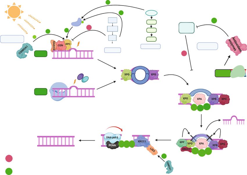
2                                                                                      Oxidative Medicine and Cellular Longevity

DNA adducts caused by UV light is the nucleotide excision            ity and mortality (15-20%) than NMSC [13, 20]. Two signifi-
repair (NER) pathway [8]. NER pathway deficiencies lead to            cant hazard factors that are related to the pathophysiology of
the development of various genetic disorders, including xero-        numerous cutaneous carcinogenesis are environmental (like-
derma pigmentosum (XP), the main focus in our review. XP is          wise named modifiable) and hereditary (additionally named
a rare autosomal recessive genodermatosis varying in terms of        nonmodifiable) factors [21, 22]. The most well-known envi-
nomenclature from (XP-A to XP-G and XP-V) depending on               ronmental hazard factor for all skin malignancy types is the
the type of mutation affecting one of eight different XP genes         exposure to UVR, which can damage DNA in skin cells like
[9–11]. XPC has the most significant number of patients, with         keratinocytes, melanocytes, and fibroblasts [17, 18]. In multi-
80 to 90% of cases, depending on the country of origin. How-         cellular life forms, the regulation of gene expression is attained
ever, an ongoing debate about the heterogeneity of XP-C              via signaling transduction pathways, mediating the develop-
patients in terms of the clinical manifestations (nonmelanoma        ment of well-organized physiological processes fundamentally
and/or melanoma skin cancers) does exist. The XPC gene is            engaged with skin cells development, proliferation, division,
located on chromosome 3p25, comprising about 15 introns              death, and differentiation [23, 24]. Understanding the intracel-
and 16 exons encoding for a functional XPC protein [12].             lular signals as well as the processes through which cells
XPC is a DNA repair protein involved in DNA damage recog-            receive and incorporate extracellular stimulus is critical for
nition in the NER pathway allowing further events to occur,          the identification and advancement of novel therapeutics for
restoring the standard DNA copy. Furthermore, the XPC                cutaneous malignancies. The mechanism of UVR at the
protein might also be involved in vital parts of DNA damage          molecular level is related with the expanding various DNA
responses, including programmed cell death and cell cycle            harm signals, e.g., initiation of the p53 pathway which signifi-
checkpoints [19]. Upon the loss of XPC’s expression, and after       cantly adjusts cell physiology to intercede cell cycle arrest and
extensive UVB-induced DNA lesions, mutations might                   enact DNA repair. If DNA remains unrepaired, p53 can
develop in genes encoding for essential proteins involved in         directly trigger programmed cell death to prevent tumor
the signal transduction pathways. The latter is essential to         development [25, 26]. Strikingly, the exposure of human
convey external stimuli into the target cell. Disorganization of     keratinocytes to UVB radiation initiates the PI3K/AKT/m-
signal transduction pathways is a major cause of skin cancer         TOR-S6K1 pathway [27]. The latter is a key pathway engaged
development in XPC patients. The aim of our work is to               with an assortment of physiologic capacities linking nutrients
characterize the proteomic signature of XPC at basal state           and growth factors to metabolism, cell development, prolifer-
and after UVB irradiation. In this article, we will review signal-   ation, angiogenesis, survival, apoptosis, and protein and lipid
ing pathways disrupted in skin cancer to understand how skin         production. This pathway is dysregulated in various malig-
cancers develop in XPC patients. Furthermore, we will also dis-      nancies including melanoma and nonmelanoma skin tumors
cuss about signaling pathways affecting NER’s activity, proteins      [28, 29]. In this part, we will discuss the molecular signaling
linked to signaling pathways influencing wild-type XPC’s func-        in NMSC and cutaneous melanoma (CM).
tion, and a little information about XPC loss and skin cancer.
                                                                     2.1. Signaling Pathways in NMSC. Basal cell carcinoma
2. Signaling Pathways Disrupted in Skin Cancer                       (BCC) and cutaneous squamous cell carcinoma (SSCs) are
                                                                     derived from dysregulated keratinocytes present in the basal
Skin cancer is one of the most common cancer types occur-            layer of the epidermis, both establishing the principle types of
ring in the overall population of people, especially in the          nonmelanoma skin cancer (NMSC) [30, 31]. The morpho-
white populace; with over a million cases distinguished in           logical highlights of BCC comprise the appearance of a gath-
the world every year [13–15]. Skin cancer classification and          ering of tumors that are made out of cells having cellular
nomenclature depend on the type of cells from which they             constituents like basal epidermal cells in the undifferentiated
arise and the clinical outcome. The two most common                  state. A significant component of BCC is the palisade course
subtypes of nonmelanoma skin cancer (NMSC) are basal cell            of action of epidermal cells in the tumor fringe that isolates
carcinomas (BCCs) and cutaneous squamous cell carcinomas             the tumor from the encompassing stroma. These cells regu-
(SSCs) both originating from the basal layer of the skin             larly give the tumor nodular shape or structure a band or
epidermis [16]. Although they share many similarities, they          string encompassing it. Contrasted with their typical part-
have different incidence rates and etiology. BCCs are consid-         ners, the tumor cells have less chromatin-rich nucleus and
ered to be the most frequent (80%-85%) followed by SCCs              cytoplasm, favoring mitotic division and also apoptotic cell
(15-20%). BCC is slow-growing and rarely metastatic (less            demise, reflecting steady development of the tumor. Clinical
than 0.01%), and most of them are often treated by surgery.          manifestation of BCC may show up in various morphological
Even though mortality is low, this threat causes morbidity           examples: nodular or cystic, shallow, infiltering, and sclerotic
and a colossal weight on social insurance frameworks around          or pigmented, which also differ in their site event [32, 33].
the world [17–19]. On the other hand, the prognosis of SCCs          The second most common type of NMSC is generally SSCs
is worse because they can be invasive with a significant              [34]. SSCs can metastasize from the epidermis to the dermal
propensity to metastasize (2-5%). Besides, a high percentage         layers as well as to the local lymph nodes where around 5% of
of SCC patients develop second primary skin cancer within 5          patients developed metastatic tumors [35]. Clinical introduc-
years of diagnosis. The third main class of skin cancer, malig-      tion including however not constrained to hyperkeratotic
nant melanoma, originating from the melanocytes, is less             plaque also leads to the arrangement of nodular mass or
frequent (less than 10%) but is associated with higher morbid-       ulceration on the skin, which might be related to torment,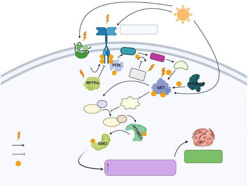
Oxidative Medicine and Cellular Longevity                                                                                           3

pruritus, or blood draining [36]. Actinic keratosis (AK) and          signaling pathway (e.g., PTCH1, SMO, and SUFU) will result
Bowen’s sickness are two premalignant types of SSCs, which,           in an increased expression of GLI1 leading to constitutive pro-
if not treated well, would develop malignant transformation           liferation that favors skin tumorigenesis. The SHH pathway is
phenotype [37]. Even though most of the SSCs are effectively           mostly associated with the etiology of BCC. Furthermore,
removed by surgical intervention, around 20% of skin cancer           mutations occurring in the PTCH1 gene have been character-
deaths are linked to SSCs [38]. Given its expanding frequency         ized in BCC patients with rare genetic disorders including
and poor prognosis, SSCs is emerging as a public medical prob-        xeroderma pigmentosum (XP) as well as in sporadic BCC.
lem. Understanding the molecular pathways relying behind              Around 60% of BCC and XP patients have shown that most
skin cancer development as a result of UVR exposure is quite          of PTCH point mutations acquire the UV signature (i.e.,
complicated. UVR can be classified as a carcinogen capable of          C → T and CC → TT transitions at dipyrimidine sites) [46].
initiating and promoting skin cancer due to its capacity to           Besides, UVR induced mutations in the PTCH1 gene account
reach the basal layer of the epidermis, triggering alterations in     for around 50% of sporadic BCCs where large and small dele-
keratinocytes. Initiation of skin tumors in response to UVR           tions occur within this gene, leading to uncontrolled cell cycle
exposure can occur through the activation of signaling path-          progression, inhibition of apoptosis, induction of angiogene-
ways that favor the survival stimulus in keratinocytes, opposing      sis, and proliferation which are hallmarks for NMSC [47].
the programmed cell death pathway. Activation of these signal-        Furthermore, it was also shown that SSCs has the potential
ing pathways can occur through direct DNA damage of critical          to overexpress Sonic Hedgehog (SHH), PTCH, and the most
genes that might act either as oncogenes or tumor suppressor          important target of HH signaling GLI1 [48, 49]. In addition,
genes, activation of transmembrane receptors involved in sig-         mouse models having SSCs have also shown that PTCH1
nal transduction cascade events like receptor tyrosine kinases        had the potential to act as an oncogene, if overexpressed
(RTKs), or via the elevation of inflammatory responses and             [50]. These alterations inactivate PTCH’s work and permit
immunosuppression mechanisms [39, 40]. The upregulation               constitutive actuation of the SMO-GLI pathway, which is by
of these pathways can then promote carcinogenesis by favoring         all accounts adequate for NMSC advancement [45]. UVR
the proliferation of these damaged cells [41]. Understanding          can also cause mutations in SMO and SUFO genes causing
the dysregulation of the signaling pathways at the molecular          aberration in the HH signaling pathway [51].
level as a result of UVB exposure would pave the way towards
specific targeted therapy to treat NMSC. Upregulation of the           2.1.2. Wnt/β-Catenin Signaling Pathway. The Wnt (Wingle-
Hedgehog pathway has been shown as an essential component             ss/INT-1) signaling is another pathway playing vital roles in
required for NMSC progression; however, other nonstandard             cellular expansion and proliferation as well as hair growth
pathways, for example, Wnt/β-catenin, p53, p16, COX-2,                and development in the skin. Similar to the HH pathway,
CDKI2A, PI3K/AKT/mTOR-S6K1, and Ras-Raf-MEK-ERK                       those two pathways crosstalk to sustain normal physiological
signaling pathways have been also involved in the pathogenesis        processes in the human body. Besides, dysregulation of the
of NMSC (Figure 1).                                                   Wnt pathway is implicated in the development of various
                                                                      cancers including NMSC [52]. β-catenin is a vital actor medi-
2.1.1. Hedgehog Signaling Pathway. Sonic Hedgehog (SHH), a            ating the downstream effects of the Wnt signaling pathway.
highly conserved pathway, is associated with organogenesis,           β-catenin is usually implicated in the formation of adherens
development patterning, tissue growth, mitogenesis, homeo-            junction between cells found in the skin [53]. In the absence
stasis, tissue repair, and hair follicle growth and sebaceous         of Wnt, β-catenin is sequestered in the cytoplasm by various
glands in the skin [42]. The canonical HH pathway comprises           protein regulators including adenomatous polyposis coli
the HH ligands as Indian HH, Sonic HH, and desert HH, the             (APC), axin, and glycogen synthase kinase β (GSK3β), where
transmembrane receptor proteins patched (PTCH1 and                    β-catenin is phosphorylated by GSK3β and targeted to pro-
PTCH2) which are classified as tumor suppressor genes,                 teasomal degradation to ensure its control and regulation.
smoothened (SMO), and glioma-associated oncogene (GLI)                When Wnt is coupled to its receptors, LDL receptor-related
transcription factors 1, 2, and 3 (GLI1, GLI2, and GLI3)              protein (LRP-6), and frizzled belonging to a family of 7-
[43]. This pathway is enacted when HH ligands bind to and             transmembrane receptors at the surface of the cell mem-
inhibit PTCH1, thereby initiating SMO downstream effects               brane, GSK3β becomes inactivated. This causes β-catenin
which in the absence of HH ligands is repressed by PTCH1              to remain dephosphorylated, thus preventing its proteasomal
[44]. SMO translocates and accumulates in the primary cilium          degradation. β-catenin can thereafter translocate to the
where this pathway befalls, allowing further cascade events to        nucleus, where it exerts its function as a transcriptional coac-
occur which in turn prompts the departure of the GLI                  tivator binding to TCF (T cell factor) motif where TCF then
proteins, sequestered in the cytoplasm with various proteins          binds and turns on the expression of Wnt target genes.
negatively controlling them like p53, PKA, and PKC-δ and              Obviously, the Wnt signaling pathway appears to act as a
the suppressor of fused (SUFU). Then, GLI proteins enter into         key controller in NMSC’s progression. Various genomic
the nucleus and turn on the expression of GLI-associated              and transcriptomic investigations have revealed that the
genes, which are responsible of producing proteins responsible        Wnt pathway is being dysregulated in SCCs. Perhaps the
for the cellular destiny, associated with proliferation, viability,   most punctual examination utilizing genomic hybridization
angiogenesis, and self-renewal. GLI1 exerts a negative feed-          discovered amplification of chromosome arms 7q, 8q, 11q,
back loop that autoregulates HH signaling by modulating               and 17q which includes Wnt and frizzled genes in SCC lines
PTCH1 [45]. UVR induced mutations at any level of the HH              suggesting its link with SCC progression [54]. Another study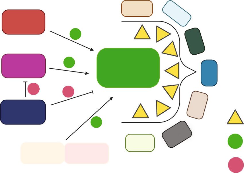
4                                                                                                      Oxidative Medicine and Cellular Longevity

                         UV                                                                                                           B
                           B                                                                                                     UV

                                                                                                               Wnt
                                 SHH
         HH pathway                                                                                                            LRP6
                                                                                                                                          Wnt pathway

                                                           Keratinocyte
                                                                                                                       P
                                                                                        Inflammatory                  P
                                                                                        response                     P
                                                                            COX-2

                                                                                                                     Ub

                                     P   P
                                                                                                                           P
                                                                                                                     Ub          Proteosomal
                              SUFO                                                                                               degradation
      Symbols:

            UVB targets                                        Cell growth + survival                                            Elevation by COX-2
                                                                    Angiogenesis
            Mutation                                            Migration/Invasion                                         BCC              cSCC

            Inhibition

            Translocate
            Expression                       Promoter Target gene                       Promoter Target gene
                                                                                                                               Development
            Activation
                                                                    Overexpression                                              of NMSCs
            Inactivation
                                                                       Nucleus

Figure 1: Development of NMSCs due to the dysregulation of Wnt, HH, p53, and COX-2 signaling pathways in keratinocytes triggered by
UVB radiation. UVB radiation can cause alterations in PTCH1, SMO, and SUFO in the HH pathway and p53 and β-catenin in Wnt pathway
that will eventually drive constitutive expression of downstream effectors which are GLI1, β-catenin, and COX-2 favoring the overexpression
of target genes (protooncogenes) like N-MYC leading to uncontrolled cell growth, survival, angiogenesis, and migration and invasion
required for NMSCs (BCC, SCCs). “Created with http://BioRender.com/.”

showed the increase of Wnt ligands and receptors at the                       thereby leading to the constitutive activation of this pathway
mRNA level using gene expression array analyses in SCC                        allowing the development of skin carcinogenesis [60].
samples [55]. Mutations in APC which is classified as a tumor
suppressor gene will eventually lead to the destabilization of                2.1.3. p53, p16, and COX-2 Signaling Pathways. At any time,
the complex sequestering β-catenin allowing its constitutive                  the cell has several systems for arresting the cell cycle if the cas-
translocation to the nucleus. In addition, mutations of β-                    cade of events does not occur in an orderly fashion, like when
catenin or mutations in axin would all lead to the constitutive               the DNA is damaged. The TP53 gene is classified as a key
activation of the Wnt signaling pathway, thereby driving                      tumor suppressor gene playing vital roles in cell cycle regula-
extensive expression of Wnt target genes which favors the                     tion. p53 is a proapoptotic protein, belonging to the Bcl-2
development of BCCs [56]. Increased expression of Wnt                         protein family that guards the genome to ensure its integrity
proteins has also been shown as a significant activator of this                and stability by orchestrating DNA damage responses [61].
pathway observed in BCCs [57, 58]. Dysregulation of the                       Exposure to mutagenic environmental factors would eventu-
Wnt/β-catenin signaling favors the upregulation of BIRC5/-                    ally create DNA lesions sensed by p53, which activates cell
Survivin which is implicated in inhibiting caspases mainly                    cycle checkpoints to initiate DNA repair. If repair systems
caspase-3 and caspase-7 thus preventing apoptosis leading to                  were incapable of resolving the error, p53 can halt the cell cycle
the immortality of tumor cells [59]. Also, UVB can be an                      events and trigger programmed cell death in order to prevent
important cause for the rise of β-catenin levels acting on                    the development of tumors. Extensive exposure to UVB
cyclooxygenase-2 (COX-2) thus augmenting the production                       radiation absorbed by the keratinocyte’s DNA will eventually
of prostaglandin E2 (PGE2) which in turn increases the                        cause mutations in the TP53 gene, leading to its inactivation
inflammatory response that favors β-catenin’s elevation                        thus favoring the development of skin cancers. TP53 gene
Oxidative Medicine and Cellular Longevity 5
mutation can be present in around 50% of BCC and approxi- via intracellular signaling cascades. The upregulation of
mately 90% of SCC cases [128]. Biopsy analysis revealed that EGFR has been reported in SCCs [70]. Skin tumorigenesis
sun-exposed skin showed C → T transversions in DNA requires EGFR activation in keratinocytes caused by UVR
sequences of TP53 named as p53 patches [62]. The recurrence [71]. The generation of reactive oxygen species (ROS) due
of C to T transitions most commonly occur at the trinucleo- to UVR exposure could be responsible for the rapid activa-
tide sequence 5 ′ -PyCG in the TP53 gene in UV-induced skin tion of EGFR rather than through growth factor binding
tumor [63]. Hotspot mutation sites present in the TP53 gene [72]. An important regulator of the EGFR activity is
are numerous caused mainly by UVB radiation. UVB lamps receptor-type protein tyrosine phosphatase- kappa (RPTP-
inducing skin tumors in mice have identified a hotspot muta- κ), maintaining EGFR in its inactive state, unphosphorylated.
tion at codon 270 correlated to a sequence change from 5 ′ Under UVR exposure conditions, ROS triggers oxidative
-TCGT to 5 ′ -TTGT [64]. Furthermore, mutations in codon inhibition of RPTP-κ thus leaving EGFR uncontrolled [73].
177 of the TP53 gene are classified as a specific marker for Protooncogene tyrosine-protein kinase Src may be also
BCC development, whereas mutations in codon 278 appear activated by UVR thus phosphorylating EGFR promoting
to be explicit for SCCs [50]. The expression level of p53 can carcinogenesis [74]. EGFR activated via UVR stimulates
be used as a prognostic marker for various skin cancers. p16 various downstream effectors, including the PI3K/AKT/m-
is another important key protein encoded by the CDKN2A TOR-S6K1 pathway. Initiation of this pathway occurs upon
gene which is a tumor suppressor gene regulating negatively the activation of RTKs and EGFR leads to the actuation of
the cell cycle progression in the G1-S transition by inhibiting phosphatidylinositol 3-kinase (PI3K), which in turn phos-
cyclin-dependent kinase 4 (CDK4). Mutations in the CDK2NA phorylates phosphatidylinositol [4, 5]-bisphosphate PIP2 to
gene would drive the cell cycle progression thereby allowing the phosphatidylinositol [3, 4, 5]-trisphosphate PIP3. In the acti-
constitutive proliferation of skin cells and the development of vation of protein kinase B (AKT), a serine/threonine-specific
skin tumors. UVR is associated with causing alterations within protein kinase occurs through the binding of PIP3 to the N-
this gene. Exon 2 of the CDK2NA gene had six distinct muta- terminus of AKT, allowing its translocation to the inner
tions: 5 out of 21 patients suffering from squamous lesions leaflet of the plasma membrane where it becomes phosphor-
and 1 out of 28 BCCs patients. The UVR signature presented ylated by phosphoinositide-dependent kinase-1 (PDK1), and
in exon 2 of this gene included two C : G to T : A transitions by the mammalian target of rapamycin (mTORC2). Regula-
and two transversions [65]. Cyclooxygenase-2 (COX-2) is a tion of AKT’s function occurs via phosphatase and tensin
key enzyme that catalyzes the conversion of arachidonic acid homolog deleted from chromosome ten (PTEN) which is
to prostaglandins playing an important role in increasing the classified as a tumor suppressor gene. AKT is an essential
inflammatory responses [66]. The major product produced part of this pathway required to convey the signals that are
upon COX-2 activation is the prostaglandin E2 (PGE2) which responsible for the regulation of various cellular processes.
is highly synthesized after UV exposure due to COX-2 activity Furthermore, AKT then activates mTORC1 through Rheb-
[67]. This prostaglandin exerts various effects at the biological GTP. mTORC1 phosphorylates downstream p70S6 kinase 1
level by suppressing programmed cell death and enhancing (S6K1) involved in protein synthesis, cell cycle progression,
cellular proliferation [68]. There is a strong correlation between cell growth, and survival (Figure 2). According to recent
the overexpression of COX-2 and NMSC development. A studies, UVR can regulate PTEN’s function in keratinocytes
major cause of COX-2 overexpression could be due to UV-B [75]. Exposure to UVR causes modifications in the PTEN
radiation exposure and the dysregulation of various signaling gene [76]. UVR also can inhibit PTEN’s function through
pathways through their permanent activation, like PI3K, ROS, favoring AKT activation thus the development of skin
MAPK, and NF-κB, that drives uncontrolled cellular expansion tumors [77]. At the level of BCC, even though little is known
and proliferation. Under normal physiological conditions, with regard to PTEN’s function in BCC, upregulation of the
wild-type p53 has a negative impact on COX-2 by decreasing PI3K/AKT pathway could be due to PTEN gene mutation
its expression. Besides, there was no significant reduction in [78]. Decreased PTEN expression levels lead to a threatening
the COX-2 levels in p53 mutant [69]. This could be a major change of the skin actuated by UVA (315–400 nm), as shown
reason for understanding why COX-2 levels are high in various by the development of SCC in nude mice [75]. UVR-induced
cancers including BCC and SCCs and low in normal epithelial AKT actuation can occur via an autocrine manner or ROS
cells including keratinocytes. Upon their mutation, p53 and activating growth factor receptors bearing RTK activity
p16 comprise two unique pathways that could turn on the [79]. It is worth mentioning that the PI3K/AKT signaling
COX-2 activity to increase PGE2 levels favoring NMSCs. pathway also affects other signaling pathways including
Ras-Raf-MEK-ERK which due to the crosstalk presence could
2.1.4. PI3K/AKT/mTOR Signaling Pathway. Transmembrane also be dysregulated favoring skin tumorigenesis.
receptors are embedded in the cell membrane, participating
in various biological processes. One huge family of such 2.2. Signaling Pathways in Cutaneous Melanoma. The overall
receptors is receptor tyrosine kinases (RTKs). Epidermal rate of cutaneous melanoma (CM) has been increasing every
growth factor receptor (EGFR) is categorized as an RTK. year at a progressively rapid rate contrasted with some other
The standard pathway begins upon the coupling of epidermal types of skin cancers [80]. CM originates from the transfor-
growth factor (EGF) on its receptor inducing receptor dimer- mation of melanocytes which are derived from neural crest
ization and phosphorylation of tyrosine residues in the cyto- cells located mainly in the epidermis of the skin [81].
solic portion of the receptor to mediate downstream effects Although it is the least common form comprising around6 Oxidative Medicine and Cellular Longevity
B
UV
EGFR Activation by UVB
PIP2
SRC PIP3
P P P
P P PDK
1
te
in ocy P PTE
N
P
rat
Ke
P
P P
GTP TSC1/2
Cytoplasm
Rheb
GTP
Rheb
Symbols:
UVR P
P
Activation
Inhibition Skin carcinogenesis
development
P Phosphorylation Cell survival, protein synthesis, cell
cycle progression, proliferation
Figure 2: The key targets of UVR in the PI3K/AKT/mTOR-S6K1 pathway. UVB exposure can cause various alterations in the
PI3K/AKT/mTOR-S6K1 pathway where any functional loss of PTEN or RPTP-κ or hyperactivity of EGFR, Src, or AKT would eventually
lead to constitutive cell survival, protein synthesis, cell cycle progression, and proliferation, which are the hallmarks of skin carcinogenesis.
“Created with http://BioRender.com/.”
1% of total skin cancers, CM is classified as the most aggres- present at the genetic level as alterations in B-RAF were
sive form due to its high metastatic capacity [82]. The occur- detected in ~60% while for N-RAS reveals ~15-30% of total
rence of cutaneous melanoma varies enormously among melanomas [86]. Both of them are classified as main oncogenes
nations, and these diverse frequency designs are attributed to essential for the mitogen-activated protein kinase (MAPK)
varieties in skin phototype, just as contrasts in sun exposure. pathway and mutations within these oncogenes would turn
Additionally, and in contrast to other solid tumors, melanoma them on constitutively, dysregulating the MAPK pathway, thus
generally influences youthful and moderately aged people favoring melanomas. Besides, B-RAF mutations were predom-
(middle age at analysis, 57 years). The frequency increments inantly found in body areas harmed by the sun but at irregular
directly after the age of 25 years until the age of 50 years, and intervals while for N-RAS they were present in damaged body
afterward, starts decreasing, especially in females [83]. The areas that were continuously exposed to the sun [87, 88].
major cause of CM development relies on the degree of sun’s Codon 600 present within B-RAF’s gene appears to be the most
exposure where UV-B radiation is the most implicated factor commonly mutated site where a single-base substitution allows
for inducing melanomas as an environmental factor where it the conversion of valine to glutamic acid producing a constitu-
could be direct when melanocyte’s DNA absorbs UVB photons tively active protein version. In addition, the N-RAS gene also
or indirect in which fluorophores including flavins and por- appears to have mutations at codon 61 converting glutamine
phyrins absorb UVB photons and generate ROS that encom- to lysine [88]. Furthermore, the involvement of UV signature
passes mainly hydrogen peroxides and superoxide anions mutation at these sites is still unknown and not fully under-
leading to the disruption of signaling pathways at the molecular stood. A third newly discovered oncogene as being linked to
level [84]. CPDs resulting from UV-B radiation are the most cutaneous melanomas is RAC1. The latter is linked to the
common types of DNA lesions. TT > TC > CT > CC is the PI3K/AKT pathway where any activating mutation in RAC1
order of their formation from the highest (TT) to lowest would dysregulate this pathway and favor cutaneous mela-
(CC) frequency [85]. Another cause related to CM could be noma development. The most common mutation involvesOxidative Medicine and Cellular Longevity 7
codon 29 converting proline to serine amino acid [89]. This of this pathway in the regulation of NER’s work is yet disput-
mutation harbors a UV-signature mutation which is C > T able. AKT can activate the mouse double minute 2 homolog
transition where UVR is most probably responsible for this (MDM2) which is a negative regulator of p53 thus favoring
mutation [90]. Patients suffering from xeroderma pigmento- its downregulation [94]. Along these lines, AKT1 has the
sum or familial retinoblastoma disorders are at higher risk for potential to inhibit NER because of the reduction in the levels
developing CM. Dysregulation of signaling pathways in CM of XPC and damage specific DNA binding protein 2 (DDB2)
had been widely reported where the main signaling pathways which are dependent on TP53 for their expression at the tran-
include PI3K/AKT/mTOR, Ras-Raf-MEK-ERK, and TGF-β scription level and are considered key effectors in the NER
signaling pathways augmenting the aggressiveness of CM. pathway [95]. Another method of hindrance is interceded by
BRAF, NRAS, CDKN2A, CDK4, and other RTKs like ROS1, the AKT1-dependent subcellular localization of XPC tran-
ALK, MET, RET, and NTRK1 are also other candidates for mel- scriptional repressors (P130) thereby inhibiting NER [96].
anoma development to be considered [91, 92]. Besides, AKT1 can boost TC-NER through the phosphoryla-
tion of EP300 which acts as a histone acetyltransferase that
3. Signaling Pathways Modulating NER undergoes chromatin remodeling processes to regulate tran-
scription events loosens up the chromatin to permit the
Nucleotide excision repair (NER) is a DNA repair pathway recruitment of key repair players including XPC [97].
involved in the removal of bulky DNA adducts caused by
exposure to UV light. NER can be divided into 2 subpath- 3.2. CSNK2A1 (CK2α1) Signaling Pathway Regulating NER.
ways: global-genome nucleotide excision repair (GG-NER) Casein kinase 2 (CSNK2A1) is a serine/threonine kinase
and transcription-coupled nucleotide excision repair (TC- involved in various signaling pathways and has the potential
NER) differing in the strategy of DNA damage recognition. to modulate NER’s activity. Recent studies demonstrated that
Upon UVB exposure, damage recognition in the GG-NER CSNK2A1 could be a potential regulator of the single- and
occurs via XPC and other coupled key players like Rad 23 double-stranded breaks of the DNA [98, 99]. X-ray repair
homologue B (RAD23B) and centrin 2 (CEN), while for cross-complementing protein 1 (XRCC1), a DNA repair pro-
TC-NER, this occurs via RNA polymerase II present in the tein playing a critical role in the NER particularly at the level
expressed regions allowing the further recruitment of of the DNA synthesis and ligation step in the NER, appeared
cockayne syndrome proteins A and B (CSA and CSB). The to be a target for CSNK2A. The latter improves the stability
upcoming steps are similar for both subpathways where the of the XRCC1-ligase III complex by triggering phosphoryla-
second step involves the opening of the DNA double helix tion of XRCC1 which positively influences NER’s activity
where transcription factor II H (TFIIH) participates and [99, 100]. Also, XPC appears to be another target for
XPD and XPB helicases progress to unwind the DNA before CSNK2A1 by which phosphorylation can occur on the
repair. The third step involves DNA damage confirmation amino acid serine at position 94, thus enhancing photoprod-
via replication protein (RPA1), XPA, and XPG. RPA1 binds ucts repair at the level of the NER [101].
to the single-stranded DNA preventing its rewinding and
XPA confirms the damage. Upon verification, the fourth step 3.3. MAPK Signaling Pathway Regulating NER. The mitogen-
which is excision involves the recruitment of XPF and activated protein kinase (MAPK) pathway is responsible for
excision repair cross-complementation group 1(ERCC1). various cellular processes. This pathway comprises different
Excision at the 5 ′ and 3 ′ sites of the damaged part of the signaling cascades of which the Ras-Raf-Mek-extracellular
DNA occurs via nucleases which are XPF and XPG. Finally, signal-regulated kinase 1 and 2 (ERK1/2) is responsible for
the fifth step involves DNA synthesis and ligation. DNA conveying the signal into the nucleus upon RTK activation.
polymerase delta (Polδ) with other important factors like The standard pathway begins upon the coupling of epidermal
proliferating cell nuclear antigen (PCNA) polymerize new growth factor (EGF) on its receptor inducing receptor dimer-
complementary bases; then after synthesis, the ligation step ization and phosphorylation of tyrosine residues in the cyto-
is required to seal the gaps between nucleotides by DNA solic receptor, then binding of GRB2 which is an adaptor
ligase 3 (LIG3) cooperating with X-ray repair cross- protein and SOS, the Ras guanine nucleotide exchange factor
complementing protein 1 (XRCC1) which acts as a linker located near Ras protein. SOS exchanges GDP bound to Ras
between Polδ and LIG3 bringing them into close proximity by GTP bound to Ras making it active. Activated Ras binds
ending up with the restoration of the normal DNA version and turns on Raf (serine/threonine kinase) which in turn
[93]. NER’s function can be modulated and influenced by activates MEK having dual activity as a serine/threonine
various signaling pathways that might impact its repair activ- and tyrosine kinase activates MAPK (JNK, ERK1/2, or
ity in terms of removing DNA adducts either positively or MAPK14) that dimerizes and translocates to the nucleus to
negatively. PI3K/AKT1, CSNK2A1 (CK21), MAPK, and activate ternary complex factor (TCF) that enables further
NFE2L2 (NRF2) are the main signaling pathways implicated activation of transcription factors. Besides, MAPK14 is
(Figure 3). responsible for halting the cell cycle progression as well as
regulating programmed cell death, while MAPK1 is responsi-
3.1. PI3K/AKT1 Signaling Pathway Regulating NER. As men- ble for controlling cellular proliferation and differentiation
tioned previously, this pathway is switched on via the RTKs processes [102]. Activation of ERK1/2 has been shown to
mediating the activation of PI3K1 which in turn activates decrease lesions [103]. Upon UVR exposure, EP300 is
several signaling cascades to activate AKT1. The inclusion enlisted to the harm site in the heterochromatin and is8 Oxidative Medicine and Cellular Longevity
+
+ RAS-GTP
0 PI3K
Raf +
30 P
EP
− PDK1
ROS
+ + MEK
P AKT
CSNK2A1 pathway ERK
Survival MARK pathway NRF2 pathway
AKT pathway
–
GG-NER
1) Damage recognition ARE NRF2
A
CS B
CS
TFIIH
3) DNA damage confirmation
RNA pol II
TC-NER
2) Unwinding of the double helix TFIIH
RPA
5) DNA synthesis and ligation
Normal DNA version
RPA
RPA
+
− Inhibits NER
4) Excision of the damaged region
+ Enhances NER
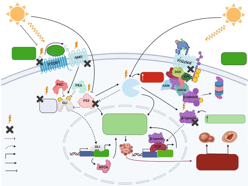
Figure 3: PI3K/AKT1, CSNK2A1 (CK2 α 1), MAPK, and NFE2L2 (NRF2) signaling pathways regulating NER’s pathway. The PI3K/AKT1 and
MAPK pathways regulate XPC positively via EP300, the CSNK2A1 pathway also can affect positively both XPC and XRCC1-Lig3 complex,
and the NRF2 pathway can enhance NER’s activity positively by inducing the expression of antioxidant enzymes inhibiting ROS. On the other
hand, AKT also can inhibit XPC under certain conditions. “Created with http://BioRender.com/.”
phosphorylated by MAPK14, MAPK1, and AKT1 expanding ditions, NFE2L2 will be translocated to the nucleus where it
histone acetyltransferase (HAT) action to add acetyl groups interacts with musculoaponeurotic fibrosarcoma (MAF) tran-
to H3 and H4 histones prompting chromatin unwinding thus scription factors and bind to antioxidant-responsive element
favoring the recruitment of XPC, DDB2, and other players of (ARE) present within the DNA sequence [109]. Transcription
the NER pathway [104]. of this region will encode for various important antioxidant
enzymes (such as glutathione S-transferase (GST), catalase,
3.4. NFE2L2 (NRF2) Signaling Pathway Regulating NER. peroxidase, and superoxide dismutase) which positively influ-
Nuclear factor erythroid 2-related factor 2 (NRF2), also ence NER’s activity [110–112].
known as nuclear factor erythroid-derived 2-like 2, is a tran-
scription factor classified as a basic leucine zipper (bZIP) 4. XPC and Signaling Pathways
responsible for controlling the expression of genes exhibiting
antioxidant activities (such as the NFE2L2 gene) to protect XPC is a key player of the GG-NER pathway involved in
against oxidative attacks causing inflammation and lesions sensing helical distortions formed upon DNA damage. XPC
[105]. Oxidation of lipids, caused by oxidative damage can is formed of around 940 amino acids and possess various
block the activity of NER in terms of DNA photolesion repair domains involved in the interaction with the damaged parts
[106]. Furthermore, the inflammation-inferred monochlora- of the DNA as well as other repair factors to proceed in the
mine (NH2CL) represses NER by means of the restraint of repairing process [113]. Although the role of XPC in signal-
p53’s phosphorylation [107]. It was additionally shown that ing pathways is not yet well understood, a recent study has
the exposure of epithelial cells to H2O2 diminishes the NER demonstrated the association between XPC and proteins
ability to less than half of its total activity (Oxidative Medicine and Cellular Longevity 9
TSPAN6 EM
P2
MAP3K5
+ ? ?
TJP
1
?
+
PTEN
SPCS1
XPC
?
−
−
?
RAB
1A
?
PTP4A2 ?
? Unknown effect
+ TR
AV
4
SRGN + Positive effect
PRKACB PRKACA
− Negative effect
Figure 4: Proteins related to signaling pathways regulating XPC’s activity. MAP3K5, PTEN, PRKACB, and PRKACA appeared to have a
positive effect, while for PTP4A2 showed a negative effect on XPC’s activity. SRGN, TRGV4, RAB1A, SPCS1, TJP1, EMP2, and TSPAN6
are newly identified interactors but with unknown effect on XPC’s activity. “Created with http://BioRender.com/.”
for the recruitment of XPC and TFIIH to the harmed DNA protein in the endoplasmic reticulum, and EMP2 has roles
locales [115]. Mitogen-activated protein kinase kinase kinase in blastocyst implantation [118]. Although these proteins are
5 (MAP3K5) who is responsible for the initiation of the p38 identified as interactors with XPC, their impact on XPC’s role
pathway has been identified as an interactor with XPC, thus are unrevealed yet.
acting as a positive regulator. Furthermore, PTEN also
appeared to positively influence XPC at the transcriptional 5. XPC and Skin Cancers
level [116]. Protein tyrosine phosphatase type IVA, part 2
(PTP4A2), otherwise called PRL2, is a phosphatase having XP is caused by mutations in genes involved in the NER
an oncogenic potential and acting as a repressor for PTEN pathway, and its naming comes from the genes which are
leading to its downregulation, thus allowing the further pro- mutated (XP-A to XP-G and XP-V). Patients acquiring XP
gression of the AKT signaling pathway. Taken all together, have a risk to develop skin cancers 10,000-fold more than
inhibiting PTEN would negatively influence XPC at the tran- normal patients because of their high sensitivity to UVR.
scriptional level, thus affecting DNA damage recognition in For both XP and non-XP having skin cancers, dipyrimidine
GG-NER. Cyclic adenosine monophosphate-dependent pro- sites appear to be the most targeted region where mutations
tein kinase A (cAMP-dependent PKA) is a member of the occur. For non-XP tumors, mutations are mostly C to T
protein kinases family that has quite essential roles at the transitions while for XP tumors they are basically CC to TT
cellular levels like glucose, lipid, and glycogen metabolism. pair mutations. In addition to that, the most important
PKA comprises 4 subunits: two regulatory and two catalytic distinction between NMSC from XP and non-XP patients is
subunits present in all of its variants. XPC was shown to asso- their number and the time of appearance. While non-XP
ciate with two variants of the catalytic subunit of PKA—PR- NMSC occurs when the patients are generally at late stages
KACA and PRKACB belonging to the PKA pathway. of life (50–60 years), XP NMSC appears at early stages of life
PRKACB or PRKACA could exert their effects on the GG- (3–5 years old) and with a higher incidence rate [119]. Out of
NER by either driving XPC to the nucleus or acting at the XP cases, XPC patients are characterized by having a non-
chromatin level facilitating remodeling events. Other pro- functional global genome repair but a normally functional
teins related to signaling pathways include serglycin (SRGN), transcription coupled repair because XPC is not required
T-cell receptor gamma variable 4 (TRGV4), member RAS for the TC-NER subpathway. UVB-inducing mutations in
oncogene family (RAB1A), signal peptidase complex subunit the nonexpressed regions of the whole genome would then
1 (SPCS1), tight junction protein 1 (TJP1), epithelial mem- lead to neoplastic transformation [120]. XPC knockout in
brane protein 2 (EMP2), and tetraspanin 6 (TSPAN6) mice has been shown to favor the development of spontane-
appeared also to associate with XPC [114] (Figure 4). SRGN ous as well as UV-induced skin cancers [121, 122]. A recent
and TRGV4 play quite important roles in immune responses, study demonstrated that the downregulation of XPC favors
TJP1 is implicated in the formation of tight junctions as well the reprograming of the cellular metabolic processes thus
as migration processes, RAB1A has roles in proteins and allowing the generation of ROS via NADPH oxidase-1
vesicles trafficking between the endoplasmic reticulum and (NOX1). When activated, the dissociated subunits of NOX1
the Golgi apparatus, TSPAN6 has been found to act as an acti- will merge forming an active enzymatic complex to produce
vator of the NF-κB pathway [117], SPCS1 a transmembrane superoxide (O2-) from O2, requiring NADPH as a substrate10 Oxidative Medicine and Cellular Longevity
[123]. Besides, the rise in the levels of ROS might cause muta- the increase in skin cancer incidence. Note that 20,000 Euro-
tions in tumor suppressor genes inactivating them as well as peans die every year from skin cancer and at least 100-200
activating mutations in oncogenes, where normal cell cycle times more are treated for skin cancer, mostly by surgery.
processes become disrupted and these cells could evade So, skin cancer poses considerable burden on patients and
programmed cell death, and finally progresses to develop health care services worldwide and the situation will only get
cancer cells. NOX-induced ROS production could lead to the worse with the aging of the population. Targeting the dysreg-
activation of the PI3k/AKT as well as kinases responsible for ulated signaling pathways at the molecular level might be quite
survival responses like Src and transcription factors (NF-κB) promising to decrease the photosensitivity as well as prevent-
and could act as an inhibitor of PTEN and other phosphatases ing skin cancer occurrence in XPC patients. In this review, we
which are responsible for regulating negatively the PI3K/AKT focused on the signaling pathways dysregulated in NMSC and
pathway. Thus, XPC loss-driving metabolic disruption could CM, signaling pathways modulating NER’s function, proteins
be a key for SCC development [124, 125]. The p53 protein linked to signaling pathways affecting XPC’s activity, and little
can affect the function of DNA repair systems especially information present about XPC’s loss and skin cancers. It is
NER either directly or indirectly. A recent study had generated worth mentioning that gene therapy could be also quite prom-
XPC (-/-) p53(-/-) mutant mice models and recognized an ele- ising to replace the XPC mutant gene by a wild-type version,
vated exacerbation in UVB-induced keratosis and accelerated but this technology requires a lot of optimization.
skin cancer appearance compared with mice that are only
XPC(-/-) homozygous mutants having a wild-type p53(+/+)
[126, 127]. Keratosis is a precancerous condition that could Conflicts of Interest
rise into SCCs. Normal patients having skin cancers have
around 50% of p53 mutations while this percentage rises to The authors declare that they have no conflicts of interest.
90% in patients having skin cancer and XP disease including
XPC. This increase in mutations is attributed to UVB expo-
sure where UV signatures impact the TP53 gene which can Acknowledgments
be a hallmark favoring skin cancer progression. P16INK4a AN was supported by a grant from the graduate school EUR
and ARF are classified as two tumor suppressor proteins, Biology, Chemistry and Health from Grenoble Alpes Univer-
encoded by INK4a/ARF locus, which works together to regu- sity. WR is supported by ANR grant PG2HEAL (ANR-18-
late various cell cycle processes including the p53 and RB CE17-0017) and ANR “Investissements d’avenir” program
pathways. Furthermore, a recent study developed mouse (ANR-15-IDEX-02).
strains lacking XPC (-/-) and INK4a/ARF (-/-) and were
subjected to UVB. The results had shown a quite significant
elevation in the stimulation of epithelioid cell melanomas in References
XPC (-/-) and INK4a/ARF (-/-) mice when being compared
with XPC (-/-) and INK4a/ARF (+/+) mice [128]. Mutations [1] E. Mullaart, P. H. Lohman, F. Berends, and J. Vijg, “DNA
induced by UV appeared in PTCH and p53 genes in XP and damage metabolism and aging,” Mutation Research,
non-XP patients having BCC [129]. Thus, deciphering the vol. 237, no. 5-6, pp. 189–210, 1990.
dysregulated signaling pathways in XPC patients will draw a [2] N. L. Williams, N. J. Amato, and Y. Wang, “Replicative
correlation on how skin cancers develop in these patients. bypass studies of α-anomeric lesions of 2 ′ -deoxyribonucleo-
sides in vitro,” Chemical Research in Toxicology, vol. 30,
no. 5, pp. 1127–1133, 2017.
6. Conclusion
[3] A. Slominski and J. Pawelek, “Animals under the sun: effects
Till date, XPC patients still lack an effective treatment and of ultraviolet radiation on mammalian skin,” Clinics in Der-
follow preventative strategies. The setting up of a new matology, vol. 16, no. 4, pp. 503–515, 1998.
disease-modeling strategy of the highly prone skin cancers, [4] H. L. Lo, S. Nakajima, L. Ma et al., “Differential biologic
xeroderma pigmentosum, using the patient-derived induced effects of CPD and 6-4PP UV-induced DNA damage on the
induction of apoptosis and cell-cycle arrest,” BMC Cancer,
pluripotent stem cells (iPSCs) will allow us to understand skin
vol. 5, no. 1, p. 135, 2005.
cancer etiology, to identify risk factors in individuals, to
[5] T. Lindahl and B. Nyberg, “Heat-induced deamination of
discover protein biomarkers for the initiation, progression,
cytosine residues in deoxyribonucleic acid,” Biochemistry,
and invasion of skin cancer, and finally, to design novel ther- vol. 13, no. 16, pp. 3405–3410, 1974.
apeutic or prevention strategies. This strategy will have several
[6] T. Lindahl and A. Andersson, “Rate of chain breakage at
outcomes situated at different levels, like understanding the apurinic sites in double-stranded deoxyribonucleic acid,”
difference in the etiology of UVR-induced BCC, SCC, and Biochemistry, vol. 11, no. 19, pp. 3618–3623, 1972.
melanomas in order to establish an effective prevention strat- [7] J. H. Hoeijmakers, “Genome maintenance mechanisms for
egy, deciphering the relationship between genotoxicity, DNA preventing cancer,” Nature, vol. 411, no. 6835, pp. 366–374,
repair capacity, and cancer incidence. The discovery of skin 2001.
“tumorgenesis-initiation” biomarkers or markers for high risk [8] J. A. Marteijn, H. Lans, W. Vermeulen, and J. H. Hoeij-
(such as DNA photoproducts, cytokines, and inflammatory makers, “Understanding nucleotide excision repair and its
factors) will allow a better identification of people at risk and roles in cancer and ageing,” Nature Reviews. Molecular Cell
improvement of preventive interventions in order to limit Biology, vol. 15, no. 7, pp. 465–481, 2014.Oxidative Medicine and Cellular Longevity 11
[9] C. Masutani, R. Kusumoto, A. Yamada et al., “The XPV (xer- [26] K. Abeyama, W. Eng, J. V. Jester et al., “A role for NF-
oderma pigmentosum variant) gene encodes human DNA kappaB-dependent gene transactivation in sunburn,” The
polymerase η,” Nature, vol. 399, no. 6737, pp. 700–704, 1999. Journal of Clinical Investigation, vol. 105, no. 12, pp. 1751–
[10] J. E. Cleaver, “Cancer in xeroderma pigmentosum and related 1759, 2000.
disorders of DNA repair,” Nature Reviews Cancer, vol. 5, [27] M. S. Leo and R. K. Sivamani, “Phytochemical modulation of
no. 7, pp. 564–573, 2005. the Akt/mTOR pathway and its potential use in cutaneous
[11] E. A. De Weerd-Kastelein, W. Keijzer, and D. Bootsma, disease,” Archives of Dermatological Research, vol. 306,
“Genetic heterogeneity of xeroderma pigmentosum demon- no. 10, pp. 861–871, 2014.
strated by somatic cell hybridization,” Nature: New Biology, [28] H. Pópulo, J. M. Lopes, and P. Soares, “The mTOR signalling
vol. 238, no. 81, pp. 80–83, 1972. pathway in human cancer,” International Journal of Molecu-
[12] B. Pani and E. Nudler, “Mechanistic insights into transcrip- lar Sciences, vol. 13, no. 2, pp. 1886–1918, 2012.
tion coupled DNA repair,” DNA Repair, vol. 56, pp. 42–50, [29] R. A. Saxton and D. M. Sabatini, “mTOR signaling in growth,
2017. metabolism, and disease,” Cell, vol. 169, no. 2, pp. 361–371,
[13] J. D'Orazio, S. Jarrett, A. Amaro-Ortiz, and T. Scott, “UV 2017.
radiation and the skin,” International Journal of Molecular [30] S. Surdu, “Non-melanoma skin cancer: occupational risk
Sciences, vol. 14, no. 6, pp. 12222–12248, 2013. from UV light and arsenic exposure,” Reviews on Environ-
[14] H. W. Rogers, M. A. Weinstock, A. R. Harris et al., “Incidence mental Health, vol. 29, no. 3, pp. 255–264, 2014.
estimate of nonmelanoma skin cancer in the United States, [31] K. A. Burton, K. A. Ashack, and A. Khachemoune, “Cutane-
2006,” Archives of Dermatology, vol. 146, no. 3, pp. 283– ous squamous cell carcinoma: a review of high-risk and met-
287, 2010. astatic disease,” American Journal of Clinical Dermatology,
vol. 17, no. 5, pp. 491–508, 2016.
[15] T. Gracia-Cazaña, S. González, C. Parrado, Á. Juarranz, and
Y. Gilaberte, “Influence of the exposome on skin cancer,” [32] M. Kasper, V. Jaks, D. Hohl, and R. Toftgård, “Basal cell car-
Actas Dermo-Sifiliográficas, vol. 111, no. 6, pp. 460–470, cinoma - molecular biology and potential new therapies,” The
2020. Journal of Clinical Investigation, vol. 122, no. 2, pp. 455–463,
2012.
[16] L. Feller, R. A. G. Khammissa, B. Kramer, M. Altini, and
J. Lemmer, “Basal cell carcinoma, squamous cell carcinoma [33] C. S. Wong, R. C. Strange, and J. T. Lear, “Basal cell carci-
and melanoma of the head and face,” Head & Face Medicine, noma,” BMJ, vol. 327, no. 7418, pp. 794–798, 2003.
vol. 12, no. 1, p. 11, 2016. [34] M. Alam and D. Ratner, “Cutaneous squamous-cell carci-
[17] B. K. Armstrong and A. Kricker, “The epidemiology of UV noma,” The New England Journal of Medicine, vol. 344,
induced skin cancer,” Journal of Photochemistry and Photobi- no. 13, pp. 975–983, 2001.
ology B: Biology, vol. 63, no. 1-3, pp. 8–18, 2001. [35] A. Azimi, K. L. Kaufman, M. Ali, J. Arthur, S. Kossard, and
[18] Y. Liu, H. Wang, T. Lin et al., “Interactions between cigarette P. Fernandez-Penas, “Differential proteomic analysis of
smoking and XPC-PAT genetic polymorphism enhance actinic keratosis, Bowen's disease and cutaneous squamous
bladder cancer risk,” Oncology Reports, vol. 28, no. 1, cell carcinoma by label-free LC-MS/MS,” Journal of Derma-
pp. 337–345, 2012. tological Science, vol. 91, no. 1, pp. 69–78, 2018.
[19] L. O'Driscoll, J. McMorrow, P. Doolan et al., “Investigation of [36] L. Lansbury, F. Bath-Hextall, W. Perkins, W. Stanton, and
the molecular profile of basal cell carcinoma using whole J. Leonardi-Bee, “Interventions for non-metastatic squamous
genome microarrays,” Molecular Cancer, vol. 5, no. 1, p. 74, cell carcinoma of the skin: systematic review and pooled anal-
2006. ysis of observational studies,” BMJ, vol. 347, no. nov04 1, 2013.
[37] N. F. Agamia, D. M. Abdallah, O. Sorour, B. Mourad, and
[20] M. Watson, D. M. Holman, and M. Maguire-Eisen, “Ultravi-
D. N. Younan, “Skin expression of mammalian target of rapa-
olet radiation exposure and its impact on skin cancer risk,”
mycin and forkhead box transcription factor O1, and serum
Seminars in Oncology Nursing, vol. 32, no. 3, pp. 241–254,
insulin-like growth factor-1 in patients with acne vulgaris
2016.
and their relationship with diet,” The British Journal of Der-
[21] A. Pérez-Sánchez, E. Barrajón-Catalán, M. Herranz-López, matology, vol. 174, no. 6, pp. 1299–1307, 2016.
and V. Micol, “Nutraceuticals for skin care: a comprehensive
[38] B. Gurney and C. Newlands, “Management of regional meta-
review of human clinical studies,” Nutrients, vol. 10, no. 4,
static disease in head and neck cutaneous malignancy.1.
p. 403, 2018.
Cutaneous squamous cell carcinoma,” The British Journal of
[22] B. A. Gilchrest, M. S. Eller, A. C. Geller, and M. Yaar, “The Oral & Maxillofacial Surgery, vol. 52, no. 4, pp. 294–300,
pathogenesis of melanoma induced by ultraviolet radiation,” 2014.
The New England Journal of Medicine, vol. 340, no. 17,
[39] H. N. Ananthaswamy and W. E. Pierceall, “Molecular mech-
pp. 1341–1348, 1999.
anisms of ultraviolet radiation carcinogenesis,” Photochemis-
[23] S. Huang and P. J. Houghton, “Targeting mTOR signaling for try and Photobiology, vol. 52, no. 6, pp. 1119–1136, 1990.
cancer therapy,” Current Opinion in Pharmacology, vol. 3,
[40] C. Nishisgori, “Current concept of photocarcinogenesis,”
no. 4, pp. 371–377, 2003.
Photochemical & Photobiological Sciences, vol. 14, no. 9,
[24] S. Wullschleger, R. Loewith, and M. N. Hall, “TOR signaling pp. 1713–1721, 2015.
in growth and metabolism,” Cell, vol. 124, no. 3, pp. 471–484, [41] E. Strozyk and D. Kulms, “The role of AKT/mTOR pathway
2006. in stress response to UV-irradiation: implication in skin car-
[25] B. L. Diffey, “Solar ultraviolet radiation effects on biological cinogenesis by regulation of apoptosis, autophagy and senes-
systems,” Physics in Medicine and Biology, vol. 36, no. 3, cence,” International Journal of Molecular Sciences, vol. 14,
pp. 299–328, 1991. no. 8, pp. 15260–15285, 2013.You can also read