Tattoos - more than just colored skin? Searching for tattoo allergens
←
→
Page content transcription
If your browser does not render page correctly, please read the page content below
Review Article
Submitted: 17.8.2020                                                            DOI: 10.1111/ddg.14436
Accepted: 14.10.2020
Conflict of interest
None.

                                                                Tattoos – more than just colored skin?
                                                                Searching for tattoo allergens

Katharina T. Weiß1, Ines                                        Summary
Schreiver2, Katherina                                           During tattooing, a high amount of ink is injected into the skin. Tattoo inks contain
Siewert2, Andreas Luch2, Birgit                                 numerous substances such as the coloring pigments, impurities, solvents, emulsifiers,
Haslböck1, Mark Berneburg1,                                     and preservatives. Black amorphous carbon particles (carbon black), white titanium
Wolfgang Bäumler 1                                              dioxide, azo or polycyclic pigments create all varieties of color shades in the visible
                                                                spectrum. Some ingredients of tattoo inks might be hazardous and allergenic chemi-
(1) Department of Dermatology and
                                                                cals of unknown potential.
Allergology, University of Regensburg,
                                                                     In Germany, about 20 % of the general population is tattooed and related ad-
Regensburg, Germany
                                                                verse reactions are increasingly reported. Since tattoo needles inevitably harm the
(2) Department of Chemical and
                                                                skin, microorganisms can enter the wound and may cause infections. Non-allergic
Product Safety, German Federal
                                                                inflammatory reactions (for example cutaneous granuloma and pseudolymphoma) as
Institute for Risk Assessment (BfR),
                                                                well as allergic reactions may emerge during or after wound healing. Especially with
Berlin, Germany
                                                                allergies occurring after weeks, months or years, it remains difficult to identify the
                                                                specific ingredient(s) that trigger the reaction.
                                                                     This review summarizes possible adverse effects related to tattooing with a focus
                                                                on the development of tattoo-mediated allergies. To date, relevant allergens were
                                                                only identified in rare cases. Here we present established methods and discuss current
                                                                experimental approaches to identify culprit allergens in tattoo inks – via testing of the
                                                                patient and in vitro approaches.

                                                                                                       In this review, the term tattoo refers to permanent tat-
Decorative permanent tattoos – a
                                                                                                  toos also including so-called permanent make-up (PMU).
widespread phenomenon today                                                                       Permanent make-up is usually made by beauticians to deco-
                                                                                                  rate especially the periorbital and perioral regions [1]. Henna
For thousands of years people have tattooed their skin for                                        tattoos are not part of this article as these are not injected
various reasons. In contrast to some cultures like Polynesian                                     into the human skin.
tribes, the meaning of tattoos in the Western world seemed                                             Tattoo inks may cause a variety of health problems,
more vague over the past centuries and was mainly associa-                                        many particularly related to the skin but some also relevant
ted with low social status [1, 2].                                                                to other organs (Figure 1). Adverse effects comprise delayed
     Nowadays, tattooing has become very popular worldwi-                                         wound healing, infections, toxic or even mutagenic proces-
de. Recent polls show that about 20 % of people in Germany                                        ses, as well as granulomatous and allergic reactions [6].
and 29 % in the United States of America are tattooed [2–4].
The many tattooed role models including soccer, pop and                                           The fate of tattoo inks in the human
movie stars have led to a broader cultural acceptance of tat-
tooed skin. Tattoos are applied in special parlors or in pri-                                     body
vate locations. They are black or multi-colored and located
on almost all areas of the human body – including mucous                                          Usually, tattoo inks are passively dragged into the dermis of
membranes and eyeballs. A survey showed that about 60 %                                           the skin by solid needles with the help of tattoo machines.
of tattoos are completely or partly black [2, 5].                                                 After tattooing, a part of the injected tattoo inks leaves the

© 2021 The Authors. Journal der Deutschen Dermatologischen Gesellschaft published by John Wiley & Sons Ltd on behalf of Deutsche Dermatologische Gesellschaft. | JDDG | 1610-0379/2021/1905
                                                                                                                                                                                                  657
This is an open access article under the terms of the Creative Commons Attribution-NonCommercial License, which permits use, distribution and reproduction in any medium, provided the original
work is properly cited and is not used for commercial purposes.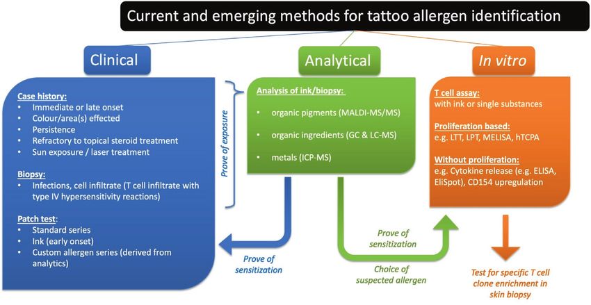
Review Article                   Tattoo allergens

      Figure 1 Possible adverse effects of tattoos. Effects may occur locally in the tattooed skin or spread systemically.

      skin via the wounded surface. Pigment particles remain in
                                                                                                      Ingredients of tattoo inks and related
      the dermis and absorb light in a specific spectral range cau-
      sing the color of the tattoo. Another fraction of the injected                                  chemical hazards
      tattoo ink is removed from the skin passively via lymph or
      blood vessels or is actively transported by migrating cells. As                                 Tattoo inks are suspensions that may contain up to 100 dif-
      a result, tattoo pigments are found in the local lymph nodes                                    ferent chemical compounds that may or may not be added
      but are likely also transported to other organs such as liver,                                  intentionally. The coloring pigment is mixed with solvents,
      lungs or kidney [1, 2, 7, 8]. Experiments with mice suggest                                     preservatives, and various other substances. Although they
      that pigment particles of tattoo suspensions are picked up by                                   are injected into the human body, tattoo inks usually do not
      Kupffer cells inside the liver as indicated by the detection of                                 need to fulfil specific safety requirements in contrast to other
      particles via electron microscopy [9].                                                          substances that are inserted into the human body, such as
           After skin healing, histological investigations showed                                     medical drugs or implants. The exact list of ingredients, if
      that pigment particles are located in the cytoplasm of dif-                                     known at all, depends on the practice of each manufacturer.
      ferent cells, including fibroblasts and macrophages [1, 2,                                      Tattooists and beauticians purchase the tattoo inks directly
      10]. A recent investigation in mice proposed a pigment cap-                                     from suppliers or through the internet [1].
      ture-release-recapture model. When tattoo pigment-laden                                              Potential hazardous properties of chemicals in tattoo
      macrophages die during the course of adult life, neighboring                                    inks include carcinogenic, immunotoxic, and sensitizing
      macrophages recapture the released pigments and seem to                                         properties (Figure 1). Tattoo inks containing hazardous che-
      ensure the macroscopic stability and long-term persistence                                      micals are frequently found on the European market. The
      of tattoos [1, 11].                                                                             major substances of concern detected in analyzed samples
           Experiments with pig and human skin revealed that the                                      are polycyclic aromatic hydrocarbons (PAH) (43 %), primary
      concentrations of red pigments, placed in the skin by tattoo                                    aromatic amines (PAA) (14 %), heavy metals (9 %) and pre-
      machines, range from about 0.60 to 9.42 mg pigment per                                          servatives (6 %) [1, 6].
      skin cm² [12]. In mice, about 30 % of intradermally injected
      red pigment disappeared from skin within six weeks after                                        Pigments
      tattooing [2]. This percentage increased to 60 % when solar
      radiation was additionally applied to the animals’ skin [13].                                   The chemical industry synthesizes pigment molecules which
      In general, the concentration of insoluble tattoo pigments                                      form tiny, solid state particles with diameters smaller than
      in skin gradually decreases over the years [14]. The soluble                                    100 nanometers (nanoparticles) up to a few micrometers
      parts of the ink will likely be excreted in the first days after                                [15]. Therefore, additional hazards may arise from nanotoxi-
      tattooing.                                                                                      cological effects which should be investigated [16].

658   © 2021 The Authors. Journal der Deutschen Dermatologischen Gesellschaft published by John Wiley & Sons Ltd on behalf of Deutsche Dermatologische Gesellschaft. | JDDG | 1610-0379/2021/1905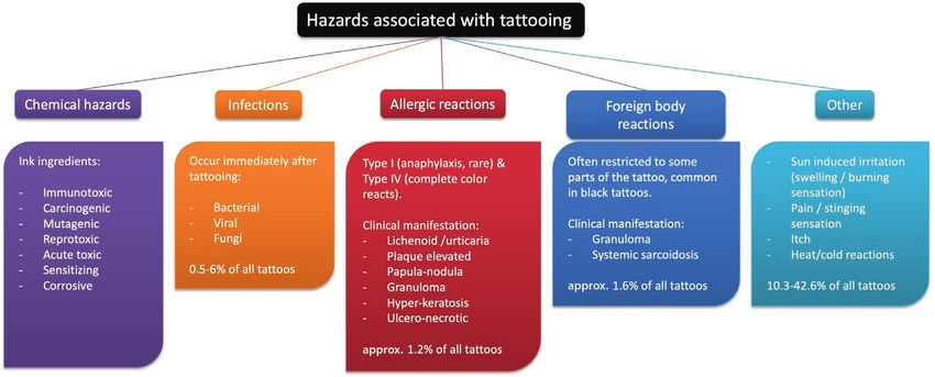
Review Article Tattoo allergens
Tattoo pigments are frequently also called dyes, but the Colored inorganic pigments
term “dyes” should be not be used since dyes are water-so-
luble and would not allow permanent skin decoration. The Most colored inorganic pigments are based on iron oxides in
permanence of a tattoo in the skin is achieved by using inso- the colors yellow (FeO(OH)), red (Fe2O3), and black (Fe3O4).
luble pigments. Since iron ore often contains heavy metals such as nickel, these
Pigments are usually sold as powder to the ink manu- display common impurities of iron oxide pigments. Nickel
facturers. Wholesalers bypass a small amount of these in- compounds are classified as carcinogens by the IARC [23].
dustrial pigments as ready-to-use ink products directly to Another group of pigments is based on heavy metals
tattooists. To achieve the respective color, tattoo inks may such as cadmium sulfide (CdS, yellow), mercury sulfide
contain different inorganic or organic pigments or a mix- (HgS, red), chromium oxide (Cr2O3, green), or cobalt spinel
ture of both. (CoAl 2O4, blue) [6]. The IARC classified these heavy metals
as group 1 (carcinogenic to humans: chromium(VI), cadmi-
Black pigments um and cadmium compounds) or group 2B (organic-mercury
compounds) carcinogens. The use of heavy metal-based pig-
Most black pigments are usually produced by imperfect com- ments declined due to their hazardous nature.
bustion of hydrocarbons yielding soot – a mixture of black In the European alert system for dangerous goods
inorganic carbon particles that contain polycyclic aromatic (RAPEX), 28 % of tattoo inks showed heavy metal contents
hydrocarbon molecules (PAH). Carbon black is classified as above the threshold values defined in the Council of Euro-
possibly carcinogenic to humans (group 2B) by the Interna- pe Resolution (CoE ResAP) (2008)1 on requirements and
tional Agency of Research in Cancer (IARC) [2, 17]. This criteria for the safety of tattoos and permanent make-up
evaluation is based on the increased occurrence of lung can- [24]. Alerts were related in particular due to the presence of
cer upon particle inhalation or the increased incidence of skin arsenic, barium, cadmium, chromium(VI), copper, lead, zinc,
cancer from carbon black extracts in animal models. The lat- and nickel. In addition to toxic and mutagenic effects, some
ter leads to the assumption that especially impurities cause metals (nickel, mercury, chromium, and cobalt) might act
the observed carcinogenic properties. Up to 201 μg PAH per as allergens eliciting cutaneous or systemic contact allergies
g ink were found in different commercially available black [25]. Nickel, cobalt, and chromium are among the most com-
tattoo inks which is far more than the currently proposed mon sensitizers with positive patch test reactions in about
limit of 0.5 μg PAH per g tattoo ink by the European Union 18 %, 6 % and 3 % of patients in Europe, respectively [26].
[18]. PAH occur freely in the ink suspension or attached to
the surface of carbon black particles [2]. PAH are metaboli- Organic pigments
cally activated to their respective diol-epoxides, which can
bind DNA and lead to mutagenicity, resulting not only in Nowadays, more than 80 % of the colored tattoo inks cont-
carcinogenicity but also in effects on lymphocyte activation ain industrial organic pigments [1, 6, 22]. Organic pigments
and macrophage differentiation [19–21]. provide a huge variety of colors spanning the whole rainbow.
Such pigments exhibit a strong light absorption yielding high
White pigments color strength and brilliance in the skin which likely is the ma-
jor reason for their application in tattoos. The polycyclic pig-
Titanium dioxide is an effective opacifier that is usually ments are generally condensed aromatic or heterocyclic ring
applied in white inks and for changing the color strength systems. Two important examples of heterocyclic pigments
of other tattoo pigments [22]. Titanium dioxide occurs are phthalocyanine (green, blue) and quinacridone pigments
in rutile, anatase, and brookite crystal structures, but (bluish red, pink, violet) [1, 22]. However, another group,
only the first two are being used as pigments. However, the azo pigments, are still most commonly used. They span
titanium dioxide, particularly as anatase, exhibits photo the yellow to red color range and are composed of condensed
catalytic activity under ultraviolet irradiation generating aromatic amines, which are often carcinogens or sensitizers.
reactive oxygen species, not radical. In 2010, the IARC The synthesis of such pigments requires complex chemical
classified titanium dioxide as group 2B carcinogen by in- processes in which also rosins, polymers, and surfactants
halation meaning it is possibly carcinogenic to humans might be added to adjust their size and dispersion properties.
[1, 17]. The mechanism of carcinogenicity is thought be a The resulting pigment may contain different educts, by-pro-
so-called over-load effect when the concentration reaches ducts, and other non-specified compounds. Due to their use
a level where the particles can no longer be sufficiently during synthesis, PAA are the most important contaminants
cleared from the lungs. This effect is therefore unlikely to of organic pigments and their concentration is highest in inks
occur in other organs, such as skin. containing azo pigments. Primary aromatic amines represent
© 2021 The Authors. Journal der Deutschen Dermatologischen Gesellschaft published by John Wiley & Sons Ltd on behalf of Deutsche Dermatologische Gesellschaft. | JDDG | 1610-0379/2021/1905
659Review Article Tattoo allergens
either impurities of these inks (free aromatic amines) or de- [32]. Among these banned substances were 1,2-benzisothi-
gradation products after sunlight or laser irradiation [27]. azol-3[2H]-one, 2-octyl-4-isothiazolin-3-one, phenol, and
Since some of these amines are classified as carcinogenic, mu- even the carcinogen formaldehyde [1]. Isothiazolinones are
tagenic or reprotoxic substances they must neither be present strong allergens and have a high rate of sensitization in the
nor released from azo pigments in tattoo and PMU products European population [26]. Other cases also indicate that
[1, 24]. shellac, a commonly used binder in tattoo inks, may provoke
allergic reactions [33].
Light exposed pigments and influence of laser Furthermore, N-nitrosamines were found in the samples
[32]. N-nitrosamines are impurities formed by the reaction
treatment on tattoos
of secondary amines with nitrite. Many N-nitrosamines are
One important mechanism for the disintegration of particles carcinogenic even in small concentrations which has been
in tattooed skin is the light induced decomposition of pigments proven in animal testing [1, 7].
molecules. It may continuously occur whenever tattooed skin Recently, the deposition of metal debris from the tattoo
is exposed to light sources emitting wavelengths which can needle in skin and local lymph nodes has been reported [34].
be absorbed by the pigment molecules [27]. Laser treatment Since the tattoo needles contain high amounts of nickel and
of tattoos is frequently performed by dermatologists in daily chromium, the metal debris may pose an additional risk for
practice. When exposed to laser light during tattoo removal, sensitization or allergy formation.
an additional process may occur: the tattoo pigment particles
may be fragmented to smaller pieces. Hitherto, the mechanis- Adverse skin reactions to tattoos
ms of action have been assumed to involve heat production in
the tattoo particle from absorbed light energy. Due to the high Starting with the placement of a tattoo, a range of adverse
light intensity and short pulse durations applied, the heating reactions can affect human health (Figure 1). Adverse reac-
of pigment particles is very rapid, yielding high local tempe- tions may be localized in the skin or involve other organs
ratures of up to several hundred degrees. The subsequent ra- of the body and can develop within a wide time range from
pid thermal expansion causes fragmentation of the particles, immediately after tattooing to years or even decades later.
accompanied by shock waves [28]. In addition, the heating
and fragmentation of pigment particles may lead to cleavage Prolonged wound healing
of chemical bonds within the pigment molecule, producing
new chemical compounds within the skin [1]. The release of Tattooing leaves the skin littered with numerous holes rea-
hazardous decomposition products such as benzene and car- ching through the epidermis roughly into the mid of the der-
cinogenic aromatic amines was reported by us and others after mis. The depth of the holes varies depending on the technique
sunlight and/or laser irradiation [13, 29, 30]. of the tattoo artist and the equipment used. The width of a
hole depends on the tattoo needles, which frequently show
Other ingredients diameters of 0.2 to 0.4 mm and which are assorted in bund-
les of up to 50 single needles. After skin injury, wound hea-
Tattooing pigment powder is almost impossible, so the pig- ling starts immediately to restore skin integrity.
ment must be added to a fluid medium. However, pigment The process of normal wound healing is divided into
particles are insoluble in water and therefore require disper- different phases [35]. After hemostasis, the immediate in-
sion in aqueous or alcoholic solvents with the help of emulsi- flammatory phase lasts up to three days. Neutrophils are the
fiers, binders (e.g. polyvinylpyrrolidone, polyethylene glycol) first immune cells that infiltrate wounded tissues, arriving in
and thickening agents to avoid particle sedimentation [1, 6]. large numbers in response to damage-associated molecular
In addition, antifoam agents are added to avoid foam produc- patterns (DAMPs) released from injured and necrotic cells.
tion while shaking the suspension (e.g. polydimethylsiloxa- The neutrophils release different cytokines, chemokines, and
ne). The concentration of pigments in tattoo ink suspensions growth factors that signal through wound-associated immu-
is usually between 10–30 % by volume [1]. ne cells and epithelial cells to promote healing [36]. Neutro-
A study from 2011 revealed that tattoo inks may contain phils prevent infection by eradicating microbes that entered
substances like the sensitizer dibutyl phthalate, the genotoxin the skin.
hexachloro-1,3-butadiene, or 9-fluorenone [31]. 9-Fluoreno- Subsequent recruitment of circulating monocytes into si-
ne is cytotoxic, generating reactive oxygen species upon light tes of tissue damage occurs. Monocyte-derived macrophages
exposure [1]. or dendritic cells manage tissue repair, regulate angiogene-
In Switzerland, preservatives banned for use in cos- sis, clear cellular debris, and recruit additional immune cells
metics were found in up to 18 % of tattoo samples tested [36]. This late inflammation phase lasts up to ten days with
660 © 2021 The Authors. Journal der Deutschen Dermatologischen Gesellschaft published by John Wiley & Sons Ltd on behalf of Deutsche Dermatologische Gesellschaft. | JDDG | 1610-0379/2021/1905Review Article Tattoo allergens
macrophages contributing to wound repair and tissue remo- treatable. Pathogens dumped into deeper skin layers can cause
deling by clearing apoptotic neutrophils [37]. Such a milieu more severe infections requiring a more extensive medical tre-
could be affected in the presence of tattoo inks, leading to an atment. Should pathogens enter blood vessels, systemic infec-
altered immune response in tattooed skin areas and a chan- tions may result [40]. The severity of an infection depends on
ged process of wound healing. the virulence of the pathogen, the immune status of the person
Skin also contains tissue-resident macrophages which being tattooed, and underlying diseases [41].
are non-migratory and respond to injury or infection by A recent review summarized 67 cases of local skin in-
sensing DAMPs [36]. Langerhans cells of the epidermis and fections and systemic complications with bacteria such as
other antigen presenting cell types may internalize antigens Corynebacterium diphtheriae, Pseudomonas aeruginosa,
from the ink and migrate to lymph nodes in which these are or Staphylococcus aureus, that were published between
then presented to T cells. 1984 and 2015 [41]. Another review searched the literature
At last, re-epithelialization is an important step because from 1991 to 2011 and listed 13 publications reporting viral
it restores the barrier function of the skin [36]. When surgical infections including vulgar warts or hepatitis C as well as 25
wounds of specific depths were created in pig skin, re-epithe- publications reporting bacterial infections with, for example,
lialization started from residual appendage structures [38]. Mycobacterium tuberculosis, Mycobacterium leprae, nontu-
After recovery of the skin barrier, the remodeling phase for berculous mycobacteria, or community-associated methicil-
rebuilding normal skin structures may last several months. lin-resistant Staphylococcus aureus (MRSA) [42]. In 2017, a
In conclusion, the wound healing of skin is an effecti- case of death after septic shock was reported that was cau-
ve but complex process that is orchestrated by immune cells sed by a Vibrio vulnificus infection after bathing in seawater
whose functions critically depend on the microenvironment with a fresh tattoo [43].
in the wound. In contrast to medical procedures such as use Sources for microbial pathogens include the own skin
of injection needles, microneedling, or ablative laser treat- surface of tattooed individuals, hands and equipment of the
ments, small tattoo wounds are filled with various cytotoxic tattooists, and an unhygienic handling of the skin after tat-
chemicals from the ink. Unfortunately, there is a lack of stu- tooing. Another source are tattoo inks of which up to 11 %
dies on whether and to which extent tattoo inks might ham- are contaminated with microorganisms [6]. Hence, even if a
per normal wound healing after tattooing. Itching, crusts, tattoo reaction only occurs in one color and thus points to
and delayed wound healing are frequently reported in sur- an allergy, microbial infections should always be ruled out.
veys, indicating that irritants, sensitizers or even microorga-
nisms may disturb this process [39]. Non-allergic inflammatory reactions
Infections From time to time, tattoos are associated with the develop-
ment of long-term local immune responses manifesting as
Tattooing produces numerous holes in skin facilitating the granulomatous and pseudolymphomatous reactions with
entry of microbial pathogens. Consequently, mild-to-moderate the majority being foreign-body granulomas (Figures 1, 2a)
superficial skin infections may occur that should be easily [44]. However, granulomatous lesions in a tattoo can be a
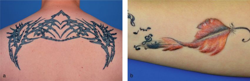
Figure 2 Common adverse tattoo reactions. Granulomatous reaction in a black tattoo (a). Type IV hypersensitivity tattoo
reaction in the red colored part of a tattoo presenting as a plaque-like reaction (b).
© 2021 The Authors. Journal der Deutschen Dermatologischen Gesellschaft published by John Wiley & Sons Ltd on behalf of Deutsche Dermatologische Gesellschaft. | JDDG | 1610-0379/2021/1905
661Review Article Tattoo allergens
manifestation of systemic sarcoidosis affecting the lungs and triggering allergic contact dermatitis (ACD) or drug expo-
eyes (uveitis) [44–46]. sure (drug hypersensitivity) [54, 55]. Both CD4+ and CD8+
Cutaneous pseudolymphoma represents a variable group T cells seem to be involved [56, 57].
of benign reactive T or B cell lymphoproliferative processes The development of a type IV hypersensitivity reaction is
simulating cutaneous lymphoma both clinically and histolo- usually separated into a sensitization phase and an elicitation
gically. The pathogenesis of this disease is still unknown, but phase. During the sensitization phase, dendritic cells migrate
since 1903 tattoos have been reported to be associated with to draining lymph nodes and prime specific naïve T cells that
these reactions [44, 47]. proliferate and differentiate into circulatory and tissue-resi-
Furthermore, tattooing can trigger certain dermatoses dent effector and memory populations [58, 59]. Dendritic cell
on the tattooed skin (isomorphic phenomenon of Koebner), migration and maturation may be triggered by the chemical
including psoriasis, lichen planus, cutaneous lupus erythe- allergen itself (sterile inflammation) or by concomitant he-
matous, and pyoderma gangraenosum [48, 49]. terologous immune stimulation [55]. During the elicitation
phase, T cells initiate and orchestrate the inflammatory reac-
Allergic reactions tion involving various effector molecules, immune and other
cells [59–61].
While their diagnosis remains difficult, allergic reactions to Typically, ACD manifests within approximately 1–3
tattoos have been reported with varying degrees of diagno- days after topical chemical contact and ceases after remo-
stic evidence. val of the culprit allergen. In tattoos, however, the potential
allergen may be permanently present which can pose seri-
Antibody-mediated tattoo reactions ous problems, for instance by inducing chronicity of the in-
flammation. The timing with which delayed-type hypersen-
Only few studies suggest a role for antibodies in chemical- sitivity reactions occur may vary from shortly after tattoo
induced hypersensitivity reactions [50]. Regarding tattoos, application up to several years later [2]. Partly, this depends
rare cases that likely involve antibody-mediated reactions on whether the individual is already sensitized to a specific
have been reported [51–53]. A first case in the recent me- compound. Additionally, some allergens require metabolism,
dical literature describes a 30-year-old woman with a color degradation or the presence additional (heterologous) immu-
tattoo made in 1993 and a black tattoo made in 1999 [51]. ne stimulation to initiate sensitization.
One month after the last tattoo, additional color was added The clinical patterns normally associated with allergic
and twelve hours later she began to experience a “burning” reactions are papulonodular, plaque-like, lichenoid, hyperke-
sensation, which evolved into hives covering multiple areas ratotic or ulceronecrotic (Figure 2b). The clinical picture also
of her body. The patient was subsequently skin prick tested includes contact urticaria-like reactions or photo-allergic
to 13 different ink colors and was positive to two of them, a reactions [62]. Concomitant reactions in older, red-colored
purple and a blue ink, indicative of an IgE-mediated hyper- tattoos may be triggered.
sensitivity [44]. Usually, allergic reactions occur locally with involvement
A second patient without any known allergies experi- of the entire tattooed area with the triggering color, but some
enced anaphylaxis six hours after application of a second authors describe cases of generalized rashes or eczemas, espe-
tattoo, which began with swelling and redness on multiple cially in previously sensitized patients. These reactions appe-
body sites [52]. A third case of delayed anaphylaxis occurred ar early (within 1–2 days after tattooing) and tend to resolve
after laser treatment for tattoo removal [53]. This patient had without treatment after a few weeks or months, indicating
an allergic cutaneous reaction at a distant, untreated tattoo that soluble tattoo ink components may be involved. Other
after the first laser treatment followed by an anaphylactic authors describe cases of generalized reactions upon attempt-
episode three days after the second laser treatment. ing to eliminate the pigment with laser treatment and cases of
photo-allergic reactions in tattoos with yellow ink containing
Type IV hypersensitivity reactions cadmium [63, 64]. Furthermore, allergic reactions without
generalized rashes can be initialized by laser treatment, and
Allergic reactions to tattoos are mainly thought to be medi- putatively by sun exposure, due to the release of sensitizers
ated by T cells, classified as delayed type IV hypersensitivity [65–67]. This so-called photoallergy should not be confused
reactions according to Gell's and Coombs’ historic classifica- with phototoxicity which involves transient reactions during
tion. With their T-cell receptor (TCR), T cells likely recogni- exposure of the tattoo to sunlight [68].
ze epitopes of chemically modified self-antigens formed in a In some rare cases, putative allergic reactions occurring
process called haptenization. Haptenization has been espe- in a tattoo may also be triggered by implant materials [69, 70].
cially described in the context of topical chemical exposure These reactions resolve with the removal of the implant.
662 © 2021 The Authors. Journal der Deutschen Dermatologischen Gesellschaft published by John Wiley & Sons Ltd on behalf of Deutsche Dermatologische Gesellschaft. | JDDG | 1610-0379/2021/1905Review Article Tattoo allergens
The majority of chronic tattoo reactions are associated autoimmunity. Additionally, granulomatous reactions in the red
with red or reddish colors, for example pink, orange, violet part of a tattoo with additional symptoms in the eyes have been
and bordeaux [39, 42, 71]. Azo pigments were the most fre- reported after influenza vaccination containing thimerosal,
quent pigments identified in skin biopsies of tattoo allergy which is a mercury-containing preservative [74]. The authors
patients with reddish colors [72]. suspected an allergy elicitation by thimerosal, which induced
Differential diagnosis of a type IV hypersensitivity to- a general inflammatory response with subsequent lung sarcoi-
ward a tattoo should always consider other late reactions dosis and hypersensitivity to the red, organic tattoo pigment.
such as granulomatous foreign body reactions, systemic di- Granuloma formation from vaccines containing thimerosal
seases (for example sarcoidosis or connective tissue disease), have also been reported in non-tattooed persons.
microbial infections, and pseudo-lymphomatoid reactions. Regarding the treatment with allopurinol, contradicto-
ry findings have been published. A 34-year-old patient with
Tattoos, medication, and (auto)immune multiple morbidities developed systemic eosinophilia with a
prominent eruption in a tattoo after the addition of allopu-
reactions
rinol to the list of medication. Allopurinol is known to be a
Adverse reactions at the site of a tattoo can also occur after cause of this drug-induced hypersensitivity syndrome. On the
systemic drug treatment for different unrelated indications. other hand, allopurinol is a treatment option for general and
Swelling, granuloma formation and pain in tattoos have been tattoo-associated granuloma and sarcoidosis – putatively by
reported after treatment with BRAF and MEK inhibitors such either acting as radical scavenger or by preventing the forma-
as dabrafenib or trametinib for malignant melanomas occur- tion of foreign-body giant cells [75].
ring outside of the tattooed area [73]. These inhibitors prevent In general, drugs or diseases that affect the immune sys-
excessive cell growth of tumor cells but may also stimulate tem may also trigger reactions in tattoos. For example, people
Figure 3 Potential strategy for the diagnosis of tattoo-related allergies. The clinical case history may provide information on the
characteristics of the suspected allergen, for example, an association with light exposure, pigments, or soluble ink ingredients.
Combined with chemical analysis, the presence of an allergen in the biopsy or ink and a positive patch test may provide a hint
on the identity of the allergen. Still, the patient might be coincidentally sensitized to the substance of the positive patch test
while another allergen might be causing the reaction in the skin. In vitro methods may show pathogenic T cell populations
in the patient compared to non-allergic controls. Finding increased numbers of allergen-reactive T cell clones in the inflamed
tattoo would almost certainly indicate a type IV hypersensitivity reaction to a substance present in the biopsy/ink. With the aid
of high-throughput sequencing technologies, this would deliver proof that the skin reaction is indeed caused by the suspected
substance.
© 2021 The Authors. Journal der Deutschen Dermatologischen Gesellschaft published by John Wiley & Sons Ltd on behalf of Deutsche Dermatologische Gesellschaft. | JDDG | 1610-0379/2021/1905
663Review Article Tattoo allergens
infected with HIV may develop a type IV hypersensitivity at least some allergens may already be present in the ink. For
reaction in tattoos after starting anti-retroviral therapy [76]. tattoo hypersensitivity reactions with a late onset, external
factors, especially light inducing photochemical cleavage of
Diagnostic tools for allergic tattoo tattoo pigments in situ in the skin, may contribute to allergen
formation from pre-haptens or pro-haptens [27, 68, 71, 79].
reactions, associated challenges, and The downside of patch testing is that it can only provi-
new approaches de a correlation between sensitization and a substance the
patient had contact with, for instance, nickel sensitization
Diagnosing allergic reactions to tattoo inks is very chal- correlating with nickel in a tattoo ink. Patch tests are accom-
lenging since diagnostic tools and knowledge about culprit panied with the uncertainty that other substances that were
allergens, not to speak of relevant epitopes recognized by the also present in the tattoo ink may have been the main reason
involved TCR, is very limited. Current diagnostic tests and for the allergic reaction. They are therefore no incontrover-
outlines for ongoing experimental research approaches for tible proof for a causal finding.
the identification of tattoo allergens are presented in Figure 3.
Skin biopsy
Patch testing
Combining clinical aspects and histological findings from
Patch testing is the diagnostic gold standard for ACD. It is not skin biopsies can help clinicians to exclude serious entities
clear whether this test is useful for diagnosing delayed-type such as cutaneous infections by atypical microorganisms,
hypersensitivity reactions provoked by allergens such as tat- systemic diseases, or lymphomatous tumor infiltrations [49].
too ingredients that are inoculated into the dermis. Theoreti- Additionally, biopsies can prove that mononuclear cell in-
cally, an intradermal test would be a more specific diagnostic filtrates, for example T cells, persist in a pattern consistent
tool for these reactions since it would more closely resemble with those observed in ACD. However, such histologic pat-
the in vivo situation, but it is not free of risk. The applied terns are not definite and may also represent, for instance, ir-
pigments may permanently remain in the dermis after the test ritant contact dermatitis. In 2015, Høgsberg et al. investiga-
and chronic reactions could be triggered. As a consequence, ted 19 biopsies from chronic tattoo reactions to red pigment
performing these tests is considered neither appropriate nor or mixtures or nuances of red [80]. The predominant histolo-
ethical [77]. gical pattern was interface dermatitis with increased counts
In order to investigate contact allergies to tattoo of T cells and Langerhans cells which fits with delayed-type
pigments, a new test block was recently introduced by the hypersensitivity reactions.
Deutsche Kontaktallergiegruppe (block 47). This block
contains 24 substances, e.g. o-phenylphenol, iron(II) sulfa- Biopsy and ink analysis
te and shellac [78]. As only few studies reflect the utility of
patch testing in this situation, it is not completely clear which The diagnosis of allergens is particularly difficult regarding
allergens should be included in specific patch tests. Many tattoo inks. As stated above, tattoo inks are complex mix-
published articles only report single clinical cases with posi- tures of insoluble pigments with solvents, thickening agents,
tive patch test results and very few combine it with adequate surfactants, preservatives, and other impurities or additives.
clinical and analytical correlation. In temporary and early-onset allergic reactions, soluble com-
In 2014, Serup et al. performed a patch test study of 90 pounds in the tattoo ink may play a role. In these cases, the
patients with tattoo reactions [71]. The general outcome of ink of the patient might give positive results in a patch test
the study showed that patients with reactions in their tattoos [65]. With later tattoo reactions – sometimes occurring years
presented mainly negative or inconsistent results when patch after tattooing – only substances still residing in the skin can
tested with common allergens, textile dyes, problematic tat- trigger the allergy. In terms of pigment-derived allergens, the
too inks, and even individual culprit inks, if available [71]. suspects are most likely metals, organic impurities, or decom-
However, if the ink was obtained weeks or months after position products of organic pigments as well as metabolites
tattooing, there is a fair chance that the product lot or com- thereof. To affirm an allergen, it is necessary to prove that the
position had changed. Negative patch test results also indi- patient was exposed to this substance and is being sensitized
cate that most allergens causing tattoo reactions are formed against it. This can be done by analyzing a skin biopsy from
inside the dermis, which can be slow and may require weeks, the tattoo or the used ink. The latter is quite unreliable, espe-
months, or years [71]. In a recent study, 37 % of allergic reac- cially with late onset allergies occurring months or years after
tions occurred during the first month after tattooing leaving tattooing with the original ink not being traceable or having
a majority of 63 % to late reactions [72]. This indicates that an altered composition in the meantime. Past investigations
664 © 2021 The Authors. Journal der Deutschen Dermatologischen Gesellschaft published by John Wiley & Sons Ltd on behalf of Deutsche Dermatologische Gesellschaft. | JDDG | 1610-0379/2021/1905Review Article Tattoo allergens
identified the metals chromium, mercury, cobalt, and nickel Likely, T cells recognize allergen-modified peptides presen-
as likely causes of allergic tattoo reactions correlating a patch ted by major histocompatibility complex (MHC) molecules
test with chemical analysis of skin biopsies [81–84]. These of antigen-presenting cells besides other possible mechanisms
metals can be frequently found in inorganic pigments. The [91, 92]. This has been illustrated for the experimental model
analysis of metals in skin biopsies or ink is comparably easy allergen 2,4,6-trinitrobenzenesulfonic acid (TNBS). This
and standard patch test preparations exist. model antigen was recognized by CD8+ T cells mainly if atta-
With organic pigments, many structural variations can ched to a lysine at position 4 of an MHC-presented peptide in
be created by chemical synthesis and might also be in use for a murine contact hypersensitivity model [93]. Alternatively,
tattooing. To date, about 104 organic pigments are known to chemicals may bind non-covalently to the TCR-peptide-
be used in tattoo inks [85]. Without knowing the exact iden- MHC interface, termed “pharmacological interaction” in
tity of the pigment embedded in a patient's skin, diagnostics case of drugs, which sometimes occurs in a HLA allele-res-
of the underlying allergen compares to searching for a needle tricted manner [54, 94].
in a haystack. The importance of the different possible epitopes in the
Pigment analytics from biological tissue or inks has allergic immune response remains unknown and the for-
so far been carried out with high-performance liquid mation of the correct epitopes can hardly be controlled by
chromatography (HPLC) techniques [86] or matrix-assisted in vitro assays. As a pragmatic solution, antigen presenting
laser desorption/ionization mass spectrometry (MALDI-MS) cells and T cells from blood are incubated with chemically
[87, 88]. HPLC methods are very much limited by the solubi- modified proteins or peptides. Alternatively, chemicals are
lity of the pigments but can provide quantitative data. Only added directly into the medium of the coculture [93, 95].
a limited amount of pigments is soluble in standard orga- Since TCRs are both highly specific but also cross-reactive
nic solvents used for HPLC analysis. MALDI-MS analysis [96], T cell in vitro tests may work even if epitopes formed
is easily applicable to a wide range of pigments but detection with skin peptides are missing.
limits are in the range of percentages and vary for each pig- The detection of rare activated T cells in a plethora of
ment. MALDI was proven suitable for the detection of visible irrelevant bystander cells is difficult. Every individual has
amounts of pigments – such as those present in tattooed skin over 100 million mainly unique T cell clonotypes with only
– in a study analyzing the pigments in 104 skin biopsies of few being specific for a given epitope [97]. To detect rare
allergic tattoo patients [72]. Here, the inks collected from the allergen-specific T cells in blood, most labs use proliferati-
tattoo artists did not always match the pigments found in the on-based methods, for instance the lymphocyte proliferation
skin of the patients. or transformation tests LPT/LTT, the memory lymphocyte
Although the pigments suspected to cause tattoo allergies immunostimulation assays MELISA, or the human T cell
can be narrowed down by chemical analysis, possible descen- priming assays hTCPA [57, 98]. To facilitate proliferation,
dent sensitizing decomposition products of these pigments as cytokine skewing cocktails may be added or regulatory T cells
well as by-products or polymers used during their synthesis may be depleted [99, 100]. Non-proliferation-based methods
may also play a role in allergy development. For most of the- usually detect cytokines, for instance via enzyme-linked im-
se substances, no standard patch test formulations exist to munosorbent or spot assays (ELISA and ELISpot). Except for
prove that the patient is sensitized against it. If the pigment priming assays, these approaches only detect memory T cells
in the patient's skin is known beforehand, a specific set of that proliferate or express the respective cytokines. Naïve
substances may be tested (Figure 3). T cells and less proliferative or non-cytokine-producing me-
mory cells are not detected, an issue that may introduce bias.
Detection of allergen-specific T cell responses Recently, a new method providing fast and compre-
hensive access to antigen-specific naïve and memory CD4+
in vitro
T cells has been published, which is based on induced CD154
T cell responses to suspected tattoo allergens may be analyzed (CD40L) expression a few hours after in vitro stimulation
in vitro similar to other chemical sensitizers triggering ACD. [101, 102]. We and others started to adopt this method to
For the development of such tests, several challenges have to detect, for example, nickel-specific or β-lactam-antibiotic-
be overcome. Firstly, relevant allergen-induced antigens need specific T cells [103, 104]. With those T cell assays, increased
to be formed which are recognized by specific T cells. percentages of specific activated T cells after treatment with
Generally, knowledge on how chemicals induce T cell chemicals can be detected.
responses is still extremely limited [54, 55, 89]. Parkinson For a test to be diagnostic, the analysis of non-aller-
et al. showed for a few sensitizers that high and low abundant gic control individuals is mandatory. For example, in case
proteins may become modified [90] while their respective of p-phenylenediamine (PPD)-induced allergy, T cell proli-
importance for T cell epitope formation remains unknown. feration was found increased in allergic individuals [95].
© 2021 The Authors. Journal der Deutschen Dermatologischen Gesellschaft published by John Wiley & Sons Ltd on behalf of Deutsche Dermatologische Gesellschaft. | JDDG | 1610-0379/2021/1905
665Review Article Tattoo allergens
In contrast, the oxidation product of PPD, the so-called presentations of cutaneous reactions to tattoo inks, including
Bandrowski's base, induced proliferation in allergic and allergic reactions, have been reported in the literature. It is
non-allergic individuals and is thus not suitable for allergy very important for physicians to be aware of these reactions
detection [95]. In case of nickel, activation of many CD4+ and of the possibility of eliciting further reactions through
T cells has been observed already in non-allergic donors, treatment with lasers or other modalities. We highly recom-
which we found to be due to the activation of T cells carrying mend pharmacological, toxicological, and epidemiological
a TCR with a TRAV9-2 gene segment or a CDR3 histidine studies to clarify the possible impact of tattooing on human
[57, 104]. This activation was still topped by donors with health.
acute nickel ACD and may therefore be used to identify some To shed light on the matter, a collaboration between
allergic patients as an alternative to patch tests in the future. the German Federal Institute for Risk Assessment (BfR)
Additionally, advances in high-throughput sequencing tech- in Berlin and the Department of Dermatology of the Uni-
nologies may be further exploited to link antigen-specific versity of Regensburg was established. Our team develops
T cell clonotypes identified in blood to those in inflamed skin new methods for the detection of hypersensitivity reactions
[58]. An overlap of T cells from the blood reactive to the an- in tattoos assisted by chemical analysis of tattoo allergens.
tigen with a local enrichment of these cells in the inflamed Our goal is to improve diagnostics in cutaneous tattoo re-
skin can causally link suspected tattoo allergens with allergic actions and to enable risk assessment of potential allergens
symptoms, which is not possible with patch tests. in tattoo inks that should be banned from the market. Data
Results from previous analytics and patch tests may help on patients with tattoo allergies are collected at the Depart-
in selection of suspected chemical allergens for in vitro assays ment of Dermatology, University Hospital Regensburg, for
and sequencing approaches to achieve a causal proof for the subsequent analysis of the pigments in the skin and identifi-
allergen in the skin (Figure 3). cation of the allergen via custom patch tests and potentially,
by T cell assays.
Treatment of cutaneous reactions to
tattoo inks Acknowledgements
Open access funding enabled and organized by Projekt
Clinical complications toward tattoos can be managed with DEAL.
different strategies [44, 105]. Treatment measures may span
from conservative to invasive procedures and depend on the Correspondence to
severity and location of the lesions. A conservative treatment
may include topical, oral, and/or intralesional steroids, oral Katharina T. Weiß, MD
anti-histamines, and protection of the tattoo from sunlight Department of Dermatology and Allergology
[49, 71]. University Hospital Regensburg
Invasive methods may permanently damage the skin by Franz-Josef-Strauß-Allee 11
scar formation. They include cryotherapy, electrosurgery, 93053 Regensburg Germany
surgical excision, dermabrasion, chemical destruction via
acid, or ablation with a non-Q-switched carbon dioxide la- E-mail: mailto.katharina.weiss@ukr.de
ser. Q-switched laser therapy is not indicated when tattoos
References
show signs of an allergic reaction, because the therapy itself
1 Bäumler W. Chemical hazard of tattoo colorants. Presse Med
can initiate or worsen hypersensitivity reactions. Probably,
2020; 49: 104046.
these reactions originate from the laser, which destroys the 2 Bäumler W. Absorption, distribution, metabolism and excre-
pigment-containing cells, resulting in the pigment becoming tion of tattoo colorants and ingredients in mouse and man:
extracellular [44, 71]. The immune system may recognize the known and the unknown. Curr Probl Dermatol 2015; 48:
allergen-induced epitopes after hapten formation from the 176–84.
released pigments or its breakdown products [106]. 3 Borkenhagen A, Brähler E. Verbreitung von Tätowierungen,
Piercing und Körperhaarentfernung in Deutschland. press re-
lease University of Leipzig: University of Leipzig, 2017.
Conclusions and recommendations 4 Shannon-Missal L. Tattoo Takeover: Three in ten Americans
have tattoos, and most don't stop at just one. 2016. Available
Depending on its size, several grams of tattoo ink consisting from: https://theharrispoll.com [Last accessed January 04,
of various chemical compounds are injected into the skin 2021].
while applying a tattoo. Causes of the systemic effects of tat- 5 Klugl I, Hiller KA, Landthaler M, Bäumler W. Incidence of
too inks mainly remain unclear to date. Furthermore, many health problems associated with tattooed skin: a nation-wide
666 © 2021 The Authors. Journal der Deutschen Dermatologischen Gesellschaft published by John Wiley & Sons Ltd on behalf of Deutsche Dermatologische Gesellschaft. | JDDG | 1610-0379/2021/1905Review Article Tattoo allergens
survey in German-speaking countries. Dermatology 2010; 221: 23 IARC. Arsenic, metals, fibres and dusts, Lyon (France), 2012.
43–50. 24 Europe CCo. Resolution (2008)1 on requirements and criteria
6 Piccinini P, Pakalin S, Contor L et al. Safety of tattoos and for the safety of tattoos and permanent make-up. 2008. Avail-
permanent make-up. Final report. 2016. Available from: able from: https://rm.coe.int/16805d3dc4 [Last accessed Janu-
https://ec.europa.eu/jrc/en/publication/eur-scientific-and ary 04, 2021].
-technical-research-reports/safety-tattoos-and-permanent- 25 Aquino M, Rosner G. Systemic contact dermatitis. Clin Rev Al-
make-final-report [Last accessed January 04, 2021]. lergy Immunol 2019; 56: 9–18.
7 Soran A, Kanbour-Shakir A, Bas O, Bonaventura M. A tattoo 26 Uter W, Amario-Hita JC, Balato A et al. European Surveillance
pigmented node and breast cancer. Bratisl Lek Listy 2014; 115: System on Contact Allergies (ESSCA): results with the Euro-
311–2. pean baseline series, 2013/14. J Eur Acad Dermatol Venereol
8 Balasubramanian I, Burke JP, Condon E. Painful, pigmented 2017; 31: 1516–25.
lymphadenopathy secondary to decorative tattooing. Am J 27 Engel E, Spannberger A, Vasold R et al. Photochemical cleav-
Emerg Med 2013; 31: 1001 e1–2. age of a tattoo pigment by UVB radiation or natural sunlight. J
9 Sepehri M, Sejersen T, Qvortrup K et al. Tattoo pigments are Dtsch Dermatol Ges 2007; 5: 583–9.
observed in the Kupffer cells of the liver indicating blood- 28 Bäumler W, Weiss KT. Laser assisted tattoo removal – state
borne distribution of tattoo ink. Dermatology 2017; 233: of the art and new developments. Photochem Photobiol Sci
86–93. 2019; 18: 349–58.
10 Ferguson JE, Andrew SM, Jones CJ, August PJ. The Q-switched 29 Hering H, Sung AY, Röder N et al. Laser irradiation of organic
neodymium:YAG laser and tattoos: a microscopic analysis of tattoo pigments releases carcinogens with 3,3'-dichlorobenzi-
laser-tattoo interactions. Br J Dermatol 1997; 137: 405–10. dine inducing DNA strand breaks in human skin cells. J Invest
11 Baranska A, Shawket A, Jouve M et al. Unveiling skin macro- Dermatol 2018; 138: 2687–90.
phage dynamics explains both tattoo persistence and strenu- 30 Schreiver I, Hutzler C, Laux P et al. Formation of highly toxic
ous removal. J Exp Med 2018; 215: 1115–33. hydrogen cyanide upon ruby laser irradiation of the tattoo
12 Engel E, Santarelli F, Vasold R et al. Modern tattoos cause high pigment phthalocyanine blue. Sci Rep 2015; 5: 12915.
concentrations of hazardous pigments in skin. Contact Der- 31 Lehner K, Santarelli F, Vasold R et al. Black tattoo inks are a
matitis 2008; 58: 228–33. source of problematic substances such as dibutyl phthalate.
13 Engel E, Vasold R, Santarelli F et al. Tattooing of skin results Contact Dermatitis 2011; 65: 231–8.
in transportation and light-induced decomposition of tattoo 32 Hauri U. Inks for tattoos and PMU (permanent make-up) /Or-
pigments–a first quantification in vivo using a mouse model. ganic pigments, preservatives and impurities such as primary
Exp Dermatol 2010; 19: 54–60. aromatic amines and nitrosamines. State Laboratory of the
14 Lehner K, Santarelli F, Penning R et al. The decrease of pig- Canton Basel City 2011.
ment concentration in red tattooed skin years after tattooing. 33 Gonzalez-Villanueva I, Hispan Ocete P, Silvestre Salvador
J Eur Acad Dermatol Venereol 2011; 25(11): 1340–5. JF. Allergic contact dermatitis caused by a black tattoo ink
15 Hogsberg T, Loeschner K, Lof D, Serup J. Tattoo inks in general in a patient allergic to shellac. Contact Dermatitis 2016; 75:
usage contain nanoparticles. Br J Dermatol 2011; 165: 1210–8. 247–8.
16 Zhang M, Jin J, Chang YN et al. Toxicological properties of 34 Schreiver I, Hesse B, Seim C et al. Distribution of nickel and
nanomaterials. J Nanosci Nanotechnol 2014; 14: 717–29. chromium containing particles from tattoo needle wear in hu-
17 IARC. Carbon black, titanium dioxide, and talc, Lyon (France), mans and its possible impact on allergic reactions. Part Fibre
2010. Toxicol 2019; 16: 33.
18 Regensburger J, Lehner K, Maisch T et al. Tattoo inks contain 35 Rittie L. Cellular mechanisms of skin repair in humans and
polycyclic aromatic hydrocarbons that additionally generate other mammals. J Cell Commun Signal 2016; 10: 103–20.
deleterious singlet oxygen. Exp Dermatol 2010; 19: e275–81. 36 Brazil JC, Quiros M, Nusrat A, Parkos CA. Innate immune cell-
19 vanGrevenynghe J, Rion S, Le Ferrec E et al. Polycyclic aromat- epithelial crosstalk during wound repair. J Clin Invest 2019;
ic hydrocarbons inhibit differentiation of human monocytes 129: 2983–93.
into macrophages. J Immunol 2003; 170: 2374–81. 37 Elliott MR, Koster KM, Murphy PS. Efferocytosis signaling
20 Bizub D, Wood AW, Skalka AM. Mutagenesis of the Ha-ras on- in the regulation of macrophage inflammatory responses. J
cogene in mouse skin tumors induced by polycyclic aromatic Immunol 2017; 198: 1387–94.
hydrocarbons. Proceedings of the National Academy of Sci- 38 Miller SJ, Burke EM, Rader MD et al. Re-epithelialization of
ences of the United States of America 1986; 83: 6048–52. porcine skin by the sweat apparatus. J Invest Dermatol 1998;
21 Davila DR, Romero DL, Burchiel SW. Human T cells are highly 110: 13–9.
sensitive to suppression of mitogenesis by polycyclic aro- 39 Serup J, Sepehri M, Hutton Carlsen K. Classification of tattoo
matic hydrocarbons and this effect is differentially reversed complications in a hospital material of 493 adverse events.
by alpha-naphthoflavone. Toxicol Appl Pharmacol 1996; 139: Dermatology 2016; 232: 668–78.
333–41. 40 Satchithananda DK, Walsh J, Schofield PM. Bacterial endocar-
22 Bäumler W, Eibler ET, Hohenleutner U et al. Q-switch laser ditis following repeated tattooing. Heart 2001; 85: 11–2.
and tattoo pigments: first results of the chemical and photo- 41 Dieckmann R, Boone I, Brockmann SO et al. The risk of bacte-
physical analysis of 41 compounds. Lasers Surg Med 2000; 26: rial infection after tattooing. Deutsches Arzteblatt Interna-
13–21. tional 2016; 113: 665–71.
© 2021 The Authors. Journal der Deutschen Dermatologischen Gesellschaft published by John Wiley & Sons Ltd on behalf of Deutsche Dermatologische Gesellschaft. | JDDG | 1610-0379/2021/1905
667Review Article Tattoo allergens
42 Wenzel SM, Rittmann I, Landthaler M, Bäumler W. Adverse 61 Kish DD, Li X, Fairchild RL. CD8 T cells producing IL-17 and
reactions after tattooing: review of the literature and IFN-gamma initiate the innate immune response required for
comparison to results of a survey. Dermatology 2013; 226: responses to antigen skin challenge. J Immunol 2009; 182:
138–47. 5949–59.
43 Hendren N, Sukumar S, Glazer CS. Vibrio vulnificus septic 62 Serup J, Carlsen KH, Sepehri M. Tattoo complaints and com-
shock due to a contaminated tattoo. BMJ Case Rep 2017; 2017. plications: diagnosis and clinical spectrum. Curr Probl Derma-
44 Islam PS, Chang C, Selmi C et al. Medical complications of tat- tol 2015; 48: 48–60.
toos: a comprehensive review. Clin Rev Allergy Immunol 2016; 63 Zemtsov A, Wilson L. CO2 laser treatment causes local tattoo
50(2): 273–86. allergic reaction to become generalized. Acta Derm Venereol
45 Kluger N. Sarcoidosis on tattoos: a review of the literature 1997; 77: 497.
from 1939 to 2011. Sarcoidosis Vasc Diffuse Lung Dis 2013; 30: 64 Mataix J, Silvestre JF. Cutaneous adverse reactions to tat-
86–102. toos and piercings. Actas Dermosifiliogr 2009; 100:
46 Rorsman H, Brehmer-Andersson E, Dahlquist I et al. Tattoo 643–56.
granuloma and uveitis. Lancet 1969; 2: 27–8. 65 Gaudron S, Ferrier-Le Bouedec MC, Franck F, D'Incan M. Azo
47 Ullmann J. Über eigentümliche Geschwulstbildung in einer pigments and quinacridones induce delayed hypersensitivity
Tätowierungsmarken. Monatsch prakt Dermatol 1903; 37: in red tattoos. Contact Dermatitis 2015; 72: 97–105.
49–52. 66 Bernstein EF. Laser tattoo removal. Semin Plast Surg 2007; 21:
48 Kluger N, Esteve E, Fouere S et al. Tattooing and psoriasis: a 175–92.
case series and review of the literature. Int J Dermatol 2017; 56: 67 van derBent SAS, deWinter RW, Wolkerstorfer A, Rustemeyer
822–7. T. Red tattoo reactions, a prospective cohort on clinical as-
49 Simunovic C, Shinohara MM. Complications of decorative tat- pects. J Eur Acad Dermatol Venereol 2019; 33: e384–e386.
toos: recognition and management. Am J Clin Dermatol 2014; 68 Hutton Carlsen K, Serup J. Photosensitivity and photodynamic
15: 525–36. events in black, red and blue tattoos are common: A ‘Beach
50 Singleton H, Popple A, Gellatly N et al. Anti-hapten antibodies Study’. J Eur Acad Dermatol Venereol 2014; 28: 231–7.
in response to skin sensitization. Contact Dermatitis 2016; 74: 69 De Cuyper C, Lodewick E, Schreiver I et al. Are metals involved
197–204. in tattoo-related hypersensitivity reactions? A case report.
51 Lee-Wong M, Karagic M, Silverberg N. Anaphylactic reaction Contact Dermatitis 2017; 77: 397–405.
to permanent tattoo ink. Ann Allergy Asthma Immunol 2009; 70 Cobb HK, Shinohara MM, Huss JT et al. Systemic contact der-
103: 88–9. matitis to a surgical implant presenting as red decorative tat-
52 Jungmann S, Laux P, Bauer TT et al. From the tattoo studio to too reaction. JAAD Case Rep 2017; 3: 348–50.
the emergency room. Dtsch Arztebl Int 2016; 113: 672–75. 71 Serup J, Hutton Carlsen K. Patch test study of 90 patients
53 Hibler BP, Rossi AM. A case of delayed anaphylaxis after laser with tattoo reactions: negative outcome of allergy patch test
tattoo removal. JAAD Case Rep 2015; 1: 80–1. to baseline batteries and culprit inks suggests allergen(s) are
54 Thierse HJ, Gamerdinger K, Junkes C et al. T cell receptor (TCR) generated in the skin through haptenization. Contact Derma-
interaction with haptens: metal ions as non-classical haptens. titis 2014; 71: 255–63.
Toxicology 2005; 209: 101–7. 72 Serup J, Hutton Carlsen K, Dommershausen N et al.
55 Meng X, Yerly D, Naisbitt DJ. Mechanisms leading to T-cell Identification of pigments related to allergic tattoo reactions
activation in drug hypersensitivity. Curr Opin Allergy Clin Im- in 104 human skin biopsies. Contact Dermatitis 2020; 82:
munol 2018; 18: 317–24. 73–82.
56 Vocanson M, Hennino A, Chavagnac C et al. Contribution of 73 Reinhard R, Gebhardt C, Schmieder A et al. Recurrent tattoo
CD4(+)and CD8(+) T-cells in contact hypersensitivity and al- reactions in a patient treated with BRAF and MEK inhibitors. J
lergic contact dermatitis. Expert Rev Clin Immunol 2005; 1: Eur Acad Dermatol Venereol 2017; 31: e375–e377.
75–86. 74 Psaltis NM, Gardner RG, Denton WJ. Systemic sarcoidosis and
57 Cavani A, Mei D, Guerra E et al. Patients with allergic contact red dye granulomatous tattoo inflammation after influenza
dermatitis to nickel and nonallergic individuals display differ- vaccination: a case report and review of literature. Ocul Im-
ent nickel-specific T cell responses. Evidence for the presence munol Inflamm 2014; 22: 314–21.
of effector CD8+ and regulatory CD4+ T cells. J Invest Dermatol 75 Borok J, Hau J, Worswick S. Adult with morbilliform rash and
1998; 111: 621–8. tattoo bullae. Dermatol Online J 2016; 22.
58 Gaide O, Emerson RO, Jiang X et al. Common clonal origin of 76 Gamba CS, Lambert Smith F, Wisell J, Brown M. Tattoo reac-
central and resident memory T cells following skin immuniza- tions in an HIV patient: Autoeczematization and progressive
tion. Nat Med 2015; 21: 647–53. allergic reaction to red ink after antiretroviral therapy initia-
59 Schmidt JD, Ahlstrom MG, Johansen JD et al. Rapid allergen- tion. JAAD Case Rep 2015; 1: 395–8.
induced interleukin-17 and interferon-gamma secretion by 77 Silvestre JF, Gonzalez-Villanueva I. Diagnostic approach for
skin-resident memory CD8(+) T cells. Contact Dermatitis 2017; suspected allergic cutaneous reaction to a permanent tattoo. J
76: 218–27. Investig Allergol Clin Immunol 2019; 405–13.
60 Kaplan DH, Igyarto BZ, Gaspari AA. Early immune events in 78 Deutsche-Kontaktallergiegruppe. Epikutantestreihen der
the induction of allergic contact dermatitis. Nat Rev Immunol DKG. 2020. Available from: https://dkg.ivdk.org/testreihen.
2012; 12: 114–24. html#a029 [Last accessed January 04, 2021].
668 © 2021 The Authors. Journal der Deutschen Dermatologischen Gesellschaft published by John Wiley & Sons Ltd on behalf of Deutsche Dermatologische Gesellschaft. | JDDG | 1610-0379/2021/1905You can also read


