Novel Human Tenascin-C Function-Blocking Camel Single Domain Nanobodies - Frontiers
←
→
Page content transcription
If your browser does not render page correctly, please read the page content below

ORIGINAL RESEARCH
                                                                                                                                               published: 15 March 2021
                                                                                                                                       doi: 10.3389/fimmu.2021.635166

                                            Novel Human Tenascin-C
                                            Function-Blocking Camel Single
                                            Domain Nanobodies
                                            Sayda Dhaouadi 1 , Rahma Ben Abderrazek 1 , Thomas Loustau 2† , Chérine Abou-Faycal 2† ,
                         Edited by:         Ayoub Ksouri 1† , William Erne 2† , Devadarssen Murdamoothoo 2 , Matthias Mörgelin 3 ,
                 Manuela Mengozzi,          Andreas Kungl 4,5 , Alain Jung 6 , Sonia Ledrappier 6 , Zakaria Benlasfar 1 , Sandrine Bichet 7 ,
Brighton and Sussex Medical School,
                                            Ruth Chiquet-Ehrismann 7‡ , Ismaïl Hendaoui 7 , Gertraud Orend 2 and
                    United Kingdom
                                            Balkiss Bouhaouala-Zahar 1,8*
                        Reviewed by:
                                            1
                Bhalchandra Mirlekar,        Laboratoire des Venins et Biomolécules Thérapeutiques, Institut Pasteur de Tunis, Université Tunis El Manar, Tunis, Tunisia,
                                            2
University of North Carolina at Chapel       Université Strasbourg, INSERM U1109 – The Tumor Microenvironment group, Hôpital Civil, Institut d’Hématologie et
                    Hill, United States     d’Immunologie, Fédération de Médecine Translationnelle de Strasbourg (FMTS), Strasbourg, France, 3 Colzyx AB, Lund,
                      Antonio Maurizi,      Sweden, 4 Institute of Pharmaceutical Sciences, Karl Franzens University Graz, Graz, Austria, 5 Antagonis Biotherapeutics
            University of L’Aquila, Italy   GmbH, Graz, Austria, 6 Tumor Bank Centre Paul Strauss, Strasbourg, France, 7 Friedrich Miescher Institute for Biomedical
                                            Research, Basel, Switzerland, 8 Faculté de Médecine de Tunis, Université Tunis el Manar, Tunis, Tunisia
                  *Correspondence:
            Balkiss Bouhaouala-Zahar
      balkiss.bouhaouala@fmt.utm.tn;        The extracellular matrix (ECM) molecule Tenascin-C (TNC) is well-known to promote
   balkiss.bouhaouala@pasteur.utm.tn
                                            tumor progression by multiple mechanisms. However, reliable TNC detection in tissues
     † These   authors have contributed     of tumor banks remains limited. Therefore, we generated dromedary single-domain
                    equally to this work
                            ‡ Deceased
                                            nanobodies Nb3 and Nb4 highly specific for human TNC (hTNC) and characterized the
                                            interaction with TNC by several approaches including ELISA, western blot, isothermal
                   Specialty section:       fluorescence titration and negative electron microscopic imaging. Our results revealed
         This article was submitted to      binding of both nanobodies to distinct sequences within fibronectin type III repeats of
                          Inflammation,
               a section of the journal
                                            hTNC. By immunofluroescence and immunohistochemical imaging we observed that
              Frontiers in Immunology       both nanobodies detected TNC expression in PFA and paraffin embedded human
       Received: 29 November 2020           tissue from ulcerative colitis, solid tumors and liver metastasis. As TNC impairs
        Accepted: 19 February 2021          cell adhesion to fibronectin we determined whether the nanobodies abolished this
         Published: 15 March 2021
                                            TNC function. Indeed, Nb3 and Nb4 restored adhesion of tumor and mesangial
                            Citation:
      Dhaouadi S, Ben Abderrazek R,         cells on a fibronectin/TNC substratum. We recently showed that TNC orchestrates
  Loustau T, Abou-Faycal C, Ksouri A,       the immune-suppressive tumor microenvironment involving chemoretention, causing
           Erne W, Murdamoothoo D,
         Mörgelin M, Kungl A, Jung A,
                                            tethering of CD11c+ myeloid/dendritic cells in the stroma. Here, we document that
  Ledrappier S, Benlasfar Z, Bichet S,      immobilization of DC2.4 dendritic cells by a CCL21 adsorbed TNC substratum was
    Chiquet-Ehrismann R, Hendaoui I,        blocked by both nanobodies. Altogether, our novel TNC specific nanobodies could offer
   Orend G and Bouhaouala-Zahar B
     (2021) Novel Human Tenascin-C          valuable tools for detection of TNC in the clinical practice and may be useful to inhibit the
      Function-Blocking Camel Single        immune-suppressive and other functions of TNC in cancer and other diseases.
                  Domain Nanobodies.
          Front. Immunol. 12:635166.        Keywords: nanobody, extracellular matrix, tenascin-c, tumor biomarker, interaction modeling, diagnostic tool,
    doi: 10.3389/fimmu.2021.635166          therapeutic tool, fibronectin type III repeat

Frontiers in Immunology | www.frontiersin.org                                      1                                            March 2021 | Volume 12 | Article 635166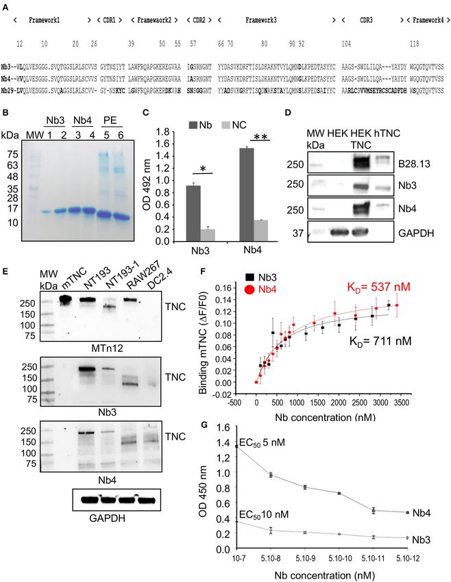
Dhaouadi et al.                                                                                                      Anti-tenascin-C Nanobodies

INTRODUCTION                                                            (36, 37). Thus, better molecular tools are needed for specific
                                                                        and sensitive recognition and potential targeting of TNC. To
Tenascin-C (TNC), discovered over three decades ago, is one             overcome these limitations, recombinant nanobodies (Nbs) with
of the ECM molecules that is highly expressed in tumors                 their remarkable characteristics (i.e., high stability, solubility
such as breast, colorectal and gastric cancers (1–4). High TNC          and specificity and low immunogenicity) may provide a
expression levels correlate with shortened lung metastasis-free         solution (38–43).
survival in breast cancer and overall survival in glioma patients           Here, we have generated two “best in class” nanobodies (Nb3
(5, 6). TNC is a large modular hexameric glycoprotein (7).              and Nb4) that recognize specifically TNC with high affinity
Each TNC subunit displays a central oligomerization domain,             by ELISA and staining of formalin fixed and fresh frozen
followed by 14.5 epidermal growth factor (EGF)-like repeats             tissues, emphasizing novel opportunities for early diagnosis
(three disulfide bridges per EGF-repeat), 17 fibronectin type           and potential monitoring of cancer progression. Therefore,
3 (FNIII) repeats (eight constant and nine additional repeats           these nanobodies may be useful for applications in routine
domains that are subject to alternative splicing) and a globular        cancer diagnosis and for future in vivo targeting of TNC in
fibrinogen domain (7). At physiological level, TNC is transiently       cancer. On the other hand, as TNC impairs cell adhesion on a
expressed during organogenesis (8) and its expression is largely        fibronectin substratum, we determined whether the nanobodies
restricted to a few sites in the adult organism such as in some         abolished this function of TNC. Indeed Nb3 and Nb4 restored
stem cell niches, tendons, and reticular fibers of lymphoid             adhesion of human osteosarcoma and mesangial cells on the
organs (9). Interestingly, high TNC levels are also found in milk       fibronectin/TNC substratum. Interestingly, we observed that
of breast feeding HIV+ mothers (10). At pathological level,             immobilization of DC2.4 dendritic cells on a TNC substratum
TNC was shown to act at multiple levels to promote tumor                in context of CCL21 (22) was blocked by Nb3 and Nb4. Finally,
progression into cancer by enhancing survival, proliferation and        by modeling the Nb/TNC interaction we determined the putative
invasion of tumor cells, driving the formation of new but poorly        amino acid residues involved in complex formation. Altogether,
functional blood vessels and to corrupt anti-tumor immunity,            we demonstrated that our two novel TNC-specific nanobodies
altogether enhancing metastasis. In addition to tumors, TNC is          display valuable characteristics for detection of TNC in situ, and
also highly up-regulated in wound healing, fibrosis and chronic         revealed their potential as therapeutic tools for inhibition of
inflammation (11, 12). Recently, high TNC levels were also              immune-suppressive and other functions of TNC.
associated with more severe COVID19 symptoms (13). Using
stochastic tumorigenesis models with engineered high and low
levels of TNC it was formally proven that TNC indeed is a               MATERIALS AND METHODS
promoter of tumor progression (14). TNC is inducing and
activating a wide range of cellular signaling pathways such as
                                                                        Purification of Recombinantly Expressed
Wnt, Notch, JNK and TGFβ (14–17). TNC also acts on stromal              hTNC
and immune cells thereby promoting tumor angiogenesis and               HEK 293/hTNC cells, previously stably transfected with the
immune escape (18–22). The distinct spatio-temporal expression          human TNC coding sequence (hTNC) were used to produce
pattern of TNC is highly regulated (23). In vitro studies               hTNC as previously described (44, 45). Briefly, cells were
demonstrated that various stimuli such as EGF, TGFβ, b-FGF,             cultured in Dulbecco’s Minimal Essential Medium (DMEM,
and TNF-α, can induce expression of TNC in breast cancer                catalog number 11995040 Gibco Life Technologies, Inc., Paisley,
stroma (5, 24). In cancerous breast tissues, EGF induced TNC            Scotland) supplemented with 10 % (v/v) fetal calf serum (FCS,
via its receptor EGFR which activated oncogenic Ras signaling.          catalog number 2-01F90-I BioConcept, Allschwil, Swizerland),
Mammary tumor cells also produced transforming growth factor            10.25 µg/mL G418 and 1.5 µg/mL puromycin under a 5% CO2
β1 (TGFβ1), which induced TNC expression in the surrounding             atmosphere at 37◦ C. The recombinant hTNC was purified from
stroma (25, 26). Due to defective autophagy TNC seems to                the conditioned medium lacking FCS as previously described
be highly abundant in triple negative breast cancer (27). An            (44). Briefly, fibronectin was removed from the conditioned
overview of factors regulating TNC expression is presented in           medium by gelatin-agarose affinity chromatography (46, 47), and
Giblin et al. (23).                                                     the flow through was purified by a nickel affinity chromatography
    Given its high expression in cancer tissues as well as              column (48). The purity of the protein was checked by Coomassie
its inflammation promoting actions, several efforts have been           Blue stained 7% SDS-PAGE and by western-blot, under reducing
launched to specifically detect TNC in situ as well as to inhibit       and non-reducing conditions. The concentration of hTNC was
its main pathological effects. These approaches included down           determined by Bradford assay (catalog number 500-0006 Bio-
regulation of TNC expression with siRNA or aptamers (28–                Rad Laboratories, Hercules, CA, USA).
32) and the use of TNC-specific antibodies for the delivery of
drugs or radiotherapy (33–35). Moreover, numerous monoclonal            E. coli Strains and Vector
antibodies recognizing TNC have been developed. However, all            The phage display vector pMECS of 4,510 bp was utilized
generated tools have their intrinsic limitations and caveats. In        to construct the VHH library, hosted in E. coli strain TG1
particular, antibodies may not reach the target tissue or can           (generously provided by Prof. Serge Muyldermans, VUB Brussel,
raise an immune response and formalin fixation, usually used            Belgium). This phagemid vector contains a sequence encoding
in routine pathology service, can impair epitope recognition            a PelB leader signal to secrete the cloned VHH-encoded Nb in

Frontiers in Immunology | www.frontiersin.org                       2                                    March 2021 | Volume 12 | Article 635166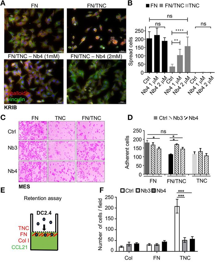
Dhaouadi et al.                                                                                                     Anti-tenascin-C Nanobodies

the periplasm with two C-terminal Hemagglutinin (HA) and 6X             GeneON, Germany) and used to infect exponentially growing
Histidine (6X His) tags for VHH-detection, when hosted in E. coli       TG1 E. coli. Following the third round of biopanning, individual
strain WK6 (49).                                                        colonies were randomly picked. VHH expression was induced
                                                                        with 1 mM isopropyl-D-thiogalactopyranoside (IPTG, catalog
Generation of the Phage-Display                                         number 2900245 5PRIME, Germany) in the periplasmic bacterial
VHH-Library                                                             compartment. Solid phase ELISA of each periplasmic extract was
The anti-hTNC nanobody phage-display VHH-library was                    carried out on hTNC (1 µg/mL), using a mouse anti-HA antibody
constructed as previously described with slight modifications           (catalog number H9658 Sigma Aldrich, MO, USA) and goat anti-
(39, 49, 50). Briefly, 3 days after the last boost of antigen           mouse IgG-peroxidase antibody (catalog number A9044 Sigma
injection, 150 mL of anti-coagulated blood sample was collected         Aldrich, MO, USA).
from the jugular vein of the immunized dromedary as
recently detailed (51). Peripheral blood mononuclear cells              VHH Sequence Analysis
(PBMCs) were extracted by density gradient centrifugation               The VHH sequences of clones that scored positive in periplasmic
using Lymphoprep (catalog number 17-829 LONZA, Basel,                   extract-ELISA were determined using the Genomic platform
Switzerland). Subsequently, total RNA was extracted and                 of Institut Pasteur de Tunis facilities (ABI Prism 3100 genetic
purified. An amount of 40 µg of total RNA was reverse                   analyzer; Applied Biosystems, Foster City, CA, USA). The VHH
transcribed into cDNA with oligo-dT primer and the SuperScript          nucleotide sequences were obtained using the ABI PRISMTM
II First-Strand Synthesis System for RT-PCR (catalog number             BigDye Terminator v3.1 Cycle Sequencing Reaction Kit (catalog
18064-014 Invitrogen, Carsbad, CA, USA). Thereafter, cDNA               number 4337454 Applied Biosystems, USA).
fragments were used as template to amplify heavy-chain IgG
encoding variable domains using specific primers [CALL001               Production, Purification, and
(5′ -GTCCTGGCTGCTCTTCTACAAGG-3′ ) and CALL002                           Characterization of hTNC-Specific Nbs
(5′ -GGTACGTGCTGTTGAACTGTTCC-3′ )]. The 700 bp PCR                      Recombinant vectors of selected positive clones with highest
fragment (VHH-CH2 without CH1 exon, corresponding to                    binding capacity to hTNC were used to transform WK6
heavy-chain antibodies) was purified from a 1% agarose gel              electrocompetent cells. Nb production was performed in shake
using the Qiaquick gel extraction kit (catalog number 28704             flasks by growing each recombinant bacteria in Terrific Broth
Qiagen, Hilden, Germany). Subsequently, these sequences were            medium (TB, catalog number 743-29175 BD Biosciences, FL,
used as template in a nested PCR to amplify VHH-only variable           USA) supplemented with ampicillin (100 µg/mL) and 0.1%
domains with nested-PCR primers [SM017 (5′ -CCAGCCGGCC                  glucose. The Nb periplasmic expression was subsequently
ATGGCTGCATGGTGCAGCTGGTGGAGTCTGG-3′ )                        and         induced with 1 mM IPTG, O/N at 28◦ C. The periplasmic extract
PMCF (5′ -CTAGTGCGGCCGCTGAGGAGACGGTGACCTG                               obtained by osmotic shock was loaded on a His-Select column
GGT-3′ )], annealing at the Framework 1 and Framework 4                 (NiNTA, catalog number 1018544 Qiagen, Hilden, Germany).
regions, including NcoI and NotI restriction sites, respectively        The His-tagged hTNC-specific Nbs were eluted with 500 mM
(catalog numbers R0193T and R3189M New England Biolabs,                 imidazole (catalog number I-0125 Sigma Aldrich, MO, USA)
UK, respectively). The PCR product was ligated into the pMECS           and an amount of 5 µg was checked on a 15% SDS gel upon-
phagemid vector (T4 DNA Ligase, catalog number 15224-041                PAGE (Bio Rad), following dialysis towards PBS with a 12 kDa
Invitrogen, Carsbad, CA, USA) using a molar ratio 1:3 in                cut-off membrane (catalog number D9527-100FT Sigma Aldrich,
favor of the inserts. Freshly prepared electro-competent E. coli        MO, USA). The final yield was determined using Bradford
TG1 cells were transformed by the ligated product and plated            assay (catalog number 500-0006 Bio-Rad Laboratories, Hercules,
overnight (O/N) on selective Luria-Bertani Miller (LB) media            CA, USA) and the molar concentration was estimated using
supplemented with (100 µg/mL) ampicillin (catalog number                the theoretical extinction coefficient of the VHH sequence. The
271896 Sigma Aldrich, MO, USA) and glucose 2% (catalog                  specificity of the purified anti-hTNC nanobodies was assessed by
number G8270 Sigma Aldrich, MO, USA). Colonies were                     ELISA. Briefly, 0.5 µg/mL of hTNC was coated onto microtiter
recovered from the overnight-incubated plates at 37◦ C. Library         plates O/N at 4◦ C and unspecific sites were blocked with
size was estimated by serial dilutions.                                 1% (w/v) gelatin (catalog number 48723 Fluka Analytical,
                                                                        USA) supplemented with PBS/0.05% Tween-20 at 37◦ C for
Selection of Anti-hTNC Nanobodies (Nbs)                                 2 h. Affinity-purified Nb was added (5 µg/mL, 1 h). Following
A representative repertoire of the VHH library was displayed            a washing step, bound Nb was detected with a mouse anti-
on phage particles using M13KO7 helper phage infection                  HA antibody (catalog number H9658 Sigma Aldrich, MO, USA)
(catalog number 170-3578 New England, BioLabs, UK). Three               and revealed with a goat anti-mouse IgG-peroxydase conjugate
consecutive rounds of immuno-affinity selection were carried            (catalog number A9044 Sigma Aldrich, MO, USA).
out on 96-well microtiter plates (catalog number M5785-1CS
Sigma Aldrich, MO, USA) pre-coated with hTNC (1 µg/panning,             Assessement of Nb Binding Affinity
O/N at 4◦ C). After each round of biopanning, bound phage               The assessement of Nb binding affinity was performed using
particles were eluted (100 mM triethylamine, pH 10.0, catalog           two methods: (i) indirect ELISA was carried out using a
number T0886 Sigma Aldrich, MO, USA) and immediately                    serial Nb dilution ranging from 5 × 10⁻⁷ to 5 × 10⁻¹²
neutralized with 1 M Tris-HCl, pH 7.4 (catalog number CE234             M, as described above; (ii) Isothermal Fluorescence Titration

Frontiers in Immunology | www.frontiersin.org                       3                                   March 2021 | Volume 12 | Article 635166
Dhaouadi et al.                                                                                                       Anti-tenascin-C Nanobodies

(IFT) was performed using recombinant murine TNC (mTNC,                  Immunofluroescence Assay
700 nM, 0.01% Tween-20), as previously described with slight             Glioblastoma cell xenografts had previously been generated by
modifications (52). Briefly, the Nb concentration varied from 500        subcutaneous injection of 2 × 10⁶ U87MG or U87MG-shTNC
to 3,500 nM. The fluorescence emission spectra for mTNC/Nb               (TNC knockdown) cells into the flank of a nude mouse (56).
complexes were collected and subsequently subtracted from                Frozen (−80◦ C) sections were cut (7 µm thickness), fixed with
emission spectra for mTNC and the resulting curves were then             4% paraformaldehyde (PFA) (catalog number 30525-89-4 Sigma
integrated. The mean values resulting from three independent             Aldrich, MO, USA) for 15 min at RT, and permeabilized with
measurements were plotted against the concentration of the               0.5% Triton X-100 in PBS and blocked with 10% normal donkey
added Nb. The resulting binding isotherms were analyzed by               serum (NDS) in PBS for 2 h at RT (catalog number 017-000-
nonlinear regression using the program Origin (Microcal Inc.,            121 Jackson ImmunoResearch Inc, USA). Sections were co-
Northampton, MA, USA). The following equation describes the              stained with the Nb and B28.13 antibody diluted in PBS, 10%
bimolecular association reaction, where Fi is the initial and Fmax       NDS O/N at 4◦ C, rabbit anti-HA antibody (ab236632 abcam,
is the maximum fluorescence values. The KD is the dissociation           UK, 1:1,000 dilution, 90 min at RT) and, donkey anti-mouse
constant, and [mTNC] and [Nb] are the total concentrations of            antibody labeled with Texas Red fluorophore (catalog number
the mTNC and the Nb ligand, respectively:                                PA1-28626 Invitrogen, Carsbad, CA, USA), and donkey anti-
                                                                         rabbit antibody labeled with Alexa Fluor 488 Green fluorophore
    F = Fi+ Fmax [KD + [mTNC] + [Nb]                                     (catalog number AB-2313584 Jackson Immuno Research Inc,
          q
             KD + [mTNC] + Nb 2 − 4 [mTNC] [Nb]                          USA) were used (1:1,000 dilution for 90 min at RT). After each

        −                                       ]                        antibody incubation, sections were washed five times with PBS.
                           2[mTNC]                                       For staining of cell nuclei, sections were incubated with 4’, 6-
Negative Electron Microscopy Imaging                                     diamidino-2-phenylindole (DAPI, 0.2 µg/mL, catalog number
The Nb/ hTNC interaction complexes were visualized by negative           32670 Sigma Aldrich, MO, USA) for 10 min at RT. Slides were
staining and electron microscopy as previously described (53).           sealed with a polymerization medium (FluorsaveTM Reagent,
Each Nb (20 nM) was conjugated with 5 nm colloidal gold                  Calbiochem) underneath the coverslips and stored at 4◦ C until
particles (AuNPs) according to routinely used procedures (54).           analysis. Pictures were taken with an AxioCam MRm (Zeiss)
AuNP-Nb conjugates were incubated with hTNC (20 nM) for                  camera and Axiovision software.
30 minutes (min) at room temperature (RT) and subsequently
negatively stained with 2% uranyl acetate. Specimens were
assessed and electron micrographs were taken at 60 kV
                                                                         Human Tumor Samples and Analysis by
with a Phillips EM-410 electron microscope using imaging                 Immunohistochemistry
plates (Ditabis).                                                        Surgically removed tongue tumors, Formalin-Fixed Paraffin-
                                                                         Embedded (FFPE) embedded in FFPE, were retrieved from
Western Blot Analysis of TNC Specific Nb                                 the tumor bank of the Centre Paul Strauss (Strasbourg,
Cell lysates (40 µg) from HEK293, HEK293/hTNC, (20 µg)                   France). The FFPE-embedded 17/18G percutaneous needle
NT193 and subclone NT193-1 cells (generated by limited                   biopsy of an hepatic metastasis derived from carcinoma of
dilution), RAW267 macrophages (ATCC) and DC2.4 dendritic                 the gall bladder (CGB) was collected as part of a study
cells (22) in RIPA buffer (catalog number R0278 SIGMA                    involving human participants approved by the Mongi Slim
Aldrich, MO, USA) or purified human hTNC (hTNC, 100                      University Hospital (MSUH) Committee on Medical Ethics
ng) and murine TNC (mTNC, 50 ng) were boiled at 100◦ C                   (La Marsa, Tunisia) and the Ethikkommission Nordwest-und
for 5 min, before loading on a 4–20% gradient SDS/PAGE                   Zentralschweiz (Switzerland). Informed consent was obtained for
gel (catalog number 456-8095 Mini-PROTEAN TGXTM , Bio-                   all subjects. Characteristics of patients with oral squamous cell
Rad Laboratories, Hercules, CA, USA), then, transferred                  carcinoma (OSCC), or CGB liver metastasis, are summarized in
onto a PVDF membrane (catalog number 1620174 Bio-                        Supplementary Table 1.
Rad Laboratories, Hercules, CA, USA). After blocking with                    Immunohistochemical staining of OSCC samples was
5% milk, PBS/0.1% Tween-20 (catalog number 1706404 Bio-                  performed on serial 5 µm deparaffinized tumor sections. For
Rad Laboratories, Hercules, CA, USA) the membrane was                    hTNC staining, intrinsic peroxidase was blocked by incubating
incubated O/N at 4◦ C with Nb3 or Nb4 (2 µg/mL). After                   sections with 3% hydrogen peroxide for 15 min and antigen
three washing steps, the membrane was first incubated with               retrieval was performed in Sodium Citrate (10 mM) buffer pH
a mouse anti-HA antibody (1 h 30 min at RT), and then with               6.0 at 95◦ C. Sections were blocked in 5% goat serum for 1 h,
the anti-mouse IgG horseradish peroxidase conjugate diluted at           then incubated ON/4◦ C with rabbit anti-TNC antibody (#19011,
1:1,000 (catalog number AB_772209, NXA931, Amersham GE                   Millipore, 1 µg/mL) or anti-hTNC Nb (2 µg/mL). After PBS
Helthcare, USA). Immunocomplexes were revealed with ECL                  rinsing, sections were incubated with biotinylated goat anti-
(catalog number 28 980926 Amersham GE healthcare, USA).                  rabbit or goat anti-lama antibodies (1 h at RT) then avidin-biotin
A prestained protein ladder (10–250 kDa, catalog number 06P-             (PK-4000, VECTASTAIN ABC Kit, Vector Lab, California,
0211 Euromedex, France) was used. Mouse monoclonal antibody              USA). Staining was revealed with 3, 3 ′ -Diaminobenzidine
B28.13 (1 µg/mL), raised against hTNC was used as a positive             developing solution (SK-4100, DAB, Vector Lab, California,
control (55).                                                            USA) then sections were counterstained with hematoxylin.

Frontiers in Immunology | www.frontiersin.org                        4                                    March 2021 | Volume 12 | Article 635166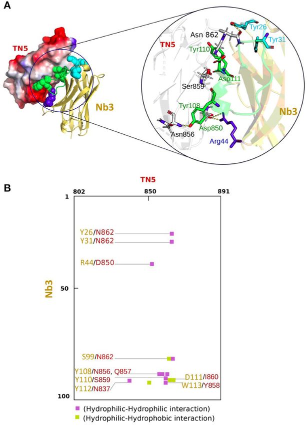
Dhaouadi et al.                                                                                                           Anti-tenascin-C Nanobodies

After embedding in aqueous mounting medium, sections were               Statistical Analysis
examined using a Zeiss Axio Imager Z2 microscope. Pictures              Statistical differences were analyzed by a two-way ANOVA
were taken with an AxioCam MRm (Zeiss, Axiovision) camera.              test or a Kruskal-Wallis t-test, Student’s test and Dunn’s post-
The image acquisition setting (microscope, magnification, light         test. Statistical analyses were performed using the GraphPad
intensity, exposure time) was kept constant per experiment and          Prism software. p-values < 0.05 or < 0.005 were considered as
in between conditions. The origin of the tumor sample, patient          statistically significant. Data are expressed as the mean ± SEM.
gender, TNM stage, presence of metastasis and sampling date are
depicted in Supplementary Table 1.                                      Structural Modeling of the Nb – TNC
   Immunohistochemical stainings of the CGB liver metastasis            Interaction
sample were performed on a Ventana Discovery Ultra instrument           For the Nb - TNC structral interaction modeling the Rosetta
(Roche Diagnostics). The procedure RUO Discovery Universal              Antibody application (57, 58) from “ROSETTA 3.8”1 was used.
was used with 40 min CC1 pre-treatment and anti-TNC                     Selection of the top 10 Model was done according to Rosetta
B28.13 (1:5,000), Nb3 (1:100) or Nb4 (1:50) were applied                scoring based on system energy. Structural information about the
manually and incubated for 1 h at 37◦ C. For Nb3 and Nb4,               5th TNC fibronectin type III repeat (TN5) was extracted from
a rabbit anti-HA antibody (C29F4, Cell Signaling), used as              the protein data bank deposited under the PDB code: 1TEN. The
a linker to detect the nanobodies, was applied manually                 TN5 structure was refined using the ROSETTA relax application
(1:200) and incubated for 1 h at 37◦ C. Then, an anti-mouse             which then was used to map potential interaction sites in the
antibody used for B28.13 (ImmPRESS reagent kit peroxidase               selected Nb through the ZDOCK docking tool, version 3.0.2 (59).
anti-mouse Ig MP-7402, Vector Laboratories) or an anti-                 Structure and complex interactions were then visualized via the
rabbit antibody used for Nb3 and Nb4 (ImmPRESS reagent                  molecular visualization PyMOL software (60).
kit peroxidase anti-rabbit Ig MP-7401, Vector Laboratories)
were applied manually (200 µl) and incubated for 32 min at
                                                                        RESULTS
37◦ C. Finally, the ChromoMap DAB kit (Roche Diagnostics)
was used for detection and slides were counterstained with              Generation of an Immune VHH Library to
Hematoxylin II and Bluing Reagent (Roche Diagnostics)                   Produce hTNC- Specific Nanobodies
for 8 min.                                                              An immune response against hTNC had been elicited in the
                                                                        immunized dromedary as previously described (51). From
Boyden Chamber Transwell                                                PBMC, total RNA was extracted and cDNA was prepared.
Chemoretention Assay                                                    This cDNA was used as template to perform the first
Boyden chamber transwell chemoretention assay of DC2.4                  PCR using primers CALL001 and CALL002 specific for the
dendritic cells toward CCL21 was carried out as described               variable domains of the heavy-chain isotypes (IgG1, IgG2,
previously (22). The bottom of the chamber was filled with              and IgG3), subsequently leading to the co-amplification of
DMEM containing human CCL21 (200 ng/mL, catalog number                  VH and VHH coding domains (Supplementary Figure 1A).
366-6C-025 R&D Systems, Minneappolis, USA). The lower                   As expected, because of the presence of CH1 domain in
surface of the transwells was coated with purified horse                conventional antibodies (IgG1) and different hinge size in non-
fibronectin (FN) (37), rat collagen type I (Col I, catalog              conventional antibodies (HCAbs), three PCR products were
number 354236 BD Biosciences, FL, USA) or hTNC at a final               observed by agarose gel electrophoresis: the 900, 790, and 720
concentration of 1 µg/cm2 and incubated O/N with blocking               bp fragments corresponding to the VH-CH1-Hinge-CH2 of
solution alone or with the Nb, respectively. DC2.4 (5 × 10⁵ )           the IgG1 and the VHH-Hinge-CH2 exons of the IgG2 and
cells resuspended in 150 µL of 1% FBS-complemented DMEM                 IgG3, respectively (Supplementary Figure 1A). Selective only-
were placed into the top chamber of the transwell system. After         VHH fragments were successfully amplified using nested PCR
5 h of incubation at 37◦ C in 5% CO2 , cells on the lower side of       specific-primers and cloned in pMECS phagemid (see above).
the insert were fixed with PFA and stained with DAPI before             The obtained hTNC-specific VHH library was estimated to
cell counting.                                                          contain approximately 3 × 10⁶ CFU/mL independent clones. The
                                                                        insert size of 19 randomly chosen clones was investigated by PCR.
Adhesion Assay                                                          The library insertion rate with a VHH insert of the expected size
The adhesion assay was carried out on human KRIB                        was 78.94% (Supplementary Figure 1D).
osteosarcoma and human MES mesangial cells. Precisely,                     A representative aliquot of the TG1 cells harboring the
96-well plates were coated with 1 µg/cm2 FN, hTNC or with               VHH antibody repertoire was rescued with the M13KO7 helper
FN and hTNC together. Nbs were added at 500 nM after the                phage to produce phage particles expressing the nanobodies.
coating for 1 h at 37◦ C. Plates were rinsed with PBS and the           Following three rounds of phage display selection on solid-
non-coated plastic surface was blocked with 1% BSA for 1 h.             phase coated hTNC, enrichment for hTNC-specific phage
After blocking, KRIB osteosarcoma and MES were plated for 3             particles was observed from the first round of panning onwards
and 2 h, respectively, at 37◦ C in a humidified atmosphere with         (Supplementary Table 2). Twenty-five periplasmic extracts
5% CO2 . After incubation, non adherent cells were removed by
PBS washing and spread cells were stained with cristal violet           1 The Rosetta software, https://www.rosettacommons.org/software/license-and-

and counted.                                                            download

Frontiers in Immunology | www.frontiersin.org                       5                                         March 2021 | Volume 12 | Article 635166
Dhaouadi et al. Anti-tenascin-C Nanobodies
selected from randomly picked individual clones of biopanning but also of TNC in the supernatant from HEK293/TNC cells.
rounds were checked by ELISA. Only clones scoring positively Parental HEK293 cells did not express TNC and also showed no
by ELISA without binding to unspecific proteins (AahI and BotI signal with Nb3 nor Nb4 (Figure 1D).
toxins, respectively) were retained (Supplementary Figure 1E). As human and murine TNC are highly conserved (62) we
In total, eight recombinant clones displaying highest recognition used Nb3 and Nb4 for detection of murine TNC by western blot.
and binding to the hTNC from the sequenced VHHs were Whereas, the monoclonal anti-TNC antibody MTn12 recognized
selected. Two recombinant clones displayed identical amino- recombinant mTNC expressed in HEK293/TNC cells indicated
acid sequences (Nb3 and Nb5). The Nb3, Nb4, and Nb29 were by the appearance of multiple bands, Nb3 and Nb4 did not
selected for further investigation. As illustrated in Figure 1A, recognize these TNC species (Figure 1E). There were several
all hTNC-specific Nb sequences (Nb3, Nb4, and Nb29) exhibit higher molecular weight bands of TNC recognized by MTn12 in
the VHH hallmark amino acid residues in the framework-2 recombinant TNC, NT193, and RAW267 cells. However, one or
region (FR2 Phe42, Glu49, Arg50, and Gly52) and the conserved more of these bands at 250 kDa were well-recognized by Nb3 and
Trp at position 118 of the anchoring region. According to their Nb4 in NT193 cells but poorly in HEK293 and RAW267 cells.
common CDR1 sequence, Nb3 and Nb4 are most likely derived On the contrary TNC proteoforms (between 150 and 250 kDa)
from the same V-D-J rearrangement and share the same B-cell that were recognized by Nb3 and Nb4 in NT193-1, RAW267 and
progenitor (40). The difference in CDR3 length of the Nb29 DC2.4 cells were not recognized by MTn12 (Figure 1E). There
corresponding sequence is remarkable (21 amino-acid residues). are several explanations for this result. First it is conceivable
Interestingly, the Nb29 has a total of six cysteins (at positions that the epitope recognized by MTn12 (which is not known)
23, 33, 103, 108, 117, and 119) and therefore harbors two is different to that recognized by Nb3 and Nb4. Second Nb3
additional interloop disulfide bonds, in addition to the FR1/FR3 and Nb4 recognize a particular TNC conformation as epitope
conventional ones (Cys23/Cys103). Nevertheless, because of its which is lost upon denaturation by SDS and boiling. Third,
moderate binding to hTNC and low yield, Nb29 was not further glycosylation may have an impact on the conformation of the
investigated here. epitope recognized by Nb3 and Nb4. Recently it was shown that
The CDR3 residue length within the Nb3 and Nb4 sequences N-glycosylation in TNC (in particular within TN5) impacted
(17 amino-acid residues) was identical and no difference was binding of the envelope protein of HIV (10). Apparently the
noticed. The only divergence in the Nb3 and Nb4 sequences was conformation of the epitope recognized by Nb3 and Nb4 is
observed at position 2 (Leu substituted by Val in FR1), at position still available in NT193 and the other murine cells despite
52 (Gly substituted by Ala in CDR2) and at position 92 (Asp denaturation as seen in Figure 1E. These bands are specific for
substituted by Gly in FR3), respectively, suggesting an interaction TNC since unspecific anti-HA bands are below 85 kDa (63).
with a common epitope on TNC. In conclusion, Nb3 and Nb4 may recognize a conformational
epitope in TNC that could be sensitive to denaturation and/or
N-glycosylation which has to be further investigated in the future.
Production and Purification of hTNC
Specific Nanobodies Assessment of Nb3 and Nb4 Affinities for
Only clones that scored highly positive
(Supplementary Figure 1E) were further used for flask
TNC by Isothermal Fluorescence Titration
By Isothermal Fluorescence Titration (IFT), we investigated the
production and Immobilized Metal Affinity Chromatography
binding of Nb3 or Nb4 to fluorescently tagged mTNC until signal
purification (IMAC), according to Hmila et al. (61). Briefly,
saturation and determined the dissociation constant (KD ) as 711
production of each soluble anti-hTNC Nb was accomplished
× 10−9 M (Nb3) and 537 × 10−9 M (Nb4) that indicates a
by transformation of E. coli WK6 cells with the corresponding
robust interaction (Figure 1F). In addition, ELISA assays were
recombinant phagemid. The amber stop codon located between
performed with Nb molar concentrations ranging from 5 × 10⁻⁷
the VHH insert and the gene III within the pMECS phagemid,
to 5 × 10⁻¹² M and revealed specific binding with a 50% Effective
resulted in expression of the Nb as soluble protein in the
Concentration (EC50 ) of both Nb3 and Nb4 binding to hTNC
periplasm compartment of E. coli, leading to rapid IMAC
at 10 and 5 nM, respectively, again revealing a strong interaction
purification of the Nb. As expected, the Coomassie-stained
(Figure 1G).
SDS/PAGE gel revealed the apparent molecular weight of 14 kDa
(Figure 1B). The Nb3 and Nb4 production yields were estimated
ranging from 0.6 to 0.8 mg/L, respectively, when flask cultured Detection of the Nanobody Binding Site in
in TB medium. No bands indicative of contaminants or Nb TNC by Negative Electron Microscopy
degradation were detected. We investigated the location of the antigenic epitope in hTNC
Nb4 (5 µg/mL) displayed a higher ELISA binding titer by negative electron microscopy where the nanobodies were
toward hTNC (0.5 µg/mL, OD492nm = 1.526), compared coupled to gold beads. Upon incubation of hTNC with gold
to Nb3 (5 µg/mL, OD492nm = 0.913) and to the irrelevant beads-bound nanobodies, we detected the gold particles along
nanobody (anti-BotI toxin nanobody, 5 µg/mL, OD492nm = the length of the hTNC monomers and quantified them
0,118) (Figure 1C, Supplementary Figure 2). Furthermore, (Figure 2A). We observed a high number of Nb3 and Nb4
immunoblotting assays revealed that both nanobodies showed binding in the middle of the hTNC monomer resembling
a specific recognition of not only the purified hTNC (100 ng) binding of several soluble factors in the fifth fibronectin type
Frontiers in Immunology | www.frontiersin.org 6 March 2021 | Volume 12 | Article 635166Dhaouadi et al. Anti-tenascin-C Nanobodies
FIGURE 1 | Specificity of the purified nanobodies for hTNC (A) Comparative alignment of amino acid sequences of nanobodies Nb3, Nb4, and Nb29 showing four
amino acid hallmark changes at positions F42, E49, R50, and G52 according to the IMGT Scientific chart analysis for the V-Domain. Positioning of CDRs1-3 and
Frameworks 1–4 are indicated. (B) SDS-PAGE analysis of the purified nanobodies Nb3 and Nb4. The nanobodies were expressed in bacteria and purified bacterial
lysate was separated on a 15 % SDS-PAGE gel that was stained with Coomassie blue. Lanes represent MW: Prestained molecular weight marker, size indicated in
kDa. 1, 2: Nb3 eluates 1 and 2. 3, 4: Nb4 eluates 1 and 2. 5: Purified periplasmic extract (PE) from Nb3 after induction. 6: Purified periplasmic extract from Nb4 after
induction. Nb3 and Nb4 are visible at 15 kDa, the respective molecular weight of a nanobody. (C) Binding specificity assessement of Nb3 and Nb4 An amount of 50
ng hTNC was coated onto microtiter plates, and 500 ng (100 µl) nanobodies were added. After incubation with a mouse anti-HA antibody and then anti-mouse HRP,
absorbance at 492 nm was measured by an ELISA reader. NC, no coating. Values were the means of 3 independent experiments. Mean ± SEM, *p < 0.05, **p <
0.01, Student’s t-test. (D) Western blot analysis of Nb3 and Nb4 An amount of 40 µg of total cell lysate from parental HEK293 (HEK) (devoid of TNC) and HEK:TNC
(Continued)
Frontiers in Immunology | www.frontiersin.org 7 March 2021 | Volume 12 | Article 635166Dhaouadi et al. Anti-tenascin-C Nanobodies
FIGURE 1 | (engineered to express hTNC) and 100 ng of purified hTNC were analyzed by Western blot for detection of TNC by B28.13 (monoclonal anti-hTNC
antibody) or Nb3 and Nb4 (2 µg/mL). GAPDH was used as loading control. Representative result, n = 3. (E) Western blot of 50 ng of purified mTNC (22) and 20 µg of
total cell lysate of the indicated murine cells with GAPDH as loading control. Detection of TNC with the MTn12 antibody or Nb3 and Nb4, respectively. Representative
result, n = 2. (F) Determination of the effective concentration (EC50) at which 50% of epitopes in hTNC are occupied by Nb3 (diamonds) and Nb4 (squares),
respectively. The experiment was done in triplicates. Mean ± SEM. *p < 0.05, Two-way ANOVA test. (G) Binding affinities of Nb3 and Nb4 for recombinant mTNC as
measured by isothermal fluorescence titration. The experiment was done three times.
FIGURE 2 | Identification of interaction sites of Nb3 and Nb4 in hTNC. (A) Binding of gold-labeled Nb3 and Nb4 to hTNC was determined by negative staining and
transmission electron microscopy. The hTNC molecule in the absence or presence of Nb3 or Nb4 is depicted. Black dots represent binding sites for Nb3 and Nb4 in
hTNC. (B) Quantification of Nb3 and Nb4 binding according to the position (nm) on TNC Representation of a TNC monomer with oligomerization domain (triangle) to
form hexamers as seen in (A), FNIII repeats (gray boxes, constant domains, white boxes, alternative domains) and fibrinogen like domain (circle). Representative result
of three independent experiments, displaying the quantification of 500 micrographs.
III repeat (FNIII) in TNC [TN5 (64)]. Nb3 and Nb4 showed carcinoma of the gall bladder (CGB) (Supplementary Table 1)
a similar binding pattern suggesting that both nanobodies with Nb3 and Nb4 and observed a strong immunoreactivity
recognize the same or overlapping epitopes in hTNC, presumably of the stroma, similar to the staining observed with the
TN5 (Figure 2B). anti-TNC antibody B28.13 (Figure 3B). This stromal staining
resembled that of TNC expression in biliary tract cancers,
including CGB liver metastasis as seen by conventional
Nanobodies Nb3 and Nb4 Recognize hTNC IHC (68).
in Fresh Frozen and Paraffin-Embedded Next we addressed recognition of murine and human TNC
Human Tissues in tissues by IHC and IF. Therefore, we stained U87MG
Next, we investigated whether Nb3 and Nb4 recognized glioblastoma xenografted tumors where it was previously noticed
hTNC in FFPA tissues. Therefore, we stained human colon that human TNC was largely more abundant than murine TNC
tissue from an ulcerative colitis (UC) patient and noticed by IHC and IF (56). We observed a fibrillar TNC signal in the
a staining pattern that resembled published TNC expression U87MG tumors by IHC with Nb3 and Nb4 (Figure 3C) that
in this tissue (65–67), (Supplementary Figure 3A). We also overlapped with that of the B28.13 antibody signal, confirming
stained tissue from human tongue tumors (OSCC) with Nb3 specificity of the nanobodies for TNC (Figure 3D). As the
and Nb4 and with a commercial rabbit polyclonal anti- U87MG tumors also express murine TNC but at much lower
TNC antibody on an adjacent section, and observed similar abundance (56), we stained U87MG tumors with a knockdown
staining patterns for TNC reminiscent of tumor matrix tracks for human TNC in the grafted tumor cells and did not see a signal,
(TMT) that have previously been described [Figure 3A, (22)]. suggesting that Nb3 and Nb4 at the chosen dilution recognize
We also stained a liver metastasis from a patient with a predominantly human TNC (Figure 3E).
Frontiers in Immunology | www.frontiersin.org 8 March 2021 | Volume 12 | Article 635166Dhaouadi et al. Anti-tenascin-C Nanobodies
FIGURE 3 | Detection of TNC in tissues by Nb3 and Nb4 IHC (A–C) and IF analysis (D,E) with Nb3 and Nb4 (A–E), B28-13 (B–E) and a polyclonal anti-TNC antibody
(Anti-TNC) (A). (A) Human OSCC (FFPE), (B) liver metastasis from a gall bladder carcinoma (FFPE), (C–E) U87MG tumors, (C) FFPE, (D,E) PFA fixed tissue. Scale bar,
100 µm (A), 200 µm (B), 70 µm (C) and 50 µm (D,E).
Nanobody Nb4 Counteracts the Supplementary Figure 4). However, upon addition of Nb4 to
Anti-adhesive Properties of TNC on a cells plated on FN/TNC we observed that cells spread in a
Nb4 dose dependent manner with actin stress fibers and focal
FN/TNC Substratum
adhesions that looked similar to those in cells plated on FN
By using human osteosarcoma KRIB cells, we investigated
(Figures 4A,B, Supplementary Figure 4). As in KRIB cells, both
whether Nb4 had an impact on cell rounding by TNC on a
Nb3 and Nb4 restored adhesion of mesangial cells (MES) on
FN/TNC substratum. Previously, we had shown that cells are
a FN/TNC substratum suggesting that Nb3 and Nb4 blocked
inhibited by TNC to spread on a combined FN/TNC substratum
binding of TNC to FN (Figures 4C,D).
since TNC competed syndecan-4 binding to FN (44, 69). Here,
we plated KRIB cells on FN, FN/TNC and TNC, respectively with
or without Nb4. By staining with pholloidin (polymerized actin) Nanobodies Nb3 and Nb4 Abolished DC2.4
and anti-vinculin (focal adhesions), we confirmed cell spreading Chemoretention by TNC/CCL21
on FN and cell rounding on FN/TNC and TNC, respectively. Previously, we had shown that in combination with CCL21 TNC
While cells had some actin stress fibers and focal adhesions immobilized dendritic DC2.4 cells (22). Here we used a Boyden
on FN, addition of Nb4 did not change that (Figures 4A,B, chamber transwell migration assay to investigate whether Nb3
Frontiers in Immunology | www.frontiersin.org 9 March 2021 | Volume 12 | Article 635166Dhaouadi et al. Anti-tenascin-C Nanobodies
FIGURE 4 | Nb3 and Nb4 interfere with TNC functions. (A,B) Cell spreading
assay followed by quantification of KRIB cells plated on FN, a mixture of FN
and hTNC (top panels) or a mixture of FN and hTNC incubated with Nb4
(bottom panels). After 2 h, the KRIB cells were fixed and stained with phalloidin
(red) to detect polymerized actin, or an anti-vinculin antibody (green) to detect
FIGURE 5 | Modeling of the three dimensional topology of the Nb3/TN5
focal adhesion complexes and the nuclear marker DAPI (blue). Higher
interaction complex and information about amino acid contacts. (A) Model of
magnifications are shown in Supplementary Figure 4. Spread cells were
the Nb3/TN5 complex TN5 (red) and Nb3 (yellow) are shown with contacting
counted (B). N = 3 experiments, n = 2 wells. **p < 0.01, ***p < 0.001.
amino acids in blue and green (left). Magnification (circle) represents a spatial
Two-way ANOVA test. (C,D) Adhesion of mesangial cells (MES) 2 h after
view on identified amino acid residues (three letter code, position in TN5 and
plating on FN, TNC or FN/TNC without (Ctrl) or with Nb3 and Nb4, respectively
Nb3 indicated by a number, respectively) generating relevant hydrophobic,
followed by quantification (D). N = 3 experiments, n = 2 wells. *p < 0.05.
electrostatic and H-bond interactions (right). TN5 is represented as white
Two-way ANOVA test. (E) Schematic representation of the Boyden chamber
surface with electrostatic surface coloring. Nb3 is presented in transparent
transwell chemoretention assay with DC2.4 toward CCL21 in the bottom well.
yellow. The most implicated amino acid residues forming H-bonds are labeled
The lower surface of the insert was coated with FN, Col I, or hTNC,
with a dashed yellow line. (B) Contact interaction map of TN5 with Nb3
respectively. (F) Quantification of DC2.4 cells on the coated surfaces upon
representing amino acids 802–891 in TNC (top) and amino acids 1–100 in Nb3
migration toward CCL21 (5 h after plating) and pretreatment or not (Ctrl) with
(left). Violet (hydrophilic-hydrophilic), green (hydrophobic-hydrophobic) and
Nb3 and Nb4, respectively. Note that Nb3 and Nb4 significantly abolished
yellow (hydrophilic-hydropholic) boxes represent the properties of the
DC2.4 cell retention by TNC/CCL21. N = 2 experiments, n = 4 wells. Mean ±
interaction.
SEM, Kruskal-Wallis test and Dunn’s post-test. ***p < 0.005, ****p < 0.0001.
Three Dimensional Topology of the
and Nb4 impacted chemoretention by TNC. Therefore, we coated Nb3/TN5 Interaction Complex
the lower surface of the insert with FN, collagen I (ColI) or We adopted a computational structure analysis strategy in order
TNC and added Nb3 or Nb4, respectively, and measured DC2.4 to identify amino acid residues involved in the interaction of Nb3
cell migration toward CCL21 placed in the lower chamber. We with TN5. The 1f2x (chain L), 3l95 (chain B), 5ocl (chain G)
measured cells adhering on the coated surfaces in the presence or and 5vak (chain B) structures were used as principal template
absence of the nanobodies and observed first a high number of for Nb3-frameworks, CDR1, CDR2 and CDR3, respectively. The
cells being tethered on TNC but not the other coatings. Second, number of generated Nb3 models was set to 1,000. The top
we noticed that the number of adherent cells dropped on TNC ten model best scores, ranked according to an energy-based
to that of the other coatings upon addition of Nb3 and Nb4 scoring were selected from 10 clusters composed of 100 structures
whereas no difference was seen with the other matrix coatings per cluster then visually double checked (to detect structural
(Figures 4E,F). anomalies) using the molecular visualization PyMOL software.
Frontiers in Immunology | www.frontiersin.org 10 March 2021 | Volume 12 | Article 635166Dhaouadi et al. Anti-tenascin-C Nanobodies
The TN5 structure (1TEN) was included in the Nb3 molecular Staining of FFPE tissues remains a challenge in the clinical
docking simulation. The positively and negatively charged practice due to frequent masking of epitopes. Also access of
residues are in blue and red, respectively, whereas the neutral antibodies to their epitopes used in functionalized antibody
side-chains are indicated in white, showing clearly separated assisted drug delivery remains a challenge. Due to their intrinsic
charges on the TN5 surface. Using the docking approach, we characteristics, such as small size, high stability and good
generated the top ten possible binding sites, ranked according specificity, nanobodies constitute promising agents to overcome
to an energy-based scoring and filtered them to get an unique some of these limitations (85). Indeed, in a recently patented
complex presenting the best molecular orientation with the most study covering another group of TNC specific nanobodies (86,
stable position (Figure 5A). In order to assess the main amino 87), the authors demonstrated that the radioactive coupled
acid residues involved in the molecular Nb3-TN5 interaction, nanobody 64 Cu-NJTs detected micrometastasis in tumor mice by
we used the COCOMAPS (bioCOmplexes COntact MAPS) web life imaging. These results promise that TNC specific nanobodies
application server. A predicted intermolecular contact map of the could be used for delivery of therapeutic compounds into tumors
Nb3-TN5 complex is illustrated in Figure 5B with a cut-off of or into other tissues with high TNC content.
3 Å, highlighting the most crucial residues mainly implicated in In this report we aimed at the development of nanobodies
the complex interaction. The predicted binding site residues in directed against hTNC to detect TNC in FFPE tissues and to block
TN5 are as follow: N862, D850, N856-Q857, S859, I860, N837, TNC functions. We purified recombinant TNC of human origin
Y858 interacting with Nb3 at position Y26, Y31, R44, S99, Y108, and used this molecule to elicit a potent immune response in the
Y110, D111, Y112, W113. Interestingly, the CDR2 and CDR3 are dromedary. A substantial proportion of polyclonal heavy chain
predicted to dominate the interaction with TN5. Details of crucial IgG subclasses bound to TNC and recognized TNC by IF staining
residues involved in H bounds as proton donors or acceptors are in tumor tissue (51). This encouraged us to generate nanobodies.
described in Supplementary Table 3. A VHH library from this dromedary was generated that met the
required quality control standards (88, 89) and allowed us to
isolate nanobodies that specifically recognized TNC. Although,
DISCUSSION the titer of dromedary antibodies against hTNC was significantly
high, the in vitro selection of anti-hTNC binders from this
The matrix, a highly abundant component of tumors, could specific VHH library allowed us to retrieve only eight binders
be considered as a good tumor biomarker as matrix is often after three rounds of bio-panning. A possible explanation of this
more stable than e.g., antigens expressed by tumor cells (70, 71). limited sequence diversity of binders might be attributed to our
Furthermore, matrix seems to be accessible to antibodies and screening condition as we immobilized hTNC which potentially
antibody derivatives in therapy (33). Detection of abundant has masked the epitopes or prevented nanobodies to bind due to
tumor specific matrix could be useful for monitoring tumors sterical hindrance. To retrieve nanobodies recognizing additional
and their progression. In this context, TNC is an intriguing TNC sequences, future biopanning could be done by using
matrix molecule, as it is highly expressed not only in tumors, soluble TNC.
but also in fibrosis and chronic inflammation often correlating In this paper, we have generated eight human TNC specific
with disease progression (4, 11, 72, 73). Hence, detection of high nanobodies and the two best in class candidates (Nb3, Nb4)
TNC levels in tissues and body fluids of patients with e.g., cancer in terms of binding strength and specificity were further
or rheumatoid arthritis is a promising strategy (21, 22, 62). The characterized in more detail. The amino acid sequences of the
large isoform of TNC, highly abundant in cancer tissue, may three TNC-specific Nb revealed a high degree of identity with
even be a good address for the delivery of drugs into the tumor human VH sequences of family III; however the VHH imprints
(1). In particular, TNC-specific antibodies were shown to be a were clearly present (90, 91).
means for tumor targeting. In the past, several TNC-specific Nb3 and Nb4 recognized specifically TNC by
monoclonal antibodies were developed as well as aptamers and immunoblotting, ELISA and negative EM imaging and by
antibody fragments (scFv) that are currently undergoing clinical staining of PFA and FFPE fixed human tissues. We identified
evaluation (37, 74–78). The anti-human TNC G11 antibody binding of Nb3 and Nb4 in the center of the TNC monomer
(79) was used for targeting TNC in glioma xenografts upon around TN5 which may be a particularly exposed site in TNC as
coupling with 18 F-fluorodeoxyglucose (37). Phase I and II clinical TN5 was shown to bind several soluble factors (22, 64). Future
trials were performed with the F16 anti-TNC antibody in studies have to address whether TN5 is particular in raising
glioma patients (80, 81), breast cancer (82, 83) and Hodgkin’s an immune response. It is interesting to note N-glycosylation
lymphoma (35). Coupling the TNC specific F16 antibody to IL- sites in TN5 and that N-glycosylation is important for the
2 (Teleukin R ) was used to deliver IL-2 into the cancer tissue envelope proteins of HIV to bind TNC (10). In this context it
(82, 84). Moreover, since 2013, a phase II clinical trial using will be interesting to learn more about the antigenic epitope
Teleukin R labeled with 131 Iodine is in progress in melanoma properties of the NJTs nanobodies and to see whether the
patients (EudraCT 2012-004018-33) (35). These antibodies did epitopes are different.
not show any adverse effects and may be useful for tumor Both nanobodies exhibited a TNC specific staining in all tested
imaging. One needs to await the outcome of the clinical studies to tissues confirming the aptitude of Nb3 and Nb4 to recognize
see whether targeting TNC with these antibodies can also reduce native TNC in situ. As our study is limited to a few examples as
tumor growth and potentially tumor progression (78). proof of concept, more stainings of FFPE embedded tissues have
Frontiers in Immunology | www.frontiersin.org 11 March 2021 | Volume 12 | Article 635166Dhaouadi et al. Anti-tenascin-C Nanobodies
to be done in the future. It is important to compare Nb3 and Nb4 2010/39043.2) of the Hautepierre hospital (Strasbourg, France)
stainings with established anti-TNC antibody staining protocols and Centre Paul Strauss have approved the study on human
to determine whether staining patterns are similar or different. In Ulcerative Colitis samples and human OSCC, respectively. The
silico, 3D modeling of the interaction of Nb3 with TN5 revealed patients/participants provided their written informed consent to
the potential contribution of CDR2 and CDR3 in the interaction participate in the study.
with critical hydrophobic amino acid residues in TN5. Future site
directed mutagenesis experiments have to evaluate the predicted AUTHOR CONTRIBUTIONS
nanobody-TN5 interaction sites in particular taking into account
a potential role of N-glycosylation. SD contributed by library construction, nanobody selection,
The Nb3 and Nb4 nanobodies may be suitable for a sandwich characterization of the nanobodies, and writing the
ELISA assay. There is a need for a robust ELISA assay to detect manuscript. RB contributed to the library construction and
TNC in body fluids such as blood and urine to be used as nanobody selection. WE, TL, CA-F, and AKs contributed
parameters for earlier diagnosis of diseases with high TNC levels by characterization of the TNC blocking functions of
(11, 12, 92, 93). Only few commercial ELISA kits are available. the nanobodies. DM, AKs, SB, and IH contributed to
A frequently used one recognizes the FNIIIB domain that is not the characterization of the nanobodies. MM contributed
present in all TNC proteoforms and thus may miss TNC species to characterization of the nanobody TNC interaction
lacking this domain (94). Thus, an advantage of using Nb3 and by negative electron microscopy. ZB contributed to the
Nb4 for ELISA is that they recognize an epitope in the constant dromedary veterinary management and immunizations.
FNIII domains (likely in TN5) thereby potentially detecting more RC-E contributed by funding, supervision, and validation.
TNC isoforms. GO contributed by funding, supervision, validation,
To respond to the need of high Nb3 and Nb4 yields for manuscript writing, and review and editing. BB-Z
applications mentioned above the expression conditions have to led the project, contributed by funding, supervision,
be optimized in the future as the production yields of Nb3 and validation, manuscript writing, and review and editing.
Nb4 varied and were not very high. All authors contributed to the article and approved the
Finally, we observed that Nb3 and Nb4 recognized not only submitted version.
human TNC but also murine TNC as seen by immunoblotting
and IFT with a KD value in the three digits nanomolar range FUNDING
comparable to other molecules binding TNC [TGF-β1, KD =
20.3 nM (64); CCL21, KD = 58 nM (22); FN III13, KD =128 nM This work was funded by grants from Institut Pasteur Tunis,
(44)]. Therefore, these nanobodies may be useful for preclinical PRF D4P1 project, Tunisia, to BB-Z, Friedrich Miescher Institute,
models assessing tumor growth by life imaging, delivery of drugs Basel, Swizerland to RC-E and ANR-AngioFib, ANR-ACKITEC,
into tissues with high TNC levels or even to inhibit TNC actions INCa/Ligue contre le Cancer ECMpact, University Strasbourg
in tumors as we observed that both Nb3 and Nb4 inhibited and INSERM to GO and personal fellowships to WE (French
TNC-induced cell rounding, and TNC specific retention of Ministry of Research MRT) and DM (Association pour la
immune cells in the matrix. Thus, our nanobodies could be recherche sur le cancer ARC).
suitable to inhibit TNC functions in cancer cell migration and
invasion and to ablate immune-suppressive functions of TNC ACKNOWLEDGMENTS
in cancer. As a major binding site for the envelope protein in
HIV was found in TN5 (10), Nb3 and Nb4 may be useful to We like to thank Dr. H. Bannour, M. Vet for his invaluable help
modulate this interaction. Finally, Nb3 and Nb4 may also be on dromedary immunization. We thank Prof. Lotfi Hendaoui
useful to target TNC actions in COVID19 as high TNC levels and Prof. Ahlem Lahmar for providing the CGB hepatic
correlated with severity of the disease symptoms (13). Our results metastasis FFPE tissue and the associated clinicopathological
provide a rationale for a future clinical evaluation of the hTNC- diagnosis. We like to acknowledge Agnes Neuville (Department
specific Nbs. of Pathology, HUS Hautepierre) for providing the UC specimens
and Tristan Rupp for having generated the U87MG tumors.
DATA AVAILABILITY STATEMENT We are sincerely sad that we cannot share the accomplished
work with RC-E who had initiated this project together
The raw data supporting the conclusions of this article will be with BB-Z.
made available by the authors, without undue reservation.
SUPPLEMENTARY MATERIAL
ETHICS STATEMENT
The Supplementary Material for this article can be found
The Institutional Review Board of the Centre de Ressources online at: https://www.frontiersin.org/articles/10.3389/fimmu.
Biologiques (Association française de normalization: 2021.635166/full#supplementary-material
Frontiers in Immunology | www.frontiersin.org 12 March 2021 | Volume 12 | Article 635166You can also read