Neural pathways of olfactory kin imprinting and kin recognition in zebrafish
←
→
Page content transcription
If your browser does not render page correctly, please read the page content below
Cell and Tissue Research (2021) 383:273–287
https://doi.org/10.1007/s00441-020-03378-4

    REVIEW

Neural pathways of olfactory kin imprinting and kin recognition
in zebrafish
Gabriele Gerlach1,2,3 · Mario F. Wullimann4,5

Received: 8 October 2020 / Accepted: 3 December 2020 / Published online: 30 January 2021
© The Author(s) 2021

Abstract
Teleost fish exhibit extraordinary cognitive skills that are comparable to those of mammals and birds. Kin recognition based
on olfactory and visual imprinting requires neuronal circuits that were assumed to be necessarily dependent on the interac-
tion of mammalian amygdala, hippocampus, and isocortex, the latter being a structure that teleost fish are lacking. We show
that teleosts—beyond having a hippocampus and pallial amygdala homolog—also have subpallial amygdalar structures. In
particular, we identify the medial amygdala and neural olfactory central circuits related to kin imprinting and kin recognition
corresponding to an accessory olfactory system despite the absence of a separate vomeronasal organ.

Keywords Accessory olfactory system · Amygdala · Crypt cells · Imprinting · Kin recognition · Social behavior ·
Vomeronasal system

Imprinting and kin recognition                                             To reduce potential errors of learning a wrong template
is widespread                                                              this process of learning mostly occurs early in life when
                                                                           the chances to be with relatives is much higher than later
The ability to treat kin differently from non-kin may be                   when mobility has increased. Such learning within a narrow
achieved by using different mechanisms of kin recognition                  time window and the often life-long memory is called kin
and is a key driver for kin selection. One type of kin                     imprinting. It is used in a wide context of social behavior to
recognition is phenotype matching which describes an                       identify the mother in larger groups, aggregate with siblings,
individual learning a template of kin and being able to later              cooperate with related conspecifics for hunting or breeding
recognize even unfamiliar kin by matching visual, acoustic,                and to avoid inbreeding (Wyatt 2010).
or olfactory components of conspecifics with this template.                    Organisms can also imprint on a variety of non-conspecific
                                                                           (Lorenz 1935) and even abiotic objects (Horn 1998). Take the
                                                                           long-known and spectacularly complex home finding to natal
* Mario F. Wullimann                                                       streams of adult salmonids. In the final stage, this behavior at
  wullimann@bio.lmu.de
                                                                           least partially relies on early imprinting on olfactory cues (i.e.,
     Gabriele Gerlach                                                      free amino acids) of their place of birth (Ueda 2012; 2019; Bett
     gabriele.gerlach@uni‑oldenburg.de
                                                                           and Hinch 2016; Ueda et al. 2016; Dittman et al. 1996; Kamran
1
     Institute of Biology and Environmental Sciences, Carl-von-            et al. 2018; 2019). These early imprinted cues apparently have
     Ossietzky University, 26129 Oldenburg, Germany                        nothing to do with kin. Another example are marine Anemone
2
     Helmholtz Institute for Functional Marine Biodiversity                fishes that live and breed on coral stocks in close proximity of
     Oldenburg (HIFMB), 26129 Oldenburg, Germany                           an anemone. The hatchlings are believed to imprint on chemical
3
     Centre of Excellence for Coral Reef Studies and School                cues of the anemone that they encounter during hatching. After
     of Marine and Tropical Biology, James Cook University,                an open sea planktonic life stage they can use olfactory cues of
     QLD 4811 Townsville, Australia                                        an anemone to find a suitable place for settlement (Arvedlund
4
     Graduate School of Systemic Neurosciences & Department                and Nielsen 1996).
     Biology II, Ludwig-Maximilians-Universität Munich,                       There are various examples of kin-biased behavior and
     82152 Planegg‑Martinsried, Germany
                                                                           recognition among teleosts (for review, see Gerlach and
5
     Max-Planck-Institute for Neurobiology,                                Hinz 2012), mostly revealed by behavioral experiments.
     82152 Planegg‑Martinsried, Germany

                                                                                                                                  13
                                                                                                                             Vol.:(0123456789)
274                                                                                       Cell and Tissue Research (2021) 383:273–287

A few examples shall illustrate this point: mangrove              Zebrafish kin related behavior and olfactory
killifish prefer to associate with their kin (Edenbrow and        periphery
Croft 2012). Larval guppies (poecilids) can distinguish kin
from non-kin (Hain and Neff 2007; Hain et al. 2017) and           Zebrafish kin imprinting and kin recognition
tend to form shoals with siblings (Piyapong et al. 2011).
Closely related mollies (also poecilids) associate with kin       Research in zebrafish represents one of the best-defined and
using visual and chemical cues (both alone are sufficient)        most coherently documented cases of imprinting on kin
and show aggressive behavior towards non-kin (Makowicz            and kin recognition in vertebrates. This work ranges from
et al. 2016). These three cases represent cyprinodontiforms.      behavioral experiments, molecular genetics, immunohisto-
Also, salmoniform brook trouts form kin groups early,             chemical identification of underlying neural structures at
but dissolve when approaching the breeding stage (Meli            peripheral and central nervous levels, neuronal tract-tracing
and Fraser 2013). Brown trout fry (O’Farrell et al. 2012)         of pathways involved, as well as experimental activation of
and Atlantic salmon juveniles (Rajakaruna et al. 2006)            neuronal structures along these pathways using the neuronal
associate with kin as shown by MHC class I or MHC class           activity related marker phosphorylated extracellular signal
II allele similarity, respectively. Within perch-like fishes,     regulated kinase (pERK).
male adult bluegill sunfishes (Lepomis macrochirus)                  The behavioral aspects of kin imprinting, the general
recognize “their” newly hatched offspring using chemical          olfactory epithelial sensory cell composition, their receptor
cues (Neff 2003) and their offspring later recognizes kin         molecules and—if known—ligands, their differential pri-
from non-kin (Hain and Neff 2006). Adult male cichlids            mary projections to the olfactory bulb, as well as the analysis
(Pelvicachromis taeniatus) prefer their own odor over that        of which sensory cells and olfactory glomeruli are involved
of other individuals (Thünken et al. 2009) and larva show         in kin imprinting and recognition have been reviewed
greater group cohesion with kin than with other individuals,      recently by Gerlach and colleagues (Gerlach et al. 2019).
but cohesion decreases with elevated competition (Hesse           Thus, these topics will be only briefly introduced here.
and Thünken 2014; Thünken et al. 2020). In another cichlid           Depending on temperature (25°C), zebrafish larvae
(Neolamprologus caudopunctatus), small females tend to            hatch from the chorion at day 4 dph and 5 dph (days
form shoals with kin while larger individuals disperse (van       posthatching). Shortly thereafter, the imprinting process
Dongen et al. 2014; review on cichlids by Keller-Costa            starts while larvae are only starting to be mobile. Larvae
et al. 2015). Adult female sticklebacks differentiate male kin    that were exposed to the visual cues of siblings at 5 dph
from non-kin based on olfaction alone (Mehlis et al. 2008),       and at 6 dph to olfactory cues of siblings that shared the
whereas in male aggressive interplays, no difference in           same MHC class II alleles became imprinted on siblings.
behavior is apparent towards kin or non-kin males (Mehlis         An olfactory preference for siblings is the result when
et al. 2009). In some cypriniforms (such as the zebrafish, see    zebrafish were still sexually immature juveniles, but as
below), larval shoaling based on kin recognition is present.      adults, females avoided the scent of brothers and males
Kin recognition-based group association has been suggested        were interested in olfactory and visual cues of females,
recently in two coral reef living species of damselfish           but did not care about relatedness (Gerlach and Lysiak
(percomorph pomacentrids) (Miller-Sims et al. 2008; Buston        2006). These time windows for imprinting on visual
et al. 2009; review Gerlach et al. 2019). Regarding the           and olfactory cues are short and last only for 1 to 2 days
special case of kin recognition in the context of shoaling, an    (Gerlach et al. 2008). If imprinting does not occur during
important issue is to test whether experimental results mimic     this period, zebrafish do not express any kin recognition
naturally occurring kin groupings (Krause et al. 2000).           later on. Interestingly, larvae only imprint on kin but
    In any case, these examples from taxa as diverse              not on non-kin cues; when they were exposed to visual
as salmoniforms, cypriniforms, gasterosteiforms,                  or olfactory cues of non-kin during this sensitive phase,
cyprinodontiforms or cichliforms and other percomorphs            they did not imprint and do not show any preference for
are clear evidence that kin recognition is widespread among       the experienced non-kin (Hinz et al. 2013a). The only
freshwater and maybe marine teleosts. However, although it        exception occurs when non-kin share the same MHC
is likely that this “knowledge” about kin relationship arises     class II alleles with the zebrafish larvae. In this case,
during early life history through a process of imprinting,        zebrafish imprinted on MHC class II identical zebrafish
details on the involved peripheral sensory mechanism, let         non-kin larvae and showed a preference for kin later
alone on the central neural underpinnings of imprinting and       in life (Hinz et al. 2013b). To understand the chemical
kin recognition are notoriously evasive in all these cases. In    composition of the olfactory cues, we tested larvae from
contrast, the beauty of the zebrafish example is that we have a   different zebrafish pairs and MHC class II alleles for
good understanding of the entire life-history of the imprinting   their response on MHC ligands which have been shown
process and the later behavioral and neural outcome.

13
Cell and Tissue Research (2021) 383:273–287 275
to influence olfactory choice in sticklebacks beforehand. the olfactory epithelial surface (crypt cells are equipped
In one of these “family” groups of larvae, we observed a with both a few cilia and microvilli), and Kappe cells
significant olfactory preference for the MHC ligand mix which have microvilli (Ahuja et al. 2014). Both are
consisting of 5 different MHC ligands. Surprisingly, by named after their peculiar cellular morphology. Kappe
adding these ligands at 6 dph, we could also trigger an cells are only known in teleosts, but crypt cells occur
olfactory preference for kin when larvae had had visual in teleost and cartilaginous fish (Ferrando et al. 2006).
contact to kin at 5 dph. We concluded that this mix of Interestingly, these two OSN cell types project with their
MHC ligands was the olfactory cue used for imprinting. axons to only one glomerulus each in the mediodorsal
These results of behavioral choice tests and screens olfactory bulb, i.e., crypt cells to mdG2 (Fig. 1e) and
for identifying MHC similarity asked for a closer look Kappe cells to mdG5 (reviewed in Gerlach et al. 2019).
at the neuronal processes that happen at 6 dph when We will have a closer look at what is known about
olfactory imprinting takes place. Based on RT-qPCR crypt cells, their expressed olfactory receptor, the
or microarrays, the expression of olfactory receptor potential role of crypt cells in triggering social behavior
genes did not differ in imprinted and non-imprinted fish and their response to kin odor. Oka and colleagues (Oka
(Gerlach et al. 2019). Thus, we concluded that perhaps et al. 2012) showed that the olfactory receptor ora4
different from salmons (Dittman et al. 2015; Madsen belonging to the v1r-like ora genes is expressed in all
et al. 2019), imprinting does not change the frequency crypt cells whereas the remaining five ora genes were
or composition of olfactory receptors in the nasal not found in this cell type. However, this receptor alone
epithelium, but other mechanisms must be responsible. cannot explain the differential responses to kin and
Therefore, we will have a look at the peripheral level of non-kin. More receptors have to be involved to explain
olfactory perception. the remarkable abilities to differentiate between many
different MHC ligands. It is still not clear whether
Zebrafish olfactory periphery MHC class II similarity between recipient and signaler
changes the intensity of ligand binding. A ligand–MHC
Olfactory chemoreception is dependent on the binding of protein–odorant receptor interaction perhaps evoked by
an odorant to its corresponding receptors located on micro- odorant-binding proteins could enhance the solubility of
villi or cilia of olfactory sensory neurons (OSNs), with hydrophobic odors and facilitate the transport of odors to
subsequent signal transmission to the central nervous sys- receptor sites (Pelosi 1994) and might lead to a stronger
tem. Various types of OSNs are embedded in the olfactory neuronal activation compared with that in larvae that are
epithelium (Fig. 1b; for recent reviews, see Olivares and exposed to peptide ligands of non-kin odor. In addition
Schmachtenberg 2019; Gerlach et al. 2019). to crypt cells, we showed that a small subpopulation of
Two major populations of OSNs are present in mOSNs responded to kin odor. Considering that MHC
teleosts—ciliated OSNs (cOSNs) and microvillous peptides are a component of kin odor, these mOSNs
OSNs (mOSNs)—resembling OSNs present in tetrapod might express a V2R receptor and bind to such MHC
main and vomeronasal olfactory systems, respectively peptides. This is also consistent with the fact that our
(Fig. 1b, c; reviewed by Eisthen 1997; Korsching 2016; MHC-related GCaMP2 activation was seen in the
Gerlach et al. 2019). The cOSNs have deeply located dorsolateral olfactory bulb and not in the mediodorsal
cell nuclei in the olfactory epithelium and long dendrites bulb area (reviewed in Gerlach et al. 2019). The nature
with apical cilia as well as an axon on the opposite of the kin signal processed in crypt cells remains elusive.
side. In zebrafish, cOSNs terminate profusely in the While the total number of crypt cells did not differ
dorsolateral and ventromedial part of the olfactory bulb in imprinted versus non-imprinted zebrafish larvae, a
(Kress et al. 2015). Microvillous OSNs (mOSNs) have significantly higher number of crypt cells was activated
usually less deeply located cell nuclei and therefore after kin odor stimulation in imprinted compared with
shorter dendrites than cOSNs, and they have microvilli non-imprinted larvae and compared with imprinted con-
extending from their apical surface and an axon on the trol larvae stimulation in our pERK activity assay (Biechl
other cell side (Yamamoto and Ueda 1979; Thommesen et al. 2016). No difference in activation was found within
1983; Hansen and Finger 2000; Kress et al. 2015). The non-imprinted larvae. As the observed higher neuronal
zebrafish olfactory bulb targets of mOSNs are some activity in specific OSNs (crypt cells and a subpopula-
dorsal, in particular mediodorsal, and ventrolateral tion of mOSNs) in imprinted larvae did not correspond
glomeruli (Kress et al. 2015). Some (and maybe all) with an increased number of these OSNs, olfactory
teleosts feature two more OSN types: crypt cells (Hansen imprinting might be explained by a change in binding
and Finger 2000; Ahuja et al. 2013; Kress et al. 2015), sensitivity of the odorant receptor itself. Another mecha-
which are spherical or pear-shaped cells located close to nism leading to differential neuronal activity might be
13276 Cell and Tissue Research (2021) 383:273–287 13
Cell and Tissue Research (2021) 383:273–287                                                                                                                277

◂Fig. 1  Teleostean and mammalian amygdala and accessory olfactory system. a Lateral view of adult zebrafish (Danio rerio) brain with indication of
 section levels shown for olfactory bulb (d/e) and telencephalon (f/g). b Schema shows the three main classes of teleostean/zebrafish olfactory sensory
 neurons (OSNs). Crypt cells, as all other OSN types, are widely distributed over the entire olfactory epithelium (modified after Kress et al. 2015). c
 Double-label immunohistochemistry for calcium-binding proteins in adult zebrafish olfactory epithelium demonstrates that calretinin (as also calbindin1
 and parvalbumin, both not shown here) is absent in crypt cells which are solely characterized by S100 (modified after Kress et al. 2015). d, e Transverse
 sections through adult zebrafish olfactory bulb show parvalbumin (d) and S100 (e) immunostained fibers (modified after Kress et al. 2015; see there for
 details). Lower key green letters in d designate groups of olfactory bulb glomeruli, white letters designate calretinin/calbindin1-free glomerular groups,
 e.g., the mediodorsal bulbar glomeruli. Apparent parvalbumin fibers in mdG2 originate from a subpopulation of microvillous OSNs, not from crypt cells.
 e S100 fibers from crypt cells converge into one glomerulus, i.e., mdG2. Red elements at bulbar periphery are glial. f, g Neuroanatomical identification
 of the intermediate nucleus of ventral telencephalon (Vi) as the homolog of the medial amygdala using nuclear stain DAPI (f) and immunohistochemistry
 for Otpa (g) (modified after Biechl et al. 2017). h, i Schematics of primary and secondary olfactory pathways in adult zebrafish (modified after Biechl
 et al. 2017). h Neuronal activity as quantified with pERK at three successive synaptic levels from peripheral OSNs in the olfactory epithelium to two
 consecutive central nervous targets, including the olfactory bulb glomerulus mdG2 (immunohistochemically identified with S100 antibody because the
 projections of S100 immunopositive crypt cells terminate there; see text and e) and the intermediate nucleus of the telencephalon Vi (immunohistochemi-
 cally identified with Otpa antibody for many of its cell bodies, see text and g) after kin odor stimulation of kin-imprinted and non-imprinted larvae. The
 counted pERK-positive cells were located in the olfactory epithelium, around the mdG2 and within Vi. Red tickmarks indicate statistically significant
 changes in activated cell numbers seen at each level (see Biechl et al. 2017 and text for details). Adult secondary olfactory projections of mediodorsal bul-
 bar area are indicated with solid black lines (targets shared with projections of entire olfactory bulb) and dashed black lines (targets specifically attributed
 to mediodorsal bulbar area; see literature below). i Projections of adult zebrafish mediodorsal olfactory bulb area (including mdG2) shown after lipophilic
 tracing substance DiI injection (Biechl et al. 2017). Additional tracer injections into tuberal hypothalamus (TH) demonstrate furthermore a teleostean
 accessory olfactory pathway via subpallial amygdalar nuclei Vp/Vi. Olfactory bulb projections shown as dashed lines to telencephalic targets are selec-
 tive for mediodorsal olfactory bulb (our data are shown in red lines, confirmed and extended by additional data from Ahuja et al. 2013; Kress et al. 2015;
 Sato et al. 2005; Braubach et al. 2012; Miyasaka et al. 2009; Gayoso et al. 2012; Turner et al. 2016). j Transverse Nissl-stained frontal section through left
 mouse forebrain (Mus musculus, section by courtesy of Dr. Alex Kaiser) with major telencephalic and diencephalic regions indicated. Frame indicates
 region shown schematically in k. k Mouse amygdala and olfactory (piriform) cortex. Blue: olfactory (piriform) cortex and endopiriform nuclei. Green:
 pallial amygdala (lateral, basolateral, basomedial, and cortical amygdala). Red: subpallial amygdala (amygdalo-striatal transition area, central, and medial
 amygdala, intra-amygdalar bed nucleus of stria terminalis) (redrawn and simplified after Martínez-García et al. 2012). Main olfactory epithelium input
 reaches via main olfactory bulb the piriform cortex and most (pallial) cortical amygdalar nuclei (but not the large posteromedial nucleus, not shown in k)
 as well as the dorsal part of the (subpallial) medial amygdala. Vomeronasal organ input reaches via accessory olfactory bulb the pallial part of the anterior
 amygdala (not shown), the entire medial amygdala and part of the cortical amygdala (its anterior nucleus, CoA, and, in particular, its large posteromedial
 nucleus (not shown). No vomeronasal input reaches the posterolateral cortical amygdalar nucleus, CoPL. Abbreviations: AStr, Amygdalo-striatal transi-
 tion area; BL, Basolateral (or basal) amygdala; BM, Basomedial (or accessory basal) amygdala; BSTa, Intra-amygdalar part of bed nucleus of the stria
 terminalis; CaPu, Caudate-putamen (striatum); CC, Crista cerebellaris; CCe, Corpus cerebelli; CeA, Central amygdala; CilCells, Ciliated cells (olfactory
 sensory neurons); CoA, Anterior part of cortical amygdala; CoPL, Posterolateral part of cortical amygdala; Cr, Crypt cells; DEn, Dorsal endopiriform
 nucleus; Dl, Lateral zone of dorsal telencephalic area; Dm, Medial zone of dorsal telencephalic area; Dp, Posterior zone of dorsal telencephalic area; EG,
 Eminentia granularis; GlPa, Globus pallidus; Ha, Habenula; Had, Dorsal part of Ha; Hav, Ventral part of Ha; L, Lateral amygdala; LI, Hypothalamic
 lobus inferior; LL, Lateral line nerves; LVe, Lateral ventricle; mdG, Mediodorsal group of olfactory bulb glomeruli; mdG2, Mediodorsal olfactory bulb
 glomerulus 2; MeA, Medial amygdala; MO, Medulla oblongata; MS, Medulla spinalis; oc, Optic chiasma; OB, Olfactory bulb; oc, Optic chiasma; OE,
 Olfactory epithelium; Olf (pir) cortex, Olfactory (piriform) cortex; oln, Olfactory nerve; Pit, Pituitary; Po, Preoptic region; SC, Spinal cord; Tel, Telen-
 cephalon; TeO, Optic tectum; TH, Tuberal hypothalamus; TLa, Torus lateralis; Vd, Vi, Vp, Vs, Vv, Dorsal, intermediate, postcommissural, supracommis-
 sural, ventral nucleus of ventral telencephalic area; VEn Ventral endopiriform nucleus; VE, ventral entopeduncular nucleus; VLo/LX, Vagal lobe. Cranial
 nerves: I Olfactory nerve, II Optic nerve, IV Trochlear nerve, VII Facial nerve, VIII Octaval nerve, X Vagal nerve

 based on inhibition. Oka et al. (2004) showed in mice                                In crucian carp, Carassius carassius, crypt cells have
 that odorants can inhibit odorant responses of olfac-                             been found to respond to sex pheromones and differed in
 tory receptors (ORs), which is evidence of antagonism                             numbers according to the reproductive season (Hamdani
 between odorants at the receptor level. Behavioral and                            et al. 2008). During winter, few crypt cells were observed
 psychophysical studies demonstrated that mixing some                              at any location within the sensory epithelium. In spring,
 odorants led to the emergence of novel perceptual quali-                          the majority of crypt cells were located deep in the
 ties that were not present in each component, suggesting                          epithelium not yet exposed to the environment. However,
 that odorant mixture interactions occurred at some levels                         during the summer spawning season, crypt cells were
 in the olfactory system (Jinks and Laing 2001; Wiltrout                           positioned at the epithelial surface. Quantification of
 et al. 2003). Thus far, the ligand that binds to ora4 is still                    the density and relative position of crypt cells in the
 unknown. However, based on our data, the ligand of ora4                           lamellae of the common carp revealed that their density
 is contained in kin odor, but it is unlikely that ora4 is the                     increases significantly with sexual maturity in both
 specific receptor for MHC peptides in zebrafish (Boschat                          males and females (Adair et al. 2018). This example of a
 et al. 2002; Isogai et al. 2011).                                                 similar biological role of crypt cells in a closely related

                                                                                                                                                   13
278 Cell and Tissue Research (2021) 383:273–287
(cyprinid) species compared to zebrafish demonstrates discussion of lateral versus ventral pallial amygdalar origin,
that crypt cells might be adapted to slightly different see Wullimann (2017).
contexts in the socio-sexual realm in teleosts. A second major part of the pallial amygdala comprises
the cortical amygdala (CoA, CoPL; Fig. 1k; CoPM, not
shown). Similar to the olfactory (or piriform) cortex
Central pathways for kin imprinting and kin (Fig. 1k), the anterior (CoA) and posterolateral (CoPL)
recognition in zebrafish cortical amygdalar nuclei receive sensory input from the
main olfactory epithelium via the main olfactory bulb
The presence of various olfactory sensory neuron (OSN) (unlike the remaining pallial amygdala, e.g., the CoPM,
types, their morphological and molecular characterization, L, BL, BM).
and differential projection patterns to the olfactory bulb In contrast, the central amygdala (CeA; Fig. 1k) is
have been summarized above for the zebrafish. The cen- subpallial in nature. Thus, it arises embryonically from the
tral olfactory pathways shall be analyzed next by particu- caudal ganglionic eminence (sometimes considered the
larly focussing on zebrafish brain regions that have a role most caudal divisions of both lateral and medial ganglionic
in social contexts such as kin recognition. A usual suspect eminences; García-López et al. 2008) and, as a result, the
in all things concerning social interactions in vertebrates is central amygdala consists mostly of inhibitory GABAergic
the accessory olfactory system that involves in amniotes a neurons. The latter also applies to the main part of the
pathway from the vomeronasal organ via an (accessory) part subpallium, the basal ganglia (caudate-putamen and globus
of the olfactory bulb to the medial amygdala, and from there pallidus; CaPu/GlPa, Fig. 1k) which arise from lateral and
to the medial hypothalamus (see below) and we will discuss medial ganglionic eminences, respectively. The central
its identification in the zebrafish. amygdala is free of any olfactory bulb input.
A second major part of the mammalian subpallial amyg-
The comparative context: the mammalian amygdala dala is the medial amygdala. Its entire superficial surface
and telencephalon receives sensory vomeronasal organ input via the accessory
olfactory bulb (Fig. 1k). Moreover, only the dorsal part of
Development and neuroanatomy. In order to understand the medial amygdala is joined by main olfactory epithelial input
comparative significance of recent neurobiological results on via main olfactory bulb. In turn, vomeronasal input joins
kin imprinting and kin recognition in the zebrafish, the general main olfactory epithelium input in (pallial) anterior corti-
neurobiological context of the mammalian amygdala needs cal amygdala (CoA) and the small (subpallial) bed nucleus
some consideration. The mammalian amygdala—although of the accessory olfactory tract (BAOT), and is even the
small in comparison to cortex and basal ganglia (see mouse sole olfactory input to the cortical amygdalar posteromedial
brain section shown in Fig. 1j)—is increasingly recognized as nucleus (CoPM) (the latter two nuclei not shown in Fig. 1k).
an agglomeration of various subnuclei (Fig. 1k). These have Another (non-olfactory) part of the subpallial amygdala is
complex patterns of inputs and intrinsic circuitry, as well as the (central and medial) extended amygdala which includes
outputs devoted to different functions (Pitkänen et al. 1997; in addition to its small intra-amygdalar part (BSTa; Fig. 1k)
Swanson and Petrovich 1998, Martínez-García et al. 2008; a series of nuclei (bed nuclei of stria terminalis (BNST))
2009a, b; 2012; Tovote et al. 2015). The rodent amygdala com- extending anteriorly towards the vicinity of the anterior
prises more than twenty nuclei and these are of two embry- commissure (Martínez-García et al. 2012).
onic origins: pallial (cortex-like; green in Fig. 1k) or subpallial Amygdalar connectivity and function. The basolateral
(basal ganglia-like; red in Fig. 1k). amygdalar complex has input and output connections with
An important mammalian pallial amygdalar complex is sensory thalamus and isocortex/hippocampus (Doron and
formed by three nuclei which are the lateral (L), basolateral Ledoux 1999; 2000; Martínez-García et al. 2009a; 2012)
(= basal) (BL, with three divisions), and basomedial which provides for ongoing (thalamus) and highly processed
(= accessory basal) (BM; with three divisions) nuclei multisensory (isocortex) as well as declarative memory
(Fig. 1k), summarily also called basolateral amygdala or (hippocampus) information. The basolateral complex has
complex (Martínez-García et al. 2009a). This large part of the a critical role in conditioned fear learning and fear memory
pallial amygdala arises embryonically from the most ventral (LeDoux 2000; Tovote et al. 2015). Its behavioral output
pallial division and, thus, produces excitatory glutamatergic (via anterior BM + anterior and posterior BL) is mediated
cells. Also all remaining pallial divisions are characterized via central amygdala to lateral hypothalamus and various
by autochthonous generation of glutamatergic cells, i.e. the brainstem centers (e.g., dorsal vagal complex and periaq-
lateral (olfactory or piriform cortex), the medial pallium ueductal gray) regulating fear and anxiety related bodily
(hippocampus) and the dorsal pallium (isocortex). For a recent (autonomic) and behavioral (motor) reactions, respectively
13Cell and Tissue Research (2021) 383:273–287 279
(Martínez-García et al. 2009a; 2012). Important for auto- vertebrates (tetrapods) and, in particular, among amniotes
nomic responses is that the central amygdala receives also (i.e., mammals, birds, reptiles). However, the understanding
ascending visceroceptive/gustatory inputs from brainstem of the amygdala in bony and cartilaginous fishes remained
(parabrachial and solitary tract nuclei) and thalamus. Minor elusive for longer. This changed with elegant experiments
direct isocortical and hippocampal inputs to the central involving discrete pallial lesions and behavioral testing in
amygdala also exist. goldfish by Cosme Salas and Fernando Rodríguez and their
A second main output system arises in both basolateral colleagues. This work established that teleostean pallial
complex (anterior and posterior BM) and cortical amygdala medial and lateral divisions (Dm and Dl; Fig. 1f) have func-
(CoA, CoPL, CoPM; the latter three nuclei are all dominated tions highly similar to the amniote pallial amygdala (Dm,
by olfactory input; see above) and runs via medial amygdala highly reminiscent of mammalian basolateral complex and
to medial hypothalamus (including preoptic region) and sep- its role in fear learning; Portavella et al. 2002; 2004) and
tum to guide socio-sexual and defensive behaviors (Martínez- hippocampus (Dl, related to place memory; Rodríguez
García et al. 2009a; 2012). In contrast to older views, also et al. 2002; et al. Salas et al. 2003; Bandoh et al. 2011).
this system involving the medial amygdala includes effects on Importantly, these behavioral associations of Dm and Dl fit
autonomic, in addition to behavioral, responses. Amygdalar generally the long known phenomenon of pallial eversion in
pathways to medial hypothalamus represent the stria termi- teleosts (Wullimann 2009; see below).
nalis; those to lateral hypothalamus are called ansa lenticu- The recognition of Dm as the teleostean pallial amygdala
laris. The various bed nuclei of stria terminalis (BNST; see fostered a preoccupation with it, but also a neglect of the
above) are bidirectionally interconnected with either central subpallial amygdala in fish. Thus, many confirmatory studies
or medial amygdala (therefore together with them called the on the teleostean pallial amygdala (Dm) followed (e.g., Lau
extended amygdala) and contribute efferent axons to hypo- et al. 2011; von Trotha et al. 2014; Ruhl et al. 2015; Silva
thalamic and other forebrain targets of the medial amygdala et al. 2015; Lal et al. 2018). In contrast, the teleostean medial
or to long descending connections of the central amygdala amygdala remained elusive (e.g. Perathoner et al. 2016) or
(Martínez-García et al. 2009a; 2012). was even concluded to be absent in teleosts. In the latter
The pathway from vomeronasal organ, via accessory view, the teleostean postcommissural subpallium (Vs, Vp;
olfactory bulb and medial amygdala to medial hypothala- see below) corresponds entirely to mammalian central amyg-
mus is also referred to as the accessory olfactory system, dala and BNST (Ganz et al. 2012; Maximino et al. 2013).
as opposed to the main olfactory system running from main In order to trace the teleostean medial amygdala, the
olfactory epithelium via main olfactory bulb to olfactory postcommissural (i.e., lying posterior to the anterior
cortex (Martínez-García et al. 2009b). As can be judged commissure) rodent telencephalon first needs consideration
from the foregoing, this is a highly simplified concept. In (Fig. 2a; two levels are shown). At these levels, the
reality, the cross-connectivity between main olfactory and caudal ganglion eminence (CGE) is seen to provide
vomeronasal organ central connections is of stunning com- by radial migration GABAergic cells to the subpallial
plexity (Martínez-García et al. 2009a; 2012). amygdala (shown is the medial amygdala; MeA; Bupesh
The cortical amygdala (in addition to the subpallial et al. 2011a, b; Abellán et al. 2013; Morales et al. 2020).
medial amygdala) provides a strong pallial olfactory con- These GABAergic cells are necessarily characterized by
notation to the socio-sexual/defensive system, but lacks expression of specific transcription factors, such as Dlx1/2,
largely isocortical interconnections, though it has a substan- Isl1, and Ascl1a, which are typical of the entire subpallium
tial output (of CoPM) to the hippocampus. The basolateral and most of hypothalamus (Fig. 2a; see legend for citations
(BM, BL) and cortical amygdala (CoPM) further project for gene expression). However, also tangential migration
to striatum in order to provide for reward and motivational of inhibitory GABAergic cells from subpallial to pallial
behavioral contexts in motor output. Thus, through highly areas is common. The most extensive example is that of
complex intrinsic and extrinsic interconnectivity, the amyg- cortical GABAergic interneurons, all of which arise from
dalar outputs finally lead into hypothalamus and brainstem the embryonic (subpallial) ganglionic eminences (Marín
guiding appropriate motor, endocrine and autonomic pro- and Rubenstein 2001). Similarly, the basolateral amygdalar
cesses related to emotional behaviors (fear, anxiety, aver- complex comprises 20% GABAergic neurons (Tovote
sion vs reward, motivation, attraction) (Pitkänen et al. 1997; et al. 2015), presumably having tangentially migrated there
Martínez-García et al. 2009b, 2012; Tovote et al. 2015). from the subpallial ganglionic eminences.
Comparative search for amygdalar subdivisions. In an In turn, excitatory glutamatergic cells migrate tangen-
admirably deep going discussion of molecular genetic, neu- tially into the mouse medial amygdala from nearby brain
rochemical, developmental and functional data, Martínez- structures, such as ventral pallium (VP), the hypothalamic
García and colleagues (2009a) provided a well supported supraopto-paraventricular region (SPV), and the eminen-
proposal of homologies of all amygdalar subdivisions in land tia thalami (EmT) (García-Moreno et al. 2010; Bupesh
13280 Cell and Tissue Research (2021) 383:273–287 et al. 2011a, b; Abellán et al. 2013; Morales et al. 2020). additional expression of transcription factors Foxg1/Sim1 Cells in these three regions all share gene expression typical (Fig. 2a). Thus, the mammalian/rodent medial amygdala is also of pallial (cortical) areas, such as Ngn1/2,Nrd (genes a mosaic of GABAergic subpallial cells complemented by that are essential in glutamatergic neuronal development), glutamatergic neuron types from extrinsic sources (ventral and neuronal differentiation genes, such as Tbr1/2, shared pallium, SPV, EmT). by cortex and eminentia thalami (Fig. 2a). The glutamater- gic cells in medial amygdala that arise from the supraopto- The teleostean amygdala and telencephalon paraventricular area SPV are selectively characterized by expression of Orthopedia (Otp) (Fig. 2a). Furthermore, We will now discuss the zebrafish medial amygdala. As Pax6 expressing medial amygdalar cells arising by tangential mentioned above, the recognition of Dm as the teleostean migration from the eminentia thalami (EmT) were described pallial amygdala was paralled by a lack of convincing argu- in the mouse (Bupesh et al. 2011b). All these glutamatergic ments for teleostean subpallial amygdalar divisions. The cellular contributions to the medial amygdala arise through obvious start of this discussion is at the olfactory periphery tangential migration, whereas medial amygdalar GABA cells because (main and vomeronasal) olfactory input character- from the caudal ganglionic eminence arrive by radial migra- izes the medial amygdala in land vertebrates. tion (Bupesh et al. 2011a). This fact identifies the medial Main and vomeronasal olfactory epithelium. Most amni- amygdala principally as subpallium because radial migration otes, and to a certain degree amphibians (Syed et al. 2017), is decisive for identifying brain divisions as seen in the case usually have a separate main olfactory epithelium (MOE) of massive tangential invasion of subpallial cells into mam- and a vomeronasal organ (VNO). Notably, absence or reduc- malian cortex (see above and Wullimann 2020). Further- tion of a VNO is seen in humans and other primates, as more, the mouse medial amygdala has two different popula- well as in bats and whales (Eisthen 1992). The respective tions of Otp cells arising in SPV, one without and one with central pathways of MOE and VNO are well segregated at 13
Cell and Tissue Research (2021) 383:273–287 281
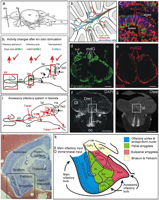
◂Fig. 2 Development of the amygdala in early mouse and zebrafish brain. the olfactory cortex (lateral pallium) and cortical amygdala
a Rostral (left panel) and caudal (right panel) transverse sections of and, secondly, the VNO projects via accessory olfactory
postcommissural telencephalic mouse and zebrafish brains with critical
pallial and subpallial gene markers indicated. Gene expression for
bulb (AOB) mostly to the medial amygdala and from there
mouse after Osório et al. 2010; Lo et al. 1991; Ma et al. 1997; Horton to the medial hypothalamus. These pathways are addressed
et al. 1999; Torii et al. 1999; Fode et al. 2000; Puelles et al. 2000), as the main and accessory olfactory systems, respectively
Morales et al. 2020; for zebrafish after Mueller et al. 2008; Herget (see above).
et al. 2014, Affaticati et al. 2015; Mueller and Wullimann 2016 (see
there for details). Dotted red arrows indicate topological lateromedial
How to discern these two pathways in teleost fishes
course of pallial ventricular surface. Solid yellow arrow indicates where a separate MOE and VNO are never discernible? As
tangential migration of glutamatergic ventral pallial cells into mouse discussed above (see “Zebrafish kin related behavior and
medial amygdala. Solid orange arrow indicates tangential migration olfactory periphery” section) both microvillous and ciliated
of Otp positive glutamatergic cells from hypothalamic supraopto-
paraventricular region (SPV) into medial amygdala (MeA; mouse)/
OSNs exist together with additional OSN types (Kappe cells,
intermediate nucleus of ventral telencephalon (Vi; zebrafish). Solid crypt cells) widely scattered over one large main olfactory
green arrow indicates radial migration of subpallial GABAergic epithelium and are not segregated into vomeronasal
cells into medial amygdala (mouse)/intermediate nucleus of ventral (microvillous OSNs) and main olfactory (cililated OSNs)
telencephalon (Vi, zebrafish). b Subpallial gene expression in early
precommissural and commissural zebrafish telencephalon identifies
epithelia or organs. Teleostean olfactory bulb projections
within the larval subpallium a ventral division (Sv; adult ventral reach all subpallial subdivisions and, in the pallium, the
nucleus of ventral telencephalon, Vv, the septum homolog) and a posterior zone of the dorsal telencephalon (Dp) and part of
dorsal division (Sd, adult dorsal nucleus of ventral telencephalon, Vd, the most ventral lateral zone (reviewed in Wullimann and
the basal ganglia homolog). Sd is further genetically subdivided into
ventral (Sdv) and dorsal (Sdd) subdivisions, representing the pallidal
Mueller 2004; Vernier and Wullimann 2009; Fig. 1i). We
and striatal homologs, respectively (and embryonic mammalian medial will not discuss the pallial olfactory connections here, but
and lateral ganglionic eminences). At the (commissural) level of the rather focus on the subpallium and the question whether
anterior commissure, the posterior division of the larval zebrafish dorsal there is a teleostean medial amygdala and a functional
subpallial division (Sdp) starts. In the adult zebrafish brain, it represents
at this level the supracommissural nucleus of the ventral telencephalon
accessory olfactory system. Before that, a short overview
(Vs). Panels in b modified after Mueller et al. (2008). The larval Sdp on the teleostean telencephalon seems appropriate.
continues into postcommissural levels (seen in a left panel), represented The zebrafish telencephalon. How does the teleostean/
in the adult zebrafish brain by the postcommissural nucleus of the zebrafish telencephalon relate in comparative terms to the
ventral telencephalon (Vp). Only recently, a most posterior ventral
telencephalic (subpallial) area characterized by otp positive cells
amniote/mammalian one? The teleostean precommissural
was recognized (Herget et al. 2014; Biechl et al. 2017) and identified (i.e., in front of the anterior commissure) nuclei of the
as the intermediate nucleus of the ventral telencephalon (Vi) in larval ventral telencephalic area (subpallium) include the adult
and adult zebrafish brains (see a, right panel, and Fig. 1). Note that ventral (Vv) and dorsal nucleus (Vd) (corresponding to
the course of radial glia fibers is indicated in the zebrafish pallium. c
This mind experiment illustrates how a postcommissural early rodent
larval ventral/dorsal part of subpallium, Sv/Sd; Fig. 2b).
telencephalon is transformed into a postcommissural early zebrafish These were long suspected to correspond to septal and
telencephalon by everting pallial masses. The most medial rodent striatal divisions of the subpallium, respectively (review
pallial division (MP/hippocampus; note position of letter X and arrow by Northcutt and Braford 1980). Eventually, the adult
indicating eversion) is virtually rolled out laterally resulting in the
topography of the postcommissural teleostean/zebrafish telencephalon
zebrafish Vd (= larval Sd) was shown to receive ascending
by maintaining topological relationships of major telencephalic input (Rink and Wullimann 2001) from dopaminergic
divisions (see text). Compare a similar figure in Wullimann (2009) posterior tubercular cells corresponding to the anterior part
for the precommissural telencephalon. Abbreviations: ac, anterior of the vertebrate mesodiencephalic dopamine cell complex,
commissure; CGE, caudal ganglion eminence; Dl, lateral zone of dorsal
telencephalic area; Dm, medial zone of dorsal telencephalic area; DP,
an input typical of the amniote striatum (reviewed in
dorsal pallium; EmT, eminentia thalami; Hy, hypothalamus; lfb, lateral Wullimann and Umeasalugo 2020). The adult Vv projects to
forebrain bundle; LVe, lateral ventricle; MeA, medial amygdala; MP, midline hypothalamus typical of the amniote septum (Rink
medial pallium; Po, preoptic area (zebrafish); POA, anterior preoptic and Wullimann 2004).
area (mouse); PTh, prethalamus; Sd larval dorsal part of subpallium;
Sdd, dorsal subdivision of Sd (striatum homomog); Sdv, ventral
Furthermore, molecular genetic data (differential expres-
subdivision of Sd (pallidum homolog), Sdp, posterior subdivision of Sd sion of Lhx6/7) showed that the zebrafish Sd can be sub-
(subpallial amygdala homolog); SPV, supraopto-paraventricular region; divided into a ventrally located larval pallidum (Sdv) and
Sv, larval ventral part of subpallium; TelCh, tela chorioidea, Th, (dorsal) striatum (Sdd) dorsally to it (Fig. 2b left panel; discussed
thalamus; VE, ventral entopeduncular nucleus; Vi, intermediate nucleus
of ventral telencephalon; VP, ventral pallium
in Mueller et al. 2008). The posterior continuation of the
larval subpallium at commissural (Sdp; Fig. 2b right panel)
and postcommissural telencephalic levels (Sdp; Fig. 2a) cor-
responds in the adult brain to the supracommissural (Vs)
least up to the olfactory bulb and partly beyond it (see above and postcommissural (Vp) nuclei of the ventral telencephalic
details for rodents; Fig. 1k). The two main pathways lead area, respectively. Thus, it had been hypothesized for a long
firstly from the MOE via main olfactory bulb mostly into time that Vs/Vp correspond to the teleostean subpallial
13282 Cell and Tissue Research (2021) 383:273–287
amygdala (i.e., the central and medial amygdala) generally been described by Levine and Dethier (1985) as one of
for teleosts (Northcutt and Braford 1980) and for zebrafish various olfactory bulb projection targets in the goldfish
in particular (Mueller et al. 2008). However, more refined and was recognized in this same position in channel catfish
arguments for either central or medial amygdala in teleosts (Bass 1981). Various studies in zebrafish have established
remained elusive as mentioned already. that the dorsomedial olfactory bulb area receives
Recent work on the expression of orthopedia (otp) exclusively microvillous OSN and crypt cell input (Gayoso
revealed in the zebrafish larval and adult hypothalamic et al. 2012; Braubach et al. 2012; Ahuja et al. 2013; Kress
preoptic area the supraoptic-paraventricular region (SPV) et al. 2015). Furthermore, only one single mediodorsal
previously seen in other vertebrates (Herget et al. 2014; glomerulus (mdG2) receives S100-positive axons from
Affaticati et al. 2015) (see above). The SPV represents the all crypt cells and from a subpopulation of microvillous
core of the hypothalamic stress axis containing numerous OSNs, the latter are at the same time parvalbumin positive
types of neuropeptidergic neurons (paraventricular nucleus (Kress et al. 2015; Biechl et al. 2017). Moreover, crypt
in amniotes corresponds to magnocellular preoptic nucleus cells (and a subpopulation of S100-negative microvillous
in teleosts) and the teleostean SPV is also characterized by OSNs) are selectively activated by kin odor in imprinted
different gene expression than the surrounding hypothalamus zebrafish larvae in activation experiments involving pERK
(Herget et al. 2014; Affaticati et al. 2015), as similarly as a read-out (Fig. 1h; see below and Biechl et al. 2016).
discussed above for the mouse (Fig. 2a). Many Otp-(protein) This connectional and functional information on a specific
positive cells migrate from this hypothalamic preoptic pathway from microvillous/crypt cells to a special (i.e.,
expression site seemingly tangentially into the most posterior dorsomedial) division of the zebrafish olfactory bulb
ventral zebrafish telencephalon (Fig. 1g). The zebrafish SVP conforms to the first synaptic step of an accessory
furthermore has Otp-positive cells that co-express Foxg1/ olfactory (“vomeronasal”) system.
Sim1 (Affaticati et al. 2015) and, presumably, such cells also Next, the higher order connections were studied by put-
migrate into the telencephalon as in the mouse (see above). ting neuronal tracer DiI into the zebrafish mediodorsal bulb
There is a general consensus that the pallial masses area (Biechl et al. 2017; Fig. 1i). In addition to central pro-
in teleosts are everted, i.e. the teleostean medial pallium jections common to the entire olfactory bulb (solid red lines
(hippocampus homolog) therefore comes to lie laterally to Vd/Vv/Dp in Fig. 1i), some are specific to the mediodor-
(Dl), whereas the ventral pallium (pallial amygdala sal olfactory bulb (dashed lines to Vs/Vp, and Vi). Thus,
homolog) remains medially (Dm; Fig. 2a, the red dotted it appears that the entire subpallial zebrafish telencephalon
arrows indicate the ventricular surface from ventral into receives secondary olfactory projections. Importantly, those
medial pallium and they run in reversed direction due to to Vi are from the mediodorsal olfactory bulb and represent
eversion). The mind experiment shown in Fig. 2c illustrates the second synaptic step of an accessory olfactory system.
the eversion process in the postcommissural telencephalon We then administered DiI to the medial tuberal hypothal-
by virtually transforming the early rodent telencephalon amus and found retrograde label in Vi (and Vp) (Fig. 1i;
into a postcommissural early zebrafish telencephalon. If Biechl et al. 2017). This is the third and final synaptic step
one takes the medial beginning of the rodent medial pallium or requirement for an accessory olfactory system.
(point X) and virtually everts it laterally (note black arrows Parallel neuronal activation experiments investigated
in Fig. 2c), the pallial masses end up in the topography of a olfactory bulb cells surrounding the only glomerulus
teleostean telencephalon. Hereby topological relationships (mdG2, recognizable in pERK stainings through S100
of pallial masses are maintained. Note in particular that the double-label, Fig. 1e) that receives S100 positive crypt
caudal ganglion eminence conforms topologically to the cell/microvillous OSN input. Indeed, olfactory bulb cells
posterior division of the dorsal subpallium (Sdp) which fits around mdG2 were activated by kin odor in imprinted lar-
its identification as medial amygdala. A similar figure can be vae in significantly higher numbers than in non-imprinted
found for the precommissural telencephalon in Wullimann larvae (Biechl et al. 2017). Finally, a significant difference
(2009). was also found for pERK activated Otp-positive cell num-
The search for the zebrafish medial amygdala and bers in the posterior telencephalic subpallial area that was
the accessory olfactory system. We then investigated newly identified intermediate nucleus of the ventral telen-
the telencephalic Otp-positive cells in more detail in cephalon (Vi; hypothesized above as the medial amygdala
the adult zebrafish and identified them as being in the homolog already). However, the difference was between
intermediate nucleus of the ventral telencephalon (Vi; non-imprinted fish (higher activated cell numbers) com-
Fig. 1f, g; Biechl et al. 2017) because Otp-positive cells pared to imprinted fish and control groups (lower activated
seem to qualify as diagnostic for the vertebrate medial cell numbers). Possibly, non-imprinted fish show a neuronal
amygdala (as discussed above for mammals). The Vi had response in Vi (medial amygdala) to the unknown kin odor
13Cell and Tissue Research (2021) 383:273–287 283
whereas this response is alleviated or lost in imprinted fish larvae along this neuronal pathway (Fig. 1h), as well as efferent
(Gerlach et al. 2019). projections of Vi to the medial hypothalamus (Fig. 1i) are con-
Finally, a comprehensive study on the entire vincing arguments for identifying Vi as a part of the teleostean
amygdaloid complex of zebrafish recently provided medial amygdala. To our knowledge this was the first time that
the most detailed basis to date for a functional neuroanatomical, developmental and functional data were pre-
neuroanatomical and developmental understanding of sented together in a teleost to identify an accessory olfactory
the teleostean amygdala and telencephalon in an explicit pathway associated to a socio-sexual context.
comparative context with the mammalian (mouse)
situation (Porter and Mueller 2020). The richness of Acknowledgements We would like to thank Bea Stiening for expert
technical assistance at the LMU-Munich. We also like to acknowledge
data and arguments in this study far exceed a thorough our fruitful interactions with various collaborators/co-authors, in par-
discussion here. However, the main advances regarding ticular the four graduate students who were active and received their
the amygdaloid complex are as follows. The zebrafish doctoral degrees during the SPP 1392 in our respective labs: Cornelia
medial amygdala is confirmed to include the Otp-positive Hinz and Kristin Tietje (Carl-von-Ossietzky Universität Oldenburg,
Germany) and Sigrid Kress and Daniela Biechl (Ludwig-Maximilians-
Vi described above, but additionally has Otp-negative Universität Munich).
subdivisions within Vs and Vp as well as in a most
dorsal subdivision of Vd (Porter and Mueller 2020). Funding Open Access funding enabled and organized by Projekt
The zebrafish central amygdala has several subdivisions, DEAL. Our original research reviewed in this article was funded by the
such as the subpallial central nucleus (Vc) and two Deutsche Forschungsgemeinschaft, Bonn, Germany through SPP 1392
Integrative Analysis of Olfaction, projects Wu211/2-1 and Wu 211/2-2
divisions in parts of Vd dorsal to the striatal one (Porter to MFW and projects GE 842/5-1 and GE 842/5-2 to GG. Additional
and Mueller 2020). Zebrafish bed nuclei of the stria support to MFW came from Prof. B. Grothe and the Graduate School
terminalis are identified in the telencephalic strands of for Systemic Neurosciences at the Ludwig-Maximilians-Universität
dopamine cells along the various subpallial nuclei and Munich (LMU-Munich), Planegg-Martinsried, Germany.
in part of Vs (Porter and Mueller 2020). Interestingly,
the cortical amygdala is identified within Dm and Compliance with ethical standards
the majority of Dm is confirmed as the remaining
Conflict of interest The authors declare that they have no conflict of
pallial amygdala (Porter and Mueller 2020). This interest.
paper represents clearly a milestone in comparative
teleostean forebrain research because it solves long- Ethical approval All applicable international, national, and/or institu-
standing questions of homology between mammalian tional guidelines for the care and use of animals were followed. Details
are contained in the original reports which are all cited in this review.
and teleostean telencephala and therefore long-term
pressing phylogenetic problems. However, some
interpretations based on developmental arguments Open Access This article is licensed under a Creative Commons Attri-
bution 4.0 International License, which permits use, sharing, adapta-
may need reconsideration in the light of radial versus tion, distribution and reproduction in any medium or format, as long
tantential migration. From our foregoing discussion it is as you give appropriate credit to the original author(s) and the source,
clear that we do not consider the Vi homologous to part provide a link to the Creative Commons licence, and indicate if changes
of the eminentia thalami as Porter and Mueller (2020) were made. The images or other third party material in this article are
included in the article’s Creative Commons licence, unless indicated
do. The Vi may receive cellular contributions from EmT otherwise in a credit line to the material. If material is not included in
by tangential migration as is reported for the rodent the article’s Creative Commons licence and your intended use is not
medial amygdala (see discussion above). However, permitted by statutory regulation or exceeds the permitted use, you will
similarly we do also not see Vi as homologous to the need to obtain permission directly from the copyright holder. To view a
copy of this licence, visit http://creativecommons.org/licenses/by/4.0/.
hypothalamic SPV (from which the medial amygdala/
Vi receives a substantial amount of otp-positive cell
numbers in mouse and zebrafish). We rather interpret
these two non-telencephalic areas as contributors of
References
cells to medial amygdala/Vi by tangential migration, Abellán A, Desfilis E, Medina L (2013). The olfactory amygdala in
whereas the decisive radial contribution of GABA cells amniotes: an evo-devo approach. Anatomical record (Hoboken,
to medial amygdala/Vi is subpallial and identifies it as N.J.: 2007), 296(9):1317–1332. https: //doi.org/10.1002/ar.22744
a part of subpallial amygdala (see Wullimann 2020 for a Adair BJ, Purser J, Patil JG (2018) Peripheral olfactory structures and
maturity-related crypt receptor neuron kinetics in the olfactory
discussion of the general problem). epithelium of carp Cyprinus carpio (L): implications for carnal
In summary, the general position of Vi within the subpal- vulnerability and pest management. Mar Freshw Res 69:1604–
lial telencephalon, a “vomeronasal” type olfactory input (crypt/ 1613. https://doi.org/10.1071/mf17386
microvillous OSNs) via dorsomedial olfactory bulb and activ- Affaticati P, Yamamoto K, Rizzi B, Bureau C, Peyriéras N, Pasqualini
C, Demarque M, Vernier P (2015) Identification of the optic
ity differences between imprinted and non-imprinted zebrafish
13284 Cell and Tissue Research (2021) 383:273–287
recess region as a morphogenetic entity in the zebrafish fore- double-labeling study in the rat. J Comp Neurol 425(2):257–274
brain. Sci Rep 5:8738. https://doi.org/10.1038/srep08738 (PMID: 10954844)
Ahuja G, Ivandic I, Saltürk M, Oka Y, Nadler W, Korsching SI (2013) Edenbrow M, Croft DP (2012) Kin and familiarity influence asso-
Zebrafish crypt neurons project to a single, identified mediodor- ciation preferences and aggression in the mangrove killifish
sal glomerulus. Scientific reports 3:2063. https: //doi.org/10.1038/ Kryptolebias marmoratus. J Fish Biol 80(3):503–518. https://
srep02063 doi.org/10.1111/j.1095-8649.2011.03181.x (Epub 2012 Jan 12
Ahuja G, Bozorg NS, Zapilko V, Shiriagin V, Kowatschew D, Oka PMID: 22380550)
Y, Korsching SI (2014) Kappe neurons, a novel population of Eisthen HL (1992) Phylogeny of the vomeronasal system and of recep-
olfactory sensory neurons. Scientific reports 4:4037. https://doi. tor cell types in the olfactory and vomeronasal epithelia of ver-
org/10.1038/srep04037 tebrates. Microsc Res Tech 23:1–21. https://doi.org/10.1002/
Arvedlund M, Nielsen LE (1996) Do the anemonefish Amphiprion jemt.1070230102
ocellaris (Pisces: Pomacentridae) imprint themselves to their Eisthen HL (1997) Evolution of vertebrate olfactory systems. Brain
host sea anemone Heteractis magnifica (Anthozoa: Actinidae)? Behav Evol 50(4):222–233. https://doi.org/10.1159/000113336
Ethology and Sociobiology 102:197–211 Ferrando S, Bottaro M, Gallus L, Girosi L, Vacchi M, Tagliafierro G
Bandoh H, Kida I, Ueda H (2011) Olfactory responses to natal stream (2006) Observations of crypt neuron-like cells in the olfactory
water in sockeye salmon by BOLD fMRI. PLOSone 6(1):e16051. epithelium of a cartilaginous fish. Neurosci Lett 403(3):280–282.
https://doi.org/10.1371/journal.pone.0016051 https://doi.org/10.1016/j.neulet.2006.04.056
Bass AH (1981) Organization of the telencephalon in the channel cat- Fode C, Ma Q, Casarosa S, Ang SL, Anderson DJ, Guillemot F (2000) A
fish. Ictalurus punctatus J Morphol 169(1):71–90. https://doi. role for neural determination genes in specifying the dorsoventral
org/10.1002/jmor.1051690107 (PMID: 30139188) identity of telencephalic neurons. Genes development 14(1):67–80
Bett NN, Hinch SG (2016) Olfactory navigation during spawning migra- Ganz J, Kaslin J, Freudenreich D, Machate A, Geffarth M, Brand M
tions: a review and introduction of the Hierarchical Navigation (2012) Subdivisions of the adult zebrafish subpallium by molecu-
Hypothesis. Biol Rev Camb Philos Soc 91(3):728–759. https:// lar marker analysis. J Comp Neurol 520:633–655. https://doi.
doi.org/10.1111/brv.12191 org/10.1002/cne.22757
Biechl D, Tietje K, Gerlach G, Wullimann MF (2016) (2016) Crypt García-Moreno F, Pedraza M, Di Giovannantonio LG, Di Salvio M,
cells are involved in kin recognition in larval zebrafish. Scientific López-Mascaraque L, Simeone A, De Carlos JA (2010) A neuronal
reports 6:24590. https://doi.org/10.1038/srep24590 migratory pathway crossing from diencephalon to telencephalon
Biechl D, Tietje K, Ryu S, Grothe B, Gerlach G, Wullimann MF (2017) populates amygdala nuclei. Nat Neurosci 13(6):680–689. https://
Identification of accessory olfactory system and medial amyg- doi.org/10.1038/nn.2556
dala in the zebrafish. Sci Rep 7:44295. https://doi.org/10.1038/ Gayoso J, Castro A, Anadón R, Manso MJ (2012) Crypt cells of the
srep44295 zebrafish Danio rerio mainly project to the dorsomedial glomeru-
Boschat C, Pélofi C, Randin O, Roppolo D, Lüscher C, Broillet MC, lar field of the olfactory bulb. Chem Senses 37(4):357–369. https://
Rodriguez I (2002) Pheromone detection mediated by a V1r doi.org/10.1093/chemse/bjr109
vomeronasal receptor. Nat Neurosci 5(12):1261–1262. https:// García-López M, Abellán A, Legaz I, Rubenstein JL, Puelles L, Medina
doi.org/10.1038/nn978 L (2008) Histogenetic compartments of the mouse centrome-
Braubach OR, Fine A, Croll RP (2012) Distribution and functional dial and extended amygdala based on gene expression patterns
organization of glomeruli in the olfactory bulbs of zebrafish during development. J Comp Neurol 506(1):46–74. https://doi.
(Danio rerio). J Comp Neurol 520(11):2317–2339. https://doi. org/10.1002/cne.21524
org/10.1002/cne.23075 Gerlach G, Hinz C (2012) Chemical signals and kin biased behaviour.
Bupesh M, Abellán A, Medina L (2011a) Genetic and experimental In: Brönmark C (ed) Hansson LA. Chemical Ecology in Aquatic
evidence supports the continuum of the central extended amyg- Systems, Oxford University Press, pp 58–71
dala and a mutiple embryonic origin of its principal neurons. Gerlach G, Lysiak N (2006) Kin recognition and inbreeding avoidance in
J Comp Neurol 519(17):3507–3531. https://doi.org/10.1002/ zebrafish, Danio rerio, is based on phenotype matching. Anim Behav
cne.22719 71:1371–1377. https://doi.org/10.1016/j.anbehav.2005.10.010
Bupesh M, Legaz I, Abellán A, Medina L (2011b) Multiple telen- Gerlach G, Hodgins-Davis A, Avolio C, Schunter C (2008) Kin recog-
cephalic and extratelencephalic embryonic domains contrib- nition in zebrafish: a 24-hour window for olfactory imprinting.
ute neurons to the medial extended amygdala. J Comp Neurol Proc Biol Sci 275(1647):2165–2170. https://doi.org/10.1098/
519:1505–1525: https://doi.org/10.1002/cne.22581 rspb.2008.0647
Buston PM, Fauvelot C, Wong MY, Planes S (2009) Genetic relatedness Gerlach G, Tietje K, Biechl D, Namekawa I, Schalm G, Sulmann A
in groups of the humbug damselfish Dascyllus aruanus: small, (2019) Behavioural and neuronal basis of olfactory imprinting
similar-sized individuals may be close kin. Mol Ecol 18(22):4707– and kin recognition in larval fish. J Exp Biol 222:jeb189746.
4715. https://doi.org/10.1111/j.1365-294X.2009.04383.x https://doi.org/10.1242/jeb.189746
Dittman A, Quinn TP, Nevitt GA (1996) Timing of imprinting to Hain TJ, Neff BD (2007) Multiple paternity and kin recognition mecha-
natural and artificial odors by coho salmon (Oncorhynchus nisms in a guppy population. Mol Ecol 16(18):3938–3946. https://
kisutch). Can J Fish Aquat Sci 53:434–442. https: //doi. doi.org/10.1111/j.1365-294X.2007.03443.x
org/10.1139/cjfas-53-2-434 Hain TJ, Neff BD (2006) Promiscuity drives self-referent kin recog-
Dittman AH, Pearsons TN, May D, Couture RB, Noakes DLG nition. Curr Biol 16(18):1807–1811. https://doi.org/10.1016/j.
(2015) Imprinting of hatchery-reared salmon to targeted cub.2006.07.021 (PMID: 16979557)
spawning locations: a new embryonic imprinting paradigm Hain TJ, Garner SR, Ramnarine IW, Neff BD (2017) Evolution of kin
for hatchery programs. Fisheries 40:114–123. https: //doi. recognition mechanisms in a fish. Anim Cogn 20(2):367–370.
org/10.1080/03632415.2015.1007206 https: //doi.org/10.1007/s10071 -016-1058-2 (Epub 2016 Nov 18)
Doron NN, Ledoux JE (1999) Organization of projections to the lateral Hamdani EH, Lastein S, Gregersen F, Doving KB (2008) Seasonal
amygdala from auditory and visual areas of the thalamus in the variations in olfactory sensory neurons - fish sensitivity to sex
rat. J Comp Neurol 412(3):383–409 (PMID: 10441229) pheromones explained? Chem Sens 33:119–123. https://doi.
Doron NN, Ledoux JE (2000) Cells in the posterior thalamus project org/10.1093/chemse/bjm072
to both amygdala and temporal cortex: a quantitative retrograde
13You can also read