Presbycusis: An Update on Cochlear Mechanisms and Therapies - MDPI
←
→
Page content transcription
If your browser does not render page correctly, please read the page content below
Journal of
           Clinical Medicine

Review
Presbycusis: An Update on Cochlear Mechanisms
and Therapies
Jing Wang 1,2, * and Jean-Luc Puel 1,2, *
 1   INSERM U051, Institute for Neurosciences of Montpellier, Hôpital Saint Eloi-Bâtiment INM, 80,
     rue Augustin Fliche-BP 74103, 34091 Montpellier, France
 2   Montpellier Neuroscience Institute, University of Montpellier, 163 rue Auguste Broussonnet,
     34090 Montpellier, France
 *   Correspondence: jing.wang@inserm.fr (J.W.); jean-luc.puel@inserm.fr (J.-L.P.); Tel.: +33-499-63-60-48 (J.W.);
     +33-499-63-60-09 (J.-L.P.)

 Received: 20 December 2019; Accepted: 10 January 2020; Published: 14 January 2020

 Abstract: Age-related hearing impairment (ARHI), also referred to as presbycusis, is the most common
 sensory impairment seen in the elderly. As our cochlea, the peripheral organ of hearing, ages, we tend
 to experience a decline in hearing and are at greater risk of cochlear sensory-neural cell degeneration
 and exacerbated age-related hearing impairments, e.g., gradual hearing loss, deterioration in speech
 comprehension (especially in noisy environments), difficulty in the localization sound sources, and
 ringing sensations in the ears. However, the aging process does not affect people uniformly; nor, in
 fact, does the aging process appear to be uniform even within an individual. Here, we outline recent
 research into chronological cochlear age in healthy people, and exacerbated hearing impairments
 during aging due to both extrinsic factors including noise and ototoxic medication, and intrinsic factors
 such as genetic predisposition, epigenetic factors, and aging. We review our current understanding
 of molecular pathways mediating ARHL and discuss recent discoveries in experimental hearing
 restoration and future prospects.

 Keywords: age-related hearing loss; presbycusis; causal factors; mechanisms; therapies

1. Introduction
     Aging is a progressive decline or loss of tissue and organ function over time due to the gradual
accumulation of deleterious biological changes. The aging process has three distinct components:
biological degeneration, extrinsic damage, and intrinsic damage. These factors are superimposed on
a genetic substrate and can be overshadowed by the general age-related susceptibility to diseases.
Age-related diseases are those that are observed with increasing frequency with increasing age, such as
atherosclerosis, cardiovascular disease, cancer, arthritis, cataracts, Alzheimer’s disease, presbyopia,
and presbycusis. Whereas all adult humans or animals become old, not all suffer from age-related
diseases. Age-related diseases can be conceptualized as accelerated aging resulting from the genetic
background interacting throughout life with environmental and lifestyle factors [1].
     Presbycusis, or age-related hearing loss (ARHL), is the loss of hearing that gradually occurs in most
people as they grow older. According to the World Health Organization [2], approximately one third of
people over 65 years of age are affected by disabling hearing loss. In 2025, there will be 1.2 billion people
over 60 years of age worldwide, with more than 500 million individuals who will suffer significant
impairment from presbycusis [2]. ARHL is a progressive, irreversible, and symmetrical bilateral
neuro-sensory hearing loss resulting either from degeneration of the cochlea, where sound-induced
vibrations are encoded by sensory hair cells into electrical signals in cochlear neurons that relay the
information to the brain (Figure 1), or loss of auditory nerve fibers during cochlear aging. Hearing loss

J. Clin. Med. 2020, 9, 218; doi:10.3390/jcm9010218                                      www.mdpi.com/journal/jcm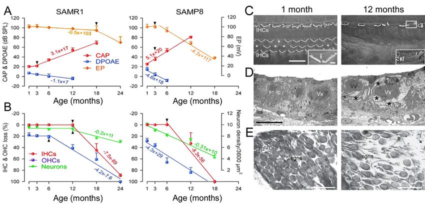
J. Clin. Med. 2020, 9, 218 2 of 22
begins inMed.
J. Clin. the2020,
high-frequency region of the auditory spectrum and spreads towards the low-frequency
9, x FOR PEER REVIEW 2 of 22
regions with age (Figure 2). The age-dependent deterioration in threshold sensitivity is generally
associated with difficulty
low-frequency in speech
regions with discrimination,
age (Figure as well asdeterioration
2). The age-dependent in sound detection andsensitivity
in threshold localization,
is generally
particularly associated
in noise. with
Males aredifficulty
generallyin more
speechseverely
discrimination,
affectedasthan
wellfemales
as in sound detection
(Figure and
2). Untreated
localization,
presbycusis canparticularly
contribute in tonoise.
socialMales are generally
isolation, moreand
depression, severely affected
dementia [3].than females (Figure 2).
Untreated presbycusis can contribute to social isolation, depression, and dementia [3].
Figure
Figure 1. Inner-ear
1. Inner-ear anatomy.(A)
anatomy. (A)Schematic
Schematic representation
representation ofofear earanatomy.
anatomy. The earear
The is divided
is divided intointo
three parts (insert): the external and middle ear transfer the sound waves to the inner ear where they
three parts (insert): the external and middle ear transfer the sound waves to the inner ear where they
are transduced into neural activity. The external ear is closed off from the middle ear by the eardrum.
are transduced into neural activity. The external ear is closed off from the middle ear by the eardrum.
In the middle ear, the eardrum is mechanically linked, by a chain of three tiny bones (the ossicles), to
In the middle ear, the eardrum is mechanically linked, by a chain of three tiny bones (the ossicles),
the oval-window membrane which closes the inner ear. Embedded in the temporal bone, the inner
to the oval-window membrane which closes the inner ear. Embedded in the temporal bone, the inner
ear comprises the balance organ or vestibule, and the hearing organ or cochlea. (B) Scanning electron
ear comprises the balance organ or vestibule, and the hearing organ or cochlea. (B) Scanning electron
micrograph of the organ of Corti. The cochlea is a coiled organ that forms a spiral. Scanning electron
micrograph
micrographs of the organ
show of Corti.linear
a narrow, The cochlea
shape of is aIHC
coiled organ that
stereocilial formsand
bundles a spiral. Scanning
a V-shape electron
of OHC
micrographs
stereocilia.show a narrow, section
(C) Transverse linear shape
of theof IHCcochlear
basal stereocilial
turnbundles andmicroscopy.
under light a V-shape ofThe
OHC stereocilia.
cochlea is
(C) made
Transverse
up of section of thewrapped
three canals basal cochlear
aroundturn under
a bony axis,light
the microscopy.
modiolus. TheseThe canals
cochleaareis the
made up of
scala
three canals (ST),
tympani wrapped around
the scala a bony
vestibuli axis,and
(SV), thethe
modiolus.
scala mediaThese canals
(SM). TheareSTthe
andscala tympani
SV are filled (ST),
with the
scala vestibuli (SV), and the scala media (SM). The ST and SV are filled with perilymph.
perilymph. The SM is filled with endolymph. The organ of Corti is situated on the basilar membrane The SM is
filled with
(bm). (B)endolymph.
= 2 mm, (C) =The10 organ
µM, (D)of=Corti
50 µm.is situated
IHCs: inneron hair
the basilar membrane
cells; OHCs: (bm).
outer hair cells = 2 mm,
(B)((B–D)
= 10 µM, (D)courtesy
(C) micrographs = 50 µm.
of Marc
IHCs:Lenoir, Inserm
inner hair U1051,
cells; OHCs:France).
outer hair cells ((B–D) micrographs courtesy
of Marc Lenoir, Inserm U1051, France).
J. Clin. Med. 2020, 9, 218 3 of 22
J. Clin. Med. 2020, 9, x FOR PEER REVIEW 3 of 22
Figure 2. Age-related
Figure 2. Age-related hearing
hearing loss
loss according
according to to the
the International
International Organization
Organization for
for Standardization
Standardization
(ISO) 7029
(ISO) 7029 standard.
standard. Shown
Shown are
are audiograms
audiograms forfor females
females (A)
(A) and
and males
males (B).
(B). The x-axis displays
The x-axis displays the
the
pure tone frequency (Hz) and the y-axis the hearing thresholds (dB HL). Each individual graph is
pure tone frequency (Hz) and the y-axis the hearing thresholds (dB HL). Each individual graph is
representative
representative ofof the
the median
median audiogram
audiogram at at aa particular
particular age
age (ranging
(ranging from
from 20
20 to 70 years
to 70 years old,
old, with
with
increments
increments ofof 10
10 years).
years).
Epidemiologic studies
studies ininlarge
largepopulations
populationsofofunscreened
unscreenedelderly elderlyadults
adults show
show that
that thethe decline
decline in
in hearing
hearing sensitivity
sensitivity accelerates
accelerates above
above the the
ageage ofto
of 20 2030toin30men,
in men,andandaboveabove
age 50agein50women
in women [4,5].[4,5].
The
The average
average hearinghearing thresholds
thresholds of menof exhibited
men exhibited a sharply
a sharply rise ofrise of hearing
hearing loss inloss
the in thefrequency
high high frequency
range,
range, whereas
whereas women’swomen’s
audiogramsaudiograms
display a display a more
more gradual gradual
sloping [4,5].sloping [4,5]. Interestingly,
Interestingly, a high proportion a highof
proportion of
participants participants
reported reported
exposure exposure
to noise, otologicto noise,
disease, otologic disease, and
and ototoxicity [4], ototoxicity
suggesting [4], thatsuggesting
the source
that
of thethe source
hearing of the hearing
impairment among impairment among unscreened
unscreened populations populations
is not exclusively is notwith
associated exclusively
aging.
associated
Basedwith aging. bone analyses correlating the patterns of hearing loss with defect location,
on temporal
Based on
Schuknecht [6]temporal
proposedbone threeanalyses correlating
major forms the patterns
of ARHL: (i) sensory of hearing loss with
presbycusis defect location,
characterized by an
Schuknecht
abrupt pure-tone[6] proposed
thresholdthree majorinforms
elevation the highof ARHL: (i) sensory
frequencies presbycusis
and hair-cell loss atcharacterized
the basal end by an
of the
abrupt
cochlea;pure-tone threshold elevation
(ii) strial presbycusis found in in the high
patients frequencies
with and hair-cell
a flat- or slightly loss at
descending the basalaudiogram,
pure-tone end of the
cochlea;
correlated(ii) strial
with presbycusis
atrophy found
of the stria in patients
vascularis; with
and (iii) a flat-
neural or slightly
presbycusis, descendingby
characterized pure-tone
a loss of
audiogram,
cochlear neuronscorrelated with atrophy
throughout of the
the entire stria vascularis;
cochlea. The precise and (iii) neural underlying
mechanisms presbycusis,the characterized
age-related
by a loss of cochlear
degeneration neuronscochlear
of the different throughout the entire
structures remain cochlea.
unclear The[7].precise
This is mechanisms
in part due tounderlying
the complexity the
age-related
of each causal degeneration
factor, but of the importantly
more different cochlearto thestructures
interaction remain
of the unclear
different [7]. This is in part
mechanistic due to
pathways
the
that complexity of each causal
can cause age-related hearingfactor, but more
loss. Sensory hair importantly to the tointeraction
cells are susceptible an accumulationof the ofdifferent
injuries
mechanistic
inflicted over pathways
time from that can cause
a number ofage-related hearingincluding
different sources, loss. Sensory hair
direct cells are susceptible
mechanical, mitochondrialto an
accumulation
oxidative injury of from
injuries inflicted
noise, over
ototoxic timesuch
drugs fromasa aminoglycosides,
number of different sources,
cisplatin, or including
other unknown direct
mechanical,
factors [8]. The mitochondrial
degenerationoxidative injury from
of spiral ganglion noise,
neurons ototoxic
(SGNs) maydrugs such as
be triggered by aminoglycosides,
the accumulation
cisplatin, or othernoise-induced
of multitudinous unknown factors loss [8]. Theafferent
of the degeneration
dendrites of [9,10].
spiral ganglion neurons
Interestingly, (SGNs)
the most may be
vulnerable
triggered by the accumulation
cochlear neurons, to both noise ofand multitudinous
aging, are those noise-induced loss of the
with high thresholds andafferent dendrites [9,10].
low spontaneous rates.
Interestingly,
Although thesethelow-spontaneous
most vulnerablerate cochlear neurons,
(SR) fibers do notto both noise and
contribute aging, aredetection
to threshold those with high
in quiet
thresholds and contribute
situations, they low spontaneous
to coding rates. Although
of transient these in
stimuli low-spontaneous rate (SR) fibers
the presence of continuous do not
background
contribute to threshold
noise [11], leading to thedetection
new conceptin quiet situations,
called hidden theyhearingcontribute to codingtone
loss. Therefore, of transient
in noise stimuli
detection in
the
maypresence of continuous
be a useful measure in background noise [11], leading
detecting age-related hearingtodeficits
the newfor concept
those called
patientshidden
who hearing
express
loss. Therefore,
difficulty, but havetone in noisenormal
relatively detection may beina quiet
thresholds useful[12].
measure in detecting age-related hearing
deficits forreview
This those patients who express
recapitulates our currentdifficulty, but have relatively
understanding of biologicalnormal thresholds
cochlear aginginon quiet [12].
hearing,
This review recapitulates our current understanding of biological cochlear
extrinsic and intrinsic risk factors that exacerbate age-related hearing function decline in animal models aging on hearing,
extrinsic and intrinsic
and in humans, risk factors
the molecular that exacerbate
pathways mediatingage-related
cochlear cell hearing function
senescence and decline in animal
degeneration as a
models and in humans, the molecular pathways mediating cochlear cell senescence and degeneration
as a consequence of aging, injury (noise, ototoxic drugs), and genetic predisposition. We discuss also
the recent discoveries in experimental hearing restoration.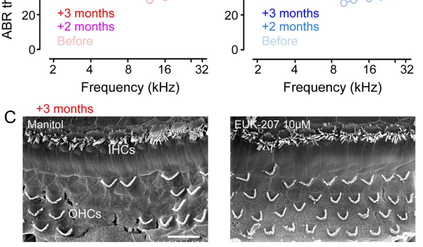
J. Clin. Med. 2020, 9, 218                                                                            4 of 22

consequence of aging, injury (noise, ototoxic drugs), and genetic predisposition. We discuss also the
recent discoveries in experimental hearing restoration.

2. Major Causal Factors of Age-Related Hearing Loss
      The clinical presentation of presbycusis, the rate of the progression, age at onset, and ultimate
severity of hearing loss varies from patient to patient. Whereas the majority of elderly patients present
clear hearing losses, a significant fraction of the geriatric population has almost normal hearing. This is
due to intrinsic (genetic predisposition, epigenetic factors, and aging), and extrinsic factors (e.g., noise-
or ototoxic drug-exposure, head trauma, cigarette smoking) that are either the sole etiology for hearing
loss, or several work in synergy with the physiopathology of presbycusis [13].

2.1. Biological Aging on Hearing

2.1.1. Aging and Hearing in Healthy People
      The clinical diagnosis of presbycusis is based on bilateral progressive loss of hearing starting
from a high-frequency region of the hearing spectrum. Loss of hearing can begin in young adulthood,
but is initially evident at 60 years for most people (Figure 2). Over time, the threshold elevation
progresses to lower and lower frequency areas. However, presbycusis studies in humans are limited
by the genetic heterogeneity and the difficulty in controlling deleterious auditory exposures over time.
Despite these limitations, it has been reported that in a cohort unscreened for noise exposure, ototoxic
drug exposure, and otologic disease history, presbycusis develops earlier and to a greater extent than
in a highly screened cohort (without history of significant noise exposure or diseases that affect the
ear) [14]. It has been suggested that the onset of hearing loss induced by biological aging is very late.
Indeed, the Mabaan tribe living in the Sudanese desert retains their hearing into old age [15]. Because
the hearing of the young Mabaans was the same as those of young people from other countries, the
good preservation of hearing in the tribe has been attributed to their quiet living environment and
generally healthy condition [16]. However, it can be argued that this difference might be caused by
genetic differences between the populations. To answer this question, Goycoolea et al. [17] compared
the hearing of natives of Easter Island, people living in a pre-industrial society, with those who had
emigrated to Chile and spent varying amounts of time in modern society. Results showed that hearing
in males that had lived or were living in Chile was significantly worse than that of males who had
lived their entire lives on Easter Island, and that the poorer hearing was related to the number of
years lived in modern society. Contrary to these early investigations, more recent studies showed that
hearing thresholds decline with age and the rate of decline accelerates with age in presbycusis patients
without noise-exposure or diseases that may affect the ear [18]. In addition, the differences of hearing
thresholds between presbycusis patients with or without noise exposure are limited [19]. These results
thus supported the belief that age is one of the major causal factors of ARHL.

2.1.2. Aging and Hearing in Animals
     To study the impact of cochlear aging on hearing, animal models are a useful tool due to their short
lifespan, controlled environments and diet composition, and limited genetic heterogeneity. Gerbils
that grew up in quiet environments [20] showed various degrees of threshold shifts with age. The
threshold shift profile was a relatively flat loss across low and mid frequencies, with the greatest losses
at the higher frequencies resembling that often seen in human presbycusis [21]. These animals also
showed a decline of the endocochlear potential [22,23] and reduced amplitudes of compound action
potentials of the auditory nerve [24]. Reduced amplitudes of compound action potentials in aging ears
suggested asynchronous or poorly synchronized neural activity in the auditory nerve of quiet-aged
gerbils [24]. Cochlear morphological examination of gerbils raised in quiet demonstrated that the most
important age-related degeneration site is the stria vascularis [7]. The degeneration of marginal and
intermediate cells of the stria vascularis began in both the base and apex of the cochlea, extending to
J. Clin. Med. 2020, 9, 218                                                                         5 of 22

the mid-cochlear regions as age increased. In addition, there was a loss of Na-K-ATPase [25] and losses
of the strial capillary area in aged animals [26]. Certainly, more work with other species aged in quiet
is needed in this area. However, existing data from quiet-aged gerbils make it clear that in gerbils,
cochlear aging impacts specifically the stria vascularis and probably the neural structures.

2.2. Genetic Predispositions
     Presbycusis shows a clear familial association. Heritability studies of presbycusis in humans
have estimated that 25% to 75% of the variance in this pathology has a genetic component [13,27,28].
Genetic polymorphisms in the genes coding detoxification enzymes, such as glutathion S-transferase
(GSTM1 and the GSTT1 null genotypes) and N-acetyltransferase 2 (NAT2*6A) [29–31] were reported
to be linked to ARHL. SOD2 promoter variants (−38C > G) of the SOD2 gene encoding a ubiquitous
mitochondrial superoxide dismutase enzyme (MnSOD) may link to the ARHL risk in men [32].
The main function of uncoupling protein 2 (UCP2) is the control of mitochondria-derived oxygen
species (ROS) [33]. In a Japanese population, UCP2 Ala55Val polymorphisms exhibited a significant
association with ARHL [34].
     An increased individual susceptibility to ARHL may rely on single nucleotide polymorphisms
in the grainyhead-like 2 gene (GRHL2), nonsyndromic sensorineural deafness type 5 (DFNA5) and
potassium voltage-gated channel subfamily q Member 4 (KCNQ4) genes, whose mutations are
responsible for DFNA28, DFNA2, and DFNA5, respectively [35–37], but also in the glutamate
metabotrophic receptor 7 gene (GRM7, e.g., OMIM ID: 604101) [38,39]. Finally, a common mtDNA
4977-bp deletion was frequently found in presbycusic patients [35].
     Some genes associated with ARHL have also been identified in mice, including age-related
hearing loss gene 1 (Ahl1), localized in chromosome 10, Ahl2 [40] on chromosome 5 (associated with
early-onset hearing loss when combined with a homozygous disease allele at the Ahl1 locus), and Ahl3
on chromosome 17 [41]. The Ahl candidate region contains several interesting candidate genes,
including genes encoding gap-junction proteins and several collagens. Mouse strains exhibiting ARHL
are also more sensitive to noise-induced hearing loss than are other strains. Collectively, polymorphisms
in some monogenic deafness-causing genes, neurotransmitter-related genes, and genes involved in
detoxification of oxidative stress and mitochondrial function are clearly associated with ARHL.

2.3. Epigenetic Factors
     Traditionally, genetics and adult lifestyle factors are considered to be among the main determinants
of aging-associated pathological conditions. Accumulating evidence, however, suggests that epigenetic
factors may contribute to these conditions [42,43]. The term epigenetics is defined as a change in
phenotype that is not caused by a change in DNA sequence [44]. Epigenetic regulation of gene
expression may change over time due to environmental exposures in common complex traits. The two
most well understood mechanisms of epigenetic alterations that lead to these phenotypic changes are
DNA methylation and histone modifications.

2.3.1. DNA Methylation
     Age-related changes in DNA methylation include global hypomethylation and region-specific
hypermethylation [45]. In the cochlea, the first evidence showing that involvement of aberrant DNA
methylation in presbycusis came from a study focused on the gap junction protein b-2 (GJB2), in the
cochlea of mimetic aging rats. In this study, Wu et al. [46] showed that hypermethylation of the
promoter region of GJB2 gene resulted in connexin 26 down-regulation and an increased risk for
presbycusis. Furthermore, Xu et al. [47] reported that hypermethylation of hearing-loss genes such as
solute carrier family 26 member 4 (SLC26A4, DFNB4) and purinergic receptor P2X 2 (P2RX2, DFNA41)
resulted in an increased risk for presbycusis in men. In addition, reduced expression of P2RX2,
KCNQ5, ERBB3, and SOCS3 genes through DNA hypermethylation in elderly women was associated
with presbycusis [48]. More recently, Bouzid et al. demonstrated that hypermethylation of CpG site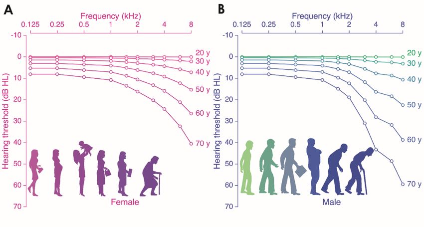
J. Clin. Med. 2020, 9, 218                                                                        6 of 22

in the cadherin-23 (CDH23) gene is likely to be associated with presbycusis in elderly women [48].
These results implicate complex pathogenic mechanisms underlying ARHL.

2.3.2. Histone Modification
     Histone proteins including H1/H5, H2A, H2B, H3, and H4 are the chief proteins of chromatin and
play an important role in maintaining the shape and structure of a nucleosome. In the last few years,
the role of histone modifications in aging and age-related diseases has emerged. Watanabe and Bloch [49]
investigated the modification of histones in the aged cochlea of mice using immunohistochemistry.
Acetylated histone H3 was detected in the spiral ganglion cells and the organ of Corti of young cochlea,
but not in those of aged cochlea. Conversely, dimethylatedhistone H3 was detected in the aged group,
but not in the young group. The degeneration was severest in the spira lganglion cells and the organ of
Corti of the basal turn. These results suggested that histone modifications may be involved in cochlear
aging regulation.

2.4. Environmental Factors
     The complexity of etiological factors for presbycusis begins with the number of environmental
risk factors, such as occupational or leisure noise, ototoxic medication (aminoglycoside, cisplatin,
salicylate, loop diuretics . . . ), cigarette smoking, and alcohol abuse [50]. However, to date, it is
not clear whether these environmental factors produce some kind of early onset and/or accelerated
progression of cochlear aging or whether they act on specific pathophysiological mechanisms. In this
part of our review, we will focus on the two most-studied environmental factors: noise exposure and
ototoxic medication.

2.4.1. Noise Exposure
      A retrospective clinical study from a large cohort of men in the Framingham Heart Study
observed that in ears with presumed cochlear damage from previous noise exposure, subsequent
progression of ARHL was exacerbated at frequencies outside the original noise-induced hearing
loss [51]. These observations suggest an age-noise interaction that exacerbates age-related hearing loss
in previously noise-damaged ears.
      Increasing evidence from animal aging models indicates that early noise exposure renders the
inner ears significantly more vulnerable to aging and may have an impact on the onset and/or
progression of ARHL [9,52,53]. Indeed, Kujawa and Liberman [52] found that noise exposure in young
CBA/CaJ mice, an inbred mouse strain used as “good hearing” mouse model, could trigger progressive
neuronal loss and exacerbate the ARHL. Furthermore, Fernandez et al. [9] showed that interactions
between noise and aging might require an acute synaptopathy to accelerate cochlear aging. In addition,
repeated exposure to a short duration sound (1 h/110 dB SPL) over a long period also led to an early
onset of ARHL (at six months of age) in Wistar rats when compared to non-exposed rats in which the
onset of ARHL was around 12 months of age [54,55]. Although the long-term effects of early noise
exposure on the aging ear are poorly understood, these clinical and experimental results indicate that
noise exposure may modify the onset and/or progression of ARHL, particularly for neural presbycusis.

2.4.2. Ototoxic Medications
     To date, the influence of other environmental risk factors such as ototoxic medications,
cigarette smoking, or alcohol abuse on ARHL is less clear and often controversial. Recently, a
large longitudinal cohort study (n = 3753) aimed at elucidating the association of ototoxic medications
exposure with the risk of developing hearing loss during the 10-year follow-up period demonstrated
that ototoxicity-age interactions may also exacerbate age-related hearing loss in older adults [56].
J. Clin. Med. 2020, 9, 218 7 of 22
3. Molecular Mechanisms of Presbycusis
Our understanding of the molecular mechanisms associated with age-related cochlear cell
degeneration and hearing loss has advanced in recent years. There are a number of pathophysiological
processes
J. Clin.that were9, reported
Med. 2020, x FOR PEERto be associated with age-related changes to functional components
REVIEW 7 of 22in the
inner ear. Here, we highlight recent advances in identifying various mechanisms involved in the “pro-,
functionalprocess”
or anti-aging components in cochlea
of the the inner(Figure
ear. Here, we Table
3 and highlight
1). recent advances in identifying various
mechanisms involved in the “pro-, or anti-aging process” of the cochlea (Figure 3 and Table 1).
Figure 3. 3.
Figure Imbalance
Imbalance between anti-aging
between anti-aging andand pro-aging
pro-aging mechanisms
mechanisms with age.with age. The
The scheme scheme
drawing
drawing
numerates several anti-aging and pro-aging mechanisms identified in the cochlear aging process.aging
numerates several anti-aging and pro-aging mechanisms identified in the cochlear
process. Anti-aging
Anti-aging mechanisms
mechanisms includeinclude estrogen,
estrogen, autophagicautophagic damageand
damage clearance, clearance, and mitochondrial
mitochondrial dynamic.
dynamic. Pro-aging
Pro-aging mechanisms
mechanisms include
include oxidative
oxidative stress,stress,
DNADNA damage,
damage, mitochondrial
mitochondrial dysfunction,
dysfunction,
senescence-like
senescence-like phenotype,
phenotype, andsenescence-associated
and senescence-associated inflammation.
inflammation.During Duringthe the
aging process,
aging process,
decreased activity of anti-aging molecules and increased activity of pro-aging properties
decreased activity of anti-aging molecules and increased activity of pro-aging properties might might lead
to accumulation
lead to accumulation of of mutations
mutationsininmitochondrial
mitochondrial DNA, DNA,increased
increasedlysosomal pH with
lysosomal pH witha resulting
a resulting
accumulation of lipofuscin and aggregates, and nuclear DNA damage,
accumulation of lipofuscin and aggregates, and nuclear DNA damage, leading to cochlear leading to cochlear cell cell
degeneration and age-related hearing loss.
degeneration and age-related hearing loss.
3.1. Pro-Aging
3.1. Pro-Aging Mechanisms
Mechanisms Identified
Identified ininthe
theCochlea
Cochlea
3.1.1. 3.1.1. Oxidative
Oxidative Stress
Stress
The oxidative stress theory of aging is based on the hypothesis that age-associated functional
The oxidative stress theory of aging is based on the hypothesis that age-associated functional
losses are due to the accumulation of reactive oxygen (ROS)- and nitrogen species (RNS)-induced
losses are due to the accumulation of reactive oxygen (ROS)- and nitrogen species (RNS)-induced DNA,
DNA, lipid, and/or protein damage. ROS and RNS are generated by several biochemical and
lipid,physiological
and/or protein damage. ROS and RNS are generated by several biochemical and physiological
processes of cellular metabolism. ROS and RNS are mainly generated by the
processes of cellular metabolism. ROS andand
mitochondria, endoplasmic reticulum, RNSperoxisomes
are mainly under
generated
both by the mitochondria,
physiological endoplasmic
and pathological
reticulum,
conditions. Fortunately, cells are able to counteract excessive ROS/RNS production via the activity of cells
and peroxisomes under both physiological and pathological conditions. Fortunately,
are able to counteract
antioxidant enzymes excessive
such as ROS/RNS
superoxideproduction
dismutases via the activity
(SODs), of antioxidant
copper/zinc superoxide enzymes
dismutase such
(Cu/Zn SOD),
as superoxide catalase, GPX
dismutases (glutathione
(SODs), peroxidase),
copper/zinc and GSR
superoxide (glutathione
dismutase reductase),
(Cu/Zn SOD),ascatalase,
well as a GPX
variety of small-molecule antioxidants such as glutathione (GSH) and thioredoxin [57].
(glutathione peroxidase), and GSR (glutathione reductase), as well as a variety of small-molecule
Increasing
antioxidants such asevidence links(GSH)
glutathione oxidative
and stress to ARHL
thioredoxin [58,59]. Low serum levels of the ROS
[57].
scavenger melatonin are significantly associated with the occurrence of high-frequency hearing loss
Increasing evidence links oxidative stress to ARHL [58,59]. Low serum levels of the ROS
in the elderly [60]. Experimental evidence also indicates that lipid peroxidation, oxidative DNA
scavenger melatonin are significantly associated with the occurrence of high-frequency hearing loss in
damage and glutathione-conjugated proteins, reduced expression of antioxidant enzymes such as
the elderly [60]. Experimental evidence also indicates that lipid peroxidation, oxidative DNA damage
catalase, and MnSOD and cytosolic SOD were associated with cochlear aging in mice [59,61,62]. In
and glutathione-conjugated
addition, mice lacking theproteins, reducedincreased
SOD1 displayed expression of antioxidant
age-related cochlearenzymes suchreduced
hair-cell loss, as catalase,
and MnSOD
thicknessand cytosolic
of the SOD were
stria vascularis, associated
and severe with cochlear
degeneration of spiralaging in mice
ganglion [59,61,62].
neurons In addition,
[63]. By contrast,
an increase in the SOD2 expression gradient in ganglion cells has been reported along the basal to
apical axis of rodent and primate cochleae [64], consistent with the differential and decreasedJ. Clin. Med. 2020, 9, 218 8 of 22
mice lacking the SOD1 displayed increased age-related cochlear hair-cell loss, reduced thickness of
the stria vascularis, and severe degeneration of spiral ganglion neurons [63]. By contrast, an increase
in the SOD2 expression gradient in ganglion cells has been reported along the basal to apical axis of
rodent and primate cochleae [64], consistent with the differential and decreased vulnerability from
high-to-low frequency regions in most ARHL. Taken together, this experimental and clinical evidence
indicates the potential involvement of oxidative stress in ARHL.
3.1.2. DNA Damage and DNA Damage Responses
Oxidative stress may cause irreversible DNA damage [65], including adducts, single-strand
breaks (SSBs), and double-strand breaks (DSBs) by base excision [66]. An early increase of
7,8-dihydro-8-oxoguanine (8-oxoG), a key biomarker of free radical-induced oxidative lesions of
mitochondrial and nuclear DNA damage [67], was observed in the cytoplasm of sensory hair cells,
supporting cells, and spiral ganglion neurons in the cochleae of SAMP8 mice (a cochlear premature
aging model), suggesting mitochondrial DNA damage [59]. More recently, we reported that the DNA
damage response could be activated in post-mitotic inner-ear cells under the stress induced by cisplatin,
an anticancer drug with an ototoxic side effect [68]. A short exposure to hydrogen peroxide (H2 O2 ) was
able to induce a premature senescence phenotype in House Ear Institute-Organ of Corti 1 (HEI-OC1)
mouse auditory cells. These displayed mitochondrial morphology damage, reduced mitochondrial
membrane potentials, an imbalance of mitochondrial fusion/fission, and an impaired mitochondrial
respiratory capacity [69]. Furthermore, using an H2 O2 exposed cochlear explant in culture and adult
SAMP8 mice in vivo, we demonstrated that ROS-induced DNA damage responses drive cochlear cell
senescence and contribute to accelerated ARHL [70].
3.1.3. Mitochondrial DNA Mutations and Dysfunction
The incidence and frequency of mtDNA point mutations and deletions increase exponentially
with age and contribute to cellular senescence in humans, monkeys, and rodents, thus providing a
causative link between mtDNA mutations and aging phenotypes in mammals [71]. In the cochlea,
it has been hypothesized that the cochlear mitochondrial redox imbalance and mitochondrial DNA
mutation and deletion might be collaboratively involved in ARHL [72]. One specific mitochondrial
common deletion (mtDNA4977) is frequently observed in the temporal bone of patients with ARHL,
and its measured levels are strongly correlated with the severity of ARHL [73]. Deletions and mutations
in mtDNA are increased in cochleae from patients with ARHL compared to those of normal-hearing
subjects [74,75]. In addition, a reduced activity of mitochondrial complex IV was observed postmortem
in spiral ganglion neurons from the temporal bone of elderly patients with ARHL [76].
Mutations in POLG-encoding DNA polymerase γ that maintains mtDNA replication fidelity [77]
or OPA1 encoding the mitochondrial dynamin-like GTPase impact mtDNA genomic stability. These
mutations are also known to cause premature hearing loss in patients and mice [78]. Mice expressing
error-prone mitochondrial DNA polymerase γ (PolgD257A) defective for proof-reading activities,
increasing the mutation frequency, led to the expression of defective respiratory chain proteins and
premature aging [79]. These Polg knockin mice also displayed early-onset ARHL, with severe loss
of SGNs and degeneration of the stria vascularis [80]. Finally, a reduced activity of complex IV was
observed in cochlear tissue of SAMP8 mice aged nine months and over [59]. Thus, accumulation of
mtDNA mutations and deletions may promote mitochondrial dysfunction and mitochondrial redox
imbalance leading to cochlear cell aging.
3.1.4. Impaired Mitochondrial Biogenesis
Mitochondrial biogenesis is the process by which cells increase their individual mitochondrial
mass and copy number to increase the production of ATP as a response to greater energy expenditure,
or during times of cellular stress [81]. We showed an overactive mitochondrial biogenesis in the cochlea
of the senescence-accelerated mouse prone 8 strain (SAMP8) at a young age that decreased in old age;J. Clin. Med. 2020, 9, 218 9 of 22
the opposite was observed in the senescence-accelerated mouse resistant 1 strain [59]. The peroxisome
proliferator-activated receptor gamma coactivator 1-alpha (PGC-1α) is a key regulator of mitochondrial
biogenesis. The overexpression of PGC-1α with its consequential increase in the transcription factors
of nuclear respiratory factor 1 and mitochondrial transcription factor A significantly decreased the
accumulation of damaged mtDNA and the number of apoptotic cells in the strial marginal cells
senescence model [82].
3.1.5. Senescence-Like Phenotype
In mitotic cells, cellular senescence is a permanent G1 arrest that is elicited in response to
different stresses [83]. The persistence and accumulation of senescent cells has been shown to
potentially play a role in the pathophysiology of aging and age-related disease [84]. Recent studies
have suggested that post-mitotic cells are also capable of entering a state of senescence and
exhibiting several senescence-associated properties, including heterochromatinization, synthesis of
pro-inflammatory interleukins, and high senescence-associated b-galactosidase activity in the brain and
retina [85,86]. Consistent with these findings, our recent results revealed that sub-lethal concentrations
of hydrogen peroxide (H2 O2 )-exposure initiated a DNA damage response together with increased
levels of key hallmarks of senescent cells, including increased expression levels of p21, and positive
senescence-associated β-galactosidase labeling cells in cochlear explants in culture, together with
increased inflammatory markers such as p38 and p-p38. Furthermore, high amounts of DNA damage
and senescence-like features were observed in the cochlear tissues of SAMP8 mice during aging and were
correlated with the accelerated ARHL observed in SAMP8 mice [70]. In addition, an accumulation of
age pigment or lipofuscin was observed in the cytoplasm of spiral ganglion neurons of SAMP8 mice [59].
Together, these results suggest that post-mitotic cell senescence may be a broader phenomenon and the
senescence-associated inflammatory secretory phenotype may negatively impact the functionality of
cochlear cells during the aging process.
3.1.6. Pro-Inflammatory Cytokines
It is now recognized that low-level chronic inflammation is correlated with the major degenerative
diseases of the elderly [87]. Even the causes of the pro-inflammatory phenotype associated with
aging and age-related disease are unclear; several molecular pathways have been identified that are
associated with both aging and low-level inflammation. The age-related oxidative stress, the age-related
accumulation of senescent cells, the senescence-associated secretory phenotype, and the decline of
autophagic activity that can trigger the inflammasome [80]. In the cochlea, high levels of Interleukin-1
Beta (IL-1β) and tumor necrosis factor (TNF) have been observed in aging cochleae [59]. Furthermore, an
association between systemic inflammation and ARHL has recently been noted in human studies [60,88,
89]. The polymorphisms of genes encoding inflammatory mediators (TNF-α rs1800630 and TNFRSF1B
rs1061624) have also been reported to contribute to the incremental risk of hearing impairment in
elderly Japanese [35].
3.1.7. Apoptosis
Apoptosis may play critical roles in cochlear aging and ARHL. Several apoptosis-related genes
change their expression with age and with hearing loss in the CBA mouse cochlear-aging model [90].
Apoptosis and mitochondrial genome mutations are implicated in ARHL and sensory hair cell
loss [91]. Deletion of the mitochondrial pro-apoptotic gene Bak prevented age-related loss of spiral
ganglion neurons and hair cells, but also ARHL in c57BL/6J mice [62]. Consistent with these results,
we also showed an increase in Bax expression and a decrease in cytochrome c oxidase expression
and activity in the cochleae of SAMP8 mice during aging [51]. Finally, the ratio of the pro-apoptotic
gene BAK1/anti-apoptotic gene BCL2 was significantly upregulated in peripheral blood samples under
ARHL and was also positively correlated with the results of the audiometric tests [92].J. Clin. Med. 2020, 9, 218 10 of 22
3.2. Anti-Aging Mechanisms
3.2.1. Mitochondrial Quality Control and Autophagy
Mitochondrial quality control and turnover is of particular importance to cochlear sensory and
neural cells because of their constant need for high levels of energy supply. Mitochondrial functionality
and integrity is achieved by the quality control mechanisms of mitochondrial dynamics (fission and
fusion events) and mitochondrial autophagy (mitophagy) [93].
Autophagy is a self-degradative process that plays a housekeeping role in removing misfolded
or aggregated proteins and damaged organelles, as well as intracellular pathogens. The autophatic
proteins Atg5, Atg4b, Atg9a, Beclin-1, and LC3B are expressed in the mouse and chicken cochlea from
developmental stages through to adulthood [94]. In the adult mouse cochlea, LC3II expression was
found to be primarily associated with SGNs [95], and its up-regulation occurred concomitantly with
an accumulation of lipofuscin, specifically in SGNs of SAMP8 mice during the aging process [59].
It has been reported that miR-34a, which has been implicated in inducing senescence, cell cycle arrest,
autophagy, and cell death [96], is also linked to ARHL in c57BL/6 mice [97]. In addition, upregulation of
miR-34a in the aging cochlea was accompanied by impairment of autophagic flux, with an accumulation
of phagophores and impaired autophagosome-lysosome fusion [98]. Together, these results suggest
that autophagy may be a housekeeping mechanism necessary for cochlear cell survival during aging.
3.2.2. Estrogen
Estrogens are the primary female sex hormones and play important roles in both reproductive
and non-reproductive systems [99]. It has been reported that estrogens possess potent antioxidant
properties and exert neuroprotective actions. The natural or surgical menopause is associated with
mitochondrial dysfunction, neuro-inflammation, synaptic decline, cognitive impairment, and increased
risk of age-related disorders [100]. Even the role of estrogen in ARHL is unclear; hearing loss is more
profound in elderly males than females [101]. In addition, women with Turner’s syndrome (45, X)
have estrogen deficiency as well as early onset presbycusis [102]. Finally, menopausal women who are
administered hormone replacement therapy have slightly better hearing than those who are not [103].
A similar sex gap in ARHL has also been observed in aged mice and rats, as the onset of ARHL appears
earlier in males than in females [104–106]. Together, this clinical and experimental evidence suggests
that estrogen may play a protective role against ARHL.
4. Pharmacotherapies
Our knowledge regarding the mechanisms of cell death provides deeper insight into various
cochlear diseases and thus supports the development of pharmacological therapies. This section
aims to emphasize the potential importance of antioxidants, mitochondrial metabolic regulators,
apoptosis inhibitors, and neurotrophins to protect cochlear cells against age-related hearing loss
(Table 1).
4.1. Antioxidants, Free Radical Scavengers, and Anti-Inflammatories
Given the role of oxidative damage in the pathogenesis of presbycusis, antioxidants may prevent the
onset or progression of ARHL. Alpha-lipoic acid is an endogenous disulfide compound synthesized de
novo in mitochondria, where it is an essential enzymatic cofactor and a powerful antioxidant molecule.
Alpha-lipoic acid has successfully prevented ARHL in rats [107]. Controversially, another study
showed that an antioxidant-enriched diet with vitamins A, C, and E, L-carnitine, and alpha-lipoic acid
significantly increased the antioxidant capacity of inner ear tissues, but did not delay the progression
of presbycusis in CBA/J mice [108]. In addition, lecithin, which plays a rate-limiting role in the
activation of superoxide dismutase and glutathione, also delayed the progression of presbycusis
in rats by protecting cochlear mitochondrial DNA [109]. Antioxidant extracts from Ginkgo biloba
leaves (EGb761, Renexin) prevented aging-related caspase-mediated apoptosis in rat cochleae [110].J. Clin. Med. 2020, 9, 218 11 of 22
J. Clin. Med. 2020, 9, x FOR PEER REVIEW 11 of 22
Finally, the synthetic superoxide dismutase/catalase mimetic EUK-207 reduced age-related loss of
hearing
ARHL, and the hair-cell
potentialdegeneration in senescence-accelerated
protective effects of antioxidants against mouse-prone
ARHL in 8 mice [70]patients
elderly (Figuresare
4
and 5). Coenzyme
controversial. Q10, or itsrole
A protective analog, coenzyme
of dietary Q10–Ter,
vitamin which
C intake are important
against ARHL was molecules
observed implicated
in elderly
inpatients
mitochondrial energy production and protection of mitochondrial membrane proteins,
[111], while no protective effect against ARHL was observed in a double-blind, randomized lipids, and
DNA from oxidative damage,
clinical trial with EGb761 [112].prevents age-related hearing loss [62]. Even if these experimental data
suggest that antioxidants
Acetylsalicylic acid might be an
(aspirin), attractive therapy
a derivative to slow
of salicylic acid,ARHL, thethe
is one of potential protective
most widely usedeffects
drugs
ofworldwide.
antioxidantsAspirin
againstdisplays
ARHL inanti-inflammatory
elderly patients areand controversial. A protective role of dietary
antioxidant properties [113]. A randomized vitamin
Cdouble-blind,
intake againstcontrolled
ARHL wasclinical
observedtrialintoelderly
assesspatients [111], therapeutic
the potential while no protective
benefitseffect against ARHL
of low-dose aspirin
was observed
in 1262 in a double-blind,
individuals age 70 yearsrandomized
old or olderclinical trial with
is currently EGb761
underway [112].
[114].
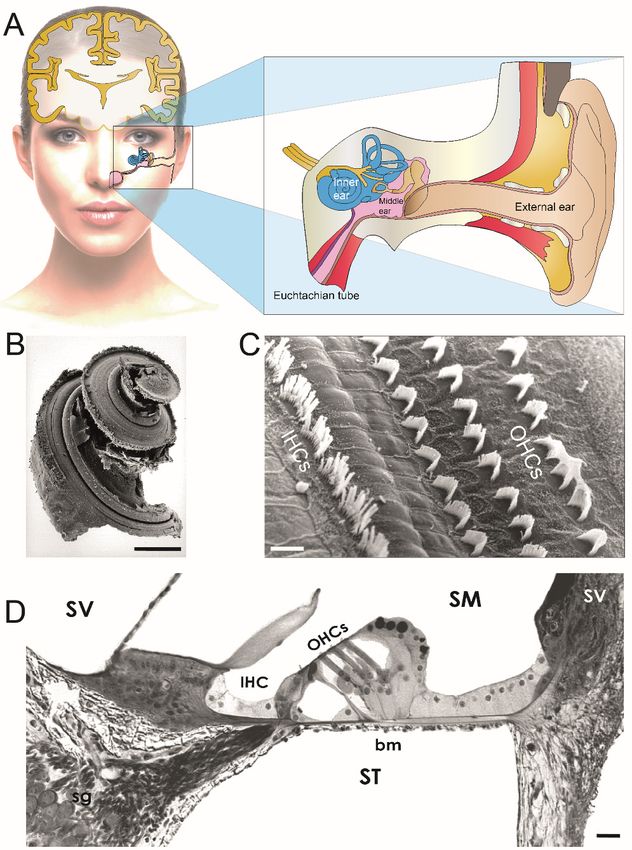
Figure4.4.Functional
Figure Functionaland andmorphological
morphologicalassessments
assessmentsininSAMP8 SAMP8and andininSAMR1
SAMR1mice. mice.(A) (A)Functional
Functional
assessment. The
assessment. The compound
compound action action potential
potential (CAP)(CAP) threshold
threshold (red (red line)
line) andand distortion
distortion product
product
otoacoustic emissions (DPOAE) amplitude (blue line) evoked
otoacoustic emissions (DPOAE) amplitude (blue line) evoked by 20 kHz tone bursts, and endocochlear by 20 kHz tone bursts, and
endocochlear potential (EP) recordings (orange line, right axis))
potential (EP) recordings (orange line, right axis)) in SAMR1 and SAMP8 mice. Fifty SAMP8 mice in SAMR1 and SAMP8 mice. Fifty
= 10 per
(nSAMP8 mice (n 1,
age: = 103, per
6, 12,age:
181,months)
3, 6, 12,and18 months)
60 SAMR1 and(n 60=SAMR1
10 per (n age:= 101, per
3, 6,age:
12, 1,
18,3,24
6, months)
12, 18, 24
months)
mice were mice
used forwere used for
functional functionalThe
assessment. assessment.
lifespan ofThe SAMR1 lifespan of SAMR1
and SAMP8 and SAMP8 was
was approximately 30
approximately
and 20 months, 30 and 20 months,
respectively. Note respectively.
the earlier and Note the increase
faster earlier and fasterthreshold
in CAP increase in andCAP threshold
decrease in
and decrease in DPOAE amplitude and EP value (arrowheads indicated
DPOAE amplitude and EP value (arrowheads indicated broken-stick nonlinearities) in the SAMP8. broken-stick nonlinearities)
Inincontrast
the SAMP8. In contrast
to SAMR1, no CAP to SAMR1,
threshold nonorCAP threshold
DPOAEs couldnorbeDPOAEs
recordedcould be recordedSAMP8
in 12-month-old in 12-month-
mice,
old SAMP8 (B)
respectively. mice, respectively.assessment.
Morphological (B) Morphological
Age-related assessment.
loss of inner Age-related
hair cells loss
(IHCs, of red
inner hairouter
line), cells
(IHCs,
hair cellsred line), blue
(OHCs, outerline),
hair and
cellsspiral
(OHCs , blue line),
ganglion and spiral
neurons (SGNs, ganglion
green line,neurons (SGNs,
right axis). Atgreen
the end line,
of
right axis). At the end of the functional assessment period, the cochleae were removed and prepared
the functional assessment period, the cochleae were removed and prepared for hair cell counting using
SEM (n =cell
for hair 5 per age perusing
counting strain)SEM
and (n SNG= 5using
per age light
permicroscopy
strain) and(nSNG = 5 per
usingagelight
per microscopy
strain). (C) Scanning
(n = 5 per
electron
age per microscopy in one and
strain). (C) Scanning 12 months
electron SAMP8inmice.
microscopy one and Few 12OHCs
months areSAMP8
lackingmice. (asterisks)
Few OHCsamong are
the three rows,
lacking but allamong
(asterisks) IHCs are thepresent
three at 1 month.
rows, but allTheIHCshigher aremagnification
present at insert 1 month.shows Thean higher
OHC
stereociliary
magnification bundle
insert with missing
shows sterocilia
an OHC (arrow). Inbundle
stereociliary a 12-month-old
with missing mouse, all OHCs
sterocilia and numerous
(arrow). In a 12-
IHCs (asterisks) have disappeared. The white box indicates
month-old mouse, all OHCs and numerous IHCs (asterisks) have disappeared. The a damaged IHC stereociliary bundle.
whiteIn the
box
insert, enlargement
indicates a damaged ofIHC
the same IHC stereociliary
stereociliary bundle. In the bundle
insert,shows fused stereocilia.
enlargement of the same Scale
IHCbar = 10 µm;
stereociliary
Insert
bundle (A) = 1 fused
in shows µm. (D) Electron Scale
stereocilia. transmission
bar = 10microscopy
µm; Insert of in the
(A)stria vascularis.
= 1 µm. At onetransmission
(D) Electron month, the
three layers of strial cells, marginal (Mc), intermediate (Ic), basal (Bc) cells,
microscopy of the stria vascularis. At one month, the three layers of strial cells, marginal (Mc), and the blood vessels (Vx)
appear normal. At 12 months, enlarged intercellular spaces and perivascular
intermediate (Ic), basal (Bc) cells, and the blood vessels (Vx) appear normal. At 12 months, enlarged edema (asterisks) are
seen. Scale bar = 10 µm. (E) Light microscopical evaluation of spiral
intercellular spaces and perivascular edema (asterisks) are seen. Scale bar = 10 µm. (E) Light ganglion loss. Shown is the
normal aspect and
microscopical density of
evaluation of neurons at 1 month,
spiral ganglion loss.and
Showna reduced numberaspect
is the normal of spiralandganglion
densityneurons
of neurons at
12atmonths. Scale bar = 50 µm. (Adapted from Ménardo et al., [59]).
1 month, and a reduced number of spiral ganglion neurons at 12 months. Scale bar = 50 µm.
(Adapted from Ménardo et al., [59]).J. Clin. Med. 2020, 9, 218 12 of 22
J. Clin. Med. 2020, 9, x FOR PEER REVIEW 12 of 22
Figure 5. Pharmacological mitigation of ROS prevents loss of hearing and hair cells in SAMP8
Figure 5. Pharmacological mitigation of ROS prevents loss of hearing and hair cells in SAMP8 mice
mice (A) SAMP8 mice and EUK-207. Shown are a SAMP8 mouse aged six months and the synthetic
(A) SAMP8 mice and EUK-207. Shown are a SAMP8 mouse aged six months and the synthetic
superoxide dismutase/catalase mimetic EUK-207. (B) Physiological assessment. The auditory brainstem
superoxide dismutase/catalase mimetic EUK-207. (B) Physiological assessment. The auditory
response (ABR) thresholds recorded before (pale red plot) and after two months (pink plot) and three
brainstem response (ABR) thresholds recorded before (pale red plot) and after two months (pink plot)
months (red plot) of Manitol treatments, or before (pale blue plot) and after two months (azure plot) and
and three months (red plot) of Manitol treatments, or before (pale blue plot) and after two months
three months (blue plot) of EUK-207 (10 µM) treatments. (C) Morphological assessment. Representative
(azure plot) and three months (blue plot) of EUK-207 (10 µM) treatments. (C) Morphological
scanning electron micrographs showing the basal regions of cochleae from Manitol-treated (left panel)
assessment. Representative scanning electron micrographs showing the basal regions of cochleae
and EUK-207-treated (right panel) SAMP8 mice after three months. Scale bar = 15 µm (Adapted from
from Manitol-treated (left panel) and EUK-207-treated (right panel) SAMP8 mice after three months.
Benkafadar et al. [70]).
Scale bar = 15 µm (Adapted from Benkafadar et al. [70]).
Acetylsalicylic acid (aspirin), a derivative of salicylic acid, is one of the most widely used drugs
4.2. Regulators of Mitochondrial Function and Metabolism
worldwide. Aspirin displays anti-inflammatory and antioxidant properties [113]. A randomized
double-blind, controlled
The sirtuin familyclinical trial to
has seven assess the
members potentialwhich
(SIRT1–7) therapeutic benefits of in
are implicated low-dose
delaying aspirin in
cellular
1262 individuals
senescence andage 70 years old
extending the or older is currently
organismal lifespanunderway
through [114].
the regulation of diverse cellular
processes, including suppression of cellular senescence and promotion of DNA damage repair [115].
4.2. Regulatorsisofa Mitochondrial
Resveratrol natural dietary Function and Metabolism
polyphenolic SIRT1 activator that is able to mimic calorie restriction
andThe to sirtuin
prolongfamilylife duration in simple organisms
has seven members (SIRT1–7) which and experimental
are implicated animal models,cellular
in delaying but is
controversial
senescence and in humansthe
extending [116,117].
organismalIn the cochlea,
lifespan dietary
through thesupplements
regulation of of resveratrol
diverse cellularsignificantly
processes,
reduced suppression
including age-related hearing loss
of cellular and hair-cell
senescence loss in C57BL/6
and promotion of DNA mice [97]. repair
damage Conversely, another study
[115]. Resveratrol is
showed that SIRT1 deficiency reduced age-related oxidative damage of cochlear hair cells and SGNs
and delayed the early onset of ARHL [118]. Caloric restriction induces upregulation of the SIRT3
gene, which, in turn, promotes the glutathione-mediated mitochondrial antioxidant defense systemJ. Clin. Med. 2020, 9, 218 13 of 22
a natural dietary polyphenolic SIRT1 activator that is able to mimic calorie restriction and to prolong life
duration in simple organisms and experimental animal models, but is controversial in humans [116,117].
In the cochlea, dietary supplements of resveratrol significantly reduced age-related hearing loss
and hair-cell loss in C57BL/6 mice [97]. Conversely, another study showed that SIRT1 deficiency
reduced age-related oxidative damage of cochlear hair cells and SGNs and delayed the early onset
of ARHL [118]. Caloric restriction induces upregulation of the SIRT3 gene, which, in turn, promotes
the glutathione-mediated mitochondrial antioxidant defense system and delays the onset of ARHL
in mice [119]. A number of potent SIRT2 inhibitors and SIRT1 activators have now been through the
first clinical trials, with evidence of safety and efficacy in psoriasis and in metabolic syndrome. This,
however, is not the case for ARHL. Looking further into the future, a better understanding of the
potential mechanisms of sirtuin-family effects in the cochlear aging process and ARHL could perhaps
one day delay ARHL in humans with the small molecule modulators of the Sirtuins.
4.3. Caspase Inhibitors
A number of specific and broad-spectrum peptide caspase inhibitors have been developed
to elucidate the role of each specific caspase in cell death. Intraperitoneal injection of the
pan-caspase-inhibitor z-VAD-FMK Z-VAD-FMK for eight weeks, starting at one week of age in DBA/2J
and A/J mice, preserved hearing by more than 10 dB SPL in the ABR thresholds and significantly
reduced OHC loss in the basal turns of the cochleae [120,121]. Even many caspase inhibitors (e.g.,
inhibitors of caspase-1, capses-2, caspase-6, and caspase-3) have been patented for their use in the
treatments of neurodegenerative diseases, cardiovascular diseases, liver diseases, and cerebral stroke.
To date, however, no caspase inhibitors have entered the market due to their toxicity and poor
pharmacokinetic profile [122].
4.4. Neurotrophins
Recent findings from temporal bones of patients aged 54 to 89 years and without an otologic disease
history indicated that the degeneration of cochlear nerve peripheral axons, despite a near-normal
hair-cell population, may be an important component of human presbycusis [123]. These data are
consisted with previous results from the same laboratory [124] showing a decline of SGNs at a
mean rate of 100 cells per year of life in human temporal bones from 100 patients aged newborn to
100 years, including only cases with normal populations of inner and outer hair cell. There were no
significant gender or inter-aural differences and no significant base-to-apex gradient in degeneration.
Although primary cochlear nerve degeneration is not expected to affect audiometric thresholds, it
may be key to problems with hearing in noise that are characteristic of declining hearing abilities
in the aging ear. Interestingly, scala tympani injection of AAV8-NT3 via cochleostomy leads to the
transduction of IHCs of the basal cochlear turn, but not of the adjacent supporting cells, and prevents
noise-induced synaptopathy in adult albino guinea pigs [125]. If an appropriate early diagnostic
method can be identified for neural presbycusis, the regeneration of terminal nerve fibers might be a
suitable therapeutic option for at least a selected group of patients.You can also read