Reactive Metabolite-induced Protein Glutathionylation: a Potentially Novel Mechanism Underlying Acetaminophen Hepatotoxicity
←
→
Page content transcription
If your browser does not render page correctly, please read the page content below
MCP Papers in Press. Published on July 13, 2018 as Manuscript RA118.000875

Reactive Metabolite-induced Protein Glutathionylation: a Potentially Novel

Mechanism Underlying Acetaminophen Hepatotoxicity

James Chun Yip Chan1, Alex Cheow Khoon Soh2, Dorinda Yan Qin Kioh1, Jianguo Li3,4,

Chandra Verma3,5,6, Siew Kwan Koh4, Roger Wilmer Beuerman4,7,8, Lei Zhou4,7,8,a, Eric Chun

Yong Chan1,9,a

Affiliations:
1
  Department of Pharmacy, National University of Singapore, 18 Science Drive 4, Singapore
117543.

                                                                                                 Downloaded from http://www.mcponline.org/ by guest on May 28, 2019
2
  School of Pharmacy, University College London, 29-39 Brunswick Square, London WC1N
1AX, United Kingdom.
3
  Bioinformatics Institute, 30 Biopolis Street, #07-01 Matrix, Singapore 138671.
4
  Singapore Eye Research Institute, The Academia, 20 College Road, Discovery Tower, Level 6,
Singapore 169856.
5
  Department of Biological Sciences, National University of Singapore, 16 Science Drive 4,
Singapore 117558.
6
  School of Biological Sciences, Nanyang Technological University, 60 Nanyang Drive,
Singapore 637551.
7
  Department of Ophthalmology, Yong Loo Lin School of Medicine, National University of
Singapore, 1E Kent Ridge Road, NUHS Tower Block Level 7, Singapore 119228.
8
  Ophthalmology and Visual Sciences Academic Clinical Research Program, Duke-NUS Medical
School, 8 College Road, Singapore 169857.
9
  Singapore Institute for Clinical Sciences, Brenner Centre for Molecular Medicine, 30 Medical
Drive, Singapore 117609.
a
 Co-corresponding authors
Dr. Eric Chun Yong Chan, Email: eric.chan@nus.edu.sg
Dr. Lei Zhou, Email: zhou.lei@seri.com.sg

Running title
Acetaminophen-induced glutathionylation in hepatotoxicity

List of Abbreviations

ANT              Adenine nucleotide translocase
APAP             Acetaminophen

                                                                                             1
ATP      Adenosine triphosphate
CAC      Carnitine/acylcarnitine carrier
CoA      Coenzyme A
CPT1     Carnitine O-palmitoyltransferase 1
CYP450   Cytochrome P450
DEDC     Diethyldithiocarbamate
DNA      Deoxyribonucleic acid
GPx      Glutathione peroxidase
Grx1     Glutaredoxin 1
GSH      Glutathione
MPTP     Mitochondrial permeability transition pore
NAPQI    N-acetyl-p-benzoquinone imine
PMCA     Plasma membrane calcium ATPase
PPARα    Peroxisome proliferator-activated receptor α
RMSD     Root mean square deviation
TRPM2    Transient receptor potential melastatin 2

                                                                   Downloaded from http://www.mcponline.org/ by guest on May 28, 2019
VDAC1    Voltage-dependent anion-selective channel protein 1

                                                               2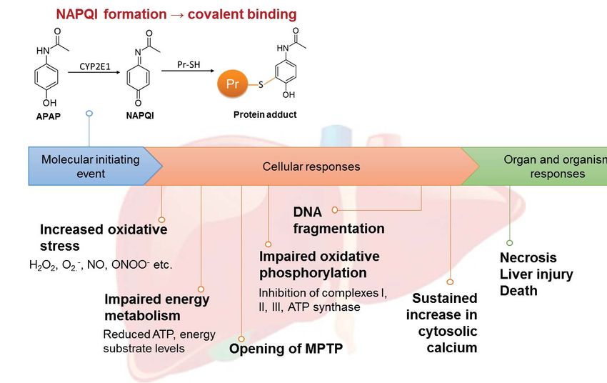
SUMMARY

Although covalent protein binding is established as the pivotal event underpinning

acetaminophen (APAP) toxicity, its mechanistic details remain unclear. In this study, we

demonstrated that APAP induces widespread protein glutathionylation in a time-, dose- and

bioactivation-dependent manner in HepaRG cells. Proteo-metabonomic mapping provided

evidence that APAP-induced glutathionylation resulted in functional deficits in energy

metabolism, elevations in oxidative stress and cytosolic calcium, as well as mitochondrial

dysfunction that correlate strongly with the well-established toxicity features of APAP. We also

                                                                                                   Downloaded from http://www.mcponline.org/ by guest on May 28, 2019
provide novel evidence that APAP-induced glutathionylation of carnitine O-palmitoyltransferase

1 (CPT1) and voltage-dependent anion-selective channel protein 1 are respectively involved in

inhibition of fatty acid β-oxidation and opening of the mitochondrial permeability transition

pore. Importantly, we show that the inhibitory effect of CPT1 glutathionylation can be mitigated

by PPARα induction, which provides a mechanistic explanation for the prophylactic effect of

fibrates, which are PPARα ligands, against APAP toxicity. Finally, we propose that APAP-

induced protein glutathionylation likely occurs secondary to covalent binding, which is a

previously unknown mechanism of glutathionylation, suggesting that this post-translational

modification could be functionally implicated in drug-induced toxicity.

                                                                                              3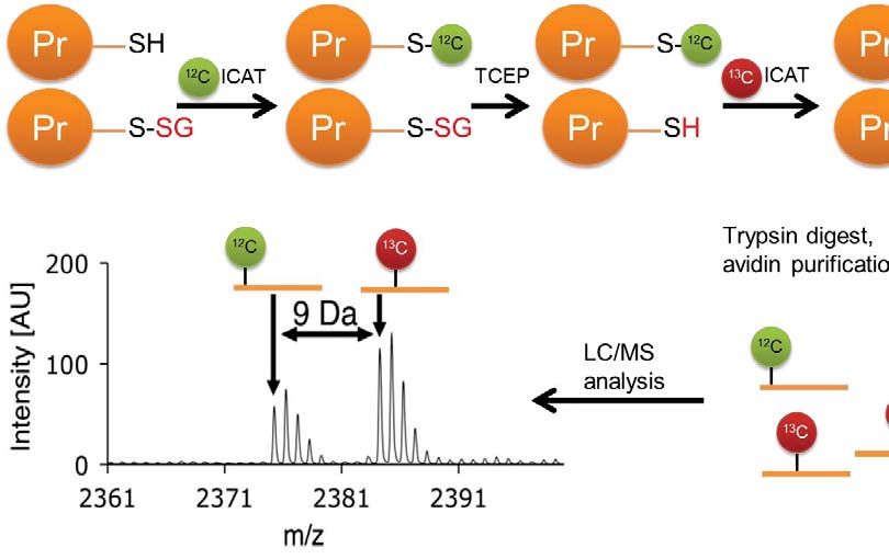
INTRODUCTION

Protein glutathionylation is a relatively understudied post-translational modification of cysteine

residues. A highly dynamic process, it involves both chemical (via reaction with reactive

oxygen/nitrogen species) and enzymatic (glutathione-S-transferase π or glutaredoxin 1 (Grx1))

mediators. Conversely, deglutathionylation is exclusively an enzymatic process mediated by

Grx1, glutaredoxin 2, sulfiredoxin and protein disulfide isomerase (1). As glutathionylation

induces a change in the size and charge of the cysteine residue, it confers various functional

changes including selective modulation of enzymatic activity and alteration in DNA binding by

                                                                                                     Downloaded from http://www.mcponline.org/ by guest on May 28, 2019
transcription factors (1). In other instances, glutathionylation does not impact protein function

discernibly but protects cysteine thiols from permanent damage due to irreversible oxidation. To

date, research in protein glutathionylation is centered on pathological conditions where oxidative

stress is featured prominently, such as cardiovascular and neurological diseases (1).

Acetaminophen (APAP), an over-the-counter antipyretic and analgesic is the leading cause of

acute liver failure in the United States (2). APAP undergoes oxidation by CYP2E1 in the liver to

form N-acetyl-p-benzoquinone imine (NAPQI), a reactive metabolite that binds covalently to

protein thiols (Fig. 1A). The crucial role of covalent binding in APAP-induced hepatotoxicity is

well-established with three key evidences: (1) toxicity is attenuated and amplified when covalent

binding is diminished and magnified by CYP450 inhibitors and inducers respectively (3), (2)

peak covalent binding preceded hepatic necrosis by 1-2 h consistently (3), and (3) patients

experiencing the most severe toxicity exhibit the highest levels of APAP adducts in plasma,

correlating with hepatic transaminase levels (4). Despite clear causality, there remains a

significant disconnect between the initial covalent adduction and the toxic sequelae. For

example, over 30 hepatic proteins including glutathione peroxidase (GPx) were reported to be

                                                                                                4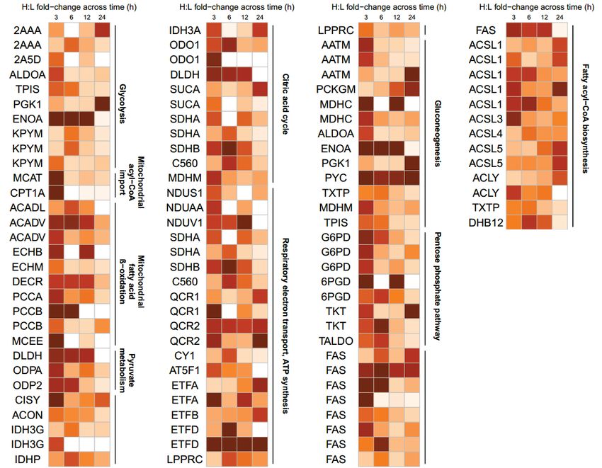
adducted, but the activities of these proteins were only moderately inhibited (5), while GPx-/-

knockout mice exposed to APAP did not exhibit attenuation of liver injury (6). Conversely,

activities of calcium-dependent ATPases, plasma membrane Na+-K+-ATPase and protein

phosphatases decrease after exposure to hepatotoxic doses of APAP, but with no evidence of

adduction by APAP (7). Therefore, covalent protein adduction is critical yet insufficient to

account for APAP-induced hepatotoxicity.

Seminal work by Nelson and colleagues indicated that APAP treatment depleted free protein

thiols, which was greater than that expected due to covalent binding of thiols alone; while

                                                                                                   Downloaded from http://www.mcponline.org/ by guest on May 28, 2019
treatment of liver homogenates with dithiothreitol restored free thiol levels (8). While APAP

causes mitochondrial oxidative stress which could account for the loss of protein thiols due to

oxidation, subsequent investigations revealed the formation of an ipso conjugate of NAPQI with

GSH. Notably, the ipso GSH adduct reacts with a protein thiol, resulting in glutathionylation

independent of a chemical reaction with oxidants (9). Similarly, an ipso protein adduct reacts

with free GSH, also culminating in protein glutathionylation (Fig. 1B). Glutathionylated regions

were also observed in liver sections of mice exposed to APAP, which overlapped with regions of

APAP protein adduction (10). These reports provide the first evidence that besides oxidative

stress, protein glutathionylation can also occur as a result of covalent binding of a reactive

metabolite to a protein thiol. Given that glutathionylation can modulate protein function, we

hypothesized that this post-translational modification could account for aspects of APAP-

induced hepatotoxicity inadequately explained by covalent adduction. Using metabolically-

competent HepaRG hepatocytes as a model for the APAP-liver injury axis, we demonstrate that

aberrant glutathionylation patterns induced by APAP can be comprehensively mapped to its

known toxicity endpoints in a time-, dose- and bioactivation-dependent manner, thus illustrating

                                                                                              5
that aberrant protein glutathionylation is potentially a novel and previously unrecognized

mechanism of drug-induced hepatotoxicity.

                                                                                             Downloaded from http://www.mcponline.org/ by guest on May 28, 2019

                                                                                        6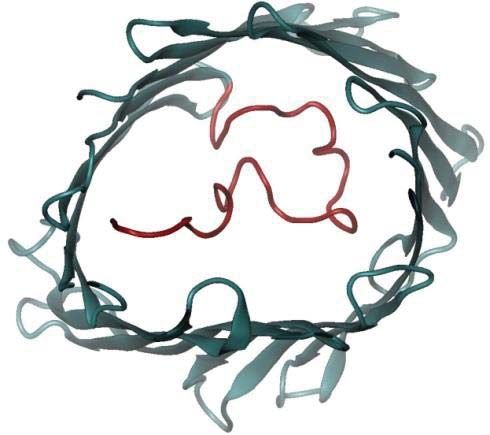
EXPERIMENTAL PROCEDURES

Reagents
Acetaminophen (APAP), bovine serum albumin, carnitine, dithiothreitol (DTT), fenofibrate,

glyceraldehyde-3-phosphate, glyceraldehyde-3-phosphate dehydrogenase (GAPDH), reduced L-

glutathione (GSH), hydrogen peroxide, ketoconazole, palmitoyl carnitine, palmitoyl-CoA,

sodium diethyldithiocarbamate trihydrate (DEDC), sodium dodecyl sulfate (SDS), trichloroacetic

acid (TCA) and tris(2-carboyxethyl)phosphine (TCEP) hydrochloride were purchased from

                                                                                                     Downloaded from http://www.mcponline.org/ by guest on May 28, 2019
Sigma-Aldrich (St Louis, MO, USA). NADPH tetrasodium salt was purchased from Santa Cruz

Biotechnology (Santa Cruz, CA, USA). Etomoxir and glutaredoxin-1 C14S mutant (Grx1) was

purchased from Cayman Chemicals (Ann Arbor, MA, USA). Diamide was obtained from Tokyo

Chemical Industries (Tokyo, Japan). N-acetyl-p-benzoquinone imine (NAPQI) was obtained

from Toronto Research Chemicals (Ontario, Canada). Glutathione reductase (GR) was obtained

from Roche Diagnostics (Basel, Switzerland). Sequencing Grade Modified Trypsin was

purchased from Promega (Madison, WI, USA). Heat-inactivated fetal bovine serum (FBS), L-

glutamine, minimum essential medium/Earle’s balanced salt solution (MEM/EBSS) and MEM

non-essential amino acids, were obtained from Hyclone (Logan, UT, USA). Penicillin G and

streptomycin were obtained from Pan Biotech (Dorset, UK). Pierce BCA protein assay kit and

mitochondrial isolation kit for cultured cells was obtained from Thermo Scientific (Waltham,

MA, USA). Phosphate-buffered saline (PBS) was obtained from Vivantis (Selangor, Malaysia).

Cleavable ICAT (isotope-coded affinity tag) reagents were purchased from ABSciex

(Framingham, MA, USA). Filter-aided sample preparation (FASP) kits were obtained from

Expedeon (Cambridgeshire, UK). Water was purified using a Milli-Q water purification system

(Millipore, Bedford, MA, USA). All other solvents and reagents used were of analytical grades.

                                                                                                 7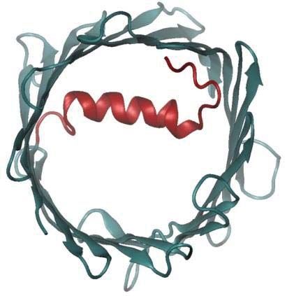
Experimental Design and Statistical Rationale

This study aimed to define protein glutathionylation patterns induced by APAP in HepaRG cells.

This was examined temporally, across different doses and in the presence of a CYP2E1 inhibitor

(Fig. 2) For the global relative quantitation of protein glutathionylation experiments, biological

triplicates of each treatment condition were collected across 3 cell passages. Each sample was

processed and measured once using mass spectrometry.

                                                                                                     Downloaded from http://www.mcponline.org/ by guest on May 28, 2019
HepaRG Cell Culture

HepaRG cells at passage 13 were obtained from Biopredic International (Rennes, France). The

cells were seeded in T-75 flasks at 2×10⁶ undifferentiated cells/flask and grown to confluence in

710 growth medium (Biopredic, Rennes, France). The cells were cultured under a humidified

atmosphere of 5% CO2 at 37°C for 14 days before passaging. Medium was renewed every 3

days. Passages 13 and 14 were neither differentiated nor used as per the vendor’s instructions.

Differentiation was induced on day 14 by replacing 710 growth medium with 720 differentiation

medium (Biopredic, Rennes, France) for a further two weeks. The cells were maintained up to 4

weeks after differentiation and were used at passage 15-18 for drug treatment and biochemical

analyses.

HepG2 Cell Culture

HepG2 human hepatocellular carcinoma cells (American Type Culture Collection, Manassas,

VA, USA) were cultured in T-175 culture flasks in culture media consisting of MEM/EBSS

                                                                                                8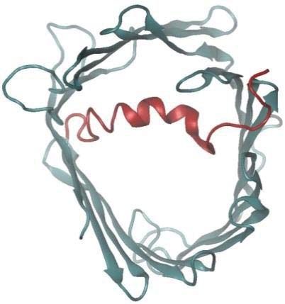
supplemented with 10% heat-inactivated FBS, 2 mM L-glutatmine, 100 U/mL Penicillin G, 100

μg/mL streptomycin and MEM non-essential amino acids. The cells were cultured under a

humidified atmosphere of 5% CO2 at 37°C. Culture medium was changed every 3 days and cells

were subcultured weekly.

Biochemical Analyses

All biochemical analyses were performed in a 96-well plate format. Undifferentiated HepaRG

cells were seeded in 96-well plates at a density of 9000 cells/well and then differentiated as

                                                                                                      Downloaded from http://www.mcponline.org/ by guest on May 28, 2019
described above. After differentiation, 720 differentiation medium which contains 1.7% DMSO

was replaced with HepaRG Tox Working Medium (Life Technologies, Carlsbad, CA, USA)

which contains 0.5% DMSO and maintained for an additional week. Cells were treated with 30

mM APAP for 3, 6, 12 and 24 h for all assays. Cell viability was assessed by the CellTiter-Glo

Luminescent Cell Viability Assay (Promega, Madison, WI, USA). Mitochondrial ROS

generation was measured using MitoSOX Red Mitochondrial Superoxide Indicator (Life

Technologies, Carlsbad, CA, USA). Mitochondrial superoxide was measured at λex = 396 nm

and non-specific oxidation at λex = 510 nm, while λem = 580 nm was used for both (11). Cellular

percentage of GSH relative to untreated control as an indicator of cell health and oxidative stress

was quantified using the luminescence-based system of GSH/GSSG-Glo Assay (Promega,

Madison, WI, USA). All biochemical assays were performed according to manufacturer’s

instructions.

                                                                                                 9
APAP Treatment of HepaRG Cells

Undifferentiated HepaRG cells were seeded in 6-well plates at a density of 2×10⁵ cells/well,

differentiated, and then maintained in HepaRG Tox Working Medium for 1 week in a total

volume of 2 mL per well as mentioned above. To investigate the time-dependency of APAP

glutathionylation, cells were treated with 30 mM APAP for 3, 6, 12 and 24 h. To determine dose-

dependence of APAP glutathionylation, cells were treated with 0.5 mM or 30 mM APAP for 3 h.

To assess the influence of CYP2E1 bioactivation on APAP glutathionylation, cells were

                                                                                                   Downloaded from http://www.mcponline.org/ by guest on May 28, 2019
pretreated with DEDC, a CYP2E1 inhibitor for 30 min. The culture media was then replaced

with DEDC-free media containing 0.5 mM APAP for 3 h. For vehicle-treated controls, cells

were treated with 1.9% ethanol for 6 h. Every vehicle or drug treatment was performed in

triplicates across 3 cell passages.

Harvesting of APAP-Treated Cell Lysate

Drug-containing culture media was removed from each well at the appropriate time-points and

cells were rinsed twice with ice-cold PBS. 500 μL of ice-cold 10% (w/v) TCA was added to each

well and kept on ice for 10 min. Cells were then scraped and the suspension was transferred into

a 1.5 mL microtube. Each well was rinsed with 500 μL of ice-cold 10% (w/v) TCA and added to

the same microtube. All tubes were kept on ice for 30 min and vortexed every 10 min. Samples

were stored at -80°C until further processing.

                                                                                             10
GluICAT Processing of Glutathionylated Proteins
Cell lysates were centrifuged at 14 000 g for 30 min at 4°C. Denaturing alkylation buffer (DAB)
containing 6 M urea, 200 mM Tris-HCI, pH 8.5, 10 mM EDTA, and 4% (w/v) SDS was freshly
prepared before use. The protein pellet was rinsed once with 500 μL ice-cold 10% (w/v) TCA,
followed by a second rinse with 200 μL ice-cold 5% (w/v) TCA. The pellet was then redissolved
in 80 μL DAB and quantified using the BCA assay. An aliquot containing 100 μg of protein was
removed and additional DAB added to a final volume of 80 μL, followed by alkylation with light
Downloaded from http://www.mcponline.org/ by guest on May 28, 2019
ICAT reagent for 2 h. Proteins were precipitated using 500 μL of -20°C cold acetone and
maintained at -20°C overnight. The acetone precipitate was centrifuged at 14 000 g for 30 min at
4°C, and the protein pellet was rinsed twice with 500 μL -20°C cold acetone to remove excess
light ICAT. The protein pellet was resolubilized in DAB and prepared for deglutathionylation as
follows: the protein solution was loaded onto a spin column and SDS was removed by washing 3
times with 6 M urea, followed by 3 washes with 50 mM ammonium bicarbonate to condition the
membrane for the subsequent deglutathionylation step. To effect deglutathionylation, the
following mixture was added: 10 mM GSH, 10 mM NADPH, 4 U/mL GR and 0.5 U/mL Grx1 in
100 μL 50 mM ammonium bicarbonate and incubated for 1 h at 37°C on the spin column.
Excess GSH was removed by washing 4 times with 50 mM ammonium bicarbonate. Newly
reduced cysteines were labelled with heavy ICAT reagent for 2 h, followed by removal of excess
heavy ICAT by washing 3 times with 50 mM ammonium bicarbonate. Proteins were then
digested with trypsin (1:30 trypsin/protein ratio) for 12-16 h at 37°C. Peptides were eluted with
50 mM ammonium bicarbonate and 0.5 M sodium chloride. Peptides were purified using avidin
11affinity chromatography. Finally the biotin tag was cleaved and samples were desalted using
UltraMicro Spin Columns (Nest Group, Southborough, MA, USA) before LC/MS/MS analysis.
LC/MS/MS Detection of GluICAT-Treated Glutathionylated Proteins
LC/MS/MS was performed using an Ultimate 3000 nanoLC system (Dionex, Thermo Fisher
Scientific, MA, USA) coupled to an ABSciex 5600 TripleTOF (ABSciex, Framingham, MA,
USA). A 50 cm × 75 μm i.d. Acclaim PepMap RSLC C18 column was employed (Dionex,
Thermo Fisher Scientific, MA, USA). This column was connected to a spray tip (New
Downloaded from http://www.mcponline.org/ by guest on May 28, 2019
Objectives, Woburn, MA), which was directly coupled to the nano-spray interface of the mass
spectrometer. Samples were loaded onto a trap column (Acclaim PepMap 100 C18, 2cm × 75μm
i.d., Dionex, Thermo Fisher Scientific, MA, USA) at a flow rate of 5 μL/min. After a 3 min wash
with loading buffer (2/98 v/v of ACN/water with 0.1% formic acid), the system was switched
into line with the C18 analytical capillary column. A step linear gradient of mobile phase B (2/98
v/v of water/ACN with 0.1% formic acid) from 5% to 25% over 10 min, 25%-60% for 9 min,
and lastly, 60%-95% over 1 min at flow rate of 300 nL/min was utilized for this analysis. Third
generation Nanospray Source was installed and other instrumentation settings were as follows:
ionspray voltage floating, 2200 V; curtain gas, 30 psi; ion source gas 1, 12 psi; interface heater
temperature, 125°C; declustering potential, 100 V. Data was acquired using TOF MS + Hi
Sensitivity product ion with Analyst TF 1.6 software (ABSciex, Framingham, MA, USA). TOF-
MS scan (experiment 1) parameters were set as follows: 0.25 s TOF MS accumulation time in
the mass range of 350 to 1250 Da was followed by MS/MS scan based on the following
parameters: mass range was set at 100 to 1500 Da; switching criteria were set to ions greater than
m/z = 350 and smaller than m/z = 1250 with charge state of 2 to 5 and an abundance threshold of
12greater than 120 cps. Former target ions were excluded for 4 s and also excluded after 1 repeat.
The maximum number of candidate ions to monitor per cycle was 20 spectra with an
accumulation time of 100 ms. All injections were performed in triplicates.
Database search parameters, acceptance criteria for protein identification and ICAT
relative quantitation
The MS data was processed using ProteinPilot software 4.5 (ABSciex, Framingham, MA, USA)
Downloaded from http://www.mcponline.org/ by guest on May 28, 2019
with database search using Uniprot (Sept 2010 release, 40516 proteins searched). Searching
parameters were as follows: (1) sample type: cleavable ICAT; (2) Cys Alkylation: none; (3)
digestion: trypsin; (4) instrument: TripleTOF 5600; (5) species: Homo sapiens; (6) ID focus:
biological modifications. All peptides identified from the database were filtered according to the
following criteria: at least 95% confidence, and possessing a heavy-to-light (H:L) ratio of at least
0.01. Peptides that did not meet the criteria were removed from further analysis. The average
H:L value across triplicate sets of peptide data from the vehicle-treated control arm were
computed. Peptides were cross-matched between those in the control list and each drug-treated
replicate, and the quotient of [H:L treated / H:L control] for each peptide in each treatment
replicate was obtained. A particular peptide was considered to be glutathionylated only if the
[H:L treated / H:L control] ratio exceeded 1.5-fold in at least 2 of 3 drug-treated replicates and
subjected to further pathway analysis described below.
Bioinformatics analysis
13Glutathionylated proteins were submitted for pathway analysis and searched by UniProt
accession number using the Reactome Pathway Knowledgebase pathway analysis tool (version
53) (15). A false discovery rate (FDR) less than 5% was applied to returned pathways. Manual
inspection of the glutathionylated proteins to identify candidates relevant to the known
manifestations of APAP toxicity was also performed. Enrichment analysis was also performed
using the PANTHER and GO Ontology database to determine the molecular function and
subcellular localization of glutathionylated proteins respectively. Proteo-metabonomic mapping
was applied to pathways meeting the FDR criteria to ascertain the relevance of these pathways to
Downloaded from http://www.mcponline.org/ by guest on May 28, 2019
APAP toxicology.
To obtain an overview of APAP-induced metabolite perturbations, we sought to capitalize on the
large number of published metabonomics studies on this subject. A comprehensive literature
search to retrieve references related to metabonomics studies of APAP toxicity was performed
using the Web of Science portal (Thomson Reuters, Philadelphia, PA, USA) with a combination
of the keywords ‘acetaminophen’, ‘paracetamol’ ‘metabolomics’, ‘metabonomics’, ‘metabolic’,
‘phenotyping’, ‘profiling’, ‘biomarkers’, ‘toxicity’, ‘hepatotoxicity’ or ‘dysfunction’. A list of
metabolite biomarkers was compiled from reports of APAP-induced metabolic perturbations and
used to manually annotate glutathionylated protein pathways obtained from the Reactome search.
Mapped proteomic-metabonomic pathways were then examined for their biological relevance
and relationship to the known manifestations of APAP toxicity.
Measurement of Mitochondrial CPT1 Activity in Untreated and Fenofibrate-Treated
HepG2 Cells
14When HepG2 cells reached approximately 80% confluency, cells were cultured for 48 h in
serum-free culture media supplemented with 150 μM fenofibrate. HepG2 cells in the untreated
arm underwent the same procedure in serum-free culture media supplemented with 0.2% DMSO.
Mitochondria were isolated from untreated and fenofibrate-treated HepG2 cells using the
mitochondrial isolation kit for cultured cells. The mitochondrial pellet was resuspended in assay
buffer consisting of 117 mM tris-buffered saline (pH 7.4), 0.28 mM GSH, 4.4 mM magnesium
chloride, 16.7 mM potassium chloride, 5 mM potassium cyanide and 0.1% bovine serum
albumin and maintained on ice. Mitochondrial protein content was quantified using the BCA
Downloaded from http://www.mcponline.org/ by guest on May 28, 2019
assay and diluted to a concentration of 310 μg/mL. For the measurement of carnitine O-
palmitoyltransferase 1 (CPT1) activity, 6.25 μg of mitochondrial suspension was mixed with 25
μM palmitoyl-CoA and pre-incubated for 10 min at 37°C with either 2 mM diamide, 2 mM
NAPQI, 20 nM etomoxir or DMSO as a vehicle control. Reactions were initiated with 25 μM
carnitine and allowed to proceed at 37°C for 10 min. Reactions were quenched using 50 μL of
ice-cold ACN containing 2 μM ketoconazole as an internal standard followed by LC/MS/MS
analysis.
LC/MS/MS Detection of Palmitoyl Carnitine as a Marker of CPT1 Activity
Samples were analyzed using an Agilent 1290 Infinity LC system interfaced with an ABSciex
Triple Quad 3500 triple quadrupole mass spectrometer equipped with a TurboIonSpray source
(ABSciex, Foster City, CA). Mobile phase A was 50 mM ammonium formate in Milli-Q water
and mobile phase B was 50 mM ammonium formate in 20/80 v/v Milli-Q water/ACN. Mobile
phases were delivered at flow rate of 0.6 mL/min. Both mobile phases were pumped through a
Waters Acquity UPLC BEH C18 130Å column (1.7 μm, 50 mm × 2.1 mm i.d.; Waters, Milford,
15MA, USA) at a flow rate of 0.6 mL/min. Elution conditions were as follows: linear gradient 70%
to 100% B (0-1 min), held at 100% B (1-2 min) and 70% B (2-2.5 min). Using an autosampler
thermostatted at 4°C, 2 μL of each sample was injected into the UPLC column maintained at
45°C. The needle was flushed with ACN for 10 s post-injection to minimize carry-over effect.
Tandem mass spectrometry was operated in the positive ion electrospray ionization (ESI +ve)
mode. MS source conditions were: curtain gas 25 psi; ionspray voltage, 3000 V; temperature,
600°C; ion source gas 1, 30 psi; and ion source gas 2, 60 psi. Compound-dependent MS
parameters were optimised using pure standards and are summarized in Supplemental Table S1.
Downloaded from http://www.mcponline.org/ by guest on May 28, 2019
Multiple reaction monitoring (MRM) transition was applied for semi-quantitation based on
analyte-to-internal standard peak area ratios. Data were acquired using ABSciex Analyst
software version 1.6, while integration of chromatographic peaks was performed using ABSciex
MultiQuant software version 3.0. Data from the biochemical analyses and CPT1 activity assay
were expressed as mean ± standard deviation (SD). Comparisons between multiple groups were
performed with one-way analysis of variance (ANOVA) followed by a post-hoc Bonferroni test,
where p < 0.05 was considered significant.
Molecular Dynamics Simulations of Native and Glutathionylated VDAC1
Native VDAC1 structure was taken from the RSCB Protein Data Bank (PDB ID: 2jk4), while
glutathionylated VDAC1 was constructed using Discovery Studio. Both proteins were embedded
in a membrane patch of 200 POPC lipid molecules. The protein molecules were modeled using
AMBER99sb force field (12), and the lipid molecules were described by AMBER14 lipid
parameters (13). TIP3P water model was used to solvate the protein-membrane system and
0.1M counter ions were added to neutralize the system. Each system was first subjected to 500
16steps of energy minimization, followed by 100 ps of position restrained molecular dynamics
simulations. Then 400 ns of molecular dynamics simulation for each protein was performed.
During the molecular dynamics simulations, both short-range electrostatic interactions and Van
der Waals interactions were calculated using a cut-off value of 0.8 nm, while the long-range
electrostatic interactions were calculated using the particle mesh Ewald methods (14). The
simulations were carried out in NPT ensemble with temperature and pressure maintained at 300
K and 1 bar, respectively. The simulations were performed using GROMACS package (15).
Downloaded from http://www.mcponline.org/ by guest on May 28, 2019
17RESULTS
Development and Validation of the GluICAT methodology
We modified the OxICAT methodology by Leichert et al. (16) to differentially label: (1) native
12
cysteines using the C-labelled (light) isotope-coded affinity tag (ICAT), and (2)
13
glutathionylated cysteines using C-labelled (heavy) ICAT. Unlike native cysteines,
glutathionylated cysteines are treated with Grx1 to reverse glutathionylation prior to labelling. In
this approach, which we termed as GluICAT, the extent of glutathionylation of a particular
Downloaded from http://www.mcponline.org/ by guest on May 28, 2019
peptide in a single sample is determined by the heavy-to-light ICAT (H:L) ratio, where a high
H:L ratio indicates a greater degree of glutathionylation (Fig. 3A). To validate GluICAT in
distinguishing native and glutathionylated proteins, we artificially glutathionylated
glyceraldehyde-3-phosphate dehydrogenase (GAPDH-SG) via treatment with hydrogen peroxide
in the presence of GSH. Based on GluICAT analysis, native GAPDH and GAPDH-SG (+Grx1)
yielded H:L ratios of 0.51 and 8.79 respectively (Fig. 3B). A protein is typically considered
redox modified (oxidized, nitrosylated or glutathionylated) if it exhibits a signal 1.5-fold greater
than that of the untreated control (16-18). The 17-fold greater H:L ratio of GAPDH-SG (+Grx1)
corroborated its greater degree of glutathionylation and validated GluICAT in identifying
glutathionylated proteins. GAPDH-SG that did not undergo Grx1-mediated deglutathionylation
(─Grx1) yielded lower H:L ratio (3.61) as compared to +Grx1 (8.79), further validating the
GluICAT method. The seemingly higher than expected H:L ratio of GAPDH-SG (─Grx1)
compared to untreated GAPDH is attributed to a lower fraction of reduced cysteine thiols tagged
with light ICAT, resulting in a smaller denominator which amplified the H:L ratio.
18Identification of Proteins Glutathionylated by APAP
We established in a preliminary experiment that APAP induces characteristic decreases in cell
viability and GSH levels, as well as an increase in superoxide formation in a time-dependent
manner across 3, 6, 12 and 24 h in HepaRG cells (Supplemental Fig. S1). To characterize
reactive metabolite-induced protein glutathionylation, we next performed a proteome-wide
screen of HepaRG cells treated with 30 mM APAP across the same 4 time points using the
Downloaded from http://www.mcponline.org/ by guest on May 28, 2019
validated GluICAT method. A total of 898 peptides corresponding to 588 proteins were found to
be glutathionylated at any one of the 4 time points monitored across 3 biological replicates. 49%
of these proteins exhibit catalytic activity (Fig. 4), while 29% are mitochondrial proteins and
13% reside in the endoplasmic reticulum. Following pathway analysis of glutathionylated
proteins, 3 major pathways emerged: (1) energy metabolism, (2) protein turnover, and (3)
defense against cellular stress. Further manual analysis uncovered proteins relevant to calcium
dynamics and mitochondrial permeability transition pore (MPTP) formation. Proteins and
peptides found to be glutathionylated by APAP temporally in these pathways are summarized in
Fig. 5 (fold-change values and peptide sequences provided in Supplemental Tables S2-5).
Collectively, these groups comprise 245 peptides and 186 proteins, approximately 27% and 31%
of total glutathionylated peptides and proteins respectively.
Proteo-metabonomic Mapping of APAP-induced Glutathionylation
While omics-level studies often yield an abundance of information, unraveling the biological
significance of these findings remains an analytical bottleneck. Given that 49% of
19glutathionylated proteins in our dataset comprised catalytic proteins, we reasoned that a
juxtaposition of these protein pathways against APAP-induced metabolic perturbations would
yield broad inferences regarding the functional consequences of glutathionylation. A total of 19
primary research articles investigating APAP-induced metabolic alterations were retrieved from
literature, which collectively reported 81 metabolites that were perturbed by APAP
(Supplemental Table S6). Despite the diversity in models (mouse, rat, pig and human) and
biological matrices (serum, plasma, whole blood, urine and liver), the perturbed metabolic
pathways are remarkably coherent, confirming the robustness of APAP toxicity metabotypes.
Downloaded from http://www.mcponline.org/ by guest on May 28, 2019
Proteo-metabonomic mapping yielded insights primarily related to the inhibition of energy
metabolism pathways. For example, fatty acids and acylcarnitine levels were consistently
elevated, while the proteins responsible for their activation, transport and downstream oxidation
were glutathionylated, suggesting that these proteins are inhibited via glutathionylation (Fig. 6A).
Similarly, the Krebs cycle and oxidative phosphorylation proteins were extensively
glutathionylated, while multiple studies reported a decrease in Krebs cycle substrates as well as a
profound decrease in ATP, suggesting glutathionylation inhibits these enzymes (Fig. 6B and C).
For other pathways, proteo-metabonomic mapping did not reveal insights into the functional
consequences of glutathionylation for several reasons: (1) low numbers of relevant metabolic
derangements (e.g. glycolysis, Fig. 6D), (2) metabolites are involved in multiple pathways,
where the metabolic perturbations could not be assigned to one particular glutathionylated
pathway (e.g. amino acids), (3) proteins interact with a broad range of endogeneous and
exogeneous substrates (e.g. Phase I and II detoxification proteins), or (4) pathways do not
intersect with small molecule metabolite fluxes (e.g. MPTP). In such cases an extensive literature
search was performed to extract further insights into the functional effect of glutathionylation.
20Glutathionylation of CPT1 Inhibits its Activity while Treatment with Fenofibrate
Attenuates the Inhibition
Although our analysis suggested that proteins involved in fatty acid activation, transport and β-
oxidation are amenable to inhibition by glutathionylation, there is currently no direct evidence to
confirm our inferences apart from the inhibition of mitochondrial carnitine/acylcarnitine carrier
(CAC) by glutathionylation (19). We measured the functional activity of CPT1 in mitochondria
Downloaded from http://www.mcponline.org/ by guest on May 28, 2019
isolated from HepG2 hepatocytes, which was subsequently exposed to the glutathionylating
agent diamide, NAPQI and etomoxir (CPT1 inhibitor; positive control). Exposure of the
mitochondria to etomoxir resulted in a 35% decrease in CPT1 activity, validating the
experimental design. Diamide and NAPQI resulted in a 40% and 74% decrease in CPT1 activity
respectively (Fig. 7A). These observations confirmed that CPT1 is functionally inhibited by
glutathionylation, suggesting that APAP-induced glutathionylation impairs the fatty acid
activation pathway.
PPARα induction was reported to protect mice against APAP toxicity (20), however the
mechanism remains unclear. We isolated mitochondria from HepG2 cells pre-treated with
fenofibrate (PPARα inducer) and measured CPT1 activity following exposure to diamide,
NAPQI and etomoxir. Remarkably, CPT1 activities in each treatment arm were at least 2-fold
higher than in the absence of fenofibrate pre-treatment (Fig. 7B). This observation is attributed to
the dampening of the inhibitory effect of glutathionylation on these enzymes via the upregulation
of fatty acid β-oxidation genes by fenofibrate. These findings further established
glutathionylation as the mechanistic link between the impairment of fatty acid β-oxidation and
the prophylactic effects of PPARα inducers.
21Glutathionylation Modulates MPTP Permeability by Shifting the Conformation of VDAC
to an Open State
Given that MPTP opening is a major event in APAP toxicity, we sought to understand the effect
of glutathionylation on the voltage-dependent anion-selective channel protein (VDAC) family
(Fig. 5D; Supplemental Table S5), which is regarded to be involved in MPTP formation. As
human VDAC1-3 share high structural homology (21), VDAC1 which possesses a crystal
Downloaded from http://www.mcponline.org/ by guest on May 28, 2019
structure was selected as a representative template for computational evaluation. VDAC1 is
known to adopt open and closed conformations (Fig. 8A), which in turn modulates pore
permeability. Molecular dynamics simulations revealed that glutathionylation of Cys232 of
VDAC1 stabilizes the open conformation, suggesting enhanced permeability to metabolites and
reactive oxygen species such as superoxides (Fig. 8B). The preference for an open state arises
from the increased hydrogen bonding between the carboxylic group of the glutathionyl segment
and the basic residues of the VDAC1 protein. The interactions of the glutathionyl segment with
the N-terminal segment (residues 1-29) stabilize its conformation, as shown in the reduced
pairwise root mean square deviation (RMSD) and the secondary structure evolution
(Supplemental Fig. S2A and B). However the pairwise RMSD for the whole protein increases
marginally, suggesting that the overall structure becomes more flexible.
Glutathionylation may Increase Cationic Binding to and Translocation through VDAC
To understand the interactions of the VDAC1 with charged molecules, the electrostatic potential
map of the VDAC1 surface was calculated (Supplemental Fig. S2C). Since the glutathionyl
22group carries a net negative charge, glutathionylated VDAC1 displays a more negative surface
potential, which reduces the free energy barrier of binding and translocation of cations such as
calcium across the pore. Molecular dynamics simulations also revealed that the location of the
negative glutathionyl group was found to be located close to the calcium binding site Glu206,
which may further enhance calcium binding.
Longitudinal Progression of APAP-induced Protein Glutathionylation
Downloaded from http://www.mcponline.org/ by guest on May 28, 2019
The highest number of glutathionylated peptides was observed at 3 h, followed by a 50%
decrease at 6 h, and was stable thereafter until 24 h (Fig. 9A). Although the total number of
glutathionylated peptides remained steady from 6-24 h, the number of glutathionylated peptides
in the 3 key pathways of energy metabolism, protein turnover and defense against cellular stress
continued to decrease over time. The temporal decrease in the number of glutathionylated
peptides that are relevant to APAP toxicity appeared to be balanced by an increase in
glutathionylation of other peptides. The percentages of peptides that did not fall into these 3
pathways were 67% at 3 h, 65% at 6 h, and increased to 78% at 12 h and 84% at 24 h.
APAP-induced Protein Glutathionylation is Dose and Bioactivation Dependent
The glutathionylation patterns associated with treatments using 0.5 mM APAP and 0.5 mM
APAP + diethyldithiocarbamate (DEDC; CYP2E1 inhibitor) were compared with that
established for 30 mM APAP at the 3 h time-point. For all three key pathways, 0.5 mM APAP
and 0.5 mM APAP + DEDC treatments resulted in a decrease in H:L fold-change values
compared with that of 30 mM APAP (Fig. 9B). Comparing 30 mM APAP against 0.5 mM
23APAP, there was a small percentage decrease (7%) in total number of glutathionylated peptides,
which became more prominent (26%) when the data was compared against 0.5 mM APAP +
DEDC treatment. When focusing on the peptides glutathionylated within the 3 key pathways, the
differences among the 3 treatments became apparent. In all 3 pathways, the number of
glutathionylated peptides decreased by approximately 50% in the 0.5 mM APAP and 0.5 mM
APAP + DEDC treatment groups when compared against 30 mM APAP. When comparing the
effect of the CYP2E1 inhibitor on APAP-induced glutathionylation (0.5 mM APAP versus 0.5
mM APAP + DEDC), there was no change in the number of glutathionylated peptides involved
Downloaded from http://www.mcponline.org/ by guest on May 28, 2019
in energy metabolism; however, the number of glutathionylated peptides involved in protein
turnover, as well as defense against cellular stress decreased by a further 30%.
24Discussion
APAP-induced hepatotoxicity is characterized by the following classical features: (1) impaired
energy metabolism, (2) elevated oxidative stress, (3) sustained increase in cytosolic calcium, and
(4) MPTP opening and DNA fragmentation, which culminate in the classical necrosis and liver
injury (Fig. 1A). While research in the past three decades has uncovered much knowledge
regarding the toxicity pathways leading to these endpoints, the precise mechanism by which
covalent binding via NAPQI initiates these cellular responses remains an unresolved enigma.
While there have been numerous proteomic studies examining changes in protein expression
Downloaded from http://www.mcponline.org/ by guest on May 28, 2019
upon exposure to APAP (22-26), thus far the identified proteins are thought to be involved in a
compensatory response to toxicity, rather than directly mediating APAP toxicity. Given the
evidence that formation of the ipso covalent adduct could induce protein glutathionylation, we
developed the GluICAT method and applied it to the HepaRG model of APAP toxicity by
McGill et al. (27) to explore the relationship between this post-translational modification and
APAP toxicity. Here our findings suggest that aberrant protein glutathionylation induced by
NAPQI, secondary to covalent binding could be responsible for initiating and driving these
toxicity phenotypes.
Impaired energy metabolism. Remarkably, nearly one-third of glutathionylated proteins in our
study were localized in the mitochondria, while mitochondrial proteins typically account for only
7% of the total liver proteome (28). The enrichment of mitochondrial proteins in our dataset
agrees with the central role that mitochondrial dysfunction plays in APAP-induced
hepatotoxicity (29). Indeed, the impairment of mitochondrial cellular energetics is the key
hallmark of APAP toxicity (30), but the underlying mechanism remains undefined. Here we
provide novel evidence that APAP-induced glutathionylation results in an extensive inhibition of
25the major energy metabolism pathways. We report for the first time that CPT1, the rate-limiting
and key regulator of mitochondrial fatty acid β-oxidation is inhibited by glutathionylation.
Besides CPT1, CAC which transports acylcarnitines into the mitochondria is also inhibited by
glutathionylation (19). The combined inhibition of fatty acid activation and uptake suggests a
significant impairment of fatty acid β-oxidation, which corroborates reports of elevated fatty
acids and acylcarnitines in APAP toxicity. Separately, pre-treatment of mice with the PPARα
inducer clofibrate, whether acute (24 h) or chronic (10 days) prior to APAP exposure conferred
protection against APAP hepatotoxicity, which was unrelated to the degree of covalent binding
Downloaded from http://www.mcponline.org/ by guest on May 28, 2019
or GSH depletion (20, 31-33). Interestingly, genes involved in fatty acid β-oxidation including
CPT1, fatty acid ligases and acyl-CoA dehydrogenases are induced by PPARα, and the PPARα
agonist Wy-14,643 resulted in protection from APAP toxicity, accompanied by a parallel
attenuation of the increase in palmitoylcarnitines (34). Using CPT1 activity as a marker of fatty
acid β-oxidation fitness, our findings demonstrate that the inhibitory effect of glutathionylation is
attenuated by the upregulation of proteins in the affected pathway.
Our inference of a similar inhibitory effect on the Krebs cycle is in agreement with literature
reports that glutathionylation of aconitate hydratase (35), 2-oxoglutarate dehydrogenase E2
subunit (36), isocitrate dehydrogenase (37) and succinate dehydrogenase (38) resulted in
enzymatic inhibition. APAP is known to deplete Krebs cycle substrates, as supplementation with
GSH and N-acetylcysteine post-APAP treatment is thought to provide energy substrates in the
Krebs cycle (39). Importantly, succinate dehydrogenase is highly sensitive to inhibition by
NAPQI (30), which we demonstrate is mediated by glutathionylation.
Lastly, mitochondrial complexes I, II and III were found to be inhibited by NAPQI (30, 40-42),
while covalent binding of APAP to ATP synthase F1 α subunit has been reported (43) which was
26accompanied by its inhibition (44). In parallel with these observations, complexes I-III and the
ATP synthase F0 complex subunit B1 was found to be glutathionylated by 30 mM APAP in our
study, and complexes I, II and ATP synthase F1 subunit α are known to be inhibited by
glutathionylation (38, 45, 46). Collectively, glutathionylation of the electron transport chain and
ATP synthase accounts for the profound decrease in ATP levels observed with APAP.
Importantly, after APAP treatment in vivo (8, 47), hepatic ATP levels were reported to decline
rapidly (maximally within 1-1.5 h) followed by a partial recovery, which is consistent with our
findings where glutathionylation of energy metabolism proteins is greatest at 3 h and declines
Downloaded from http://www.mcponline.org/ by guest on May 28, 2019
thereafter (Fig. 9A), possibly indicating restoration of ATP synthesis.
In the case of glycolysis, the numbers of mapped metabolites alone are insufficient to make a
judgement with regards to the functional consequence of glutathionylation of this pathway;
however the glycolytic enzymes identified as glutathionylated in our study were all reported to
be inhibited by glutathionylation (48, 49).
Elevated oxidative stress. Glutathionylation of complex I of the electron transport chain and 2-
oxoglutarate dehydrogenase E1 subunit is known to aberrantly amplify mitochondrial superoxide
formation (50, 51), which in turn generates peroxynitrite. Separately, APAP-induced oxidants
(including H2O2, superoxide and peroxynitrite) are believed to trigger c-jun-N-terminal kinase
activation (29), which translocates to the mitochondria and further amplifies oxidative stress,
triggering the opening of the MPTP, causing the collapse of mitochondrial membrane potential,
loss of ATP synthesis capacity, rupture of the outer mitochondrial membrane, release of
endonucleases and DNA fragmentation (29). A recent review by Jaeschke and colleagues (52)
attributed the origin of APAP-induced oxidative stress to oxidant leakage from complex I of the
electron transport chain, but the authors acknowledged that the molecular events triggering
27mitochondrial oxidative stress remain unclear. Here, we advance the explanation that aberrant
glutathionylation of complex I and 2-oxoglutarate dehydrogenase is the source of increased
superoxide formation. Importantly, the timeline of APAP glutathionylation of these proteins,
which occurs within 3 h, agrees with reports of an initial increase in mitochondrial oxidative
stress in the first 2 h of exposure to APAP (7). Finally the protein deglycase DJ-1 is known to
play a critical role in maintaining mitochondrial homeostasis and quenching oxidative stress,
thereby exerting a cytoprotective activity towards oxidative insults (53). In Parkinson’s disease
models, loss of DJ-1 results in reduced mitochondrial membrane potential, increased
Downloaded from http://www.mcponline.org/ by guest on May 28, 2019
mitochondrial fragmentation, and reduced mitochondrial connectivity (54). Glutathionylation of
DJ-1 is known to increase its degradation (55), and here we propose that besides an increase in
mitochondrial oxidative stress, glutathionylation also diminishes the ability of the mitochondria
to resist the oxidative insult, ultimately culminating in mitochondrial injury and dysfunction.
Sustained increase in cytosolic calcium. Numerous studies have demonstrated APAP-induced
calcium dysregulation, characterized by a 10-fold increase in cytosolic and mitochondrial
calcium to micromolar levels (56-58), which occurred as early as 45 min after APAP exposure
(57). This increase strongly paralleled the time-course of covalent binding (59), preceded
cytotoxicity (58) and was attributed to inhibition of PMCA via thiol oxidation, resulting in
diminished extrusion of intracellular calcium (57, 59, 60). Addition of the reducing agent
dithiothreitol was shown to reverse PMCA inhibition, restore cytosolic calcium to baseline levels
and prevent APAP-induced cytotoxicity (61). Here we demonstrate that PMCA is
glutathionylated by APAP, which functionally results in its inhibition (62). Separately,
Kheradpezhouh et al. reported that the transient receptor potential melastatin 2 (TRPM2)
mediated the hepatocellular uptake of calcium after in vitro exposure to APAP or H2O2 for 1 h,
28while calcium influx was almost completely blocked by TRPM2 inhibitors (63). NAPQI is
known to trigger NAD+ hydrolysis to ADP-ribose (64), which is the ligand that activates
TRPM2. We propose that it is the combination of both increased calcium influx via TRPM2
activation, as well as concurrent inhibition of calcium efflux via PMCA glutathionylation that
explains the increase in cytosolic calcium observed in APAP toxicity.
MPTP opening and DNA fragmentation. MPTP opening is a key feature in APAP toxicity, and
represents the ‘point of no return’ in the toxic sequelae as it results in the collapse of
mitochondrial membrane potential and translocation of endonucleases to the nucleus which
Downloaded from http://www.mcponline.org/ by guest on May 28, 2019
results in DNA fragmentation. Opening of the pore is known to be triggered by several events,
including high mitochondrial oxidative stress and elevated mitochondrial calcium (secondary to
increased cytosolic calcium). Although we observed glutathionylation of VDAC1-3 and adenine
nucleotide translocase (ANT) 1/2 (thought to either be the main pore-forming components or to
regulate pore opening), their precise contribution to MPTP opening remains an open question.
Our molecular dynamics simulations reveal for the first time that glutathionylation of Cys232 of
VDAC1 favors an open conformation, which is suggestive of pore-forming activity.
Additionally, we found that ANT was glutathionylated at 3-6 h, while VDAC was generally
glutathionylated at 12-24 h. McGill et al. reported that MPTP opening occurred at 12-24 h after
exposure to APAP in HepaRG cells (27). The simulations and time-course data suggest that
VDAC glutathionylation could be implicated in MPTP opening. Interestingly VDAC2 was found
to be adducted by NAPQI (23), which agrees with the proposed relationship between protein
adduction and glutathionylation. Overall, these evidences collectively indicate that protein
glutathionylation triggers a series of direct and indirect events that culminate in MPTP opening
and mitochondrial dysfunction.
29While not directly related to MPTP opening or APAP toxicity, the modification of the
electrostatic potential of VDAC to favor binding and translocation of cationic molecules is an
interesting observation. VDAC is known to regulate calcium permeability through the outer
mitochondrial membrane and possesses two calcium-binding sites (Glu206 and Glu76) (65). The
close proximity of the negative glutathionyl group to Glu206 is suggestive of a regulatory role in
mitochondrial calcium flux, particularly since VDAC1 (but not VDAC2 or 3) (66) provides the
route for calcium entry into mitochondria upon apoptotic stimuli. Such findings create new
research opportunities to further investigate the relationship between the closely related fields of
Downloaded from http://www.mcponline.org/ by guest on May 28, 2019
oxidative stress, mitochondrial dysfunction and apoptosis.
In contrast to energy metabolism, there are limited reports regarding the effect of
glutathionylation on protein turnover and defense against cellular stress. APAP can inhibit
protein synthesis and degradation (67), while glutathionylation of both the E2 ubiquitin-
conjugating enzyme and the 26S proteasome results in inhibition of ubiquitination and
proteasomal degradation (68, 69). Separately, glutathionylation inhibited cytosolic superoxide
dismutase 1 (70), while that of heat shock protein 70 (71) resulted in a gain in activity.
Glutathionylation of proteins in these pathways may contribute to APAP toxicity and requires
further investigation.
The ability of APAP to directly induce protein glutathionylation via covalent binding, as well as
indirectly via oxidative stress is a unique feature among known inducers of glutathionylation.
Here we provide evidence which indicates that glutathionylation that is relevant to APAP
toxicity is driven by the former rather than the latter. Firstly, there is a 35% overlap between
proteins that are known to be covalently bound to APAP and proteins that were glutathionylated
in our study (Supplemental Table S7). Additionally, Yang et al. provided evidence that regions
30of covalent binding overlapped with regions of protein glutathionylation in mice treated with
APAP (10), while Nelson and colleagues mechanistically elucidated the formation of the ipso
adduct that provokes glutathionylation (9). Secondly, the time-course of glutathionylation more
closely matches that of covalent binding than oxidative stress. APAP-induced protein
glutathionylation measured in our study peaked at 3 h (Fig. 9A), while covalent binding as
measured by APAP-cysteine conjugates peaked at 6 h (27) and oxidative stress was maximal at
12 h (Supplemental Fig. S1). Furthermore, Yang et al. reported that glutathionylation was
highest in mice between 3-6 h, followed by a decline (10). Finally, glutathionylation exhibited
Downloaded from http://www.mcponline.org/ by guest on May 28, 2019
dose- and bioactivation-dependency. Our results suggest that glutathionylation of proteins
relevant to APAP toxicity was decreased as a result of exposure to a sub-toxic dose of 0.5 mM
APAP. Although there was only a small decrease in total number of glutathionylated peptides
with 0.5 mM APAP, there was a far larger decrease for the subset of toxicologically important
peptides. When treated with CYP2E1 inhibitor (DEDC), which was reported to ameliorate APAP
toxicity by suppressing NAPQI generation, the extent of protein glutathionylation further
decreased, although it was not completely abolished for several reasons: (1) the use of DEDC
itself may introduce another source of oxidative stress (72, 73), although we attempted to limit
this by using a low DEDC concentration, and (2) NAPQI formation can also be mediated by
other CYP450 enzymes, including CYP3A4, although the contributions from these enzymes are
reported to be low (74). Taken together, our work provides further evidence that the reactive
metabolite NAPQI can directly induce protein glutathionylation, a previously unrecognized
mechanism of this modification (Fig. 1B).
While McGarry and colleagues suggested that protein glutathionylation confers a protective
effect to Gstp1/2-/- mice against APAP toxicity (75), it is difficult to compare with our findings
31for the following reasons. Our findings are drawn from a straightforward comparison between
APAP-exposed and control HepaRG cells (rather than against a more complicated background of
GSTP knockout). While the authors chose the Gstp1/2-/- mice model to investigate if
glutathionylation might account for the known resistance of Gstp1/2-/- mice to APAP toxicity, the
ability of Gstp to modulate glutathionylation is itself a potential confounding factor in
interpreting the results. Importantly, their study design is based on a comparison between
wildtype and Gstp knockout mice, thus their findings need to be interpreted in the context of
Gstp influence on glutathionylation patterns, rather than the role of glutathionylation in APAP
Downloaded from http://www.mcponline.org/ by guest on May 28, 2019
toxicity which is the research question we have investigated. Finally, we cannot discount the
possibility of species differences that led to their inference of the protective effect of
glutathionylation under Gstp-null conditions.
Although the evidences we provide here collectively point towards a potentially causative role
for glutathionylation in APAP toxicity, we recognize that more work is needed to definitively
establish the precise role of glutathionylation in the etiology of APAP toxicity. For example, the
relative contributions of direct covalent binding, protein glutathionylation and oxidative stress
(which we suggest are interlinked) to the molecular perturbations and cellular dysregulations
induced by APAP are still unknown, but need to be worked out in future investigations.
Additionally, while we have worked with an established in vitro cell model which is easier to
handle and manipulate, our findings need to be further investigated in an appropriate animal
model (e.g. mouse) which is capable of responding dynamically with corresponding
compensatory mechanisms to the toxicological insult. Finally, due to limited knowledge of the
functional consequences of glutathionylation, we have not thoroughly explored the rest of the
glutathionylated proteome found in our study. These are exciting avenues for future research,
32You can also read