Iron status influences non-alcoholic fatty liver disease in obesity through the gut microbiome
←
→
Page content transcription
If your browser does not render page correctly, please read the page content below
Mayneris-Perxachs et al. Microbiome     (2021) 9:104
https://doi.org/10.1186/s40168-021-01052-7

 RESEARCH                                                                                                                                          Open Access

Iron status influences non-alcoholic fatty
liver disease in obesity through the gut
microbiome
Jordi Mayneris-Perxachs1,2,3, Marina Cardellini4, Lesley Hoyles5,6, Jèssica Latorre1,2,3, Francesca Davato4,
José Maria Moreno-Navarrete1,2,3, María Arnoriaga-Rodríguez1,2,3, Matteo Serino7,8, James Abbott5,
Richard H. Barton5, Josep Puig1,2,3, Xavier Fernández-Real9, Wifredo Ricart1,2,3, Christopher Tomlinson5,
Mark Woodbridge5, Paolo Gentileschi10, Sarah A. Butcher5, Elaine Holmes5, Jeremy K. Nicholson5,
Vicente Pérez-Brocal11,12, Andrés Moya11,12, Donald Mc Clain13,14, Rémy Burcelin7,8, Marc-Emmanuel Dumas5,15,16,17,
Massimo Federici4 and José-Manuel Fernández-Real1,2,3*

  Abstract
  Background: The gut microbiome and iron status are known to play a role in the pathophysiology of non-
  alcoholic fatty liver disease (NAFLD), although their complex interaction remains unclear.
  (Continued on next page)

* Correspondence: jmfreal@idibgi.org
1
 Department of Endocrinology, Diabetes and Nutrition, Hospital of Girona
“Dr Josep Trueta”, Girona, Spain
2
 Departament de Ciències Mèdiques, University of Girona, Girona and
Biomedical Research Institute of Girona (IdibGi), Girona, Spain
Full list of author information is available at the end of the article

                                       © The Author(s). 2021 Open Access This article is licensed under a Creative Commons Attribution 4.0 International License,
                                       which permits use, sharing, adaptation, distribution and reproduction in any medium or format, as long as you give
                                       appropriate credit to the original author(s) and the source, provide a link to the Creative Commons licence, and indicate if
                                       changes were made. The images or other third party material in this article are included in the article's Creative Commons
                                       licence, unless indicated otherwise in a credit line to the material. If material is not included in the article's Creative Commons
                                       licence and your intended use is not permitted by statutory regulation or exceeds the permitted use, you will need to obtain
                                       permission directly from the copyright holder. To view a copy of this licence, visit http://creativecommons.org/licenses/by/4.0/.
                                       The Creative Commons Public Domain Dedication waiver (http://creativecommons.org/publicdomain/zero/1.0/) applies to the
                                       data made available in this article, unless otherwise stated in a credit line to the data.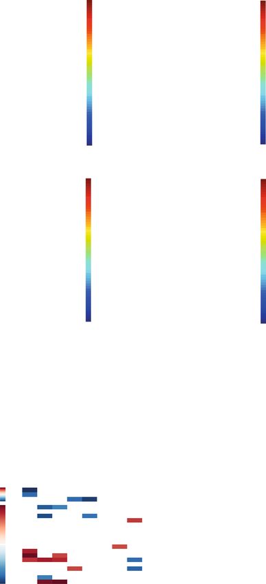
Mayneris-Perxachs et al. Microbiome   (2021) 9:104                                                             Page 2 of 18

 (Continued from previous page)
 Results: Here, we applied an integrative systems medicine approach (faecal metagenomics, plasma and urine
 metabolomics, hepatic transcriptomics) in 2 well-characterised human cohorts of subjects with obesity (discovery n
 = 49 and validation n = 628) and an independent cohort formed by both individuals with and without obesity (n =
 130), combined with in vitro and animal models. Serum ferritin levels, as a markers of liver iron stores, were
 positively associated with liver fat accumulation in parallel with lower gut microbial gene richness, composition and
 functionality. Specifically, ferritin had strong negative associations with the Pasteurellaceae, Leuconostocaceae and
 Micrococcaea families. It also had consistent negative associations with several Veillonella, Bifidobacterium and
 Lactobacillus species, but positive associations with Bacteroides and Prevotella spp. Notably, the ferritin-associated
 bacterial families had a strong correlation with iron-related liver genes. In addition, several bacterial functions
 related to iron metabolism (transport, chelation, heme and siderophore biosynthesis) and NAFLD (fatty acid and
 glutathione biosynthesis) were also associated with the host serum ferritin levels. This iron-related microbiome
 signature was linked to a transcriptomic and metabolomic signature associated to the degree of liver fat
 accumulation through hepatic glucose metabolism. In particular, we found a consistent association among serum
 ferritin, Pasteurellaceae and Micrococcacea families, bacterial functions involved in histidine transport, the host
 circulating histidine levels and the liver expression of GYS2 and SEC24B. Serum ferritin was also related to bacterial
 glycine transporters, the host glycine serum levels and the liver expression of glycine transporters. The
 transcriptomic findings were replicated in human primary hepatocytes, where iron supplementation also led to
 triglycerides accumulation and induced the expression of lipid and iron metabolism genes in synergy with palmitic
 acid. We further explored the direct impact of the microbiome on iron metabolism and liver fact accumulation
 through transplantation of faecal microbiota into recipient’s mice. In line with the results in humans, transplantation
 from ‘high ferritin donors’ resulted in alterations in several genes related to iron metabolism and fatty acid
 accumulation in recipient’s mice.
 Conclusions: Altogether, a significant interplay among the gut microbiome, iron status and liver fat accumulation is
 revealed, with potential significance for target therapies.
 Keywords: Systems medicine, Ferritin, Iron status, Gut microbiome, Non-alcoholic fatty liver disease, Shotgun
 sequencing, Metagenomics, Obesity

Background                                                      Despite this emerging evidence suggesting a role of
Non-alcoholic fatty liver disease (NAFLD) is a highly         both the gut microbiome and iron in the pathogenesis of
prevalent metabolic disease (the worldwide prevalence of      NAFLD, their complex cross-talk remains unclear.
NAFLD is 25.2% and increasing [1]) that can progress to       Therefore, in the present study, we applied an integrative
cirrhosis and hepatocellular carcinoma, being a risk fac-     systems medicine approach (faecal metagenomics,
tor for the development of type 2 diabetes and cardio-        plasma and urine metabolomics, hepatic transcripto-
vascular disease. NAFLD is complex and multifactorial,        mics) in 3 well-characterised human cohorts, combined
with iron interacting with the development of NAFLD           with in vitro and animal models, to characterize mecha-
[2] through gluconeogenic signals [3]. In the liver, iron     nisms responsible for the interaction between the gut
induces the synthesis and release of ferritin (an intracel-   microbiome and iron metabolism in NAFLD.
lular protein which stores iron), with its serum concen-
tration proportional to body iron stores, and frequently      Results
increased in patients with NAFLD [4].                         An overview of the study human cohorts and omics ana-
   As the gut microbiome causally impacts the host phe-       lyses pipeline can be found in Figure S1. Serum ferritin
nome in hepatic liver fat accumulation [5], the compos-       was measured in three cohorts: (a) a discovery cohort of
ition of the gut microbiota could influence the impact of     subjects with obesity (n = 49); (b) a validation cohort of
dietary iron on the development of NAFLD because this         subjects with obesity from Italy and Spain (n = 628); and
transition metal is a critical nutrient for both mammals      (c) an independent cohort of subjects with and without
and microorganisms [6]. Only ~ 5–15% of iron is absorbed      obesity from Spain (n = 130). Plasma and urine metabo-
and the remainder passes into the colon, where it is avail-   lomics were acquired in a subsample of both the discov-
able to the gut microbiota [7]. The microbiota is also        ery and the replication cohorts (plasma (n=48 and n=
known to affect the absorption of key minerals, with iron     328) and urine (n=47 and n=322, respectively). The
being an important micronutrient in terms of its interac-     transcriptome was analysed in a subsample of the dis-
tions with bacteria and the immune system [8].                covery and replication cohorts (n = 86). Finally, faecal
Mayneris-Perxachs et al. Microbiome   (2021) 9:104                                                                Page 3 of 18

samples from 56 women with obesity from the replica-             at the species level also revealed consistent associations
tion and validation cohort, and 130 subjects with and            with Veillonella sp. AS16, Veillonella sp. 6_1_27, Lactoba-
without obesity from the independent cohort were used            cillales, Streptococcus pneumoniae, Lachnospiraceae bacter-
to perform shotgun metagenomics sequencing.                      ium TF01-11, Bacteroides sp. GAC:633, Bacteroides
                                                                 coprophilus, Mediterranea massiliensis, Millionella massi-
Increased serum ferritin is associated with liver fat            liensis and Prevotella sp. CAG:487 in both subjects with
accumulation and the gut microbiome composition and              and without obesity. Notably, the microbiome associated
functionality                                                    with hs-CRP was markedly different to that linked to serum
In both discovery and replication cohorts, serum ferritin in-    ferritin (Figure S5a,b). In addition to the microbiome com-
creased with the severity of liver fat accumulation (Fig. 1a,    position, their functionality was also evaluated by shotgun
b). No significant associations were found between high-         sequencing in this cohort. Remarkably, analysis of bacterial
sensitivity C-reactive protein (hs-CRP) and serum ferritin       metagenomes based on KEGG functional annotation iden-
(Fig. 1c, Figure S2a,b) in any of the three cohorts. We also     tified several bacterial functions related to iron and amino
performed shotgun metagenomics, 1H NMR spectroscopy              acids transport, glutathione metabolism, heme and sidero-
and transcriptomics to characterize the faecal microbiome,       phore biosynthesis, fatty acid biosynthesis and DNA repli-
the biofluid metabolome and the liver transcriptome. Hav-        cation and repair, associated with serum ferritin
ing processed > 5 Gb of metagenomic sequence data per in-        concentrations (Fig. 1h, Additional file 3: Table S3). Add-
dividual, we derived taxonomy and gene richness as well as       itional O-PLS regression analyses based on EggNOG func-
mapping and annotation of gene functions on the inte-            tional annotations revealed similar results (Figure S5c, d
grated gene catalog for a subsample (n = 56) of women            and Additional file 4: Table S4).
with obesity from Italy and Spain [5]. Subjects within the
highest ferritin quartiles (Q3 and Q4) had decreased gene        An iron-associated transcriptome signature is linked to
richness compared with those in the lower ferritin quartile      the gut microbiome and liver fat accumulation
(Q1) (Fig. 1d). Consistently, multivariate penalized regres-     We then explored the associations of serum ferritin with
sion models adjusted for age, BMI, country and hs-CRP re-        the liver transcriptome in a subsample (n = 86) of the
vealed a significant association between serum ferritin and      discovery and replication cohorts from Italy and Spain.
the gut microbiome with significant decreases in families        Out of the 48 mRNAs identified from an O-PLS model
from the Firmicutes, Actinobacteria and Proteobacteria           (Fig. 2a), transferrin receptor (TFRC, pFDR < 1.0 ×
phyla, particularly Pasteurellaceae, Leuconostocaceae and        10−10), hepcidin antimicrobial peptide (HAMP, pFDR =
Micrococcaceae (Fig. 1e,f). Similar results were obtained        1.95 × 10⁻⁵), NCOA4 (pFDR = 0.05) and ferritin heavy
from multivariate orthogonal partial least squares (O-PLS)       chain (FTH1, pFDR = 0.003), all involved in iron status
regression and posterior validation by univariate partial        [9], were the mRNAs most associated with serum ferritin
Spearman’s correlation (pSC) analyses adjusted by age,           after further individual validation by pSC (Fig. 2b). En-
BMI, country and hs-CRP (Figure S3a–f). Notably, all iden-       richment analyses highlighted a significant over-
tified ferritin-associated bacterial families (with the excep-   representation of pathways associated with iron and glu-
tion of Leuconostocaceae) had a strong correlation with          cose metabolism (Fig. 2c). We further investigated the
iron-related genes such as TFRC, HAMP, MitoNEET, IRP1            association between the expression of several solute car-
and ferroportin (SLC40A1), measured by quantitative qRT-         rier transporters (SLCs) and the serum ferritin concen-
PCR (Figure S4). We replicated these findings using              tration (Fig. 2d, e). SLC51A, SLC11A1 and SLC6A9 had
DESeq2 analysis in an independent cohort of 130 subjects         the strongest associations with serum ferritin. After-
with and without obesity (Additional file 1: Table S1), in       wards, integrating metagenomic and transcriptomic re-
whom the majority of the associations between serum fer-         sults, we identified iron-associated transcriptome
ritin and bacterial families and genera were confirmed after     signatures linked to the microbiome and the degree of
adjustment for age, BMI, sex and hs-CRP (Fig. 1g, Figure         liver fat accumulation (Fig. 2f–k, Figure S6). From O2-
S3g, h, Additional file 2: Table S2). The most consistent re-    PLS multivariate integration between ferritin-associated
sults were the negative associations of several Veillonella,     bacterial families and transcripts, hierarchical clustering
Bifidobacterium and Lactobacillus species (from phyla Fir-       analysis and univariate pSC, we identified a clear cluster
micutes and Actinobacteria) with serum ferritin levels, and      comprising NUDT10, NNMT, MTUS1, SOCS2 and
positive associations with Bacteroides and Prevotella species    SBNO2, downregulated with increased ferritin levels
(from phylum Bacteroidetes). When we analysed the data           (Fig. 2h) and correlated to variation in different bacterial
according to the obesity status, we found consistent nega-       families that were themselves linked to serum ferritin
tive associations of serum ferritin with Lactobacillales, Pas-   (Fig. 2g, k). In a second cluster, the expression of
teurellaceae, Streptococcaceae and Mycobacteriaceae in           SEC24B, GYS2, SLC51A, TFRC, RPSX5, LOC100130078
both individuals with and without obesity. DESeq2 analysis       and ACSM5 was also mirrored by these families. A third
Mayneris-Perxachs et al. Microbiome (2021) 9:104 Page 4 of 18 Fig. 1 (See legend on next page.)
Mayneris-Perxachs et al. Microbiome        (2021) 9:104                                                                                  Page 5 of 18

 (See figure on previous page.)
 Fig. 1 Association of serum ferritin with liver fat accumulation, gene richness and the gut microbiome composition. Association of serum ferritin
 with degree of liver fat accumulation in a the discovery and b replication cohorts (Mann-Kendall trend test and Wilcoxon tests). c Association of
 hs-CRP with serum ferritin quartiles in the replication cohort (Mann-Kendall trend test and Wilcoxon tests). d Association of microbial gene
 richness with ferritin quartiles in a subsample of obese women from the discovery and replication cohorts (generalized linear model GLM). e
 Bacterial families and f genera associated with serum ferritin in a subsample of obese women from the discovery and replication cohorts. Mnet
 penalized regression models were built on bacterial data including age, BMI, country and hs-CRP as covariates. g Volcano plot of differential
 bacterial abundance and h metagenome KEGG functions associated with ferritin as calculated from shotgun metagenomic sequencing in an
 independent cohort of obese and non-obese subjects, adjusting for age, BMI, sex and hs-CRP. Significantly different taxa are coloured according
 to phylum. adaB, methylated-DNA-[protein]-cysteine S-methyltransferase; cpg; glutamate carboxypeptidase; cycA; D-serine/D-alanine/glycine
 transporter; fabA, 3-hydroxyacyl-[acyl-carrier protein] dehydratase/trans-2-decenoyl-[acyl-carrier protein] isomerase; fabM; trans-2-decenoyl-[acyl-
 carrier protein] isomerase; gshA, glutamate-cysteine ligase; nei endonuclease VIII; entF, enterobactin synthetase component F; FTR, FTH1, efeU,
 high-affinity iron transporter; hemG; menaquinone-dependent protoporphyrinogen oxidase; hutM, histidine permease; mtsC; iron/zinc/
 manganese/copper transport system permease protein; mtsA; iron/zinc/manganese/copper transport system substrate-binding protein; PARP, poly
 [ADP-ribose] polymerase; seqA; negative modulator of initiation of replication; yqjH, ferric-chelate reductase (NADPH)

cluster of genes positively associated with ferritin (USP3,                  identified several metabolites, such as ketone bodies and
SIX1, PDE7A and SNAPC2) also anti-correlated with                            gluconeogenic substrates, in both discovery (n = 48 for
those bacterial families. Remarkably, the expression of                      plasma; n = 47 for urine) and replication (n = 328 for
most of these bacterial-associated genes, changed in pro-                    plasma; n = 322 for urine) cohorts associated to the
portion to both serum ferritin levels and liver fat accu-                    identified hepatic transcriptome signatures (Fig. 2i, j)
mulation (Figure S6).                                                        linked to the microbiome and severity of NAFLD. Inte-
                                                                             gration of ferritin-associated metabolites and bacterial
The transcriptome signature was replicated in human                          families by O2-PLS regression (Fig. 3i, j) revealed strong
primary hepatocytes                                                          positive associations between histidine, tyrosine, citrul-
We then sought to validate the transcriptome findings                        line and glutamine and bacterial families negatively asso-
by studying the effects of iron and palmitic acid (PA), a                    ciated with serum ferritin (Fig. 3j–l). These families also
trigger of hepatic fat accumulation [10], in human pri-                      had strong negative associations with ketone bodies (3-
mary hepatocytes. We found that iron, PA and PA sup-                         hydroxybutyrate (3-OHB) and acetoacetate).
plementation in cells pretreated with iron led to
triglyceride accumulation in primary human hepatocytes                       Iron influences the gut microbiome composition
(Figure S7a, b). Notably, iron induced a striking increase                   To validate the cross-talk between iron status and the
in the expression of lipid metabolism genes (FABP4,                          microbiome uncovered in humans, we first tested
FABP5, FATP5) and of the fatty acid transporter CD36                         whether the dietary iron content impacts the micro-
in synergy with PA (Figure S7c–f) in parallel to upregu-                     biome in vivo in the mouse (Fig. 4a). Using 16S rRNA
lated iron-related genes (FTL and FTH) (Figure S7g, h).                      gene amplicon sequencing, we showed that variation in
   Strikingly, PA supplementation in cells pretreated with                   dietary iron dramatically reshapes the composition of
iron downregulated the expression of most of the identi-                     the gut microbiota (Fig. 4b, c). Then, we characterised
fied genes associated negatively with serum ferritin                         the impact of a high-fat diet vs. control diet with differ-
(GY2, SEC24B, MTUS1, SOCS2, SLC51A) compared to                              ent iron contents in mice using metagenomics (Fig. 4d).
PA or iron alone (Fig. 2l–s) in parallel to fat accumula-                    Bacterial biodiversity and observed species changed dra-
tion, confirming the associations observed in subjects                       matically according to fat and iron content of the diet
with different degrees of hepatic fat accumulation. The                      (Fig. 4e–g). While a high-fat diet decreased bacterial bio-
exception was SBNO2, known to be increased in proin-                         diversity under a low iron diet, the opposite as found in
flammatory responses. Conversely, genes associated                           diets with high iron content. Principal coordinate ana-
positively with serum ferritin (PDE7A) and also with                         lyses revealed different microbial community composi-
liver steatosis increased significantly after iron exposure.                 tions depending on the iron content in each diet (Fig.
                                                                             4h, i). Interestingly, the differences in the microbial com-
Metabolomics identifies gluconeogenic substrates and                         position between the high-fat and control diets de-
ketone bodies connected with the iron-related                                creased with iron content, becoming non-significant at
microbiome and transcriptomic signatures                                     high iron levels (Figure S8a–d). Several families and gen-
We then performed discovery and replication                                  era in the phylum Firmicutes inversely associated with
metabolome-wide association studies (MWAS) for fer-                          serum ferritin in patients were confirmed to be influ-
ritin in serum (Fig. 3a–d) and urine (Fig. 3e–h) using O-                    enced accordingly by the iron content of the mice diet
PLS multivariate regressions confirmed by pSC. We                            in O-PLS models (Fig. 4j–m).
Mayneris-Perxachs et al. Microbiome (2021) 9:104 Page 6 of 18 Fig. 2 (See legend on next page.)
Mayneris-Perxachs et al. Microbiome (2021) 9:104 Page 7 of 18
(See figure on previous page.)
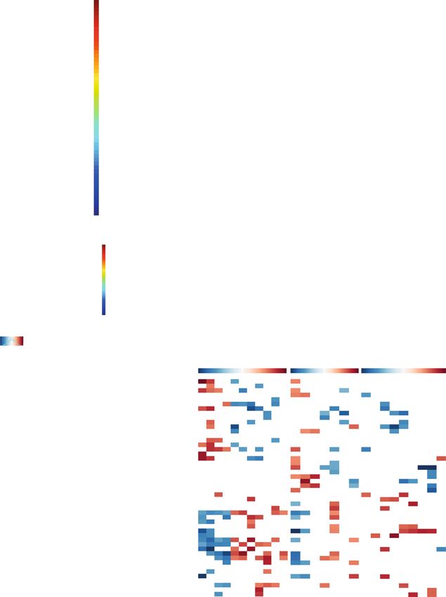
Fig. 2 Association of transcriptomic data with serum ferritin. a Permutation test for the goodness-of-fit (R2Y) and goodness of prediction (Q2Y)
obtained from the O-PLS model between serum ferritin and hepatic transcriptome in a subsample of the discovery and replication cohorts from
Italy and Spain (n = 86). b Significant transcripts associated with serum ferritin after further validation of the O-PLS significant variables by pSC
adjusting for age, sex, BMI and country. c Pathways significantly associated with serum ferritin based on mapping associated transcripts by over-
representation analysis with hypergeometric test. d Permutation tests for the O-PLS model between serum ferritin and SLCs (n = 86). e Significant
SLCs associated with serum ferritin after further validation of the O-PLS results by pSC adjusting for age, sex, BMI, and country. f O2-PLS scores for
the joint variation between microbial families and genes associated with serum ferritin. A model with 2 predictive components, and 1 orthogonal
component for the genes and bacterial families blocks, was constructed based on 7-fold cross-validation. g O2-PLS joint loadings plots, where
pcorr represents the correlation-scaled loadings from the gene block and qcorr represents the correlation-scaled loadings from the bacterial
families block. h Heatmap displaying z-scores of the ferritin-associated transcripts for each subject. Clustering was based on Euclidean distances
and Ward linkage. Genes associated with liver fat accumulation from O-PLS modelling are highlighted in bold, whereas those associated with
bacterial families from O2-PLS modelling are highlighted in colour boxes. i Heatmap for the pSC adjusted by age, BMI, sex, and country between
ferritin-associated plasma and j urine metabolites with ferritin-associated transcripts (n = 86). k Significant (p < 0.05) pSC adjusted for age, BMI
and country, between ferritin-associated families and transcripts (n = 56). Only significant associations (p < 0.05) are displayed. Significant
associations after a pFDR correction (pFDR < 0.05) are highlighted with a black box. l–n Expression of upregulated (GSK3B, PDE7A, SBNO2) and o–
s downregulated genes (GYS2, SEC24B, SOCS2, MTUS1 and SLC51A) in human primary hepatocytes after treatment with iron and palmitic acid.
Data are mean ± SEM. Comparisons by one-way ANOVA. *p < 0.05, **p < 0.01, ***p < 0.001 compared to control group based on t test. #p < 0.05,
##
p < 0.01, ###p < 0.001 compared to PA group based on t test. Ctrl, control group; PA, palmitic acid; Fe48h, pre-treatment iron 50 μM for 48h;
Fe72h, pre-treatment iron 50 μM for 72h; Fe48h + PA, pre-treatment iron 50 μM for 48h + palmitic acid 200 μM for 24 h; Fe72h + PA, pre-
treatment iron 50 μM for 72 h + palmitic acid 200 μM for 24 h
The gut microbiome affects iron metabolism is consistent with their reliance on utilizing iron from
After showing that iron availability greatly influences the host transferrin for growth and survival [11]. Recently,
gut bacterial ecosystem, we evaluated whether the hepatic lipid levels, including bile acids, have been re-
microbiota on its own might affect iron status. We lever- cently negatively correlated with Micrococcaceae in dia-
aged our previous mouse study showing that faecal betic mice [12], and the Desulfovibrionaceae family has
microbiota transplantation (FMT) triggered hepatic fat been associated to overfeeding-induced fatty liver [13].
accumulation [5] to evaluate the relationship between Consistent with our results, increases in the caecal con-
human donor ferritin levels and iron-related genes in re- tents of Coprococcus were identified in rats supple-
cipient mice (Fig. 4n). O-PLS-discriminant analysis re- mented with iron, which were suggested to mediate
vealed that microbiota from the ‘high ferritin donors’ oxidative stress and histopathological alterations ob-
group resulted in alterations in genes related to iron me- served in the liver of these animals [14]. Also in agree-
tabolism, with increases in ftl1, fth1 and slc40a1, in par- ment with our results, low serum ferritin concentrations
allel to decreased tfrc (Fig. 4o–r), which is in line with coexisted with decreased abundance of Veillonella spe-
the transcriptomic results in humans and the in vitro re- cies in ulcerative colitis patients receiving FMT from
sults from human primary hepatocytes. Also, in agree- healthy donors [15], while Veillonella genus abundance
ment with the in vitro results, the microbiota from ‘high was dose-dependently enriched after improvement of
ferritin donors’ also increased the expression of fabp4 in steatosis in NASH patients [16]. In addition, those with
recipient’s mice livers, showing the effect ferritin- > 70% reduction in liver fat had a trend towards reduc-
associated microbiota on liver lipid accumulation. tion of Methanobrevibacter, which we also found nega-
tively associated with serum ferritin levels. Significant
Discussion and drastic increases in Bacteroides and Prevotella have
In the current study, we evaluated the contribution of been observed in obese and NASH individuals, while a
the gut microbiota in iron status and liver fat accumula- progressive decrease in the abundance of Bifidobacter-
tion in a discovery and validation cohort of patients with ium was observed from healthy to NASH groups [17].
obesity and an additional independent cohort of individ- Finally, lack of iron requirements in lactic acid bacteria
uals with and without. We further identified a potential is in agreement with the negative associations observed
role of the microbiome as a regulator of iron status con- among several Lactobacillus species and the serum fer-
trolling hepatic fat deposition in animal models. Serum ritin levels.
ferritin levels were positively associated with liver fat ac- Although we used ferritin as a marker of iron store,
cumulation in parallel to a decrease in several bacterial we must take into account that it is also an acute-phase
families. At the same time, these ferritin-related families reactant which is increased under inflammatory condi-
were associated with liver genes involved in iron metab- tions [18]. Given that our study subjects were all obese,
olism. The Pasteurellaceae family had the strongest and low-grade chronic inflammation is a hallmark of
negative association with the serum ferritin levels, which obesity [19], inflammation could have an influence onMayneris-Perxachs et al. Microbiome (2021) 9:104 Page 8 of 18 Fig. 3 (See legend on next page.)
Mayneris-Perxachs et al. Microbiome         (2021) 9:104                                                                                     Page 9 of 18

 (See figure on previous page.)
 Fig. 3 Associations of metabolomic data with serum ferritin. Permutation tests for the goodness-of-fit (R2Y) and goodness of prediction (Q2Y)
 obtained from the O-PLS model between serum ferritin and a the serum (n = 48) and e urine metabolome (n = 47) in the discovery cohort, and
 b the serum (n = 328) and f urine metabolome (n = 322) in the replication cohort. Significant c, d serum and g, h urine metabolites associated
 with serum ferritin after further validation of O-PLS identified metabolites by pSC adjusting for age, sex, BMI and country. i O2-PLS scores for the
 joint variation between plasma and urine metabolites and microbial families associated with serum ferritin. A model with 2 predictive
 components, and 0 and 1 orthogonal component for the metabolites and bacterial families blocks, was constructed based on 7-fold cross-
 validation. j O2-PLS joint loadings plots, where pcorr represents the correlation-scaled loadings from the gene block and qcorr represents the
 correlation-scaled loadings from the bacterial families block. k Heatmap for the pSC adjusted by age, BMI and country between ferritin-associated
 urine and l plasma metabolites with ferritin-associated bacterial families (n = 56). Only significant associations (p < 0.05) are displayed. Significant
 associations after a pFDR correction (pFDR < 0.05) are highlighted with a black box

ferritin levels. Importantly, we did not find any signifi-                     the degree of liver fat accumulation, which we confirmed
cant association between inflammatory markers (hs-                             by supplementing human primary hepatocytes with iron
CRP) and serum ferritin, suggesting that serum ferritin                        and PA, a trigger of hepatic fat accumulation [10]. Im-
was measuring iron stores in our cohorts. To further                           portantly, excessive gluconeogenesis has been previously
rule out the effect of inflammation in the identified asso-                    associated with NAFLD in humans [22] and iron has
ciations, we adjusted our analysis by hs-CRP. Notably,                         been also shown to influence gluconeogenic signals [3].
the microbiome associated with hs-CRP was markedly                             In line with these previous results, we found alterations
different to that linked to serum ferritin, confirming that                    of genes involved in glucose metabolism. Thus, from
the observed microbiome-ferritin associations were inde-                       those mRNAs involved in the microbiome-associated
pendent of inflammation.                                                       transcriptomic signature of iron (also linked to liver fat
   In addition to identifying an iron-associated micro-                        accumulation), GYS2 (which catalyses the rate-limiting
biome signature, we also evaluated the microbiome                              step in the synthesis of glycogen) showed the strongest
functionality. Different bacterial pathways were associ-                       negative association with NAFLD (Figure S6). Disruption
ated with iron stores in the host, including heme and                          of GYS2 is known to result in impaired glucose depos-
siderophore (iron binding molecules) biosynthesis, iron                        ition and hepatic insulin resistance and liver fat accumu-
transport, glutathione metabolism and DNA replication                          lation in mice by changing de novo lipogenesis through
and repair (known to be iron-dependent). Of note, the                          increased expression of SREBP1c [23]. Insulin also sig-
bacterial cytochrome b561 function was strongly nega-                          nals to SREBP1 through inhibition of GSK3, which we
tively associated with serum ferritin levels. As cyto-                         found positively correlated with ferritin levels. In line
chrome b561 is known to be involved in iron absorption                         with these results, we found an upregulation of GSK3B
[20], this finding suggests that the bacterial cytochrome                      after treating human primary hepatocytes with iron or
b561 competes with the enzyme present in the intestine                         palmitic acid, which was exacerbated after co-treatment.
influencing iron uptake. In line, the expression of several                    Interestingly, we found a negative correlation between
bacterial iron transport and chelation functions (FTR,                         ferritin and insulin action measured through euglycemic
FTH1, efeU; yqjH; mtsC; and mtsA) was reduced in sub-                          hyperinsulinemic clamp (r = − 0.31, p = 8.9e-4). GYS2
jects with high serum ferritin levels. Similarly, bacterial                    clustered with SEC24B, which is responsible for the ER-
enzymes involved in heme biosynthesis (uroporphyrino-                          to-Golgi transport of proteins, and disrupted ER-to-
gen decarboxylase; and hemG, menaquinone-dependent                             Golgi trafficking has shown to contribute to ER stress,
protoporphyrinogen oxidase) and siderophore biosyn-                            hepatic injury and NAFLD [24, 25]. These transcripto-
thesis (entF, enterobactin synthetase component F) were                        mics findings were supported by metabolomics results.
also strongly negatively associated with serum ferritin. It                    Therefore, we identified some ferritin-associated metab-
is also worth noting the strong associations of host                           olites (sarcosine, citrulline, glutamate) that have been
serum ferritin levels with bacterial functions involved in                     previously linked to iron-induced impairment of glucose
glutathione biosynthesis (gshA, glutamate-cysteine ligase)                     metabolism [26]. In agreement, we found that subjects
and glutathione precursors (cpg, glutamate carboxypepti-                       with higher ferritin concentrations had lower serum
dase). Interestingly, the plasma and liver levels of gluta-                    levels of glutamine, alanine and glycerol, the main sub-
thione are depleted in NAFLD patients and altered                              strates used for liver gluconeogenesis, which is docu-
glutathione metabolism has been identified as a prevail-                       mented by liver transcriptomics (Fig. 2b and Figure S6).
ing feature in NAFLD [21].                                                     Notably, glutamine had a strong positive association
   We also evaluated the associations of serum ferritin                        with Leuconostocaceae, one of the bacterial families most
with the liver transcriptome and the serum and urine                           negatively associated with serum ferritin levels. However,
metabolome. Remarkably, we identified iron-associated                          the most consistent effect was the negative association
transcriptome signatures linked to the microbiome and                          of serum ferritin with histidine levels in both the
Mayneris-Perxachs et al. Microbiome   (2021) 9:104   Page 10 of 18

 Fig. 4 (See legend on next page.)
Mayneris-Perxachs et al. Microbiome       (2021) 9:104                                                                                 Page 11 of 18

 (See figure on previous page.)
 Fig. 4 Validation studies in primary hepatocytes and FMT mice. a Scheme of the experimental design for study 1. Mice were fed for 9 weeks
 diets containing low- (LI), low-normal- (LNI), high-normal- (HNI), moderately high- (MHI) and high- (HI) iron doses. b Heatmap displaying genus
 relative abundances for each mouse. c Principal coordinate analysis (PCoA) depicting dissimilarities between groups based on unifrac distance
 metrics. d Scheme of the experimental design for study 2. Mice were fed either a high fat diet (HFD) or a no-HFD diet containing four different
 iron doses (LI, LNI, HNI, MHI) for 10 weeks. e Variations in the Shannon diversity index, f Chao1 richness estimator and g observed species of mice
 fed either a HFD or a no-HFD with different iron doses (LI, LNI, HNI, MHI). h PCoA based on Canberra distance metric for the no-HFD-fed mice
 and i the HFD-fed mice with different iron doses. Differences in microbial composition between iron doses for each diet were assessed by PERM
 ANOVA using 999 permutations. j, k Permutation tests for the O-PLS models between iron dose and bacterial families or genera in HFD-fed mice,
 respectively. l Significant families and m genera identified from O-PLS regression loadings to be associated with iron dose. n Scheme of the
 experimental design for study 3. Low-ferritin (n = 3) and high-ferritin (n = 3) microbiota human donors were selected and for each donor their
 faecal samples were transplanted n = 6–8 mice after antibiotic treatment. After 14 days following colonization gavage mice were sacrificed and
 iron and liver fat accumulation-related genes (n = 22) were measured by PCR. o Permutation test for the O-PLS-DA model between mice genes
 and the human donor group (low- or high- ferritin). p Significant mouse genes associated with donor group from O-PLS-DA regression loadings.
 q Ferroportin (Slc40a1) and r Tfrc expression according to the donor ferritin concentration

discovery and replication cohorts. Histidine has shown                      [32]. Conversely, the control of LPS signalling by SOCS2,
to supress hepatic gluconeogenesis by activation of                         another negative inflammation regular, is minimal [33],
STAT3 independent of central insulin action [27]. No-                       which is also consistent with the lack of associations that
ticeably, the host serum ferritin levels were strongly as-                  we observed among bacterial families and the liver ex-
sociated with the bacterial function histidine permease                     pression of this gene.
(hutM), and the Pasteurellaceae and Micrococcaceae                            We sought to validate these results by treating human
families, both also positively associated with the liver ex-                primary hepatocytes with iron and palmitic acid. For
pression of GYS2. These families were also strongly asso-                   those gene transcripts that were positively associated
ciated with the SLC51A expression. This gene is                             with serum ferritin in the discovery cohort (GSK3B,
involved in bile acid transport and has recently been as-                   PDE7A) including subjects with different degrees of liver
sociated with NASH [28]. Importantly, the gut micro-                        fat accumulation, we found a consistent upregulation of
biota can regulate the pool size and composition of bile                    these genes after treatment with either palmitic acid or
acids [29], which play an important role in NAFLD                           iron. Co-treatment with palmitic acid and iron exacer-
pathogenesis and progression [30]. Interestingly, most                      bated these effects. Some genes negatively associated
bile acids are conjugated to glycine and we identified                      with serum ferritin and steatosis degree, were consist-
glycine as negatively associated with serum ferritin (Fig.                  ently downregulated after treatment with iron or iron +
3c, d). In addition, glycine is a key rate-limiting compo-                  palmitic acid (SLC51A, MTUS1). However, the results
nent of heme biosynthesis, mainly supplied by the gly-                      obtained for other genes negatively associated with
cine transporter 1 (GLYT1) encoded by SLC6A9, which                         serum ferritin in this cohort with steatosis seemed coun-
was positively associated with serum ferritin (Fig. 2d, e).                 terintuitive (GYS2, SEC24B, SOCS2). Hence, contrary to
This increased glycine demand may account for the                           what we expected, treatment with iron and/or pal-
lower serum levels associated with high ferritin concen-                    mitic acid led to an upregulation of these genes in
trations. In addition, glycine is the limiting substrate in                 human primary hepatocytes. However, co-treatment
glutathione synthesis from glutamate in subjects with                       with both iron and palmitic downregulated the ex-
NAFLD [21]. Remarkably, we found a strong negative                          pression of these genes, which is in line with the re-
association between serum ferritin levels and the expres-                   sults observed in the discovery cohort. We
sion of the bacterial glycine transporter (cycA) and                        hypothesize that this downregulation could arise from
glutamate-cysteine ligase (gshA), the first enzyme in the                   an ‘hormesis effect’, i.e., an adaptative compensatory
glutathione biosynthetic pathway. Finally, serum acetone                    process following an initial disruption of homeostasis,
and 3-OHB were also positively and consistently associ-                     to compensate the initial disruptions in gene expres-
ated with serum ferritin concentration, in line with                        sion induced by palmitic acid or iron alone.
hyperinsulinemia resulting in shifted energy supply from                      Our results, based on the identification several bacter-
glucose to ketone bodies in NAFLD in parallel with in-                      ial species and metagenome functions involved in iron
creased circulating levels of the latter [31]. The serum 3-                 metabolism and NAFLD, suggested a direct impact of
OHB levels also had a strong negative correlation with                      the microbiome on iron metabolism. Hence, we further
the liver expression of SLC51A and SBNO2, both posi-                        explored the potential causative role of the gut micro-
tively associated with Pasteurellaceae and Micrococca-                      biome on iron metabolism and liver fact accumulation
ceae families. Consistently, SBNO2 expression increased                     using a mouse FMT experiment. Faecal microbiome
markedly following LPS-induced systemic endotoxemia                         transplantation from ‘high ferritin donors’ into recipient
Mayneris-Perxachs et al. Microbiome   (2021) 9:104                                                           Page 12 of 18

mice increased the expression of several genes involved       microbiome and host homeostasis in general [34] and
in iron metabolism as well as that of genes that promote      the Microbiome-Iron-Liver fat axis in particular, thereby
fatty acid accumulation such as fabp4, which is consist-      disclosing potential targets for therapy.
ent with results observed after treating human primary
hepatocytes with iron. Conversely, microbiota from ‘low
                                                              Patients and methods
ferritin donors’ increased the expression of iron-related
                                                              Detailed methods for the animals studies, 1H-NMR
Tfrc. This is agreement with our findings in human sub-
                                                              metabolomics, liver transcriptomics and faecal 16S
jects, where we found strong positive associations among
                                                              rRNA and metagenomics sequencing can be found in
Actinomycetaceae, Acidaminococcacea and Enterobacte-
                                                              Additional file 5: Supplementary methods.
riaceae families (both increased in the microbiota of
subjects with low serum ferritin levels) and the host liver
TFRC expression. These results are also consistent with       Patient recruitment and sample processing
the negative association found among metagenome func-         The discovery cohort included n = 49 obese patients
tions related to fatty acid biosynthesis (fabM and fabA)      aged 24 to 63 years old at the Endocrinology Service of
and the host serum ferritin concentrations. This could        the Hospital Universitari de Girona Dr Josep Trueta (Gi-
reflect a possible use of fatty acids from the host by the    rona, Spain). The replication cohort comprised n = 628
microbiota, avoiding the process of de novo synthesis.        obese patients aged 20 to 67 years old at the Endocrin-
Therefore, we showed that the microbiota itself could         ology Service of the Hospital Universitari de Girona Dr
recapitulate in recipient mice the phenomic hallmarks of      Josep Trueta (n = 287) and at the Center for Athero-
iron metabolism from the human donor, thereby                 sclerosis of Policlinico Tor Vergata University of Rome
impacting liver fat accumulation in the long term.            (Rome, Italy; n = 341). Sample size was not determined
   The current study presents some limitations. A minor-      by statistical methods and is comparable to other studies
ity of the bacterial population might have a role in          in the field [35–37]. All subjects gave written informed
NAFLD development but could remain undetected by              consent, validated and approved by the ethical commit-
metagenomics. On the other hand, the FMT experiment           tee of the Hospital Universitari Dr Josep Trueta (Comitè
has also its limitations because some strictly anaerobic      d’Ètica d’Investigació Clínica, approval number 2009
bacterial species might have a role in NAFLD develop-         046) and Policlinico Tor Vergata University of Rome
ment but are lost during sample collection, storage and       (Comitato Etico Indipendente, approval number 28-05-
manipulation. We showed changes in the expression of          2009). Inclusion criteria included Caucasian origin,
iron-related genes, but the impact of these changes on        stable body weight 3 months before the study, free of
iron levels of mice was not evaluated. Therefore, the         any infection 1 month preceding the study and absence
causal role of the microbiome as a regulator of iron sta-     of any systemic disease. Exclusion criteria were the fol-
tus needs to be further confirmed with the measurement        lowing: presence of liver disease (specifically tumoural
of circulating iron makers in these mice. Finally, we         disease and hepatitis C virus infection) and thyroid dys-
showed iron supplementation impacts on the micro-             function (based on biochemical work-up), alcohol con-
biome composition and that bacterial biodiversity and         sumption (> 20 g/day), hepatitis B (anti-HD virus
observed species changed dramatically according to fat        antibodies), drug-induced liver injury (using a drug
and iron content of the diet (Fig. 4e–g). It needs to be      questionnaire).
further investigated whether iron supplementation im-            A third independent cohort included obese (BMI ≥ 30
pacts on fat deposition in the liver in mice under differ-    kg/m2) patients and age- and sex-matches non-obese
ent feeding regimes.                                          subjects (BMI 18.5–< 30 kg/m2) aged 27–67 years old,
                                                              recruited at the Endocrinology Service of the Hospital
Conclusions                                                   Universitari de Girona Dr Josep Trueta (Girona, Spain).
In conclusion, combining a comprehensive systems              Exclusion included type 2 diabetes mellitus, chronic in-
medicine approach with validations in independent co-         flammatory systemic diseases, acute or chronic infec-
horts and causality assessment in pre-clinical models,        tions in the previous month; use of antibiotic, antifungal,
our findings demonstrate a significant cross-talk among       antiviral or treatment with proton-pump inhibitors; se-
gut microbiota, iron status and liver fat accumulation. In    vere disorders of eating behaviour or major psychiatric
particular, we uncover microbiome- and iron-linked            antecedents; neurological diseases, history of trauma or
metabolomic and transcriptomic signatures involving           injured brain, language disorders; and excessive alcohol
imbalances in gluconeogenic metabolites, ketone bodies        intake (≥ 40 g OH/day in women or 80 g OH/day in
and cellular transport, which altogether modulate liver       men). All subjects gave written informed consent, vali-
fat accumulation. This work highlights the crucial im-        dated and approved by the ethical committee of the
portance of the interplay between micronutrients,             Hospital Universitari Dr Josep Trueta.
Mayneris-Perxachs et al. Microbiome (2021) 9:104 Page 13 of 18
Stool, plasma and urine samples from all subjects were Microbiome analyses
obtained during the week before elective gastric bypass Bacterial population in mouse faeces from studies 1 and
surgery, during which the liver biopsy was sampled. All 3 was determined using next-generation high through-
samples were stored at − 80 °C. Liver samples were col- put sequencing of variable regions of the 16S rRNA bac-
lected in RNAlater, fragmented and immediately flash terial gene, whereas a shotgun metagenomic sequencing
frozen in liquid nitrogen before storage at − 80 °C. was employed for mice study 2 and human cohorts. De-
tailed protocols are included in supplementary materials.
Hepatic steatosis
Primary human hepatocytes culture and treatments
An ultrasound system with a 3.5 MHz convex trans- Cryopreserved primary human hepatocytes (HH) were
ducer (Siemens Acuson S2000, Mochida Siemens Med- commercially sourced (Innoprot, Bizkaia, Spain) and cul-
ical System, Tokyo, Japan) was used to scan the liver. tured with hepatocytes medium (Innoprot) supple-
Hepatic steatosis was defined as absent (grade 0: < 5% mented with 5% fetal bovine serum, 1% hepatocytes
steatosis), mild (grade 1: 5–33% steatosis), moderate growth supplement (mixture of growth factors, hor-
(grade 2: > 33–66% steatosis) or severe (grade 3: > 66% mones and proteins necessary for culture of primary he-
steatosis) using the scoring system for NAFLD [38]. Im- patocytes) and 100 U/ml penicillin and streptomycin.
ages were independently evaluated by two radiologists HH were grown on poly-L-lysine pre-coated cell dishes
blinded to clinical and laboratory data [39]. at 37 °C and 5% CO2 atmosphere following manufac-
turer’s recommendations. Cells were treated with iron
Liver histology for 48 or 72 h alone, or in combination with palmitic
Liver biopsies were previously obtained for n = 86 pa- acid (PA) for 24 h following the iron 48/72 h treatment.
tients who underwent bariatric surgery [5]. The investi- Compounds were prepared as follows: 27.84 mg of PA
gators were blind to group allocations. Liver biopsies (Sigma, San Luis, MO) was dissolved in 1 ml sterile
were analysed by a single pathologist expert in hepatic water to obtain a 100 mM stock solution. Five percent
pathology. For each liver sample, haematoxylin and bovine serum albumin (BSA) was prepared in serum-
eosin, reticulin and Masson’s trichrome staining were free DMEM and then mixed with PA stock solution for
performed. Hepatic steatosis grade was determined ac- at least 1 h at 40 °C to obtain a 5 mM solution. Iron was
cording to the scoring system for NAFLD [38]. dissolved in water. Iron was used at 50 μM for 48 or 72
h, and PA 200 μM for 24 h. BSA was used in all treat-
ments as the vehicle. All experimental conditions were
Serum ferritin performed in 4 biological replicates. After treatment,
Serum ferritin in both discovery and replication cohorts cells were washed with PBS and collected with Qiazol
was measured by microparticle enzyme immunoassay for RNA purification or fixed with paraformaldehyde 4%
(AxSYMTM; Abbot Laboratories) with intra- and inter- for Oil Red O staining. After fixation, cells were dipped
assay CVs < 6%. in isopropanol 60% before completely dried and stained
with Oil Red O (Sigma, Lyon, France) for 10 min at
1
H nuclear magnetic resonance spectroscopy-base
room temperature. Pictures were taken using an inverted
metabolic profiling
microscope.
Spectroscopic analysis of urine (n = 47 for discovery co-
Gene expression analysis using real-time PCR (cells,
hort, n = 322 for replication cohort) and plasma samples
mouse, human)
(n = 48 for discovery cohort, n = 328 for replication co-
hort) was performed on a Bruker DRX600 spectrometer Total RNA was extracted and purified using RNeasy
equipped with either a 5-mm TXI probe operating at Mini Kit (QIAGEN, Gaithersburg, MD) following manu-
600.13 MHz or a 5-mm BBI probe operating at 600.44 facturers’ protocol. Gene expression procedures were
MHz. The 90° pulse length was determined prior to each assessed using LightCycler 480 Real-Time PCR System
run and field frequency was locked using D2O as solv- (Roche Diagnostics SL, Barcelona, Spain), using Sybr-
ent. Detailed procedures are included in supplementary green technology suitable for relative genetic expression
materials. quantification. Peptidylprolyl isomerase A was used as
housekeeping.
Transcriptomics Mice studies
Transcriptomic analyses from liver biopsies have been Detailed procedure for the animal studies is included in
previously described [5] and detailed procedures are in- supplementary materials. To assess the effects of dietary
cluded in supplementary materials. iron on the gut microbiome (mice study 1), 12-week-oldMayneris-Perxachs et al. Microbiome (2021) 9:104 Page 14 of 18
male C57BL/6J mice were fed with low- (LI: 4 mg/kg), to be tuned: the regularization parameter (λ ≥ 0) con-
low-normal- (LNI: 35 mg/kg), high-normal- (HNI: 500 trolling the shrinkage of the variables, the elastic net
mg/kg), moderately high- (MHI: 2000 mg/kg) or high- mixing parameter (0 < α < 1) controlling the contribu-
(HI: 20,000 mg/kg) carbonyl iron diets for 9 weeks be- tion of ridge (α = 0) and LASSO (α = 1) penalty to the
fore sacrifice. The impact of different diets on the effects model, and the MCP penalty (γ). These were optimized
of dietary iron on the gut microbiome (mice study 2) by 10-fold cross-validation. The relationship between
was assessed by feeding mice with either a ‘no-High Fat serum ferritin quartiles and MGR was assessed using a
Diet’ (No-HFD) or a ‘High Fat diet’ (HFD) with different generalized linear model (GLM) with a multinomial
concentrations of iron representing the LI, LNI, HNI probability distribution and a cumulative logit as a link
and MHI diets. Finally, the causal role of the micro- function to account for non-normality adjusting for age
biome on iron status was evaluated by faecal microbiota and BMI using SPSS.
transplantation from low- (n = 3) and high- (n = 3) fer-
ritin donors matched for age and BMI to recipient mice Microbiome data analysis (mouse)
(mice study 3). Alpha and beta diversity indices were obtained using the
R package vegan. Trends between alpha diversity mea-
Statistical analysis sures and iron doses for each diet were assessed by non-
Ferritin distribution and normality was checked visually parametric Mann-Kendall test, whereas differences be-
and using the Kolmogorov-Smirnov and Shapiro-Wilk tween HFD and no-HFD for each iron dose were
tests and was found to be not normally distributed. assessed by non-parametric Wilcoxon-Mann-Whitney
Metagenomic, transcriptomic and metabolomic data test. To estimate the relatedness of microbial communi-
were also not normally distributed. Therefore, all univar- ties among groups, beta diversity distances between sam-
iate correlations analyses were based on partial Spear- ples were examined using principal coordinate analysis
man’s correlation (pSC) adjusting for age, BMI, sex and (PCoA). Differences in microbial composition were
country. Multivariate analyses were performed on the assessed by PERMANOVA analyses using the Adonis
log10 transformed data. Associations between serum fer- function in vegan R package with 999 permutations.
ritin and steatosis grade in the discovery and replication Families and genera responsible for differences in the
cohorts were assessed by non-parametric Kruskal Wallis microbial compositions among iron doses for each diet
test and steatosis grade 0 (absence) was compared to were identified through multivariate O-PLS modelling,
grades 1–3 using the non-parametric Wilcoxon-Mann- whereby the microbiome composition was used as the
Whitney test. Associations between microbiome, tran- descriptor matrix (X) to predict the iron dose (Y). The
scriptomic or metabolomic data with serum ferritin predictive performance of the model (Q2Y) was calcu-
quartiles were assessed by non-parametric Mann- lated using a leave-one-out cross-validation approach
Kendall test to detect monotonic trends. The Wilcoxon- and model validity was established by permutation test-
Mann-Whitney test was used to individually compare ing (1000 permutations). Variable selection was based
the lowest ferritin quartile (Q1 used as reference) with on O-PLS regression loadings adjusted for multiple test-
the higher quartiles. ing using the Benjamini-Hochberg procedure (pFDR). A
pFDR < 0.05 was used as the reference feature selection
Microbiome data analysis (human) criterium.
Taxa were filtered so that only those having a relative
abundance > 20% in at least 20% of the samples were Transcriptome and metabolome data analysis
considered for further analyses. To identify families and For transcriptomic and metabolomic data, we used a
genera associated with serum ferritin, we applied both combination of multivariate O-PLS modelling and par-
multivariate Mnet regularization regression [40] models tial Spearman’s correlation (pSC) using in-house MATL
and DESeq2 [41] analyses including age, BMI, country AB scripts. First, an O-PLS model was built. Here, the
and hs-CRP as covariates using the R package ncvreg, omics profiles were used as the descriptor matrix (X) to
phyloseq and DESeq2. The Mnet uses a combination of predict serum ferritin as the response variable (Y). Then,
ridge (l2) and minimax concave penalties (MCP) to deal variable selection was achieved combining the variable
efficiently with predictors ≥ n problems with highly cor- importance for projection (O-PLS-VIP) [42] and the O-
related predictors, which is typical of omics data. Com- PLS regression loadings adjusted for multiple testing
bining both penalties, the Mnet performs variable using the Benjamini-Hochberg procedure (pFDR). A
selection by forcing the regression coefficients of vari- pFDR < 0.05 was used as the reference feature selection
ables not actually associated with the response to 0 and criterium. However, a less restrictive threshold (pFDR <
at the same time handles multicollinearity within the 0.1 unless otherwise indicated) was used to include vari-
data. In the Mnet, there are three parameters that need ables with high VIP (> 1). Finally, each individualMayneris-Perxachs et al. Microbiome (2021) 9:104 Page 15 of 18
variable identified form multivariate models was further using the Consensus Pathway database (CPDB) [45].
validated by pSC adjusting for age, BMI, sex and Pathway significance was assessed using a hypergeo-
country. metric test and a Bonferroni procedure was applied for
multiple testing correction.
Clustering analysis
Unsupervised hierarchical clustering analysis (HCA) was
performed to identify general patterns of transcriptomic
variation among serum ferritin quartiles. Significant Supplementary Information
The online version contains supplementary material available at https://doi.
transcripts associated with serum ferritin were used for org/10.1186/s40168-021-01052-7.
sample clustering. Before clustering, data were standard-
ized as z scores across samples for each transcript. This Additional file 1:. Baseline characteristics of the independent cohort.
standardized matrix was then used in unsupervised Additional file 2:. Gut microbiome bacterial species associated with the
HCA using Euclidean distances and Ward linkage. Heat- serum ferritin. Results of differential bacterial abundance associated with
ferritin as calculated from shotgun metagenomic sequencing in an
maps and dendrograms following HCA were generated independent cohort of obese and non-obese subjects, adjusting for age,
using the heatmap.2 function from the gplots R package. BMI, sex, and hs-CRP.
In the heatmaps, a red-blue colour scale was used Additional file 3:. Metagenome functions based on KEGG annotation
whereby shades of red and blue represent higher and associated with the serum ferritin. Results of metagenome KEGG
functions associated with ferritin as calculated from shotgun
lower values, respectively, compared with the mean. metagenomic sequencing in an independent cohort of obese and non-
obese subjects, adjusting for age, BMI, sex, and hs-CRP.
Integration of datasets Additional file 4:. Metagenome functions based on EggNOG
To explore the functional associations among the micro- annotation associated with the serum ferritin. Results of metagenome
EggNOG functions associated with ferritin as calculated from shotgun
biome changes and metabolic and transcriptomic pertur- metagenomic sequencing in an independent cohort of obese and non-
bations associated with serum ferritin, were used two obese subjects, adjusting for age, BMI, sex, and hs-CRP.
approaches. First, datasets were integrated using an O2- Additional file 5: Supplementary methods. Methods for the animals
PLS approach. It is a bidirectional multivariate regres- studies, NMR metabolomics, liver transcriptomics, and 16S rRNA and
shotgun metagenomics sequencing.
sion method that allows separate joint covariance be-
Additional file 6: Figure S1. Flow chart of the study human cohorts
tween two blocks from systemic variation specific and omics analyses pipeline.
(orthogonal) to each block (X and Y) [43]. The number Additional file 7: Figure S2. Associations of serum ferritin with hs-CRP.
of components for the predictive and orthogonal blocks a) Association of hs-CRP with serum ferritin quartiles in the discovery co-
was selected based on a 7-fold cross-validation using the hort and b) an independent cohort of obese and non-obese patients
(Mann-Kendall trend test and Wilcoxon tests).
R package OmicsPLS [44]. Variable loadings for each
Additional file 8: Figure S3. Associations of serum ferritin with the gut
block were scaled as correlation coefficient (pcorr and microbiome in the human cohorts. Permutation tests for the goodness-
qcorr for X and Y, respectively) and represented in a of-fit (R2Y) and goodness of prediction (Q2Y) obtained from the O-PLS
correlation circle plot. Scaling was performed by calcu- model between serum ferritin and a) bacterial families or b) bacterial gen-
era in a subsample of obese women from the discovery and replication
lating the correlation between each variable and its asso- cohorts from Italy and Spain (n = 56). c) Significant families and d) genera
ciated component. The longer the distance to the origin, associated with serum ferritin from O-PLS regression loadings. Families
the stronger the relationship between variables. Strongly and genera associated positively and negatively associated with serum
ferritin from Mnet regression models are highlighted in dark red and
positively associated variables or groups of variables are blue, respectively. e) Significant families and f) genera associated with
projected closely to each other on the correlation circle serum ferritin after further validation of the O-PLS significant variables by
(~ 0° angle). The variables or groups of variables strongly pSC adjusting for age, BMI, country, and hs-CRP. g) Associations of bacter-
ial families and h) genera associated with serum ferritin by DESeq2 ana-
negatively associated are projected diametrically opposite lysis from shotgun metagenomic sequencing data in the independent
(~ 180° angle) on the correlation circle. Variables not cohort of obese and non-obese patients (n = 130), adjusting for age, BMI,
correlated are situated ~ 90° one from the other. Add- sex, and hs-CRP. Families and genera also associated with serum ferritin
in the discovery and replication cohorts based on Mnet regression
itionally, univariate partial Spearman’s correlations ad- models are highlighted in dark red, whereas those also identified from O-
justed for age, BMI, sex and country were calculated and PLS modelling are highlighted in dark pink.
represented as correlation heatmaps. Only significant Additional file 9: Figure S4. Associations of bacterial families with iron-
correlations (p < 0.05) are displayed. A pFDR correction related genes (discovery cohort, n = 35). Only significant correlations are
coloured. Genes were measured by real time-PCR. Bacterial families with
was used to adjust P values for multiple testing. Signifi- a significant positive association with serum ferritin concentrations are
cant associations after a pFDR correction (< 0.05) are highlighted in dark red, whereas those with a significant negative associ-
highlighted with a black box. ation are highlighted in dark blue. ChREBP, carbohydrate response elem-
ent binding protein; LCN2, Lipocailin 2; MitoNEET, Mitochondrial Inner
NEET Protein; TFRC, Transferrin Receptor; SLC40A1, Solute Carrier Family
Pathway analysis 40 Member 1 (Ferroportin); TF, Transferrin; FTH1, Ferritin Heavy Chain 1;
Differentially expressed genes associated with serum fer- HAMP, Hepcidin Antimicrobial Peptide; FTL, Ferritin Light Chain; IRP1, Iron
Regulatory Protein 1.
ritin were annotated via over-representation analysisYou can also read