Environmental Plasticity of the RNA Content of Staphylococcus aureus Extracellular Vesicles - Frontiers
←
→
Page content transcription
If your browser does not render page correctly, please read the page content below
ORIGINAL RESEARCH
                                                                                                                                                 published: 11 March 2021
                                                                                                                                         doi: 10.3389/fmicb.2021.634226

                                              Environmental Plasticity of the RNA
                                              Content of Staphylococcus aureus
                                              Extracellular Vesicles
                                              Brenda Silva Rosa Da Luz 1,2, Aurélie Nicolas 1, Svetlana Chabelskaya 3,
                                              Vinícius de Rezende Rodovalho 1,2, Yves Le Loir 1, Vasco Ariston de Carvalho Azevedo 2,
                                              Brice Felden 3*† and Eric Guédon 1*†
                                              1
                                               INRAE, Institut Agro, STLO, Rennes, France, 2 Laboratory of Cellular and Molecular Genetics, Institute of Biological
                                              Sciences, Federal University of Minas Gerais, Belo Horizonte, Brazil, 3 BRM [Bacterial Regulatory RNAs and Medicine]
                                              UMR_S 1230, University of Rennes, Inserm, Rennes, France

                                              The roles of bacterial extracellular vesicles (EVs) in cell-to-cell signaling are progressively being
                                              unraveled. These membranous spheres released by many living cells carry various
                                              macromolecules, some of which influence host-pathogen interactions. Bacterial EVs contain
                          Edited by:
                     Carlos Robello,
                                              RNA, which may serve in communicating with their infected hosts. Staphylococcus aureus,
Universidad de la República, Uruguay          an opportunistic human and animal pathogen, produces EVs whose RNA content is still poorly
                        Reviewed by:          characterized. Here, we investigated in depth the RNA content of S. aureus EVs. A high-
              Maria Rosa Garcia-Silva,
                                              throughput RNA sequencing approach identified RNAs in EVs produced by the clinical S.
      Institut Pasteur de Montevideo,
                             Uruguay          aureus strain HG003 under different environmental conditions: early- and late-stationary growth
              Lysangela Ronalte Alves,        phases, and presence or absence of a sublethal vancomycin concentration. On average,
  Carlos Chagas Institute (ICC), Brazil
                                              sequences corresponding to 78.0% of the annotated transcripts in HG003 genome were
                     *Correspondence:
                             Eric Guédon
                                              identified in HG003 EVs. However, only ~5% of them were highly covered by reads (≥90%
                     eric.guedon@inrae.fr     coverage) indicating that a large fraction of EV RNAs, notably mRNAs and sRNAs, were
                             Brice Felden
                                              fragmented in EVs. According to growth conditions, from 86 to 273 highly covered RNAs
           brice.felden@univ-rennes1.fr;
                bfelden@univ-rennes1.fr       were identified into the EVs. They corresponded to 286 unique RNAs, including 220 mRNAs.
     †
         These authors have contributed       They coded for numerous virulence-associated factors (hld encoded by the multifunctional
                    equally to this work      sRNA RNAIII, agrBCD, psmβ1, sbi, spa, and isaB), ribosomal proteins, transcriptional
                                              regulators, and metabolic enzymes. Twenty-eight sRNAs were also detected, including bona
                       Specialty section:
            This article was submitted to     fide RsaC. The presence of 22 RNAs within HG003 EVs was confirmed by reverse transcription
                      Infectious Diseases,    quantitative PCR (RT-qPCR) experiments. Several of these 286 RNAs were shown to belong
                  a section of the journal
                Frontiers in Microbiology     to the same transcriptional units in S. aureus. Both nature and abundance of the EV RNAs
         Received: 27 November 2020           were dramatically affected depending on the growth phase and the presence of vancomycin,
          Accepted: 18 February 2021          whereas much less variations were found in the pool of cellular RNAs of the parent cells.
           Published: 11 March 2021
                                              Moreover, the RNA abundance pattern differed between EVs and EV-producing cells according
                               Citation:
                                              to the growth conditions. Altogether, our findings show that the environment shapes the RNA
               Da Luz BSR, Nicolas A,
  Chabelskaya S, Rodovalho VdR, Le            cargo of the S. aureus EVs. Although the composition of EVs is impacted by the physiological
 Loir Y, Azevedo VAdC, Felden B and           state of the producing cells, our findings suggest a selective packaging of RNAs into EVs, as
      Guédon E (2021) Environmental
      Plasticity of the RNA Content of        proposed for EV protein cargo. Our study shedds light to the possible roles of potentially
               Staphylococcus aureus          functional RNAs in S. aureus EVs, notably in host-pathogen interactions.
                  Extracellular Vesicles.
          Front. Microbiol. 12:634226.        Keywords: membrane vesicle, small regulatory RNA, virulence factors, vancomycin, RNA-Seq, extracellular
    doi: 10.3389/fmicb.2021.634226            vesicle, RsaC, RNAIII

Frontiers in Microbiology | www.frontiersin.org                                       1                                            March 2021 | Volume 12 | Article 634226
Da Luz et al.                                                                                                             S. aureus EV RNA Cargo

INTRODUCTION                                                              antibiotics exposures, EVs increase S. aureus adhesion and
                                                                          cell aggregation, and contribute to biofilm formation (He
The release of extracellular vesicles (EVs) by living cells is a          et al., 2017). Recent data highlight the importance of EVs
well-established phenomenon required for intercellular                    in staphylococcal pathogenesis since EVs derived from various
communications and trans-kingdom interactions (Brown et al.,              human and animal strains of S. aureus share a conserved
2015; Toyofuku, 2019). These spherical membranous particles               EV proteome (Tartaglia et al., 2020).
vary from 20 to 300 nm in diameter and contain macromolecules                The vast majority of functional studies on bacterial EVs,
such as nucleic acids, proteins, lipids, and small metabolites.           however, challenged their proteome. Regarding the presence
Initially considered to be trash bags to eliminate unwanted               of DNAs and RNAs in EVs, most studies have been conducted
material outside of the cells, they are now widely recognized             on Gram-negative bacteria (Perez Vidakovics et al., 2010;
as protective delivery shuttles of bioactive molecules from donor         Blenkiron et al., 2016; Koeppen et al., 2016; Bitto et al.,
to recipient cells (Brown et al., 2015; Kim et al., 2015; Gill            2017; Choi et al., 2017; Malabirade et al., 2018; Yu et al.,
et al., 2019). The functional characterization of bacterial EVs           2018; Han et al., 2019). OMV-associated RNAs can include
is of interest due to their capacities to affect bacteria-host cell       messenger RNAs (mRNA), transfer RNAs (tRNA), ribosomal
interactions and bacterial pathogenesis (Kaparakis-Liaskos and            RNAs (rRNA), or small regulatory RNAs (sRNA; Biller et al.,
Ferrero, 2015; Tsatsaronis et al., 2018). Although the formation          2014; Ghosal et al., 2015; Ho et al., 2015; Sjöström et al.,
of outer membrane vesicles (OMVs) in Gram-negative bacteria               2015; Blenkiron et al., 2016; Koeppen et al., 2016; Choi et al.,
was early documented in 1966 (Work et al., 1966), the formation           2017; Dauros-Singorenko et al., 2018; Liu et al., 2018a;
of such structures was disregarded in Gram-positive bacteria              Malabirade et al., 2018; Tsatsaronis et al., 2018; Frantz et al.,
until recently. The production of EVs by a Gram-positive                  2019). EV-associated RNA cargo, notably sRNAs, can influence
bacterium, Staphylococcus aureus, was demonstrated in 2009                host-pathogen interactions, cell-to-cell communications, and
and, ever since, numereous studies confirmed EV release by                bacterial pathogenesis (Dauros-Singorenko et al., 2018;
other Gram-positive bacteria (Lee et al., 2009, 2013a; Rivera             Tsatsaronis et al., 2018; Lee, 2019; Ahmadi Badi et al., 2020;
et al., 2010; Prados-Rosales et al., 2011; Brown et al., 2014;            Lécrivain and Beckmann, 2020). For instance, OMVs from
Olaya-Abril et al., 2014; Kim et al., 2016a; Liu et al., 2018b).          Pseudomonas aeruginosa can transfer an sRNA into the human
    Staphylococcus aureus commonly colonizes the skin or nasal            airway cells, resulting in IL-8 decrease (Koeppen et al., 2016).
tract of vertebrates, without causing disease (Wertheim et al.,           Likewise, transfection of OMV-associated sRNAs from the
2005). However, it is also one of the main opportunistic pathogen         periodontal pathogens Aggregatibacter actinomycetemcomitans,
in humans, and a frequent cause of multi-drug resistant                   Phorphyromonas gingivalis, and Trepanema denticola into human
nosocomial infections (Ziebuhr, 2001). S. aureus is responsible           cells reduced host interleukine release (Choi et al., 2017). The
for a wide array of diseases, ranging from minor infections               presence of RNAs within Gram-positive EVs has been reported
in soft tissues to life-threatening diseases, such as sepsis,             for fewer species (Resch et al., 2016; Dauros Singorenko et al.,
meningitis, and pneumonia (Salgado-Pabón and Schlievert, 2014;            2017; Frantz et al., 2019; Rodriguez and Kuehn, 2020).
Tong et al., 2015). The type and severity of infections depend            Interestingly, Frantz et al. (2019) recently reported that the
on strain-specific virulence factors, mostly expressed from               EV-associated rli32 sRNA of Listeria monocytogenes can trigger
accessory genetic elements (Gill et al., 2011). Secreted and              the induction of a type I IFN response in host cells. This
surface-exposed S. aureus virulence factors weaken the host               finding supports that Gram-positive EVs can also participate
immune response, leading to bacterial immune evasion and                  to host-pathogen interactions by dedicated vesicular RNAs.
pathogenesis (Foster, 2005). EVs could be a vehicle for secretion         Data about RNA cargo in EVs released by S. aureus are scarce,
and surface-display of these molecules and, accordingly, recent           with only two recent reports. While the first provided a partial
studies indicate that S. aureus EVs carry important bacterial             RNA profile of S. aureus MSSA476 EVs without functional
survival and virulence factors, such as β-lactamases, toxins,             analyses (Joshi et al., 2021), the second showed that the
and proteins involved in adhesion to host cells (Lee et al., 2009;        uncharacterized RNA content of S. aureus Newman EVs likely
Gurung et al., 2011; Jeon et al., 2016; Askarian et al., 2018;            stimulate the potent IFN-β response observed in cultured
Tartaglia et al., 2018, 2020; Wang et al., 2018).                         macrophage cells (Rodriguez and Kuehn, 2020).
    Biologically active β-lactamase in S. aureus EVs can confer              As far as we know, our work is the first example that
a transient resistance against ampicillin to surrounding sensible         provides a detailed RNA profile associated to EVs from a
bacteria (Lee et al., 2013b). Furthermore, the presence of                reference clinical S. aureus strain, HG003. The staphylococcal
α-hemolysin inside EVs accelerates host cell death (Thay et al.,          EV RNA cargo was unveiled by high-throughput RNA sequencing
2013; Hong et al., 2014), and EV-associated exfoliative toxin             from purified EVs after release by cells grown under various
A (ETA) induces a characteristic toxicity onto human epithelial           environmental conditions. They include early- and late-stationary
cells (Jeon et al., 2016). Moreover, S. aureus-derived EVs                growth phases, with or without a sublethal concentration of
facilitate the induction and exacerbation of skin and pulmonary           vancomycin, an antibiotic used to treat multidrug-resistant
inflammations (Hong et al., 2011, 2014; Kim et al., 2012;                 infections and that influences S. aureus EV biogenesis and
Jun et al., 2017). EVs-associated molecules can be more                   functions (Hsu et al., 2011; He et al., 2017). The RNA cargo
efficient than cytoplasmic proteins to elicit an immune response          from the EVs was analyzed and compared to the RNA content
and host-cell toxicity (Hong et al., 2014). In response to                of the HG003 parental cells.

Frontiers in Microbiology | www.frontiersin.org                       2                                    March 2021 | Volume 12 | Article 634226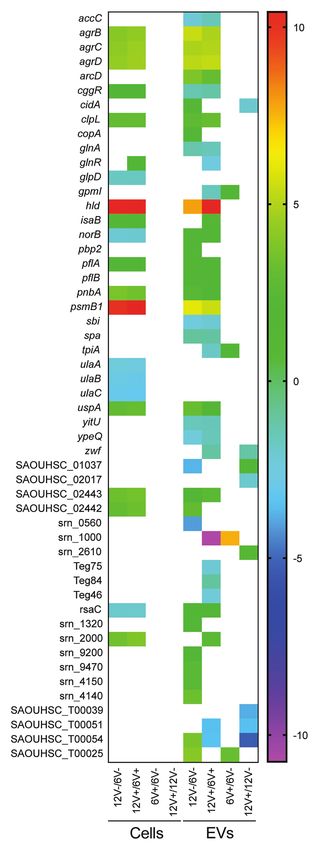
Da Luz et al.                                                                                                                  S. aureus EV RNA Cargo

MATERIALS AND METHODS                                                         with camera level at 15 and threshold at 5, while other parameters
                                                                              were adjusted as necessary.
Bacterial Strain and Growth Conditions
The S. aureus strain used in this work was the model strain                   RNA Extraction From S. aureus HG003
HG003 (Herbert et al., 2010), a NCTC8325 derivative, isolated                 Whole Cells and Its Derived EVs
in 1960 from a sepsis patient. HG003 contains functional rsbU                 RNA extraction was carried out as similar as possible for both
and tcaR genes, two global regulators that are missing in the                 cell and EV samples. Bacterial RNA extraction was performed
NCTC8325 parent strain. The HG003 genome is well documented                   from 10 ml culture pellet. The samples were mixed with glass
(Sassi et al., 2014), and this strain is widely used as a reference           beads in 300 μl lysis buffer (0.5% SDS w/v, 30 mM sodium
to investigate staphylococcal regulation and virulence (Liu et al.,           acetate; 1 mM EDTA) and 400 μl phenol (acid buffered at
2018a). HG003 strain was pre-inoculated in BHI broth and                      pH 5.0) at 65°C. Mechanical lysis was accomplished with
grown overnight at 37°C under 150 rpm/min agitation, and                      2 cycles of 30 s in Precellys at 6,500 rpm. For EV sample
then inoculated 0.1% in 500 ml of fresh BHI (125 rpm/min,                     RNA extraction, particles isolated from the equivalent of 800 ml
at 37°C) on a 1 L Scott flask. Bacterial cultures were retrieved              bacterial culture were mixed with 300 μl of lysis buffer and
after 6 h and 12 h for early- and late-stationary phases, respectively,       400 μl phenol at 65°C, the same volumes used for cell RNA
in the presence or absence of a sub-inhibitory concentration                  extraction. Since EVs lack the thick layer of peptidoglycan
(0.5 μg/ml) of vancomycin (Supplementary Figure S1).                          (PGN) found in the bacterial cell wall, mechanic lysis was
                                                                              not necessary and was achieved with lysis buffer. EV and
S. aureus EVs Isolation and Purification                                      EV-producing cell samples were incubated for 10 min at 65°C,
Cultures were submitted to EVs isolation and purification, as                 being homogenized by vortex every minute. Next, samples
previously described (Tartaglia et al., 2018, 2020). In brief, for            were centrifuged during 10 min 13,000 rpm, 4°C, and the
each condition 1 L of bacterial cell culture was centrifuged                  upper phase was recovered to a new tube. All samples were
at 6,000 × g for 15 min and filtered through 0.22 μm Nalgene                  mixed with additional 400 μl of phenol at 65°C, and the
top filters (Thermo Scientific). Then, the culture supernatant                previous steps were repeated. Then, 400 μl of phenol:chloroform
fraction was concentrated around 100-fold using the Amicon                    1:1 was added, followed by two times addition of 400 μl pure
ultrafiltration systems (Millipore) with a 100kDa filter, and                 chloroform, repeating the step of upper phase recovery, mixture
ultra-centrifuged for 120 min at 150,000 × g to eliminate the                 and centrifugation (5 min at 13,000 rpm, 4°C). Subsequently,
soluble proteins. Next, the suspended pellet was applied to a                 1.5 volumes of ice-cold 100% ethanol and 10% volume of
discontinuous sucrose gradient (8–68%) and ultra-centrifuged                  NaAc were added and the mix was stored at −20°C overnight.
at 100,000 × g for 150 min. Fractions containing EVs were                     Samples were centrifuged at 13,000 rpm for 30 min at 4°C,
recovered and washed in TBS (150 mM NaCl; 50 mM Tris-Cl,                      and the pellets were washed twice with 1 ml of cold 70%
pH 7.5) for final ultra-centrifugation at 150,000 × g (120 min).              ethanol. Finally, the pellets were dried with a SpeedVac
At last, EVs were suspended in cold TBS and kept at −80°C                     concentrator for 2 min and dissolved in RNase-free water.
until use.                                                                    The quality and quantity of the RNAs were verified by Nano
                                                                              Drop, agarose gel, and Bioanalyzer (Agilent). Samples were
                                                                              kept at −80°C until use. No RNase treatment was applied to
EVs Visualization by Electron Microscopy                                      bacterial cell or to EV samples before RNA extraction.
Negative staining electron microscopy was performed as
previously described (Rodovalho et al., 2020) to investigate
the shape and integrity of purified EVs. EVs samples were                     RNA Sequencing
diluted, and solutions containing between 1010 and 1011 particles             The RNA samples were sent to ViroScan3D® (Lyon, France)
per ml were analyzed. For this, samples were applied to glow-                 for DNA removal, ribosomal RNA depletion and RNA
discharged copper EM grids (FF200-Cu) for 30 s, followed by                   sequencing. Total RNA samples were submitted to a DNase
excess solution removal with filter paper. The same process                   treatment with RNase-Free DNase Set (Qiagen) according
was repeated with 2% uranyl acetate, and samples were observed                to manufacturer’s instructions. Then, the samples quantified
with a Jeol 1400 transmission electron microscope (JEOL Ltd.),                using the Quantifluor RNA system (Promega), and qualified
operating at 120 kV.                                                          using RNA Nano Chip on Bioanalyzer 2100 (Agilent) for
                                                                              the EV-producing cell samples, and on the SS RNA system
                                                                              on Fragment Analyzer (AATI) for the EVs samples. RNA
Determination of EVs Sizes and                                                samples were then submitted to the standard protocol Ovation
Concentrations                                                                Universal Prokaryotic RNA-Seq, Nugen, Anydeplete rRNA,
Nanoparticle Tracking Analysis (NTA) using an sCMOS camera                    library preparation. RNA quantity used for library preparation
and a Blue488 laser (Nano Sight NS300) was performed to                       are displayed in Supplementary Table S1. The quality of
assess EVs size and concentration. For that, samples were                     libraries was assessed with the Quantifluor DNA system
diluted into TBS to achieve optimal concentration and submitted               (Promega) and qualified with the HS-NGS system on Fragment
to a constant flux generated by a syringe pump (speed 50),                    Analyzer (Aati). The insert mean size of the libraries was
at 25°C. Results were retrieved from 5 × 60 s videos recorded                 0.34 kp for the EV-producing cell samples, and 0.45 kp for

Frontiers in Microbiology | www.frontiersin.org                           3                                     March 2021 | Volume 12 | Article 634226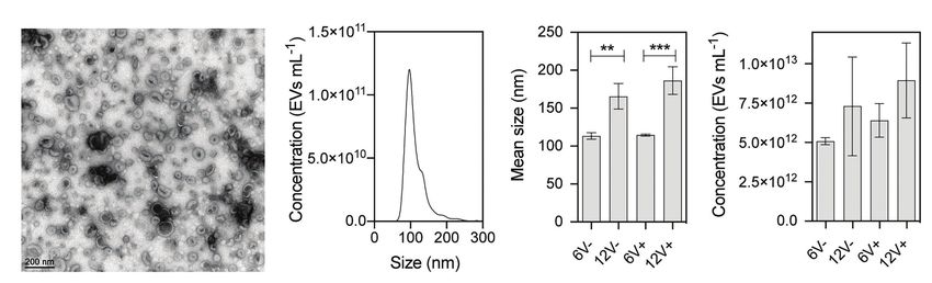
Da Luz et al.                                                                                                            S. aureus EV RNA Cargo

the EV samples (Supplementary Table S1). Sequencing was                 quantitative PCR (qPCR) are listed in Supplementary Table S2
performed with Illumina, NextSeq500, 75 cycles, single-read,            and were designed using eprimer3 software (EMBOSS). qPCR
High Output. For each experimental condition, three biological          was carried out in a 16 μl volume containing 15 ng cDNA,
replicates were sequenced. EV-producing cell samples ranged             specific primers (300 nM), and 8 μl IQ™ SYBR Green Supermix
from 9 to 27 million pair-end reads per sample, and EV                  (Bio-Rad). Reactions were run on a CFX96 real-time system
samples ranged from 30 to 67 million pair-end reads per                 (Bio-Rad, France) using the following cycling parameters: DNA
sample. Reads mapping to the reference genome ranged from               polymerase activation and DNA denaturation 95°C for 5 min,
8 to 26 million, and from 0.38 to 27 million reads for                  40 cycles of denaturation at 94°C for 15 s, and extension at
EV-producing cells and EV samples, respectively. Basic statistics       60°C for 30 s. Melting curve analysis was included to check
of the RNA-Seq data are displayed in Supplementary Table S1.            the amplification of single PCR products. Samples setups included
                                                                        biological triplicates and technical duplicates as well as negative
                                                                        controls corresponding to qPCR reactions performed without
Transcriptome Analysis                                                  cDNA (cDNA negative control) and from RT reactions obtained
The reads were cleaned and trimmed with Trim-Galore
                                                                        from EV RNAs without reverse transcriptase (RT negative
(Martin, 2011) using the default parameters. Reads were
                                                                        control). Results were analyzed with the GFX Manager software
mapped with Bowtie2 (Langmead and Salzberg, 2013) in local
                                                                        and Ct values were determined. Results with Ct equal or above
mode against two staphylococcal genomes used as references:
                                                                        40 were considered negative and only experiments with ΔCt ≥ 4
the NCTC 8325 (NC_007795.1) reference genome with sRNA
                                                                        between negative controls and RT samples were considered.
annotation from SRD (Sassi et al., 2015) and the HG003
genome (GCA_000736455.1) for the non-annotated genes in
NCTC8325 genome. Genes were counted with FeatureCounts
(Liao et al., 2014) with the strand, the multi mapping, and             RESULTS
the overlapping options.
   A list of differentially expressed RNAs was obtained by
                                                                        S. aureus HG003 Produces EVs in Different
EdgeR (Robinson et al., 2009) embedded in SARTools (Varet               Growth Conditions
et al., 2016). The threshold of statistical significance was set        Extracellular vesicles secreted by HG003 were isolated from
to 0.05, with the adjustment method of Benjamini-Hochberg.              the cell-free supernatants of bacterial cultures at early- and
RNA coverage was calculated with Bedtools coverage (Quinlan             late-stationary growth phases (6 and 12 h, respectively), as well
and Hall, 2010). RNAs with ≥ 90% coverage in at least one               as in the absence (V-) and presence (V+) of a sublethal
EV condition were kept for further indepth analysis. RNA                concentration of vancomycin (0.5 μg/ml). For that purpose,
coverage visualization was performed with the Integrative Viewer        we used centrifugation, filtration, and density gradient
Software (IGV; Thorvaldsdóttir et al., 2013) on a log scale.            ultracentrifugation, the standard method for EV isolation and
   Subcellular location prediction was performed with SurfG+            purification at high purity (Yamada et al., 2012; Dauros Singorenko
(Barinov et al., 2009). Clusters of Orthologous Groups (COGs)           et al., 2017). EV homogeneity and integrity were evaluated by
and KEGG categories were obtained using the eggNOG-                     both negative staining electron microscopy and by NTA. Electron
mapper v2 web tool (Huerta-cepas et al., 2017, 2019).                   micrographs of purified EVs revealed typical nano-sized vesicular
Functional enrichment analysis was performed with g:Profiler            structures, with cup-shaped forms in all tested conditions (Raposo
web-server (Raudvere et al., 2019; Reimand et al., 2019).               and Stoorvogel, 2013; Figure 1A and Supplementary Figure S2).
A maximum value of p 0.05 was set as a threshold for                    NTA analyses showed a typical profile of particles for all EV
significative categories.                                               samples (Figure 1B and Supplementary Figure S2). A significant
   A timepoint clustering study was conducted with the R                increase of approximately 55% in EV diameter was observed
package maSigPro (Conesa et al., 2006; Nueda et al., 2014) on           in those purified from late-stationary phase cultures, compared
highly covered EV RNAs with normalized counts by EdgeR.                 to early-stationary phase cultures (for both 6V− vs. 12V− and
In this analysis vancomycin treatment is not taken into                 6V+ vs. 12V+), whereas no significant difference was observed
consideration. The threshold of statistical significance was set        in the absence or presence of vancomycin (6V− vs. 6V+ and
to 0.05, with the adjustment method of Benjamini-Hochberg.              12V− vs. 12V+, Figure 1C). EV yield is essentially similar at
                                                                        the two growth phases, irrespective to the presence/absence of
                                                                        vancomycin (Figure 1D). In summary, S. aureus HG003 releases
RT-qPCR                                                                 EVs with variable diameters depending on the growth phase.
Reverse transcription quantitative PCR (RT-qPCR) was used               A sublethal concentration of vancomycin, however, does not
to validate RNA-seq results. EVs were isolated from the cell-           impact the EV morphology, concentration, or diameter.
free supernatants of three new independent S. aureus cultures
at late-stationary growth phases (12 h) in the absence of
vancomycin. EV RNAs were purified as mentioned above. Around            The S. aureus EVs Harbor all RNA
1.5 μg of RNAs was treated with DNAse I (Amplification Grade,           Functional Classes
Invitrogen) according to manufacturer’s instructions. cDNA              Total RNA was extracted from HG003 EVs to investigate their
synthesis was performed with the high capacity cDNA Reverse             compositions. The quality of the RNA preparations was checked
Transcription kit (Applied Biosystems). The primers used for            and validated, and the samples sequenced. RNA-seq data were

Frontiers in Microbiology | www.frontiersin.org                     4                                     March 2021 | Volume 12 | Article 634226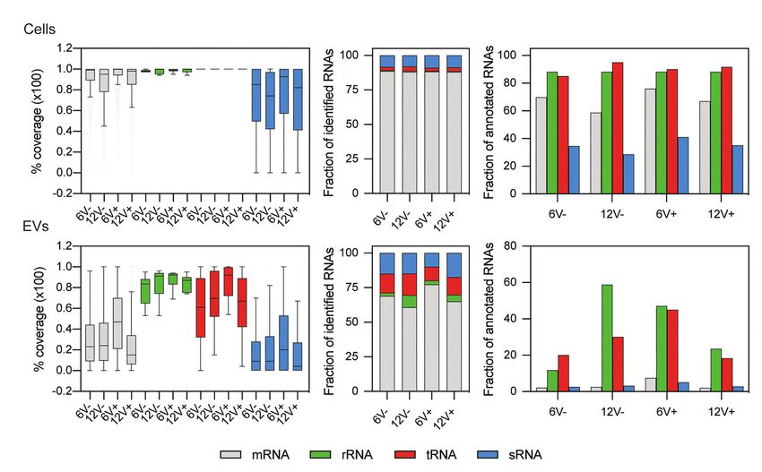
Da Luz et al.                                                                                                                                   S. aureus EV RNA Cargo

                        A                                  B                             C                          D

  FIGURE 1 | Physical characterization of purified Staphylococcus aureus HG003-secreted extracellular vesicles (EVs). (A) Representative electron microscopy
  image of negatively stained HG003 EVs. (B) Representative graph of the EV size distribution. (C) Mean EV sizes. (D) EV yields. Data were obtained from three
  independent EV replicates. Asterisks indicate statistical significance (one-way ANOVA followed by Tukey’s multiple comparisons test: **p < 0.01; ***p < 0.001).
  Early- and late-stationary growth phases (6 and 12, respectively) in the absence (V−) or presence (V+) of vancomycin.

compared between the different growth conditions, and with                             to the EV-producing cells. To analyze potentially functional
those obtained from parental HG003 cells that produced the                             RNAs in EVs, only RNAs with a coverage ≥90% were considered
EVs in each condition (i.e., the EV-producing cells). Around                           for further analysis (Supplementary Table S5). The distribution
2649 ± 238 RNAs annotated in the HG003 genome were                                     of the newly filtered RNAs was depicted in Figures 3B,C.
identified in the EVs according to growth conditions, with an                          Such a harsh quality criterion impacted mainly the RNAs from
average count of over five reads per RNA in each condition,                            the EVs, and particularly mRNAs and sRNAs. Only 3.5 ± 2.7
whereas 3120 ± 35 annotated RNAs were identified within                                and 3.4 ± 1.2% of annotated mRNAs and sRNAs, respectively,
the EV-producing cells (Supplementary Tables S3 and S4).                               were identified within EVs with such a threshold, while
All the four main RNA functional classes (tRNAs, rRNAs,                                67.9 ± 7.2% and 34.9 ± 5.0% of annotated mRNAs and sRNA
mRNAs, and sRNAs) were identified in both the purified EVs                             were identified, respectively, for EV-producing cells (Figure 3C).
and the EV-producing cells (Figure 2A). In both the EVs and                            Compared to the parental cells, the EVs were slightly depleted
the EV-producing cells, ~84% of the mapped RNAs corresponded                           into mRNAs (68.0 ± 7.0% for the EVs vs. 88.2 ± 0.3% in the
to protein-coding genes (mRNAs). The sRNAs were the second                             EV-producing cells) but, interestingly, were enriched for the
most abundant mapped RNA class (~12% of the reads). The                                other RNA functional classes including the sRNAs (14.3 ± 3.2%
remaining 4% of the mapped RNAs are tRNAs and residual                                 for the EVs vs. 8.6 ± 0.4% in the EV-producing cells, Figure 3B).
rRNAs. Note that most of the rRNAs were voluntarily removed
during the RNA purification. Most of the annotated mRNAs,                              Functional Characterization of the RNAs
tRNAs, and residual rRNAs were identified in the EV-producing
parental cells (from 86 to 96%), while this value dropped to
                                                                                       From the EVs
                                                                                       According to experimental conditions, from 86 to 273 RNAs
79.8 ± 2.7% for sRNAs (Figure 2B). These percentages were
                                                                                       with a ≥90% coverage and an average count of over 5 reads
slightly lower for EV samples (from 72 to 89%), although
                                                                                       per RNA were identified within EVs from S. aureus HG003
they remained high for the sRNAs (59.1 ± 10.2%). All these
                                                                                       (Figure 4A and Supplementary Table S5). They corresponded
RNAs detected in the purified EVs prompted us to check
                                                                                       to 286 unique RNAs and were either mRNAs (220), tRNAs
their coverages, to evaluate their integrity.
                                                                                       (28), residual rRNAs (10), and sRNAs (28). The presence of
                                                                                       some of these transcrits associated with HG003 EVs and
The S. aureus Purified EVs Contain Both                                                corresponding either to mRNAs or sRNAs was confirmed by
Fragmented and Intact RNAs From Various                                                RT-qPCR on RNAs extracted from three independent biological
Functional Classes                                                                     replicates (Figure 5). Among the mapped mRNAs, most were
For the EV-producing cells, the median values of mRNAs,                                implicated in translation, ribosomal structure and biogenesis
tRNAs, and residual rRNAs coverages were between 95 and                                (17.5%, COG J), energy production and conversion (13.6%,
100%, and were 74 and 92% for the sRNAs (Figure 3A and                                 COG C), carbohydrate transport and metabolism (COG G,
Supplementary Table S4), implying that those RNAs were                                 7.9%), transcription (5.2%, COG K), and cell wall/membrane/
mainly intact, and not degraded. The coverage profile was                              envelope biogenesis (5.2%, COG M; Figure 4B). Several COG
drastically different for the RNAs recovered from the purified                         and KEGG categories were notably enriched (p < 0.05) in the
EVs. While the coverage of the residual rRNAs varied from                              EVs compared to the EV-producing bacteria (Figure 4C).
83 to 92%, the median coverage values for the other RNA                                mRNAs expressing proteins with a cytoplasmic location prediction
functional classes ranged from 61 to 92% for the tRNAs, 15                             were more represented in the EVs (79.5%) than into the
to 47% for the mRNAs, and 4 to 20% for the sRNAs (Figure 3A).                          producing cells (72.9%; Figure 4D). Interestingly, EVs contained
These lower coverages suggested that a substantial fraction of                         several mRNAs coding for virulence-associated proteins such
mRNAs and sRNAs were fragmented in the EVs compared                                    as the immune evasion protein A and Sbi, the Atl autolysin,

Frontiers in Microbiology | www.frontiersin.org                                    5                                             March 2021 | Volume 12 | Article 634226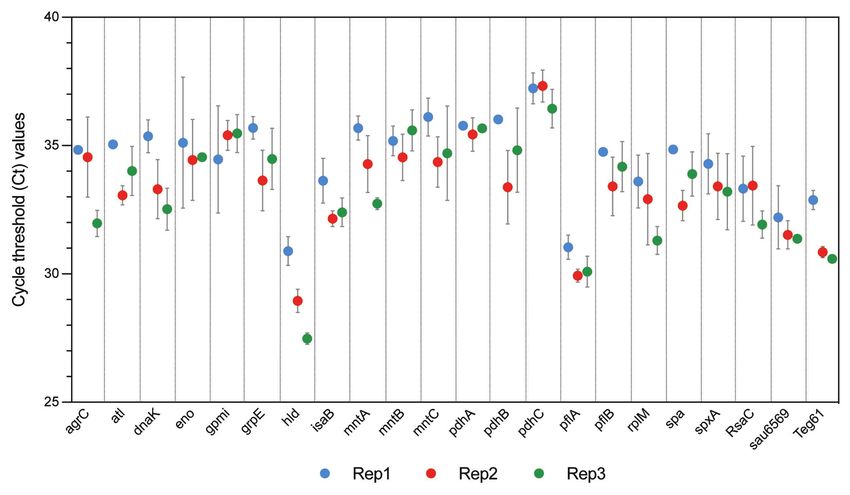
Da Luz et al.                                                                                                                            S. aureus EV RNA Cargo

                                        A                               B

  FIGURE 2 | Relative RNA composition of S. aureus HG003 and its secreted EVs. Individual colored bars represent the relative amount of each RNA class for
  mapped reads (A) and annotated RNAs (B). RNA-Seq data is the average of three independent replicates. Number of reads have been normalized with EdgeR.
  RNA classes are defined from the S. aureus genome annotation NCTC8325/HG003. Early- and late-stationary growth phases (6 and 12, respectively) in absence
  (V−) or presence (V+) of vancomycin.

                                  A                                    B                       C

  FIGURE 3 | Relative composition of highly covered RNAs from S. aureus HG003 and its secreted EVs. Colors represent the relative amount of each RNA class.
  (A) Percentage of RNA median coverage. Distribution of newly filtered RNAs with ≥90% coverage were plotted for (B) mapped reads (C) and annotated RNA.
  Number of reads have been normalized with EdgeR. RNA classes are defined from the S. aureus genome annotation NCTC 8325/HG003. Early- and late-stationary
  growth phases (6 and 12, respectively) in absence (V−) or presence (V+) of vancomycin.

Frontiers in Microbiology | www.frontiersin.org                                6                                          March 2021 | Volume 12 | Article 634226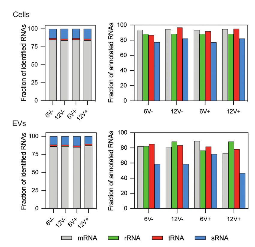
Da Luz et al.                                                                                                                                        S. aureus EV RNA Cargo

                                    A                                 B                                        D

                             C

  FIGURE 4 | Staphylococcus aureus HG003 EV RNA cargo and its modulation by different growth conditions. (A) Venn diagrams of RNA composition in EV-producing cells
  (upper panel) and EVs (lower panel) from different growth conditions. Early- and late-stationary growth phases (6 and 12, respectively) in absence (V-) or presence (V+) of
  vancomycin. (B) Prediction of Clusters of Orthologous Groups (COG) categories for mRNAs: NA, not predicted; S, function unknown; Q, secondary metabolites
  biosynthesis, transport, and catabolism; P, inorganic ion transport and metabolism; I, lipid transport and metabolism; H, coenzyme transport and metabolism; F, nucleotide
  transport and metabolism; E, amino acid transport and metabolism; G, carbohydrate transport and metabolism; C, energy production and conversion; O, post-translational
  modification, protein turnover, and chaperones; U, intracellular trafficking, secretion, and vesicular transport; M, cell wall/membrane/envelope biogenesis; T, signal
  transduction mechanisms; V, defense mechanisms; D, cell cycle control, cell division, chromosome partitioning; L, replication, recombination and repair; K, transcription;
  J, translation, ribosomal structure and biogenesis. (C) COG and KEGG categories enriched (p < 0.05) in EVs compared to EV-producing cells. (D) Subcellular localization of
  proteins encoded by mRNAs as predicted by SurfG+: Cyt, cytoplasmatic; Mbe, membrane; PSE, surface-exposed; Sec, secreted; NA, Not predicted.

the Hld δ-hemolysin encoded by the multifunctional sRNA                                   coverage ≥90% across the entire operon in both the EVs and
RNAIII, the PSMβ1 Phenol Soluble Modulin, the FntA iron-                                  the EV-producing bacteria. Figure 6 illustrates the sequencing
storage ferritin, and the MntABC iron ABC transporter. Among                              coverage of various contiguous genes within the EVs and the
the 20 tRNAs annotated in the genome, 15 were identified                                  EV-producing cells. Long mRNA operons, up to ~14,000
into the EVs (tRNAHis, tRNAAsn, tRNAGlu, tRNAArg, and tRNAAsp                             nucleotides, were detected as fully covered by reads into the
were absent). Five copies of the 16S and 23S rRNAs were also                              purified EVs, supporting the presence of highly covered RNAs
detected, implying that our rRNA depletion procedure was                                  and operons as full-length transcripts.
incomplete. Finally, 28 annotated potential sRNAs were detected
within EVs, and among the 50 or so bona fide sRNAs defined                                EV RNA Composition Varies With Growth
for the HG003 strain (Liu et al., 2018a), only RsaC was                                   Conditions
identified with a ≥90% coverage in this study. Note that                                  The RNA composition of the purified EVs was compared between
despite encoding the highly covered Hld transcript, the sRNA                              early- and late-stationary phases, and with or without vancomycin.
RNAIII presented only 71% gene coverage and therefore was                                 Eighteen percentage (n = 51) of all detected RNAs with ≥90%
excluded from analysis. Around 196 out of the 286                                         coverage were common to all the EV samples (Figure 4A and
EV-associated RNAs colocalized at the same loci onto the                                  Supplementary Table S5), implying that the RNA content of
HG003 chromosome, to form 42 clusters of 2 to 29 contiguous                               the EVs highly varied according to the growth conditions. The
genes that were experimentally shown to belong to the same                                percentage of RNAs shared by all the EV-producing cell samples,
transcriptional units (Mäder et al., 2016; Supplementary Table S6).                       however, was much higher (70%, n = 1761). The shared RNAs
Among these transcriptional units, 17 displayed a RNA-Seq                                 among the EVs included mRNAs expressing virulence factors

Frontiers in Microbiology | www.frontiersin.org                                       7                                               March 2021 | Volume 12 | Article 634226
Da Luz et al. S. aureus EV RNA Cargo
FIGURE 5 | Reverse transcription quantitative PCR (RT-qPCR) validation of S. aureus HG003 EV RNAs. RT-qPCR experiments were performed from RNAs
extracted from EV samples isolated from the cell-free supernatants of three independent S. aureus cultures at late-stationary growth phases (12 h) in the
absence of vancomycin (Rep1, Rep2, and Rep3). Quantitative PCR (qPCR) successfully amplified the coding-sequence of 19 mRNAs, and 3 sRNAs. Samples
setups included biological triplicates (Rep1, Rep2, and Rep3) and technical duplicates as well as negative controls corresponding to qPCR reactions performed
without cDNA, and from RT reactions performed without reverse transcriptase enzyme. Ct values are expressed as mean ± SD from two independent technical
replicates performed in triplicates.
(Atl and Spa), metabolic enzymes (pyruvate dehydrogenase and conditions were also detected (Supplementary Table S7). Among
cytochrome c oxidase complexes, glycolytic enzymes) and the 286 EV-associated RNAs, 110 were differentially abundant
transcriptional regulators (SpxA, CggR, and GlnR), as well as RNAs between two conditions. Variations were detected at all times
involved in translation (ribosomal proteins, rRNAs, and tRNAs; and in the absence or presence of vancomycin, although
Supplementary Table S5). The 51 common RNAs also included the growth phase appeared to have a greater impact on
9 potential sRNAs, notably RsaC involved in S. aureus oxidative RNA abundance (75 and 64 differentially abundant RNAs
stress adaptation and nutritional immunity (Lalaouna et al., 2019). were detected between early- and late-stationary phase with
For both the EVs and EV-producing cells, more RNAs or without vancomycin, respectively), than the antibiotic
were detected at 6 h (275 and 2443 for EVs and EV-producing stress (8 and 9 differentially abundant RNAs were detected
cells, respectively) than at 12 h (126 and 2161 for EVs and between presence and absence of vancomycin in early- and
EV-producing cells, respectively, Figure 4A). Likewise, more late-stationary phase, respectively; Supplementary Table S7).
RNAs were detected in the EVs in the presence (277 and A selection of RNAs with a modulation of their abundance
2,474 for EVs and EV-producing cells, respectively) than in according to the growth conditions is displayed in Figure 7.
the absence of vancomycin (140 and 2,279 for EVs and The most modulated RNAs into EVs produced from the
EV-producing cells, respectively; Figure 4A), indicating that two growth conditions were mRNAs coding for virulence-
the antibiotic modifies the RNA cargo of the EVs. These associated factors, such as agrB, agrC, agrD, psmB1, and
results also highlighted that the growth phase and the antibiotic hld with a 30- to 1300-fold change, two potential annotated
stress impacted mostly the RNA content of the EVs, but sRNAs, srn_0560, and srn_1000 with a 16- and 190-fold
much less that of the parental cells. Indeed, when we considered change, respectively, tRNAGly (SAOUHSC_T00025) and tRNAThr
the RNAs detected in only one condition (i.e., specific RNAs), (SAOUHSC_T00054), with fold changes greater than 16.
their fractions were higher in the EVs than in EV-producing Among the differentially abundant RNAs according to the
cells, and that for all the tested conditions. For example, growth phase, 32 were detected both in presence and absence
58% of RNAs found within EVs at 6 h were specific to this of vancomycin, with similar fold changes highlighting their
condition, while specific RNAs represented only 14% of all reproducible variations into EVs across different environmental
RNAs detected at 6 h in the EV-producing cells. conditions (Figure 7 and Supplementary Table S7).
Differentially expressed RNAs were also detected for the
EV RNA Abundance Varies With Growth EV-producing cells when their expression was compared
Conditions between early- and late-stationary phase both in absence
In addition to the qualitative variations observed, significant differences (n = 136) and in presence of vancomycin (n = 147), which
(Padj < 0.05) in EV RNA abundance between the experimental was expected since bacterial transcription differs qualitatively
Frontiers in Microbiology | www.frontiersin.org 8 March 2021 | Volume 12 | Article 634226Da Luz et al. S. aureus EV RNA Cargo FIGURE 6 | HG003 EVs contain long mRNA transcripts. Comparison between operon coverages between EV-producing cell and EV samples in different conditions. Early- and late-stationary growth phases (6 and 12, respectively) in absence (V-) or presence (V+) of vancomycin. RNA coverage is visualized with Integrative Viewer Software (IGV) in log scale. and quantitatively when facing different growth conditions RNAs counted for only 2% of the RNAs in the EV-producing (Supplementary Table S8). Note that no significantly cells. The abundance pattern of several RNAs differed between differentially expressed RNAs were detected according to the EVs and the EV-producing cells according to the growth the presence of vancomycin. As observed previously for the conditions (Figure 7). While some RNAs such as agrBCD, EV RNA content, the growth conditions, particularly the psmβ1, and hld mRNAs displayed the same variations of growth phase, impacted mostly the RNA abundance of the their abundance pattern in EVs and EV-producing cells EVs, but much less than of the parental cells. Indeed, 38% regardless the growth conditions. Others, such as spa and of RNAs detected within EVs displayed changes in their RsaC, were differentially abundant between the EVs and abundance between conditions, while the fraction of modulated the EV-producing cells. Frontiers in Microbiology | www.frontiersin.org 9 March 2021 | Volume 12 | Article 634226
Da Luz et al.                                                                                                                            S. aureus EV RNA Cargo

                                                                                   FIGURE 7 | EV-producing cells and EVs with a log2 fold change.
                                                                                   Comparisons comprised data with at least one of the two samples containing
                                                                                   ≥90% coverage. The log2 fold change is displayed as colored squares from
                                                                                   −2 (purple) to 10 (red). Early- and late-stationary growth phases (6 and 12,
                                                                                   respectively) in absence (V−) or presence (V+) of vancomycin.

                                                                                  Timepoint Clustering Analysis Reveals
                                                                                  Different RNA Abundance Profiles
                                                                                  Between the EVs and EV Producing Cells
                                                                                  To evaluate the influence of the growth phase on EV and EV
                                                                                  producing cell RNA composition, a negative binomial-based
                                                                                  approach, with the R package maSigPro (Conesa et al., 2006)
                                                                                  was applied. Briefly, maSigPro provides a differentially expressed
                                                                                  transcript analysis of serial data between experimental groups
                                                                                  (e.g., EV and EV producing cells). maSigPro was applied to
                                                                                  the 286 highly covered EV RNAs and identified 91 RNAs
                                                                                  with significant temporal profile changes (Padj < 0.05). RNAs
                                                                                  were clustered according to their expression profiles
                                                                                  (Supplementary Table S9). Figure 8 shows the six RNA clusters
                                                                                  obtained. Three clusters grouped transcripts with a similar
                                                                                  expression profile between EV and EV-producing cells: cluster
                                                                                  4 with 8 RNAs (including, e.g., agrBD and arcC2) and cluster
                                                                                  3 with 18 RNAs (including, e.g., hld and psmβ1) contained
                                                                                  more abundant transcripts over time in both EVs and
                                                                                  EV-producing cells, whereas cluster 6 with 14 RNAs (including,
                                                                                  e.g., fusA, tuf, and secY1) contained less abundant transcripts
                                                                                  at 12 h than at 6 h in both. Interestingly, the three other
                                                                                  clusters grouped RNAs that showed opposite expression profiles
                                                                                  over time in EVs and EV-producing cells. Cluster 1 with 10
                                                                                  RNAs (including, e.g., RsaC and pdhA), and cluster 2 with
                                                                                  24 RNAs (including, e.g., ldh1, qoxABC, and rpoBC) grouped,
                                                                                  similarly, more abundant transcripts at 12 h in EVs, and less
                                                                                  abundant transcripts at 12 h in EV-producing cells. On the
                                                                                  contrary, cluster 5 with 17 RNAs (including, e.g., ccgR and
                                                                                  sbi) grouped less abundant transcripts in EVs and more abundant
                                                                                  transcripts in EV-producing cells at 12 h. Altogether, this
                                                                                  analysis highlighted that the transcript expression pattern could
                                                                                  temporally differ between EVs and EV-producing cells.

                                                                                  DISCUSSION
                                                                                  Extracellular vesicles are universal carriers of macromolecules
                                                                                  including extracellular RNAs all along from bacteria, archaea,
                                                                                  and fungi to protists. Recent investigations on bacterial EV
                                                                                  biogenesis, release, and trafficking showed their functional
                                                                                  importance for bacterial communication and survival
                                                                                  (Tsatsaronis et al., 2018). Information regarding S. aureus
                                                                                  EV RNA cargo, however, is lagging behind. Here, we report
                                                                                  the first exploratory work on EVs released by S. aureus
                                                                                  HG003, the characterization of its EV RNA cargo under
                                                                                  different conditions, and an indepth transcriptomic comparison
                                                                                  between the EVs and the EV-producing cells.
                                                                                     Environmental conditions, such as growth phase and
  FIGURE 7 | Differential RNA abundance between the EV-producing cells and
  the EVs. Colored square shows different RNA abundance patterns between
                                                                                  environmental stresses, reportedly influence the production,
                                                                                  content and functions of EVs (Tashiro et al., 2010; Kim et al., 2016b;
                                                               (Continued)
                                                                                  Orench-Rivera and Kuehn, 2016; Askarian et al., 2018;

Frontiers in Microbiology | www.frontiersin.org                              10                                           March 2021 | Volume 12 | Article 634226
Da Luz et al. S. aureus EV RNA Cargo
A D
B E
C F
FIGURE 8 | Timepoint clustering of expression profiles between EVs and EV-producing cells. The analyses were performed with the R package maSigPro from
normalized counts by EdgeR. Statistical significance (p < 0.05) was adjusted with Benjamini-Hochberg method. Median expression values are calculated at 6 and
12 h for EVs (blue) and EV-producing cells (red) for the six clusters (from A to F).
Yun et al., 2018; Andreoni et al., 2019). Here, to investigate used here (0.5 μg/ml) is probably too low to detect any changes
the impact of environmental changes on S. aureus EV production, in EV morphology and production.
the selected conditions were the early- and late-stationary All RNA classes are detected by RNA-Seq within HG003-
growth phases, with or without a sublethal concentration of derived EVs. These include rRNAs, which were still detected,
vancomycin that does not impact growth. Differences were suggesting that the rRNA depletion carried out here was
observed regarding EV sizes. EVs derived from the late-stationary incomplete. In the absence of filtering by coverage of sequencing
growth phase were larger than those collected during the early- data, on average, 78.0 ± 7.0% of the annotated transcripts in
stationary phase. This can be due to cell-wall morphology and HG003 genome were present in the EVs (91.8 ± 1.0% for
peptidoglycan structure that are characteristics of the growth EV-producing cells). Of these, a large portion of mapped RNAs
stage in S. aureus (Zhou and Cegelski, 2012). A correlation corresponded to mRNAs. These results are consistent with
between the degree of peptidoglycan cross-linking to the cell RNA-Seq data obtained with similar criteria from OMVs in
wall stiffness and EVs release was observed for both Gram- Salmonella enterica serovar Typhimurium (S. Typhimurium)
negative and Gram-positive bacteria (Zhou et al., 1998; that harbor around 73% of the annotated transcripts including
Deatherage et al., 2009; Schrempf and Merling, 2015; up to 86% of mRNAs according to growth conditions (Malabirade
Schwechheimer et al., 2015; Wang et al., 2018). Notably, et al., 2018). A recent study addressing the sRNA content of
sub-inhibitory concentrations of penicillin decreases peptidoglycan EVs also pointed out that mRNAs are the more abundant
cross-linking, triggering an increase in S. aureus EV yields RNA species in EVs derived from S. aureus strain MSSA476
and sizes (Wang et al., 2018). Here, we show that sublethal after rRNA depletion (Joshi et al., 2021). On the contrary,
concentrations of vancomycin, an antibiotic that also targets studies with Escherichia coli revealed that EVs were enriched
peptidoglycan synthesis in S. aureus, does not impact either mainly with short RNAs, such as tRNAs (Ghosal et al., 2015;
EV morphology or EV production yields, but does change Blenkiron et al., 2016). Nevertheless, these variations may be a
their RNA content in terms of composition and abundance. result of different RNA extraction and library preparation
Vancomycin affects S. aureus EV activity starting at 1 μg/ml protocols, or simply correspond to singular characteristics of
(He et al., 2017). The sub-inhibitory vancomycin concentration EVs derived from different bacterial species. As expected, data
Frontiers in Microbiology | www.frontiersin.org 11 March 2021 | Volume 12 | Article 634226Da Luz et al. S. aureus EV RNA Cargo filtering of RNAs with ≥90% coverage decreased the number RsaC and RNAIII were also detected within S. aureus EVs of detected RNAs. However, the RNA content of the EVs, from strain MSSA476 (Joshi et al., 2021), suggesting their wider particularly the mRNAs and sRNAs (only 5.1 ± 2.8% of EV occurence in staphylococcal EVs. RNAs initially detected are still identified after the filtering), The presence of full-length, functional RNAs in EVs raises was much more affected than that of the EV-producing cells the question of their biological roles. EVs are produced to (69.0 ± 7.90% of EV-producing cell RNAs are still detected transport bioactive molecules to interact and communicate with after the filtering). The low RNA coverage in the HG003 EVs other cells. So far, most studies on S. aureus EVs investigated might perhaps reflect the absence of transcription within EVs the protein cargo. Therefore, the broad spectrum of activities and, thus, the progressive degradation of a substantial fraction associated with S. aureus-derived EVs was related to their of the EVs-associated RNAs after their formation and/or during protein content (Jeon et al., 2016). In some S. aureus strains, their purification. These findings are consistent with a recent EVs carry β-lactams that confer transient resistance to ampicillin- report showing that the predominant RNA type in EVs from susceptible E. coli and S. aureus (Lee et al., 2013b). Likewise, S. aureus Newman is 2,000 nucleotides (e.g., that do not encode the corresponding genes in their genome pbp2, copA, rpoB, rpoC, and atl). 67% of these RNAs are could also be part of a transient horizontal phenotype acquisition, organized into 42 gene clusters that are co-transcribed in which could be of use during infection to disseminate specific S. aureus (Mäder et al., 2016). Among them, 17 are full-length virulence-associated factors through the bacterial community. transcripts across entire operons in HG003 EVs, with lengths Finally, beside interactions between bacterial cells, S. aureus up to ~14,000 nucleotides. As observed for S. Typhimurium EV associated RNAs, notably sRNAs, may be involved in the OMV RNAs (Malabirade et al., 2018), these findings support host-pathogen interactions (Eberle et al., 2009; Li and Chen, that highly covered RNAs are present as full-length transcripts. 2012; Furuse et al., 2014; Sha et al., 2014; Koeppen et al., These RNAs belong to all annotated classes of RNAs. The 2016; Westermann et al., 2016; Choi et al., 2017; Frantz et al., mRNAs from the EVs encode proteins involved in transcription, 2019; Han et al., 2019; Lee, 2019; Rodriguez and Kuehn, 2020). translation, energy production and conversion, carbohydrate The 28 potential annotated sRNA detected within HG003 EVs metabolism, and cell wall biogenesis. In addition to these are potential candidates for further functional characterization, housekeeping functions, EVs also harbors mRNAs encoding especially during S. aureus-host cell interactions. S. aureus virulence-associated proteins, such as the agr operon responsible secreted EVs elicit immune responses that mimic those of the of quorum-sensing, autolysin Atl, protein A (Spa), EV-producing cells (Gurung et al., 2011; Hong et al., 2011, immunoglobulin-binding protein (Sbi), immunodominant 2014; Kim et al., 2012, 2019; Thay et al., 2013; Choi et al., staphylococcal antigen B (IsaB), δ hemolysin (Hld) encoded 2015; Jeon et al., 2016; Jun et al., 2017; Askarian et al., 2018; by the multifunctional sRNA RNAIII, and the PSMβ1 phenol- Tartaglia et al., 2018; Wang et al., 2018, 2020; Rodriguez and soluble modulin, as well as several iron acquisition systems. Kuehn, 2020). Strikingly, within their RNA cargo, several mRNAs Besides, most tRNA species are detected within the EVs, as encode immunomodulatory proteins, as Sbi, Spa and PSMβ1, well as residual rRNAs and 28 annotated sRNAs, including and may participate into the immune response triggered by bona-fide RsaC involved in S. aureus oxidative stress adaptation the protein cargo if they are ultimately translated. Yet, such and nutritional immunity (Lalaouna et al., 2019). Note that functions remain to be demonstrated for the RNA cargo of Frontiers in Microbiology | www.frontiersin.org 12 March 2021 | Volume 12 | Article 634226
Da Luz et al.                                                                                                                    S. aureus EV RNA Cargo

S. aureus EVs. mRNAs expressing PSMβ and hemolysin δ                           possess similar functions than in the bacterial cytoplasm? What
toxins from the EVs, if translated into recipient bacteria or                  are the rules for RNA sorting into HG003 EVs? What are the
host cells, could perhaps facilitate staphylococcal intracellular              roles and functions of the S. aureus EV RNA cargo? These exciting
survival, but this hypothesis should be experimentally challenged.             questions, among others, should be adressed in further studies.
    The RNA cargo of HG003 EVs, in both identity and abundance,
depends on the growth conditions. Similarly, the EV RNA cargo
of S. Typhimurium is also sensitive to environmental changes                   DATA AVAILABILITY STATEMENT
indicating that it reflects the bacterial adaptation to its environment
(Malabirade et al., 2018). It could be a faster way to transfer                The datasets presented in this study can be found in online
information of changes perceived by one cell to surrounding                    repositories. The names of the repository/repositories and
cells even before they sensed the environmental stimuli in order               accession number(s) can be found at: https://www.ebi.ac.uk/
to quickly promote group adaptation. We found, however, that                   ena, https://www.ebi.ac.uk/ena/browser/view/PRJEB40502.
the vancomycin treatment had less impact on RNA abundance
compared to the growth phase. Although the composition of
HG003 EVs represented the intracellular state of the bacterial                 AUTHOR CONTRIBUTIONS
transcriptome through global packaging, two main findings,
                                                                               BSRL, BF, SC, YL, VA, and EG conceived and designed the experiments.
however, reinforce the concept of a potential selective packaging
                                                                               VA and EG supervised the study. BSRL and SC performed the
of RNAs into EVs, as proposed for its protein cargo (Haurat
                                                                               experiments. AN performed computational analysis. BSRL, BF, SC,
et al., 2011; Cahill et al., 2015; Tartaglia et al., 2020). First,
                                                                               VRR, AN, and EG analyzed the data. VA, YL, and EG contributed
we measured an enrichment for several functional and subcellular
                                                                               to funding acquisition. BSRL and EG wrote the original draft. All
localization RNA categories in EVs when compared to
                                                                               authors contributed to data interpretation, drafting the manuscript,
EV-producing cells. Second, the relative abundance of several
                                                                               critically revising the manuscript, and approving its final version.
RNAs between two environmental conditions was different in
the EVs and the EV-producing cells. Other studies also found
that some RNA populations were enriched in EVs from Gram-
negative and Gram-positive pathogenic bacteria (Ghosal et al.,
                                                                               FUNDING
2015; Koeppen et al., 2016; Resch et al., 2016; Malabirade et al.,             This work has received financial support from INRAE (Rennes,
2018; Malge et al., 2018; Han et al., 2019; Langlete et al., 2019;             France) and Institut Agro (Rennes, France). This work was
Zhang et al., 2020). This notably includes sRNAs, which can                    part of the CARAVEL project financed by the MICA division
play regulatory activity in the host (Koeppen et al., 2016;                    from INRAE. BSRL and VRR were supported by the International
Malabirade et al., 2018; Langlete et al., 2019; Zhang et al., 2020).           Cooperation Program CAPES/COFECUB at the Federal
The enrichment of RNAs associated with bacterial diseases in                   University of Minas Gerais funded by CAPES – the Brazilian
EVs derived from many pathogenic bacteria reinforces the                       Federal Agency for the Support and Evaluation of Graduate
physiopathological role of these structures in host-pathogen                   Education of the Brazilian Ministry of Education (number
interaction and host cell invasion, which could be borne by                    88887.179897/2018-00 and 99999.000058/2017-03, respectively).
their RNA cargo as well as by their protein cargo. The selective
mechanisms of EV RNA content packaging have not yet been
elucidated. It has been proposed that RNA packing into EVs                     ACKNOWLEDGMENTS
could depend on RNA size and location (eg., nearby EVs
formation site), as well as on their affinity for other molecules              This work was conducted in the frame of BactInflam International
(eg., membrane proteins; Langlete et al., 2019). Nevertheless,                 Associated Laboratory between INRAE (France) and UFMG
such enrichment results should be interpreted with carefulness.                (Brazil). It benefited from the facilities and expertise of the
Indeed, they could also reflect a difference in RNA half-lives                 MRic-TEM platform (https://microscopie.univ-rennes1.fr). We are
between EVs and EV-producing cells, as well as a difference                    grateful to Agnes Burel (Univ Rennes, BIOSIT – UMS 3480,
in RNase activity (Langlete et al., 2019; Lécrivain and Beckmann,              US_S 018, Rennes, France) for sessions with the microscope.
2020), pointing out that RNAs with longer half-lives could                     We are also grateful to the genotoul bioinformatics platform
be protected from degradation, leading to an artifactual                       Toulouse Occitanie (Bioinfo Genotoul) for providing computing
accumulation in the EVs over time.                                             and storage resources. Sandrine Parayre (INRAE, STLO) and
    In summary, our exploratory work provides novel insights in                Sandrine Péron (INRAE, STLO) are warmly acknowledged for
S. aureus EVs by the characterization of its RNA cargo and paves               the technical assistance during RT-qPCR experiments.
the way for further functional studies. Mainly, it sheds light on
the possible roles of EV RNA cargo in intra- and inter-species
communication, in the virulence and pathogenesis of S. aureus,                 SUPPLEMENTARY MATERIAL
and as trash bags for degraded RNAs. The study of bacterial EV
RNA cargo is an emerging area of research. Evidently, as with                  The Supplementary Material for this article can be found online
all emerging fields, each advance raises further questions: Are                at: https://www.frontiersin.org/articles/10.3389/fmicb.2021.634226/
the full-length RNAs in HG003 EVs functional, and do they                      full#supplementary-material

Frontiers in Microbiology | www.frontiersin.org                           13                                      March 2021 | Volume 12 | Article 634226
Da Luz et al. S. aureus EV RNA Cargo
REFERENCES RNAs that are potent inducers of Beta interferon. Am. Soc. Microbiol. 10,
1–15. doi: 10.1128/mBio.01223-19
Ahmadi Badi, S., Bruno, S. P., Moshiri, A., Tarashi, S., Siadat, S. D., and Furuse, Y., Finethy, R., Saka, H. A., Xet-Mull, A. M., Sisk, D. M., Jurcic
Masotti, A. (2020). Small RNAs in outer membrane vesicles and their Smith, K. L., et al. (2014). Search for MicroRNAs expressed by intracellular
function in host-microbe interactions. Front. Microbiol. 11:1209. doi: 10.3389/ bacterial pathogens in infected mammalian cells. PLoS One 9:e106434. doi:
fmicb.2020.01209 10.1371/journal.pone.0106434
Andreoni, F., Toyofuku, M., Menzi, C., Kalawong, R., Shambat, S. M., François, P., Ghosal, A., Upadhyaya, B. B., Fritz, J. V., Heintz-Buschart, A., Desai, M. S.,
et al. (2019). Antibiotics stimulate formation of vesicles in Staphylococcus Yusuf, D., et al. (2015). The extracellular RNA complement of Escherichia
aureus in both phage-dependent and -independent fashions and via different coli. Microbiologyopen 4, 252–266. doi: 10.1002/mbo3.235
routes. Antimicrob. Agents Chemother. 63, e01439–e01518. doi: 10.1128/ Gill, S., Catchpole, R., and Forterre, P. (2019). Extracellular membrane vesicles
AAC.01439-18 in the three domains of life and beyond. FEMS Microbiol. Rev. 43, 273–303.
Askarian, F., Lapek, J. D., Dongre, M., Tsai, C. M., Kumaraswamy, M., Kousha, A., doi: 10.1093/femsre/fuy042
et al. (2018). Staphylococcus aureus membrane-derived vesicles promote Gill, S. R., McIntyre, L. M., Nelson, C. L., Remortel, B., Rude, T., Reller, L. B.,
bacterial virulence and confer protective immunity in murine infection et al. (2011). Potential associations between severity of infection and the
models. Front. Microbiol. 9:262. doi: 10.3389/fmicb.2018.00262 presence of virulence-associated genes in clinical strains of Staphylococcus
Barinov, A., Loux, V., Hammani, A., Nicolas, P., Langella, P., Ehrlichh, D., aureus. PLoS One 6:e18673. doi: 10.1371/journal.pone.0018673
et al. (2009). Prediction of surface exposed proteins in Streptococcus pyogenes, Groot, M., and Lee, H. (2020). Sorting mechanisms for MicroRNAs into
with a potential application to other gram-positive bacteria. Proteomics 9, extracellular vesicles and their associated diseases. Cell 9, 1–16. doi: 10.3390/
61–73. doi: 10.1002/pmic.200800195. cells9041044.
Biller, S. J., Schubotz, F., Roggensack, S. E., Thompson, A. W., Summons, R. E., Gurung, M., Moon, D. C., Choi, C. W., Lee, J. H., Bae, Y. C., Kim, J., et al.
and Chisholm, S. W. (2014). Bacterial vesicles in marine ecosystems. Science (2011). Staphylococcus aureus produces membrane-derived vesicles that induce
343, 183–186. doi: 10.1126/science.1243457 host cell death. PLoS One 6:e27958. doi: 10.1371/journal.pone.0027958
Bitto, N. J., Chapman, R., Pidot, S., Costin, A., Lo, C., Choi, J., et al. (2017). Han, E. C., Choi, S. Y., Lee, Y., Park, J. W., Hong, S. H., and Lee, H. J. (2019).
Bacterial membrane vesicles transport their DNA cargo into host cells. Sci. Extracellular RNAs in periodontopathogenic outer membrane vesicles promote
Rep. 7:7072. doi: 10.1038/s41598-017-07288-4 TNF-α production in human macrophages and cross the blood-brain barrier
Blenkiron, C., Simonov, D., Muthukaruppan, A., Tsai, P., Dauros, P., Green, S., in mice. FASEB J. 33, 13412–13422. doi: 10.1096/fj.201901575R
et al. (2016). Uropathogenic Escherichia coli releases extracellular vesicles Haurat, M. F., Aduse-Opoku, J., Rangarajan, M., Dorobantu, L., Gray, M. R.,
that are associated with RNA. PLoS One 11:e0160440. doi: 10.1371/journal. Curtis, M. A., et al. (2011). Selective sorting of cargo proteins into bacterial
pone.0160440 membrane vesicles. J. Biol. Chem. 286, 1269–1276. doi: 10.1074/jbc.
Brown, L., Kessler, A., Cabezas-Sanchez, P., Luque-Garcia, J. L., and Casadevall, A. M110.185744
(2014). Extracellular vesicles produced by the gram-positive bacterium Bacillus He, X., Yuan, F., Lu, F., Yin, Y., and Cao, J. (2017). Vancomycin-induced
subtilis are disrupted by the lipopeptide surfactin. Mol. Microbiol. 93, 183–198. biofilm formation by methicillin-resistant Staphylococcus aureus is associated
doi: 10.1111/mmi.12650 with the secretion of membrane vesicles. Microb. Pathog. 110, 225–231. doi:
Brown, L., Wolf, J. M., Prados-Rosales, R., and Casadevall, A. (2015). Through 10.1016/j.micpath.2017.07.004
the wall: extracellular vesicles in gram-positive bacteria, mycobacteria and Herbert, S., Ziebandt, A. K., Ohlsen, K., Schäfer, T., Hecker, M., Albrecht, D.,
fungi. Nat. Rev. Microbiol. 13, 620–630. doi: 10.1038/nrmicro3480 et al. (2010). Repair of global regulators in Staphylococcus aureus 8325 and
Cahill, B. K., Seeley, K. W., Gutel, D., and Ellis, T. N. (2015). Klebsiella comparative analysis with other clinical isolates. Infect. Immun. 78, 2877–2889.
pneumoniae O antigen loss alters the outer membrane protein composition doi: 10.1128/IAI.00088-10
and the selective packaging of proteins into secreted outer membrane vesicles. Ho, M., Chen, C., Goodwin, J. S., Wang, B., and Xie, H. (2015). Functional
Microbiol. Res. 180, 1–10. doi: 10.1016/j.micres.2015.06.012 advantages of Porphyromonas gingivalis vesicles. PLoS One 10:e0123448. doi:
Choi, J. W., Kim, S. C., Hong, S. H., and Lee, H. J. (2017). Secretable small 10.1371/journal.pone.0123448
RNAs via outer membrane vesicles in periodontal pathogens. J. Dent. Res. Hong, S. W., Choi, E. B., Min, T. K., Kim, J. H., Kim, M. H., Jeon, S. G.,
96, 458–466. doi: 10.1177/0022034516685071 et al. (2014). An important role of α-hemolysin in extracellular vesicles on
Choi, S. J., Kim, M. H., Jeon, J., Kim, O. Y., Choi, Y., Seo, J., et al. (2015). the development of atopic dermatitis induced by Staphylococcus aureus.
Active immunization with extracellular vesicles derived from Staphylococcus PLoS One 9:e100499. doi: 10.1371/journal.pone.0100499
aureus effectively protects against staphylococcal lung infections, mainly via Hong, S. W., Kim, M. R., Lee, E. Y., Kim, J. H., Kim, Y. S., Jeon, S. G., et al.
Th1 cell-mediated immunity. PLoS One 10:e0136021. doi: 10.1371/journal. (2011). Extracellular vesicles derived from Staphylococcus aureus induce
pone.0136021 atopic dermatitis-like skin inflammation. Allergy 66, 351–359. doi: 10.1111/j.
Conesa, A., Nueda, M. J., Ferrer, A., and Talón, M. (2006). maSigPro: a method 1398-9995.2010.02483.x
to identify significantly differential expression profiles in time-course microarray Hsu, C. Y., Lin, M. H., Chen, C. C., Chien, S. C., Cheng, Y. H., Su, I. N.,
experiments. Bioinformatics 22, 1096–1102. doi: 10.1093/bioinformatics/btl056 et al. (2011). Vancomycin promotes the bacterial autolysis, release of
Dauros Singorenko, P., Chang, V., Whitcombe, A., Simonov, D., Hong, J., extracellular DNA, and biofilm formation in vancomycin-non-susceptible
Phillips, A., et al. (2017). Isolation of membrane vesicles from prokaryotes: Staphylococcus aureus. FEMS Immunol. Med. Microbiol. 63, 236–247. doi:
a technical and biological comparison reveals heterogeneity. J. Extracell. 10.1111/j.1574-695X.2011.00846.x
Vesicles 6:1324731. doi: 10.1080/20013078.2017.1324731 Huerta-cepas, J., Forslund, K., Coelho, L. P., Szklarczyk, D., Jensen, L. J.,
Dauros-Singorenko, P., Blenkiron, C., Phillips, A., and Swift, S. (2018). The Mering, C.Von, et al. (2017). Fast genome-wide functional annotation through
functional RNA cargo of bacterial membrane vesicles. FEMS Microbiol. Lett. orthology assignment by eggNOG-mapper. Mol. Biol. Evol. 34, 2115–2122.
365, 1–9. doi: 10.1093/femsle/fny023 doi: 10.1093/molbev/msx148
Deatherage, B. L., Lara, J. C., Bergsbaken, T., Barrett, S. L. R., Lara, S., and Huerta-Cepas, J., Szklarczyk, D., Heller, D., Hernández-Plaza, A., Forslund, S. K.,
Cookson, B. T. (2009). Biogenesis of bacterial membrane vesicles. Mol. Cook, H., et al. (2019). EggNOG 5.0: a hierarchical, functionally and
Microbiol. 72, 1395–1407. doi: 10.1111/j.1365-2958.2009.06731.x phylogenetically annotated orthology resource based on 5090 organisms and
Eberle, F., Sirin, M., Binder, M., and Dalpke, A. H. (2009). Bacterial RNA is 2502 viruses. Nucleic Acids Res. 47, D309–D314. doi: 10.1093/nar/gky1085
recognized by different sets of immunoreceptors. Eur. J. Immunol. 39, Jeon, H., Oh, M. H., Jun, S. H., Kim, S. I., Choi, C. W., Kwon, H. I., et al.
2537–2547. doi: 10.1002/eji.200838978 (2016). Variation among Staphylococcus aureus membrane vesicle proteomes
Foster, T. J. (2005). Immune evasion by staphylococci. Nat. Rev. Microbiol. 3, affects cytotoxicity of host cells. Microb. Pathog. 93, 185–193. doi: 10.1016/j.
948–958. doi: 10.1038/nrmicro1289 micpath.2016.02.014
Frantz, R., Teubner, L., Schultze, T., Pietra, L., Müller, C., Gwozdzinski, K., Joshi, B., Singh, B., Nadeem, A., Askarian, F., Wai, S. N., Johannessen, M.,
et al. (2019). The secRNome of Listeria monocytogenes harbors small noncoding et al. (2021). Transcriptome profiling of Staphylococcus aureus associated
Frontiers in Microbiology | www.frontiersin.org 14 March 2021 | Volume 12 | Article 634226You can also read