A Comparative Analysis of Ash Leaf-Colonizing Bacterial Communities Identifies Putative Antagonists of Hymenoscyphus fraxineus
←
→
Page content transcription
If your browser does not render page correctly, please read the page content below
ORIGINAL RESEARCH
                                                                                                                                                 published: 29 May 2020
                                                                                                                                         doi: 10.3389/fmicb.2020.00966

                                             A Comparative Analysis of Ash
                                             Leaf-Colonizing Bacterial
                                             Communities Identifies Putative
                                             Antagonists of Hymenoscyphus
                                             fraxineus
                                             Kristina Ulrich 1 , Regina Becker 2 , Undine Behrendt 2 , Michael Kube 3 and Andreas Ulrich 2*
                                             1
                                               Institute of Forest Genetics, Johann Heinrich von Thünen Institute, Waldsieversdorf, Germany, 2 Microbial Biogeochemistry,
                                             Research Area Landscape Functioning, Leibniz Centre for Agricultural Landscape Research (ZALF), Müncheberg, Germany,
                                             3
                                               Integrative Infection Biology Crops-Livestock, University of Hohenheim, Stuttgart, Germany

                                             In the last few years, the alarming spread of Hymenoscyphus fraxineus, the causal
                                             agent of ash dieback, has resulted in a substantial threat to native ash stands in
                                             central and northern Europe. Since leaves and leaf petioles are the primary infection
                                             sites, phyllosphere microorganisms are presumed to interact with the pathogen and
                            Edited by:       are discussed as a source of biocontrol agents. We studied compound leaves from
                         Daguang Cai,
                                             susceptible and visible infection-free trees in four ash stands with a high likelihood of
           University of Kiel, Germany
                                             infection to assess a possible variation in the bacterial microbiota, depending on the
                       Reviewed by:
              Luciano Kayser Vargas,         health status of the trees. The bacterial community was analyzed by culture-independent
   Secretaria Estadual da Agricultura,       16S rRNA gene amplicon sequencing and through the isolation and taxonomic
          Pecuária e Irrigação, Brazil
                    Carolina Chiellini,
                                             classification of 2,589 isolates using matrix-assisted laser desorption/ionization time-
               University of Pisa, Italy     of-flight mass spectrometry (MALDI-TOF MS). The bacterial community structure did
                  *Correspondence:           not show significant differences. However, a set of amplicon sequence variants (ASVs)
                       Andreas Ulrich
                                             and MALDI groups belonging to Luteimonas, Aureimonas, Pseudomonas, Bacillus, and
                      aulrich@zalf.de
                                             Paenibacillus were distinctly increased in tolerant trees, which may be associated with
                   Specialty section:        the ability of the tree to resist the pathogen. The most obvious differences were observed
         This article was submitted to
                                             for Luteimonas, a genus that is also exclusively present in the healthy core microbiome.
          Plant Microbe Interactions,
               a section of the journal      In a first in vitro screen of antagonists, approximately 11% of total isolates suppressed
             Frontiers in Microbiology       the growth of H. fraxineus, but a statistical test with two different H. fraxineus strains
        Received: 28 February 2020           confirmed only the antagonistic activity of 8% of these isolates. The antagonistic isolates
           Accepted: 22 April 2020
           Published: 29 May 2020            were assigned to Bacillus velezensis, Pantoea vagans, and Pseudomonas caspiana.
                             Citation:       Overall, our study provides a set of isolates or phylogenetic groups that might be
       Ulrich K, Becker R, Behrendt U,       involved in the process that prevents the penetration and spread of H. fraxineus. In
         Kube M and Ulrich A (2020) A
                                             the next step, in planta experiments are required with a longer period of exposure to
           Comparative Analysis of Ash
              Leaf-Colonizing Bacterial      H. fraxineus to evaluate effective isolates or consortia of isolates acting through direct
       Communities Identifies Putative       antagonism or competition or indirectly by inducing resistance.
       Antagonists of Hymenoscyphus
   fraxineus. Front. Microbiol. 11:966.      Keywords: ash dieback, Fraxinus excelsior, microbiota, antagonism, dual cultures, healthy core microbiome,
      doi: 10.3389/fmicb.2020.00966          phyllosphere

Frontiers in Microbiology | www.frontiersin.org                                     1                                                 May 2020 | Volume 11 | Article 966
Ulrich et al.                                                                                                                     Ash Leaf Microbiota

INTRODUCTION                                                                 inside the leaves (Cross et al., 2017; Hanackova et al., 2017a;
                                                                             Schlegel et al., 2018).
Starting in northeastern Poland in the early 1990s, the severe                   Several studies have explored the composition of fungal
dieback of common ash (Fraxinus excelsior) with a high mortality             communities in F. excelsior leaves and shoots (Unterseher
rate has spread across the European continent and is now                     et al., 2007; Davydenko et al., 2013; Kosawang et al., 2018;
present almost throughout the entire natural distribution range              Schlegel et al., 2018). Seasonal, site-specific, and vertical changes
of European ash (Kowalski and Holdenrieder, 2009a; McKinney                  were observed in the identified fungal communities (Scholtysik
et al., 2014; Vasaitis and Enderle, 2017). The causal agent of               et al., 2013; Cross et al., 2017; Hanackova et al., 2017a), but
ash dieback is the invasive ascomycete fungus Hymenoscyphus                  distinct differences in the fungal communities on leaves of
fraxineus (T. Kowalski) (Baral et al., 2014). This pathogen is               F. excelsior trees with and without dieback symptoms were
considered to originate from Asia, where it is reported to have an           not detected (Griffiths et al., 2020). According to Schlegel
asymptotic association with Fraxinus mandshurica and Fraxinus                et al. (2016), ash leaf-inhabiting fungal endophytes produce
chinensis ssp. rhynchophylla (Zhao et al., 2013; Cleary et al., 2016).       antifungal secondary metabolites that inhibit the germination of
In Europe, it is killing ash at an alarming rate and displacing              H. fraxineus ascospores ex situ. Furthermore, fungal endophytes
the non-aggressive indigenous fungus Hymenoscyphus albidus                   with a high antagonistic activity against H. fraxineus in vitro
(Drenkhan et al., 2017).                                                     and the potential to serve as biological control agents against
    Less than 5% of trees are partially resistant or tolerant to             ash dieback were isolated (Junker, 2013; Schulz et al., 2015;
ash dieback disease, and trees of all ages are affected at various            Hanackova et al., 2017a; Kosawang et al., 2018).
site types in forest, urban, and nursery settings (Timmermann                    Although communities of fungal endophytes and epiphytes
et al., 2011; McKinney et al., 2014). While young trees often die            inhabiting F. excelsior leaves are well studied, very little is
within a few years after infection, older trees generally become             known about the bacterial community of common ash and how
chronically diseased and more susceptible to root rot diseases               the bacteria interact with the pathogen H. fraxineus. Recently,
caused by Armillaria spp. (Skovsgaard et al., 2010; Langer et al.,           Griffiths et al. (2020) analyzed the bacterial community structure
2015; Chandelier et al., 2016).                                              of F. excelsior leaves in the United Kingdom but did not
    The infection of leaves by H. fraxineus derives from the                 observe a significant effect of the H. fraxineus infection. They
windborne ascospores produced between June and September                     identified a small proportion of genera negatively correlated
in sexual fruiting bodies (apothecia) that are mainly growing                with the intensity of the H. fraxineus infection. However, studies
on the petioles of ash leaves that have fallen from infected                 of culturable bacteria and their antagonistic potential toward
trees after overwintering on soil debris (Queloz et al., 2011).              H. fraxineus are still lacking.
H. fraxineus behaves as a necrotrophic pathogen. The ascospores                  Bacteria directly attack fungal pathogens by secreting
germinate on the leaf surface and leaf petioles. After penetrating           antibiotic compounds, including heterocyclic phenazines
the cuticle and the epidermis by forming appressoria, the fungus             (Mavrodi et al., 2006) and cyclic lipopeptides (Ongena et al.,
causes dark brown/orange lesions on leaflets, which then expand               2007) or hydrogen cyanide as a volatile compound that acts
proximally and spread into petioles (Hanackova et al., 2017b).               directly on cells by blocking cytochrome c oxidase. The
After crossing the petiole–shoot junction, the pathogen may                  production of extracellular hydrolytic enzymes has an important
also spread into shoots and twigs, causing necrotic bark lesions.            role in degrading the cell wall polymers of fungal plant pathogens
Xylem vessels are occluded, and as branches are girdled by                   (Wu et al., 2018). However, the fungal pathogen is also
lesions in the cambium, the crown starts dying back. However,                suppressed through competitions for resources such as nutrients
shoot infection is the endpoint in the life cycle of H. fraxineus            or space (e.g., by siderophores), niche colonization, blockage
because fructification only very rarely occurs on dead stems                  of the potential entry points of the pathogen, or prevention of
(Gross et al., 2014). The loss of leaves in the crown of mature              the germination of its propagules, as well as indirectly through
trees proceeds over several years and leads to tree death in                 the induction of systemic resistance (Terhonen et al., 2018;
severe cases (Cleary et al., 2013; Gross et al., 2014; McKinney              Schlechter et al., 2019). Due to their protective effects, plant-
et al., 2014). The fungus overwinters among the leaf litter,                 associated bacterial isolates represent potential biocontrol agents
where it undergoes an extensive saprophytic stage. In early                  in a number of trees (Brooks et al., 1994; Barriuso et al., 2008;
summer, new apothecia appear on the pseudosclerotial leaf                    Melnick et al., 2011; Prieto et al., 2011). In the last few years,
rachises, mostly on petioles from the previous year (Kowalski                research examining the correlation between bacteria and fungi
and Holdenrieder, 2009b; Gross et al., 2014) and fire ascospores              with plant health has shifted from single strains or species to
up into the air (100,000 ascospores per cubic meter were                     a more community-based view that focuses on microbiomes
found in infected areas). These ascospores are distributed                   (Berg et al., 2017; Köberl et al., 2017; Witzell and Martin, 2018).
widely by the wind (Hietala et al., 2013; Chandelier et al.,                 Koskella et al. (2017) reported a link between the community
2014) and carried to ash leaves to complete the life cycle                   composition of bark-associated bacteria and symptoms of
(Gross et al., 2012).                                                        bleeding canker in Castanea dentata, suggesting that the tree
    Since leaves and leaf petioles are the primary infection sites           microbiota affected the spread of the disease.
for establishing new infections on trees (Cleary et al., 2013;                   In addition to stochastic colonization, active recruitment of
Schwanda and Kirisits, 2016) and H. fraxineus is able to complete            useful microbial communities (Muller et al., 2016; Lemanceau
its entire life cycle on ash leaves, the pathogen might be                   et al., 2017; Hamonts et al., 2018) is presumed to be an operational
hampered by bacteria and fungi colonizing leaf surfaces or living            mechanism for shaping the microbiota of long-living trees

Frontiers in Microbiology | www.frontiersin.org                          2                                          May 2020 | Volume 11 | Article 966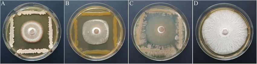
Ulrich et al.                                                                                                                  Ash Leaf Microbiota

(Witzell and Martin, 2018). Other factors are also known to                 Germany). The cells were disrupted again with the FastPrep-
modulate the plant microbiota, including local conditions, plant            24 Instrument (MP Biomedicals, Germany; 30 s at 6 ms−1 ),
age and fitness, climate, and pathogen presence (Compant et al.,             and extraction was performed using a standard protocol. The
2019). Accordingly, bacteria specific for the leaves of tolerant ash         degree of DNA degradation was monitored on 1% agarose gels.
trees in severely affected stands may be associated with the ability         The DNA purity and concentration were measured using a
of the tree to resist the pathogen or are potentially involved in           NanoDrop spectrophotometer. The V5–V6 region of the 16S
protecting the host from ash dieback. Therefore, in this study,             rRNA genes was amplified using the primers 799F and 1115R,
ash trees with visible symptoms of ash dieback were compared                which exclude the chloroplast and mitochondria DNA of the
with healthy-looking trees, which were recorded as having a                 host plant (Chelius and Triplett, 2001; Redford et al., 2010).
relatively high tolerance to H. fraxineus. We hypothesize that              The primers contained a heterogeneity spacer along with a
significant differences in the bacterial leaf microbiome exist, and           barcode sequence. The amplicons were purified with a MSB Spin
the comparison of these trees will reveal bacteria that are capable         PCRapace kit (Invitek, Berlin, Germany) and mixed at equimolar
of suppressing the pathogen. A high-throughput sequencing                   DNA concentrations. Library preparation and Illumina MiSeq
approach is combined with cultivation methods to analyze the                300-bp paired-end sequencing was performed at LGC Genomics
microbiome as completely as possible and to overcome the biases             (Berlin, Germany).
of both procedures. We further assume that plant-associated
bacteria adapted to ash trees are a reservoir for isolates with             Data Processing and Statistical Analysis
antagonistic potential. The ability to directly attack the fungal           The raw sequence data were processed with the DADA2
pathogen is tested in vitro by performing dual-culture assays.              R package v. 1.12.1 (Callahan et al., 2017) and mothur v.
                                                                            1.39.1 (Schloss et al., 2009; RRID: SCR_011947). The DADA2
                                                                            algorithm was applied for quality filtering and removing chimeric
MATERIALS AND METHODS                                                       sequences. Based on a parametric error model, the pipeline
                                                                            provides amplicon sequence variants (ASVs), in contrast to the
Study Site and Sampling Strategies                                          traditional operational taxonomic unit (OTU) approach, and
Field sampling was conducted in July 2017 at four ash forest                reduces the problem of falsely identified OTUs originating from
districts in Northeast Germany with a severe infestation of                 mis-clustered sequences (Fricker et al., 2019). The ASVs were
H. fraxineus infection: Lendershagen (54◦ 23′ N, 12◦ 83′ E; plot A),        taxonomically assigned by the naive Bayesian classifier method
Karnin (54◦ 26′ N, 12◦ 87′ E; plot B), Pennin (54◦ 25′ N, 13◦ E; plot       using the Silva reference database v. 132 (RRID: SCR_006423).
C), and Wredenhagen (53◦ 30′ N, 12◦ 58′ E; plot D). In each of the          In addition, the assignment was completed at the species level if
districts, four pairs of adjacent trees consisting of a susceptible         the ASVs exactly matched the sequence of the reference strain
and a tolerant individual (age between 60 and 80 years) were                of only one species. The taxonomic assignment was confirmed
selected. The distance between the tree pairs within the districts          with the taxonomic classification in mothur using the RDP
was at least 80 m. Healthy trees were very rarely observed in               training set 16 (RRID: SCR_006633) and the Silva database.
the long-term monitoring plots and were previously recorded                 Statistical analyses were performed using the phyloseq, vegan,
as having relatively high tolerance to ash dieback because they             and ape packages of R 3.6.0, as well as MicrobiomeAnalyst
had grown in a site where the pathogen has been present for                 (Chong et al., 2020; RRID: SCR_015022). All samples were
several years (Sündermann and Jütte, 2014). Compound leaves                 rarefied to the minimum number of sequences among all
with petioles from healthy trees were sampled by shooting down              samples. Three diversity indices (Chao 1, Shannon, and Simpson)
branches using a catapult; diseased trees were cut down to sample           were calculated. The community structure was compared by
the leaves. Representative samples were collected from the middle           applying a principal coordinate analysis (PCoA) based on an
part of the crowns from both healthy and susceptible trees. To              unweighted UniFraq distance matrix of the ASVs. Significant
exclude the effect of the pathogen H. fraxineus on the indigenous            differences between the bacterial communities were tested
microbial community, visually healthy leaves and petioles were              using analysis of similarity (ANOSIM). A taxonomic network
sampled from both susceptible and tolerant trees. Altogether,               was constructed using Cytoscape version 3.7.1 to visualize
samples from 32 trees were collected and stored in plastic bags             differences in the core microbiome (Shannon et al., 2003;
at 4◦ C for transportation and processed within 24 h.                       RRID: SCR_003032). The differential abundance at different
   In the laboratory, leaves, leaflets, petioles, and rachis were            taxonomic levels was analyzed with the metagenomeSeq tool
randomly chosen from compound ash leaves. An aliquot of the                 using the false discovery rate (FDR) for multiple test correction
samples (4 g) was ground in 4 ml of sterile 0.85% NaCl with a               (Paulson et al., 2013).
sterile mortar and pestle and used for cultivation. Another aliquot            The paired sequence reads were deposited in the public
of the samples (4 g) was ground in liquid nitrogen and stored at            repository Sequence Read Archive (SRA, RRID: SCR_004891)
−80◦ C until DNA extraction.                                                with the accession number PRJNA602193.

DNA Extraction and Amplicon                                                 Isolation of Bacteria
Sequencing From Ash Leaves                                                  For the isolation of epiphytic and endophytic bacteria, the
Total DNA was extracted from 100 mg of the ground plant                     ground plant material from each sample was serially diluted
material using the DNeasy Plant Mini Kit (Qiagen, Hilden,                   in 0.85% NaCl and plated on R2A, a nutrient-poor medium

Frontiers in Microbiology | www.frontiersin.org                         3                                        May 2020 | Volume 11 | Article 966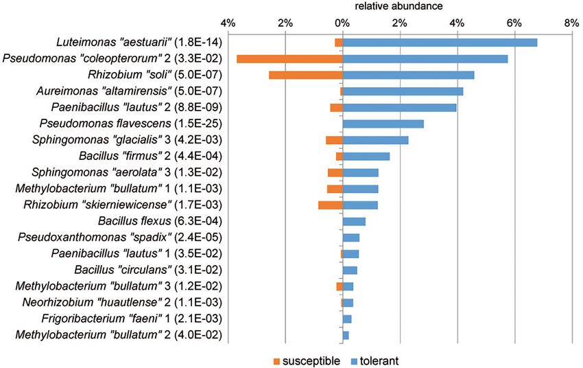
Ulrich et al.                                                                                                                  Ash Leaf Microbiota

that is suitable for the growth of diverse plant-associated              the groups were consecutively numbered. For groups assigned
bacteria (Difco, Detroit, United States). Plates were incubated          to one of our reference isolates, the species name is reported in
at 25◦ C for at least 7 days. Population densities were                  quotation marks and represents the most closely related species
determined by counting the colony-forming units (CFU),                   (Supplementary Table S1).
and the results are reported as CFU per gram of fresh                        The Pseudomonas MALDI groups were subjected to a further
weight. Approximately 80 isolates per sample were randomly               phylogenetic analysis using a multi-locus sequence analysis
selected from R2A, purified by streak dilution and stored                 (MLSA). The housekeeping genes gyrB, rpoB, and rpoD were
at −80◦ C in nutrient broth containing 40% glycerol until                chosen for this purpose and amplified and sequenced as
further processing.                                                      described by Mulet et al. (2010). Together with the 16S rRNA
                                                                         gene, a concatenated tree was constructed. The alignment
                                                                         comprised 1,432 bp of 16S rRNA, 1,088 bp of gyrB, 1,111 bp
Dereplication and Phylogenetic                                           of rpoB, and 716 bp of rpoD gene. The total length of the
Classification of Epiphytic and                                          alignment was 4,347 bp.
Endophytic Isolates                                                          For the statistical analysis, the number of isolates per sample
Matrix-assisted laser desorption/ionization time of flight mass           was normalized using the CSS method (Paulson et al., 2013).
spectrometry (MALDI-TOF MS) was used as an approved high-                Significant differences in the community composition between
throughput method for the dereplication and classification of             tolerant and susceptible trees were tested by the MetagenomeSeq
bacteria in large environmental studies (Huschek and Witzel,             tool (MicrobiomeAnalyst, PFDR < 0.05).
2019). Prior to measurement, bacterial isolates were freshly                 The 16S rRNA gene sequences of the reference isolates of the
inoculated on CASO broth (Fluka, Buchs, Switzerland) solidified           MALDI groups were deposited in NCBI (RRID: SCR_006472)
with 15 g L−1 agar and cultivated for 24 h at 25◦ C. The                 under the accession numbers MN989032–MN989182.
whole-cell measurement protocol was used to obtain mass
spectra. Briefly, ∼0.1 mg of cell material was transferred directly
from the edge of a bacterial colony to a MALDI target spot.
                                                                         In vitro Screening and Statistical
After drying at room temperature, sample spots were overlaid             Analysis of Antagonistic Activity Toward
with 1 µl of matrix solution (Bruker Daltonics, Bremen,                  H. fraxineus
Germany). The MS analysis was performed with a MicroflexTM                All bacterial isolates were screened for their antagonistic activity
LT/SH MALDI-TOF mass spectrometer (Bruker Daltonics) using               toward H. fraxineus isolate P3 using a cocultivation assay
Flex Control 3.4 software, as described in detail by Müller              described by Pane and Zaccardelli (2015). Briefly, an agar
et al. (2016). The MALDI Biotyper Preprocessing Standard                 plug (ø 5 mm) with H. fraxineus mycelium was placed in
Method and the MALDI Biotyper MSP Identification Standard                 the center of the petri dish, while four bacterial isolates were
Method were used for bacterial classification with Biotyper               spread around the edge of the plate. Plates containing the
3.1 software equipped with the MPS Library (release August               H. fraxineus mycelium alone were used as control. The test was
2018), according to the manufacturer’s instructions (Bruker              performed on PDA medium enriched with ash shoot extract
Daltonics). Isolates with a score value greater than 2.3 (highly         (30 g of ash leaves per liter of PDA) at 22◦ C. Due to its
probable species identification) were considered as identified             slow growth, H. fraxineus was inoculated 5–6 days before the
by the Bruker database. The spectra of the isolates with lower           bacteria. The antifungal activity was monitored by measuring the
scores were compared with each other and grouped based                   fungal colony radius (r) after 1 and 2 weeks until the control
on a score > 2.3. Representative strains of the unidentified              reached the edge of the plate. The inhibition of H. fraxineus
groups were taxonomically classified by sequencing almost the             was estimated using the following formula: percent growth
complete 16S rRNA gene. Briefly, total DNA was extracted                  inhibition = 100 × [(rcontrol − rcocultivation ) ÷ rcontrol ]. Isolates
from single colonies by resuspending them in 20 µl of 25 mM              with inhibition rates >30% were chosen for a subsequent
NaOH/0.25% SDS and incubating the mixture for 15 min at                  statistical assay (three replicates) with two H. fraxineus isolates
95◦ C. The 16S rRNA gene was amplified using primers 8f and               (P3 and HF23). In this approach, each bacterial isolate was
1525r (Lane, 1991) according a protocol described by Ulrich and          tested on a separate plate by spreading it around the fungus
Wirth (1999) and sequenced with the internal primers 1492r               four times. The statistical test was also used to evaluate the
(Lane, 1991) and 782r (Chun and Goodfellow, 1995) or 926r                vitality of the residual H. fraxineus mycelium after confrontation
(Liu et al., 1997). All reference strains were unambiguously             with the bacteria. For this purpose, agar plugs (ø 5 mm) of the
taxonomically assigned at the genus level with mothur using              mycelium were picked and incubated on PDA with the ash shoot
the RDP database. For a further phylogenetic classification,              extract. The growth of the fungus was assessed after 2 weeks
the closest related species were determined using EzBioCloud             by estimating the mycelium diameter and comparing it to the
(Yoon et al., 2017). After supplementing the MALDI BiotyperTM            untreated control.
database with the spectra of the reference strains, a reliable              Two-year-old ash seedlings were inoculated with either isolate
taxonomic identification of all isolates was achieved using the           using an approach described by Przybyl (2003) to verify the
results of the MALDI-TOF MS analysis. The MALDI groups                   pathogenicity of the H. fraxineus isolates. The symptoms were
were designated with respect to the assigned reference strain.           monitored for 8 weeks, and the virulence of both isolates
If several groups of the same bacterial species were identified,          could be confirmed.

Frontiers in Microbiology | www.frontiersin.org                      4                                           May 2020 | Volume 11 | Article 966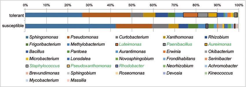
Ulrich et al.                                                                                                                                          Ash Leaf Microbiota

RESULTS                                                                                  all samples (with a mean of 58%). Within the Proteobacteria,
                                                                                         α-Proteobacteria were the most abundant class, representing 40%
Comparison of the Bacterial Community                                                    of the total sequences, followed by γ- and β-Proteobacteria at 9
                                                                                         and 6%, respectively. Other abundant phyla were Bacteroidetes
Structure by Amplicon Sequencing of the
                                                                                         (20%), Actinobacteria (17%), and Deinococcus–Thermus (3%).
16S rRNA Gene                                                                            Firmicutes accounted for only 0.33% of ASVs among all
The V5–V6 region of the 16S rRNA gene from the microbiomes                               samples. Only 1.8% of the total sequences were unable to
of 16 tolerant and 16 susceptible trees was amplified with primers                        be assigned to bacterial taxa. The results revealed a high
that exclude the host plant DNA. An evaluation of the Illumina                           phylogenetic diversity of leaf bacteria, with 241 taxa identified
sequence reads revealed an exceedingly high abundance of the                             at the genus level. Sphingomonas (18.4%), Hymenobacter (16%),
genera Bacillus (93%) or Escherichia and Pseudomonas (89%) in                            and bacteria of the order Rhizobiales (8.7%) dominated
the samples B2K and A1K (from susceptible trees), respectively,                          the total bacterial community. Other abundant genera were
suggesting that these samples were contaminated. Both samples                            Methylobacterium (5.4%), Pseudomonas (3.2%), Deinococcus
were excluded from the analyses. After quality filtering and                              (3.0%), and Massilia (2.7%).
removing potential chimeras and plastid and mitochondrial                                    Comparing the bacterial microbiomes of tolerant and
sequences from ash trees, 3,562,425 high-quality sequences were                          susceptible trees at the phylum level, Firmicutes was significantly
obtained that formed 3,634 ASVs with an average of 413 ASVs                              (threefold) increased in tolerant F. excelsior (PFDR = 0.05). At
per sample. Sequences rarefied to 44,537 reads per sample were                            the class level, γ-Proteobacteria were present at significantly
used as input for all subsequent analyses.                                               higher levels in tolerant plants (PFDR = 0.046) and were
   Alpha diversity estimates such as the Shannon index showed                            particularly enriched with sequences associated with bacteria of
no significant differences, with values of 4.02 ± 0.34 (mean ± SD)                         the family Xanthomonadales (PFDR = 0.0016) and the genus
for susceptible trees and 4.03 ± 0.35 for tolerant trees. The                            Luteimonas (PFDR = 3.9E-06). Five abundant phylotypes (>0.3%)
analysis of the beta diversity resulted in small but significant                          were significantly increased in tolerant trees at the genus level.
differences between the bacterial community structure grouped                             Luteimonas showed a 15-fold increase, Halomonadaceae sp. a 3.8-
by forest plot and health status (ANOSIM R = 0.2118,                                     fold increase, Microbacterium a 2.6-fold increase, Aliihoeflea a
P < 0.001). The differences are shown in Figure 1. The first axis                          27-fold increase, and Agrococcus a 2.2-fold increase. Some other
indicated some differences between tolerant and susceptible trees,                        genera, such as Aureimonas, were also present at higher levels
particularly in plot D. However, the pairwise comparisons did not                        (3.7-fold) in tolerant trees, but the difference was not significant.
result in any significant differences.                                                         The leaf core microbiome was determined at the genus level
   The taxonomic assignment of ASVs revealed 10 taxa at the                              and defined as those taxa that were present in at least 75%
phylum level. The phylum Proteobacteria predominated across                              of the respective samples. It included 37 abundant bacterial

  FIGURE 1 | Ordination plot showing the differences in the community structures of tolerant (t) and susceptible (s) trees across the four sampled forest districts (A–D).
  A principal coordinate analysis (PCoA) was applied based on the unweighted UniFraq distance matrix.

Frontiers in Microbiology | www.frontiersin.org                                      5                                                  May 2020 | Volume 11 | Article 966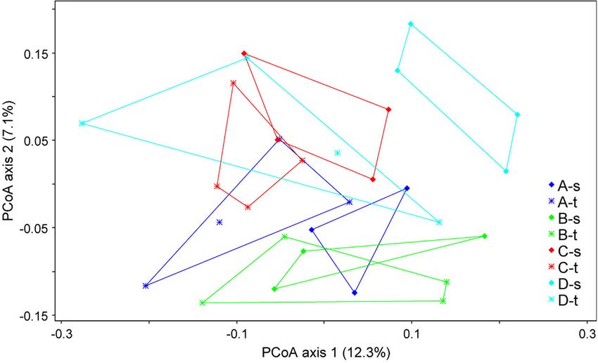
Ulrich et al.                                                                                                                                       Ash Leaf Microbiota

  FIGURE 2 | Core microbiomes of susceptible and tolerant F. excelsior leaves at the genus level. The core microbiomes (taxa occurring in 75% of all replicates of
  each group that exhibited at least 0.1% abundance in the community) were combined for the network analysis. The size of nodes corresponds to the relative
  abundance in the whole dataset, node labels display their taxonomic affiliation, and the color of the nodes indicates the respective phylum.

taxa (greater than 0.1%) for the tolerant F. excelsior trees,                          were detected only in susceptible trees. Overall, the specific
which represent 87.1% of all sequences. Susceptible plants                             bacteria for trees with each health status were less abundant than
contained only 30 genera in their core microbiome (88.2% of                            the shared microbiota.
the total sequences). The comparison of the core microbiomes of                           Based on ASVs, bacterial communities of tolerant and
tolerant and susceptible ash trees is illustrated using a clustering                   susceptible trees were compared using a differential abundance
network (Figure 2). Twenty-eight taxa shared both core                                 analysis. Figure 3 presents an overview of ASVs that were
microbiomes, including the predominant genera Sphingomonas                             significantly increased in tolerant ash trees. ASV0013, which
and Hymenobacter. Nine genera from three different phyla                                was identified as Luteimonas sp., was remarkably increased in
were specific to the tolerant trees, with the highest abundance                         tolerant trees. Several ASVs from other genera were increased;
observed for Luteimonas and Chryseobacterium. Two genera                               for instance, seven ASVs of the genus Pseudomonas and two

Frontiers in Microbiology | www.frontiersin.org                                    6                                                 May 2020 | Volume 11 | Article 966
Ulrich et al.                                                                                                                                     Ash Leaf Microbiota

  FIGURE 3 | Overview of significantly increased amplicon sequence variants (ASVs) in tolerant F. excelsior trees. The ASVs are arranged according their abundance
  (>0.07%). Statistical significance (PFDR ) is indicated in brackets.

ASVs of Aureimonas showed a significantly higher abundance                             (MALDI groups) belonging to 45 genera (Supplementary
in tolerant trees.                                                                    Table S1). At the phylum level, the culturable ash-associated
                                                                                      bacteria were dominated by Proteobacteria (81%), followed by
                                                                                      Actinobacteria (14%) and Firmicutes (5%). Proteobacteria were
Comparison of the Culturable Bacterial                                                mainly represented by α- and γ-Proteobacteria at 47% and
Communities of Susceptible and Tolerant                                               33%, respectively. In the comparison of tolerant and susceptible
F. excelsior Trees                                                                    trees, the amount of Proteobacteria was the same, whereas the
Population densities of culturable bacteria in F. excelsior leaves                    proportions of Actinobacteria (11% to 18%) and Firmicutes (9%
and petioles ranged from 5 × 10⁴ to 8 × 10⁵ CFU g−1                                   to 1%) were different to some extent, as the latter was significantly
fresh weight. Significant differences were not observed between                         increased in tolerant plants (PFDR = 0.0044). Regarding the
tolerant and susceptible trees.                                                       bacterial classes, γ-Proteobacteria (37 to 29%, PFDR = 0.022) and
   The 2,589 bacterial isolates derived from the 32 samples                           Bacilli (9 to 1%, PFDR = 0.021) were present at significantly higher
represent the common culturable epiphytic and endophytic                              levels in tolerant trees.
bacterial communities from the leaves of susceptible and tolerant                        At the genus level, Sphingomonas and Pseudomonas
F. excelsior trees. The isolates were classified into 166 phylotypes                   dominated the culturable bacterial community of ash trees

Frontiers in Microbiology | www.frontiersin.org                                   7                                                May 2020 | Volume 11 | Article 966
Ulrich et al. Ash Leaf Microbiota
FIGURE 4 | Structure of the culturable bacterial communities in tolerant and susceptible F. excelsior trees. Genera with significantly increased abundance in tolerant
trees are highlighted in green. The analysis is based on the classification of 2,589 isolates (16 replicates).
at 33 and 20%, respectively, followed by Curtobacterium, respectively). Eight of the 13 MALDI groups were clearly assigned
Xanthomonas, and Rhizobium each with 6% and Frigoribacterium to five distinct Pseudomonas species. In addition to P. flavescens
and Luteimonas with 4%. The relative abundance of bacterial and Pseudomonas cerasi, two MALDI groups were each classified
genera in trees differing in health status is shown in Figure 4. into P. coleopterorum, P. graminis, and Pseudomonas caspiana.
A comparison of bacterial communities revealed a significantly In contrast to the phylogenetic analysis based on the 16S rRNA
higher proportion of isolates in tolerant trees for the abundant gene, the Pseudomonas “congelans” group (isolate C4P022b) was
genera Luteimonas (24-fold), Paenibacillus (7-fold), and located close to the P. syringae-type strain in the concatenated
Aureimonas (46-fold), as well as genera occurring at lower tree. Similarly, the Pseudomonas “extremaustralis” group (isolate
percentages, such as Staphylococcus, Pseudoxanthomonas, and A4K089) was more closely assigned to Pseudomonas poae and
Rhodobacter. Pseudomonas trivialis than to the P. extremaustralis-type strain
A differential abundance analysis was performed at the level using MLSA. Three MALDI groups [Pseudomonas “moorei”
of MALDI groups to identify groups of isolates specific for (D4P040), P. “rhizosphaerae” (C4K059), and P. “caspiana”
tolerant trees that may be associated with the ability of the tree to 1 (B1K012)] did not clearly cluster with any Pseudomonas
resist the pathogen (Figure 5). In tolerant trees, the Luteimonas strain, suggesting that these groups probably represent new
MALDI group was increased more than 20-fold. Additionally, species. Overall, the MALDI classification and MLSA result
other groups, such as Aureimonas “altamirensis,” Paenibacillus in a similar differentiation at the species level. Within a
“lautus” 2, Pseudomonas flavescens, Sphingomonas “glacialis” 3, species, namely, P. caspiana, the differentiation corresponds
and Bacillus “firmus” 2, showed a distinctly higher abundance in to the MALDI groups. For P. graminis and P. coleopterorum,
the culturable bacterial community of tolerant trees. Of the four the intraspecies differentiation was not consistent between
Methylobacterium “bullatum” MALDI groups, three groups were the two methods.
significantly increased in tolerant trees.
The genus Pseudomonas is particularly interesting among
bacteria that are able to inhibit the activity of pathogenic fungi. It Antagonistic Potential of Bacterial
was the second-most abundant genus in the culturable bacterial Isolates – Growth Inhibition of
community of ash leaves. In tolerant trees, the proportion of H. fraxineus
pseudomonads was slightly increased (22.6%) compared to that The bacterial isolates were screened for their ability to inhibit the
in susceptible trees (17%). Altogether, 497 Pseudomonas isolates growth of H. fraxineus using cocultivation assays as a method
were obtained from the 32 leaf samples and classified into to detect putative antagonists. The screen produced 282 isolates
13 MALDI groups belonging to five different species groups (10.9% of the total isolates) that suppressed pathogen growth by
(Pseudomonas lutea, Pseudomonas rhizosphaerae, Pseudomonas at least 30%. In some cases, the inhibitory effect was combined
syringae, Pseudomonas fluorescens, and Pseudomonas straminea) with the lysis of the mycelium. The antagonistic isolates were
within the P. fluorescens lineage (Mulet et al., 2010; Peix et al., obtained in the same proportions from tolerant and susceptible
2018). For a more precise differentiation of the species groups, trees and mainly belonged to the genera Sphingomonas (38%)
at least one representative isolate was chosen from each MALDI and Pseudomonas (21%). A series of other genera (18) from
group to perform a MLSA (Figure 6). The analysis focused on three different phyla, such as Xanthomonas, Microbacterium,
Pseudomonas coleopterorum and Pseudomonas graminis because and Paenibacillus, was present at comparably low proportions
they contained the largest numbers of isolates (179 and 114, (1–7%, Supplementary Figure S1). Isolates of the genera
Frontiers in Microbiology | www.frontiersin.org 8 May 2020 | Volume 11 | Article 966Ulrich et al. Ash Leaf Microbiota
FIGURE 5 | MALDI groups of bacterial isolates with a significantly higher abundance in tolerant trees. Groups displaying significant increases are sorted according to
their abundance. The level of significance is indicated in brackets (PFDR , MetagenomeSeq). Species names enclosed in quotation marks were assigned based on
16S rRNA gene sequencing. Similarity values to the closest relative type strains are shown in Supplementary Table S1.
Pantoea and Bacillus showed the highest mean growth inhibition isolate P3 was completely killed, and the less susceptible isolate
rates (∼40%). HF23 showed a visible reduction in vitality (Table 1). A clear
Based on the primary screen, 78 isolates were assayed for antagonistic activity was also observed for Bacillus “tequilensis”
their antagonistic activity with a statistical cocultivation test C4K066b and Erwinia “billingiae” D4P109. These bacteria were
using three replicates and two H. fraxineus strains. Both isolates weaker in their ability to immediately suppress fungal growth, but
were confirmed to be virulent on young ash trees, whereas they affected the mycelium during cocultivation. For example, the
H. fraxineus P3 was more susceptible in cocultivation tests than mycelium of H. fraxineus P3 was completely lysed at the end of
HF23 (Supplementary Table S3). The evaluation was completed the confrontation with B. “tequilensis” C4K066b (Figure 7). This
by testing the vitality of the remaining fungal mycelium after lytic effect was less pronounced for HF23, resulting in a relative
confrontation with the bacteria. An overview of the activity of high mycelium vitality (64%). In contrast, E. “billingiae” D4P109
isolates from all tested MALDI groups is shown in Table 1. In completely killed both H. fraxineus isolates during cocultivation.
general, the test confirmed the results of the screen, although
several isolates, including members of the genus Luteimonas,
failed in the statistical test. Of the 78 isolates, six strains DISCUSSION
significantly inhibited the growth of both H. fraxineus isolates,
whereas 17 strains only affected P3 and eight only inhibited In ash dieback, which is caused by H. fraxineus, leaflets and
HF23 (Supplementary Table S3). The strongest effects were petioles are the main entry point for the pathogen (Cleary et al.,
observed for Bacillus “velezensis” A4P130, Pantoea “vagans” 2013; Hanackova et al., 2017b). During and directly after invasion
B3K066, and P. “caspiana” B1P055, with growth inhibition rates and penetration of epidermal tissue, that is, during the biotrophic
ranging from 41 to 55%. In particular, cocultivation with A4P130 phase of the pathogen, endophytic and epiphytic microorganisms
resulted in the formation of a sharp necrotic zone along the in both the leaf and the petiole might be able to inhibit the
edge of the mycelium of H. fraxineus P3 (Figure 7). However, progression of the pathogen (Cross et al., 2017; Hanackova
the mycelium picked outside of this zone remained viable. et al., 2017b; Mansfield et al., 2018). Therefore, microorganisms
The isolates B3K066 and B1P055 exerted a dramatic effect by colonizing the compound leaves have the potential to function
lysing the fungal mycelium during cocultivation. Accordingly, as biocontrol agents against H. fraxineus. Numerous studies have
Frontiers in Microbiology | www.frontiersin.org 9 May 2020 | Volume 11 | Article 966Ulrich et al. Ash Leaf Microbiota FIGURE 6 | Phylogenetic tree of representative strains for the Pseudomonas MALDI groups in relation to respective type strains of the genus. The tree is based on the analysis of partial sequences of four concatenated genes (16S rRNA, gyrB, rpoB, and rpoD; accession numbers/locus tags are listed in Supplementary Table S2). The dendrogram was generated with the maximum likelihood algorithm based on the general time-reversible substitution model with G + I. Numbers at branch nodes refer to bootstrap values >70%. Bar, 0.01 substitutions per nucleotide site. The designation of MALDI groups was only based on 16S rRNA gene similarity. Pseudomonas species groups were indicated as proposed by Peix et al. (2018). examined the association of fungi with H. fraxineus infections. approach based on the assumption that both groups may interact However, studies of bacteria colonizing F. excelsior are rather with the pathogen in a similar manner. In the phyllosphere, rare, although bacteria are estimated to be the most abundant and microbes are often observed both as epiphytes on the plant diverse colonists of the leaf phyllosphere. In our study, population surface and as endophytes within plant tissue (Whipps et al., densities of up to 8 × 10⁵ CFU g−1 leaf were observed, which are 2008; Hardoim et al., 2015; van Overbeek and Saikkonen, 2016). in the same range as the values measured by Weyens et al. (2009). Many bacteria have the ability to switch between endophytic We analyzed endophytic and epiphytic bacteria using a combined and free-living lifestyles and may help protect the plant from Frontiers in Microbiology | www.frontiersin.org 10 May 2020 | Volume 11 | Article 966
Ulrich et al.                                                                                                                                             Ash Leaf Microbiota

TABLE 1 | Antagonistic activity of isolates from different MALDI groups assessed by the ability to inhibit the growth of H. fraxineus and vitality test of the remaining
fungal mycelium.

Isolatea             Taxonomic assignment                    Growth inhibition rate in coculture (%)b           Vitality of the mycelium (% of the untreated control)

                                                             P3                       HF23                       P3                               HF23

C4K093             Achromobacter “denitrificans”            40.0*                     12.3                       98.4                              81.5
A4P130                  Bacillus “velezensis”               50.6*                     54.7*                      96.3                             101.7
C4K066b                 Bacillus “tequilensis”              35.1                      37.5                       0.0                               63.9
C4P040b                    Bacillus cereus                  30.0                      35.0                       98.5                              91.7
A2K052           Curtobacterium “flaccumfaciens” 2          28.4                      22.9                       0.0                               87.5
D4P109                   Erwinia “billingiae”               40.0*                     22.5                       0.0c                              0.0c
B3K063                Frigoribacterium “faeni” 5            44.4*                     37.5                       0.0                               82.9
D1P082                 Luteimonas “aestuarii”               22.5                      25.6                       64.8                              86.2
D3P076             Methylobacterium “bullatum” 1            30.0                       2.4                      100.0                              90.7
C4K020              Methylobacterium “cerastii”             25.3                       4.4                       0.0c                              94.4
D3P082           Methylobacterium “goesingense” 2           30.8                       6.1                       96.3                              90.7
C4K087b         Methylobacterium “pseudosasicola” 2         22.1                      18.8                       97.1                              90.3
C1P060             Microbacterium “hatanonis” 2             39.5*                     17.7                       95.8                              96.0
D2K023             Novosphingobium “fluoreni” 4             35.0                      34.6                      85.9c                              89.8
B3P038                 Paenibacillus “lautus” 2             45.7*                      7.3                      67.6c                              85.5
B3K066                   Pantoea “vagans”                   40.5*                     47.5*                      0.0                               80.8
B1P055              Pseudomonas “caspiana” 3                41.0*                     43.0*                      0.0                               86.8
A1P062                 Pseudomonas “cerasi”                 32.1                      36.7                       0.0                               80.5
B2K013            Pseudomonas “coleopterorum” 2             32.1                      26.8                       0.0                               1.9
D4P037               Pseudomonas flavescens                  6.7                       6.3                      27.4c                              92.9
A3P049               Pseudomonas “graminis” 1               29.6                      17.5                       0.0c                              87.2
A4K089            Pseudomonas “extremaustralis”             31.3                      21.5                       0.0c                              92.0
A2P026             Pseudoxanthomonas “spadix”               35.0                      15.7                       91.5                              84.4
A2P086                  Rahnella “victoriana”               33.3                      26.2                       0.0                               97.4
B3P075              Rhizobium “skierniewicense”             31.3                      30.0                       0.0                               86.7
B3P008              Sphingomonas “aerolata” 1               36.4                      32.9                       0.0c                             100.0
D4P108                 Sphingomonas “taxi” 1                22.5                      35.0                       98.6                             100.0
D2K022              Sphingomonas “aurantiaca”               37.5                      30.9                      76.6c                              95.2
B3K005                 Sphingomonas faeni 1                 38.7*                     32.9                       0.0                               90.3
B4K076                 Variovorax “robiniae” 1              26.7                      26.8                       96.0                              88.7
A4P033                Xanthomonas “cynarae”                 44.4*                     28.9                       0.0                               81.0
a Ofthe 78 tested bacteria, only one typical or the best isolate from each MALDI group is listed. Details are provided in Supplementary Table S2. b Growth inhibition
was tested using the H. fraxineus isolates P3 and HF23. *Significantly increased compared to a control Pseudomonas sp. PI01 (culture collection Thünen Institute,
Waldsieversdorf, Germany) that did not show antagonistic activity in cocultures (growth inhibition rates: 4.0 and 8.5). The test was performed using the Kruskal–Wallis
one-way analysis of variance on ranks followed by pairwise comparisons with Dunn’s test (P < 0.05). c Detection of the antagonistic isolate inside the recovered H. fraxineus
mycelium or instead of the mycelium.

  FIGURE 7 | Inhibition of H. fraxineus P3 growth by cocultivation with B. “velezensis” A4P130 (A), P. vagans B3K066 (B), and B. tequilensis C4K066b (C). The
  growth of H. fraxineus P3 without cocultivation served as the control (D).

Frontiers in Microbiology | www.frontiersin.org                                       11                                                  May 2020 | Volume 11 | Article 966
Ulrich et al. Ash Leaf Microbiota inside the leaves and from the leaf surface (Hardoim et al., 2008). the study by Griffiths et al. (2020), the bacterial microbiome Thus, in this context, the distinction between the two habitats analyzed in our study did also not show a direct association appears arbitrary. between the health status and the community structure, but a Bacterial communities in the ash phyllosphere were number of bacterial groups were significantly associated with the dominated by Proteobacteria, Bacteroidetes, and Actinobacteria, H. fraxineus infection. which have also been reported to be the dominant phyla in In the comparison of bacteria of tolerant and susceptible ash the phyllosphere of F. excelsior plants in the United Kingdom leaves at the phylum level, Firmicutes was significantly increased (Griffiths et al., 2020), as well as in other plants (Muller et al., in tolerant leaves. Genera of this phylum are well-known 2016). In studies exclusively analyzing epiphytic bacteria potent antagonists and agents functioning in the biocontrol of in the phyllosphere of a number of trees, α-Proteobacteria pathogenic fungi by producing various bioactive metabolites, dominated the bacterial community, consistent with our results such as iturins and fengycins, with a strong inhibitory effect (Redford et al., 2010; Laforest-Lapointe et al., 2016). Likewise, on the growth of a wide range of plant pathogens (Emmert the proportion of Firmicutes was very low. An analysis of and Handelsman, 1999; Ongena et al., 2005; Müller et al., exclusively endophytic bacteria revealed a higher abundance of 2015). In particular, isolates of Bacillus spp. are considered the γ-Proteobacteria and Firmicutes (Hardoim et al., 2015). most common groups that induce systemic resistance (ISR) At the genus level, similar to other woody plants such as (Vanpeer et al., 1991; Kloepper et al., 2004; Ongena et al., poplar and grapevine, the microbiome of F. excelsior leaves 2007) and prime plant defenses against pathogens (Elsayed et al., was dominated by Sphingomonas and Hymenobacter (Leveau 2020). At the class level, γ-Proteobacteria showed a significantly and Tech, 2011; Crombie et al., 2018). Aydogan et al. (2018) higher abundance in tolerant F. excelsior trees. Consistent with postulated that these genera may represent “hub” taxa that this result, other studies have described γ-Proteobacteria as are very important for the microbiome structure and plant– components of the microbiome of healthy oak and banana plants pathogen interactions. In addition to Sphingomonas, isolates (Sapp et al., 2016; Köberl et al., 2017). of the genus Pseudomonas dominated the culturable bacterial The most obvious differences at the genus or species level community in our study, while their proportions were very small were observed for the γ-Proteobacterium Luteimonas, which among the ASVs. This finding indicates the importance of using was significantly increased in healthy trees using both amplicon classical cultivation methods to complement high-throughput sequencing (ASV 15-fold) and cultivation (MALDI group 24- sequencing approaches in studies examining the interaction fold). Isolates of this genus have been described as phyllosphere between pathogens and the plant microbiota. bacteria of different plants (Sun et al., 2012; Comby et al., The main question to be answered in this study was whether 2017; Wemheuer et al., 2017). In antagonistic assays designed differences exist in the microbiota of tolerant and susceptible to identify biological control agents for the management of trees that may be related to the severity of ash dieback. In Fusarium head blight, Luteimonas showed a slight inhibition of the last few years, research examining the correlations between pathogen growth (Comby et al., 2017). In our study, the genus microorganisms and plant health has increased in importance Luteimonas was a component of the specific core microbiome and shifted from single strains and species to a more community- of tolerant F. excelsior plants (Figure 2). Consistent with this based view (Witzell and Martin, 2018; Schlechter et al., 2019). finding, Köberl et al. (2017) described Luteimonas as part of The identification and comparison of the core microbiomes the “healthy rhizosphere core microbiome” of banana plants in of tolerant and susceptible trees are considered particularly Fusarium oxysporum wilt-infested areas in Central America and important to detect key members of the microbial community as an indicator species of healthy plants. involved in protecting the plants from pathogens and to estimate Similar to Luteimonas, the abundance of Aureimonas was the spread of diseases in relation to microbiome interactions substantially increased in the leaves of tolerant plants (46-fold (Koskella et al., 2017; Lemanceau et al., 2017; Hamonts et al., in the culturable microbiome and 3.7-fold in ASVs), suggesting 2018; Orozco-Mosqueda et al., 2018). Koskella et al. (2017) that this genus might also be involved in the process that protects identified a link between the community composition of bark- ash plants from the pathogen. Aureimonas strains were reported associated bacteria and bleeding canker symptoms caused by to possess antifungal activity against Phytophthora nicotianae in P. syringae in horse chestnut, suggesting that tree microbiota are pineapple by González et al. (2017). Some species of this genus important to regulate the spread of the disease. In whitebark have been reported to produce siderophores (Lin et al., 2013), pine, the resistance to the fungal pathogen Cronartium ribicola providing the plant with the ability to inhibit the growth of is associated with microbiome combinations in healthy trees soilborne pathogens due to limited iron levels (Chaiharn et al., (Bullington et al., 2018). Similarly, interactions between Erysiphe 2009), and these proteins also appear to be involved in activation alphitoides, the causal agent of oak powdery mildew, and foliar of the ISR against foliar pathogens (van Loon et al., 2008). fungal and bacterial communities have been described for Significantly higher amounts of isolates from the genus pedunculate oak (Jakuschkin et al., 2016). In the context of ash Paenibacillus were detected in tolerant trees. More specifically, dieback, no significant differences were observed between the two MALDI groups of P. “lautus” were distinctly increased. structures of the endophytic fungal communities of trees with Various Paenibacillus species produce antifungal compounds and without symptoms using amplicon sequencing and culture- such as iturins and paenimyxin (Chung et al., 2000; Selim et al., dependent methods (Hanackova et al., 2017a; Schlegel et al., 2018; 2005) and excrete a variety of cell wall-degrading enzymes (Naing Kosawang et al., 2019; Griffiths et al., 2020). Consistent with et al., 2014; Yadav and Dubey, 2018). Paenibacillus species are also Frontiers in Microbiology | www.frontiersin.org 12 May 2020 | Volume 11 | Article 966
Ulrich et al.                                                                                                                    Ash Leaf Microbiota

known to elicit the ISR against pathogenic fungi (Sang et al., 2014;        activity against gram-positive bacteria, whereas gram-negative
Grady et al., 2016).                                                        bacteria were not affected (Halecker et al., 2014). However, in
    In addition to these genera that are probably involved in               our cocultivation tests, the growth of the bacterial strains was
the plant–pathogen interaction, the analysis revealed MALDI                 not inhibited. Strikingly, the H. fraxineus strains P3 and HF23
groups or ASVs of other genera, such as Pseudomonas, Bacillus,              used in this study displayed extremely different susceptibilities
Methylobacterium, and Sphingomonas, present at a significantly               to the bacteria. This difference corresponds to the highly varying
higher abundance in the phyllosphere of tolerant F. excelsior               capabilities of H. fraxineus strains, including exoenzyme profiles,
leaves. In particular, Pseudomonas is noticeable because seven              growth rates, and the production of antibiotics (Halecker et al.,
ASVs and two MALDI groups were significantly increased in                    2014; Junker et al., 2017).
the phyllosphere of tolerant plants. Bacteria of this genus are                 The strongest growth inhibition was observed for a
known to play an important role in the biological control                   B. velezensis isolate; however, the vitality of the H. fraxineus
of phytopathogenic fungi due to the production of antifungal                mycelium was not substantially altered. On the other hand,
metabolites and hydrogen cyanide (Bloemberg and Lugtenberg,                 the strong fungistatic effect might be sufficient to prevent the
2001; Haas and Defago, 2005; Fouzia et al., 2015). Some species             spread of the pathogen in ash leaves. B. velezensis is known to
control the growth of pathogens by inhibiting spore germination             possess antagonistic activity toward various phytopathogenic
(Raaijmakers et al., 1995) or by producing extracellular enzymes            fungi by producing antifungal compounds, such as bacillomycin,
that lyse components of the fungal cell wall (Compant et al.,               fengycin, and iturin (Palazzini et al., 2016; Lim et al., 2017). In
2005). In contrast to direct antagonism, different Pseudomonas               tomato, Elsayed et al. (2020) showed a shift in the composition
strains have the capacity to inhibit or suppress the activity of            of the prokaryotic community in response to inoculation with
pathogens by eliciting resistance pathways in the host plant                B. velezensis and assumed a priming effect of plant defenses
(Bakker et al., 2007).                                                      against the pathogen Ralstonia solanacearum.
    In the core microbiome, the Bacteroidetes member                            In vitro screens for antagonism with dual cultures are a
Chryseobacterium and several actinobacterial genera, such                   promising method to search for potential biocontrol candidates
as Microbacterium, were also specific for tolerant ash trees                 that directly affect the respective fungal pathogen. However, niche
(Figure 2). These genera are known not only for their abilities             competition, blocking potential entry points of the pathogen,
to promote plant growth but also as biocontrol agents targeting             and indirectly activating the plant immune system also lead to
fungal pathogens (Borah et al., 2018; Sang et al., 2018).                   a higher tolerance of the host plant (van Loon et al., 2008;
    Overall, the comparison of the microbiota of F. excelsior leaves        Berg et al., 2017; Terhonen et al., 2018). Thus, non-antagonistic
revealed several bacterial groups that are specific for tolerant             bacteria are important in plant defenses as well (De Boer
trees, such as Luteimonas, Aureimonas, Pseudomonas, Bacillus,               et al., 2006; Ardanov et al., 2012; Ab Rahman et al., 2018).
and Paenibacillus, suggesting that some of these genera might be            In recent years, inoculation with individual bacterial isolates
involved in the process that prevents the penetration and spread            has been shown to trigger a shift in the plant microbiome
of the H. fraxineus.                                                        (Andreote et al., 2010; Mitter et al., 2017). According to Ardanov
    The drivers for the differences between the microbiomes                  et al. (2012), an endophytic Methylobacterium isolate shifts the
of tolerant and susceptible plants are not yet known. Besides               structure of the entire endophytic community in potato plants.
stochastic colonization, long-living trees seem to be able to               The alteration in the microbiome correlated with the resistance to
actively recruit useful microbial communities (Witzell and                  different pathogens, whereas the bacterium itself did not possess
Martin, 2018). So the plant genotype could indirectly mediate               antagonistic activity.
the resistance toward the pathogen. However, other factors or                   In conclusion, the study of the ash phyllosphere resulted
a combination of both might be responsible for shaping the                  in the identification of bacteria specific to healthy leaves and
microbiota as well.                                                         confirmed the hypothesis that significant differences exist in
    A screen against H. fraxineus was performed to identify                 a series of bacterial groups putatively capable of suppressing
antagonistic bacteria for use in subsequent in planta assays. One           disease progression. This result was completed by a set of isolates
tenth of the total isolates suppressed the growth of H. fraxineus           that substantially inhibited H. fraxineus growth in vitro. The
in the screen, but the antagonistic activity was confirmed in a              bacteria were isolated directly from ash leaves, wherefore it can be
statistical test for only a small proportion (approximately 1–              assumed that they are able to stably establish after inoculation of
2%) of the total isolates. Comparable studies of the antagonistic           F. excelsior seedlings. In a next step, in planta experiments over a
effects of bacteria toward H. fraxineus have not been published to           longer period of exposure to H. fraxineus are required to evaluate
date. Among the culturable fungal community, a large percentage             the isolates or consortia of isolates, acting via direct antagonism
of endophytes, particularly fast-growing isolates, inhibited the            or competition or indirectly via induction of resistance.
pathogen (32 or 75%), but, on the other hand, approximately
25% of the fungi were inhibited by H. fraxineus (Schulz et al.,
2015; Hanackova et al., 2017a). The inhibition might be due                 DATA AVAILABILITY STATEMENT
to the production of different fungistatic compounds such as
viridin and hymenosetin by the pathogen (Andersson et al., 2010;            The datasets generated for this study can be found in
Halecker et al., 2014). Hymenosetin, a member of the family                 the Sequence Read Archive (SRA): PRJNA602193, NCBI:
of 3-decalinoyltetramic acid antibiotics, also exhibited strong             MN989032–MN989182.

Frontiers in Microbiology | www.frontiersin.org                        13                                          May 2020 | Volume 11 | Article 966
You can also read